Most cellulose degrading enzymes have a two-domain structure consisting of a catalytic domain and a ..... It is fairly inexpensive and it is widely recognized as a ...
VTT PUBLICATIONS 294
Structure-function relationships in fungal cellulose-binding domains Markus Linder VTT Biotechnology and Food Research
Dissertation for the degree of Doctor of Technology to be presented with due permission for public examination and debate in the Auditorium Ke 2 at the Helsinki University of Technology on the 24th of January, 1997, at 12 o’clock noon.
TECHNICAL RESEARCH CENTRE OF FINLAND ESPOO 1996
ISBN 951-38-4952-X ISSN 1235-0621 Copyright © Valtion teknillinen tutkimuskeskus (VTT) 1996
JULKAISIJA – UTGIVARE – PUBLISHER Valtion teknillinen tutkimuskeskus (VTT), Vuorimiehentie 5, PL 2000, 02044 VTT puh. vaihde (90) 4561, telekopio 456 4374 Statens tekniska forskningscentral (VTT), Bergsmansvägen 5, PB 2000, 02044 VTT tel. växel (90) 4561, telefax 456 4374 Technical Research Centre of Finland (VTT), Vuorimiehentie 5, P.O.Box 2000, FIN–02044 VTT, Finland phone internat. + 358 0 4561, telefax + 358 0 456 4374
VTT Bio- ja elintarviketekniikka, Geenitekniikka, Biologinkuja 1, PL 1500, 02044 VTT puh. vaihde (09) 4561, faksi (09) 455 2103 VTT Bio- och livsmedelsteknik, Genteknik, Biologgränden 1, PB 1500, 02044 VTT tel. växel (09) 4561, fax (09) 455 2103 VTT Biotechnology and Food Research, Genetic Engineering, Biologinkuja 1, P.O.Box 1500, FIN–02044 VTT, Finland phone internat. + 358 9 4561, fax + 358 9 455 2103
Technical editing Leena Ukskoski
VTT OFFSETPAINO, ESPOO 1996
Linder, Markus. Structure-function relationships in fungal cellulose-binding domains. Espoo 1996, Technical Research Centre of Finland, VTT Publications 294. 29 p. + app. 22 p. UCD Keywords
577.21:663.53 cellulose, cellulase, cellulose-binding domain, cellobiohydrolase, protein carbohydrate interaction, protein binding.
ABSTRACT Most cellulose degrading enzymes have a two-domain structure consisting of a catalytic domain and a cellulose-binding domain (CBD). The domains form well defined units which are separated by a distinct linker region. The CBD mediates the binding of the enzyme to the solid cellulose substrate. It is not involved in the hydrolysis, but if it is removed, much of the enzymes activity towards cellulose is lost. Typical examples of CBD containing enzymes are the major cellulases from the fungus Trichoderma reesei. The type of CBDs found in T. reesei consist of about 40 amino acids. The most studied one is from the enzyme cellobiohydrolase I (CBHI). Its structure resembles a wedge with two distinct faces. In this work the focus has been on functional studies on this type of CBDs and in particular that of CBHI. It was shown that several amino acid residues on one of the faces of the wedge, the flat face, are involved in the binding to cellulose. Several modifications on the other face, the rough face, did not to markedly affect the binding of the CBD. This led to the conclusion that the participation of the rough face to cellulose binding is very improbable. It was also shown that CBDs from different enzymes can have different binding affinities. The difference in affinity between CBHI and the higher affinity CBD of endoglucanase I was shown to be largely dependent on only one amino acid difference on the binding face. The effect of linking two domains was studied by constructing a double CBD consisting of the cellobiohydrolase II (CBHII) at the N-terminus and the CBHI CBD at the C-terminus. The two domains were linked by a 24 amino acid long linker. It was found that the linkage in the double CBD led to a substantially higher affinity towards cellulose compared to either of the two individual domains by themselves. A mechanism to explain the affinity increase based on the twodomain structure is presented. It is also proposed that a similar mechanism might affect the interaction of the wild type enzymes with cellulose. Finally, the reversibility of the CBD binding to cellulose was investigated. This is an issue which has been unclear, and is intimately linked to the function of the enzyme and applications of CBDs. The CBHI CBD was radioactively labeled with tritium, which allowed more detailed studies of its behavior than were possible before. It was shown that if a sample with CBD and cellulose at equilibrium is diluted, then
3
a new equilibrium further down the same isotherm is established. This result clearly shows that the binding is reversible. It was also shown that the CBHI CBD had an exchange rate at the surface which would not be rate limiting for a processive enzyme to move along the cellulose surface during hydrolysis.
4
PREFACE I started the work on my thesis in September 1993 at BMC in Uppsala. In October 1994 I moved to VTT to the gene-technology group where I completed it. First of all I want to thank all the people who have worked on the projects which are part of my thesis or who have made the necessary groundwork. Dr. Tapani Reinikainen was the first person involved in CBD research who I came in contact with. He helped me to get started in Uppsala and also later at VTT. In Uppsala I worked with Dr. Göran Pettersson, Dr. Gunnar Lindeberg, Dr. Jerry Ståhlberg and others to whom I am grateful their support and help. Back in Finland at VTT I started in the group of Dr. Tuula Teeri, who has since enthusiastically supported and encourged my work. In addition to the purely “material” help I have received from the above mentioned persons, the scientific discussions and exchange of ideas has been of great importance and enjoyment. My supervisor at the University of Technology, professor Simo Laakso, is thanked for his support even before the start of this work. I am also grateful to professor Hasse Söderlund who actually got me started on the work and given his support ever since. Professor Juha Ahvenainen and VTT are thanked for for the excellent working facilities. Professors Mike Penner and Olle Teleman are thanked for the discussions, which in the case of the two last articles played an important role. I also acknowledge my other co-authors, colleagues in cellulase research or otherwise connected persons; Arto Annila, Maija Mattinen, Irma Salovuori, Anu Koivula, Laura Ruohonen, Ingemar von Ossovsski, Janne Lehtio and Gunnar Hendriksson. Ingemar is especially thanked for helping with the language usage in the writing of this thesis (can you say it like this?). Merja Penttilä is thanked for her interest in the CBD work. The following institutions are thanked for financial support; Nordisk forskerutdanningsakademi (NorFA), the University of Uppsala, the Academy of Finland, the Technology Development Centre (Tekes) and VTT. Finally I want to thank my wife Tove for everything (she even moved to Sweden with me). The support of the rest of my family is also acknowledged.
Espoo September 17, 1996
Markus Linder
5
LIST OF PUBLICATIONS This thesis comprises of a summary of the present study and the following original publications which are refered to in the text by the Roman numerals given below.
I
Linder, M., Mattinen, M.L., Kontteli, M., Lindeberg, G., Ståhlberg, J., Drakenberg, T., Reinikainen, T., Pettersson, G. and Annila, A. (1995). Identification of functionally important amino acids in the cellulose-binding domain of Trichoderma reesei cellobiohydrolase I. Prot. Sci. 4, pp. 1056 1064.
II
Linder, M., Lindeberg, G., Reinikainen, T., Teeri, T.T. and Pettersson, G. (1995). The difference in affinity between two fungal cellulose-binding domains is dominated by a single amino acid substitution. FEBS Lett. 372, pp. 96 - 98.
III
Linder, M., Salovuori, I., Ruohonen, L. and Teeri, T.T. (1996). Characterization of a double cellulose-binding domain: synergistic highaffinity binding to cellulose. J. Biol. Chem. 271, pp. 21268 - 21272.
IV
Linder, M. and Teeri, T.T. (1996). The cellulose-binding domain of the major cellobiohydrolase of Trichoderma reesei exhibits true reversibility and a high exchange rate on crystalline cellulose. Proc. Natl. Acad. Sci. USA. 93, pp. 12251 - 12255.
6
CONTENTS ABSTRACT.............................................................................................................3 PREFACE ................................................................................................................5 LIST OF PUBLICATIONS......................................................................................6 LIST OF ABBREVIATIONS ..................................................................................9
1 INTRODUCTION..............................................................................................10 1.1 CELLULOSE-BINDING DOMAINS.......................................................10 1.2 AIMS OF THE PRESENT STUDY..........................................................14 2 MATERIALS AND METHODS. ......................................................................15 2.1 PRODUCTION AND PURIFICATION OF SYNTHETIC PEPTIDES...15 2.2 BINDING ASSAYS. .................................................................................15 2.3 EXPRESSION IN ESCHERICIA COLI. ..................................................15 2.4 TRITIUM LABELING OF THE CBHI CBD BY REDUCTIVE METHYLATION....................................................................................16 2.5 THEORETICAL TREATMENT OF BINDING ISOTHERMS. ..............16 3 RESULTS AND DISCUSSION. .......................................................................17 3.1 THE CELLULOSE BINDING FACE.......................................................17 3.2 THE DOUBLE CBD. ................................................................................21 3.3 THE REVERSIBILITY OF BINDING. ....................................................22 4 CONCLUSIONS................................................................................................24
7
REFERENCES.......................................................................................................25 APPENDICES
Appendices of this publication are not included in the PDF version. Please order the printed version to get the complete publication (http://www.inf.vtt.fi/pdf/publications/1996/)
8
LIST OF ABBREVIATIONS CBD CBHI CBHII EGI BMCC IMAC HPLC BSA Fmoc Cex
cellulose-binding domain cellobiohydrolase I cellobiohydrolase II endoglucanase I bacterial microcrystalline cellulose immobilized metal ion affinity chromatography high performance liquid chromatography bovine serum albumin 9-fluorenylmethyloxycarbonyl cellobiohydrolase / xylanase from Cellulomonas fimi
9
1 INTRODUCTION 1.1 CELLULOSE-BINDING DOMAINS In nature, the biological degradation of cellulose is an essential link in the carbon cycle, and thus crucial for the whole ecosystem of the earth. For technology, cellulose has been a basic raw material throughout history. Still today the use and application of cellulose is expanding and finding new forms, and in line with this, new methods of treating cellulose are emerging. The focus of this work has been on molecular aspects of how the enzymes that degrade cellulose work. Even though this is a basic study of a natural system, the links to practical applications, sometimes unexpected ones, are not distant. Cellulose has chemically a rather simple structure. The familiar solid material is formed as bundles of linear polymer chains which in turn are composed of Dglucose as the only type of monomer. The linkage between monomers is a β-1,4glycosidic bond. When the cellulose chains pack together to form the solid material, every second glucosyl residue is turned upside-down, apparently because this allows a strong inter- and intra-molecular network of stabilizing forces to be formed (Hon, 1994). If we imagine that “n” represents the basic glucosyl residue, then a cellulose chain would look like “nunununun.....”. An enzyme attacking cellulose does not recognize that “n” is the repeating unit, but it sees “nu”, or the cellobiosyl residue, as the smallest repeating unit. In Trichoderma reesei, which is an efficient cellulose-degrading fungi, the main cellulose-hydrolyzing enzyme is called cellobiohydrolase I (CBHI) because cellobiose is its main product. It can also be called an exoglucanase because it works from the ends of the cellulose chains. There is another main group of cellulases, the endoglucanases, which attack the cellulose chains at internal bonds. The reaction products of the endoglucanases is a mixture of soluble sugars and new chain ends in the middle of the long cellulose chains. Together the endo and exoglucanases work in synergy (reviewed in: Gilkes et al., 1991, Nevalainen and Penttilä, 1995). Studies of cellulases at the molecular level has reveled that these enzymes often have a structure composed of two distinct domains (Figure 1). One of the domains is the catalytic domain which contains the catalytic machinery needed to break the glycosidic bonds. The other domain is a cellulose-binding domain (CBD). One of the first enzymes in which this two-domain structure was found was CBHI (Bhikhabhai et al., 1984, van Tilbeurgh et al., 1986). Molecular cloning soon after that revealed, by comparing homologous sequences, that related CBDs exist in many other cellulases as well (Teeri et al., 1987). In fungi, all so far identified CBDs are homologous, but in bacteria several different families of CBDs have been identified (Tomme et al., 1995). Today it is a well established fact that the domain organization is a general feature of cellulases, even though the different families of CBDs are not related (Figure 2). This is also
10
Figure 1. Schematic representation of the two-domain structure typically found in cellulases. The catalytic domain is linked to the cellulose binding domain (CBD) by a distinct linker region which often is glycosylated. irrespective of where they are found, in aerobic or anaerobic environments, degrading wood litter in the ground or in animal guts (Béguin and Aubert, 1994, Denman et al., 1996). Ever since the CBDs were found there has been an intense effort to understand how the two-domain organization works and what the role of the CBD is. The fact that the natural substrate of cellulases is insoluble has complicated the study of them. When the substrate is solid, common parameters such as concentration must be reevaluated. The solid substrate also poses some special requirements on the enzymes degrading it. For example, the enzyme cannot wait for the substrate to diffuse to it, but must itself diffuse to the substrate. The first studies on the role of CBDs were conducted with truncated forms of the enzyme consisting only of the catalytic domain. These were obtained by removing the CBD from the intact enzyme by limited proteolysis. The truncated forms showed a reduced activity towards insoluble cellulose, but their activity towards soluble substrates was not affected (van Tilbeurgh et al., 1986, Tomme et al., 1988, Gilkes et al., 1988). Also when mutating the CBD in an intact enzyme to decrease its affinity, a corresponding decrease in the activity on insoluble cellulose was noted. As with the truncated enzymes there was no decrease on the activity on soluble substrates (Reinikainen et al., 1992, Reinikainen et al., 1995). The function of the CBD was proposed to be to increase the concentration of the catalytic domain near the cellulose surface, thus causing an increase in the substrate concentration for the catalytic domain, and thus a higher activity (Ståhlberg et al., 1991).
11
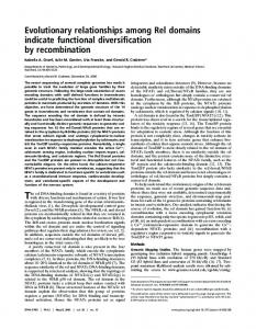
Figure 2. Schematic structures of two CBDs belonging to different families. A is the CBD of the enzyme cellobiohydrolase I from Trichoderma reesei (Kraulis et al., 1989) and B is the CBD from a exoglucanase / xylanase (Cex) from Cellulomonas fimi (Xu et al., 1995). Both CBDs have a similar function, but their structures are completely different. The Cex CBD has a β-barrel fold and the CBHI CBD has a irregular triple stranded β-sheet as the major secondary structure element. Both structures are drawn with the same absolute scale. The figures were made with the program Molscript (Kraulis, 1991).
12
Even before the two-domain structure of cellulases had become established, the relation between adsorption and hydrolytic activity had been realized. A rule stating that “the better the binding, the better the hydrolysis”, has been proposed as a general guideline (Klyosov, 1990). The rule seems to hold in many cases, for example in the above mentioned case when the CBD was mutated to decrease binding. Also when comparing enzymes from different origin, the correlation between binding and activity has generally been found to be true. An interplay between the two domains during hydrolysis has also been suggested. (Shen et al., 1991, Srisodsuk et al., 1993, Wilson et al., 1995). When the length of the linker region between the domains is changed, an effect on both the activity and binding of the enzyme is seen. This indicates that the domains act in concert, and that the spatial separation between the domains plays an important part in the mechanism of the enzyme. The two domain structure of cellulases is shared by several other solid-substrate degrading enzymes as well. Chitinases and raw starch degrading enzymes are examples of this. Mutagenesis and truncation of these enzymes in most cases give the same effects as seen in cellulases, i.e. decreased activity on solid substrates but unaffected activity on soluble substrates (Beintema, 1994, Coutinho and Reilly, 1994, Blaak and Schrempf, 1995, Iseli et al., 1993, Watanabe et al., 1992). CBDs are not only found in cellulases, but in some hemicellulose degrading enzymes as well (Gilkes et al., 1988, Doi et al., 1994, Margolles-Clark et al., 1996, Millward-Sadler et al., 1994). The CBDs do not in the absolute majority of the cases have affinity for the hemicellulose substrate, nor do the catalytic domains generally have any appreciable activity on cellulose. One potential reason for the occurrence of the CBDs in these enzymes is that they would be directed to any exposed part of cellulose and in this way further free it from hemicellulose for attack by cellulases. In a few cases it has been reported that xylan-binding domains exist in xylanases (Irwin et al., 1994, Black et al., 1995) There is evidence of additional effects of CBDs as well. In one study it was shown that addition of free CBD to a hydrolysis mixture could increase the activity of a cellulase. The effect was ascribed to the release of small particles from the cellulose and as a result the surface would be more accessible and hydrolyzable (Din et al., 1994). That a CBD can disrupt aggregates of cellulose has been noted also by changes in appearance and sedimentation rates of cellulose suspensions when CBD has been added. In general, a clear rationalization of these results has been difficult since different families of CBDs could give different effects and also different preparations of cellulose can differ greatly (Klyosov, 1990, Gilkes et al., 1993, Henrissat, 1994, Brun et al., 1995). The study of cellulases has also resulted in an novel possibility for application. Since CBD bind specifically to cellulose they can be used as affinity tags for specific immobilization. A fusion protein can be constructed by recombinant techniques where a CBD is linked to another domain such as an antibody fragment. This recombinant protein will then have the capability of binding to cellulose. By this function it can be both purified and immobilized. Cellulose is in
13
several ways advantageous for use as a carrier material. It is fairly inexpensive and it is widely recognized as a safe material for use in pharmaceutical or food processing applications. In addition it can be easily disposed (Ong et al., 1989, Reinikainen et al., 1996, Bayer et al., 1994).
1.2 AIMS OF THE PRESENT STUDY In this thesis the focus has been on studying the isolated CBDs of the fungal type, and especially the CBHI CBD. Specifically the following points were of major interest: 1. To determine side chains and regions of the CBD which are involved in the binding to cellulose. 2. To study general aspects of the binding, especially the reversibility and the effect of linkage of domains. The experimental part of thesis consists of four publications. In the first and second publication the mechanism of adhesion to cellulose was studied by amino acid substitutions. In this way several residues involved in th binding were identified. In the third publication the two-domain structure of cellulases was studied by mimicking it in a fusion protein consisting of two CBDs linked together by a linker region similar to that of cellulases. Finally, in the fourth publication, the reversibility of the binding and the exchange rate of the CBD on the cellulose surface were studied.
14
2 MATERIALS AND METHODS 2.1 PRODUCTION AND PURIFICATION OF SYNTHETIC PEPTIDES In publications I and II the CBDs used were produced by automated solid phase peptide synthesis using Fmoc chemistry. After each coupling step the unreacted amino groups were capped with acetic anhydride. This allowed the use of immobilized metal affinity chelating chromatography (IMAC) to be used in the first purification step (Lindeberg et al., 1991). Oxidation of cysteines to form disulfide bridges was performed by mixing peptide with reduced glutathione at pH 7.5 overnight at 4 °C. The final purification was performed by preparative reversed phase HPLC. The purified peptides were lyophilized and characterized by analytical reversed phase HPLC, mass-spectroscopy and amino acid analysis.
2.2 BINDING ASSAYS Lyophilized peptide was dissolved in 50 mM acetate buffer pH 5.0 containing 50 mM NaCl. The concentration of the stock solutions were determined by UV adsorption at 280 nm using extinction coefficients determined by amino acid analysis. The stock solutions were diluted with the same buffer and mixed with an equal volume of cellulose suspension. Variations to this basic assay are detailed in publication IV. Some different types of cellulose were used in the assays. In publications I, III and IV bacterial microcrystalline cellulose (BMCC) was used. Tunicin was used in publications II and III. In addition to these, Avicel and chitin were used in publication III. The adsorption was stopped by centrifugation and removal of the supernatant. In manuscript IV filtration was used. The concentration of unbound peptide was quantified by analytical reversed phase chromatography, and in publication IV also by scintillation counting.
2.3 EXPRESSION IN ESCHERICIA COLI The CBDs used in publications III and IV were produced in E. coli. The recombinant protein was a double CBD consisting of the CBD of T. reesei cellobiohydrolase II (CBHII) linked to the CBHI CBD. This was done by fusing the coding region for the first 41 residues in CBHII to the coding region of the last 57 residues of CBHI. This sequence was preceded by the pelB signal sequence of Erwina carotovora. All DNA manipulations were performed using standard protocols (Sambrook et al., 1989). The protein was secreted into the culture medium and purified by precipitation of contaminating proteins by MgSO4 addition, chromatography on a hydrophobic interaction column and finally purification by preparative reversed phase chromatography. The pure protein was then lyophilized. To obtain the individual CBDs, the double CBD was hydrolyzed in the middle of the linker region by trypsin. The identity and purity of the three
15
products were verified by amino acid analysis, analytical reversed phase chromatography and mass-spectroscopy.
2.4 TRITIUM LABELING OF THE CBHI CBD BY REDUCTIVE METHYLATION In publication IV a 3H labeled CBD was used. The labeling was performed by reductive methylation. In the reaction, dilute formaldehyde forms a Schiff base with free amines in the protein. The Schiff base is then reduced specifically with tritiated NaCNBH3 (Jentoft and Dearborn, 1979). The amine thus becomes methylated, with 3H added to some of the methyl groups. Any 3H added to the nitrogen is rapidly exchanged. In the CBHI CBD obtained from the double CBD by proteolysis there is one free amine group. This is located at the N-terminal proline residue, and thus it is a secondary amine. Thus only one methyl group was added to each CBD. The methyl group adds to the protruding region of the linker and is therefore 11 amino acids from the folded part of the protein. By mass spectroscopy it was confirmed that only one methyl group was added to each CBD and that all of the CBD had reacted. Adsorption isotherms of labeled and nonlabeled CBD showed that the methylation did not affect the binding properties.
2.5 THEORETICAL TREATMENT OF BINDING ISOTHERMS There has been much controversy about how the isotherms of cellulase adsorption to cellulose should be analyzed. Factors such as potential irreversibility (Beldman et al., 1987) or overlapping binding sites (Gilkes et al., 1992, Sild et al., 1996) have been mentioned as factors making conventional Langmuir models inappropriate. In addition it has proved very difficult to determine the saturation levels for the adsorption. In absence of this data it is impossible calculate any absolute values for the binding parameters. In this work most analysis relied on the initial slopes of the isotherms. When comparing very similar molecules, such as analogues with single amino acid substitutions, the ratio of the initial slopes are related to the difference in free energy of binding by the following thermodynamic formula: ∆G = -RT ln (K1/K2) where ∆G is the difference in free energy, R is the universal gas constant, T is the temperature , K1 and K2 are the initial slopes which are compared. In the figures with adsorption isotherms the lines were drawn using a one site Langmuir model, or if the fit was not acceptable a two site model was used.
16
3 RESULTS AND DISCUSSION 3.1 THE CELLULOSE BINDING FACE Sequence comparison of homologous CBDs shows that they contain several conserved residues and among them, four positions where the aromatic nature of the residues is strictly conserved (Figure 3). It has also been noted from structures of protein carbohydrate complexes, that aromatic residues frequently are involved in these types of interactions (Vyas, 1991, Quiocho, 1993). The structure determination of the CBHI CBD gave the first clues of how the CBD might bind to cellulose (Kraulis et al., 1989). From the structure it was noted that three of the conserved residues (Y5, Y31 and Y32) were aligned at the same face of the peptide. This led to the suggestion that this face probably was involved in the binding. In Figure 4 the structure is shown and the nomenclature explained. After the structure was solved it was shown that mutation of Y31 on the flat face resulted in a decrease in binding of the protein (Reinikainen et al., 1992). In this work the intact protein produced by heterologous expression in yeast was used. However, in the same work a mutation at the opposite, rough face, caused a similar decrease in binding. Thus it was not possible to rule out that both faces were involved in th binding. Later it was shown that the effect seen on the rough face was due to overglycosylation of the linker by the heterologous host (Reinikainen et al., 1995).
Figure 3. Sequences of the CBDs of the four major cellulases from T. reesei. Several residues show strict conservation, indicating structural or functinal importance. Sequences are from: CBHI (Shoemaker et al., 1983), CBHII (Teeri et al., 1987), EGI (Penttilä et al., 1986) and EGII (Saloheimo et al., 1988).
17
Figure 4. Alpha carbon trace of the CBHI CBD (Kraulis et al., 1989). The molecule has two distinct faces: the one containing the three tyrosines is called the “flat face” and the opposite face is called the “rough face”. In A all side chains changed in this study are indicated. In B only the sidechins at the flat face which are suggested to be involved in the binding are shown. The figure was made with the program Molscript (Kraulis, 1991).
18
In the work described in publication I, amino acid substitutions in synthetic analogues of the CBDs were used to study the role of individual amino acids. The analogues were produced by solid phase synthesis and were of the same size as the one which had been used for the structural determination. Earlier it had been shown that the synthetic analogue does not differ from the wild type in its binding properties (Johansson et al., 1989). The following substitutions were made: Y5A, Y31A, Y32A, N29A, Q34A and P16R. The first five changes were chosen since these residues occurred as conseved residues in sequence alignments as well as on the basis of their common position on the flat face. The last one was chosen because it creates a major change on the rough face and was the same as had been tested earlier and then affected the binding (Reinikainen et al., 1992). The effect of the substitutions on the structure of the CBD was investigated by two dimensional NMR. Three different parameters of the backbone protons were investigated: the changes in chemical shifts, nuclear Overhauser effects (NOEs) and coupling constants. Together these parameters gave information by which it was possible in a qualitative way to describe how the backbones had changed. The information obtained in this way is not as informative as complete structures would have been, but solving all the structures would have been a very time consuming and tedious task. A general trend noticed was that the N-terminus in general was more sensitive to mutations than other parts. This was especially evident in the case of the peptide Y5A were it was suspected that much of the tertiary structure might have been disrupted. Surprisingly, it was found that most of the structural changes in P16R also occurred in the N-terminus. Otherwise, there were indications of only small structural changes, indicating that the fold of the peptide is very stable. Especially the peptide Y31A showed only very small changes. The binding isotherms for each peptide was determined by using HPLC for quantification. The largest effects of the substitutions was observed for the Tyr residues on the flat face. Both Y5A and Y32A lost all their affinity while some affinity was retained by Y31A. The finding is in accordance with the structural data where the disruption of the back bone was greater for both Y5A and Y32A than for Y31A. Determining the role of both Gln-34 and Asn-29 was more difficult. However, by calculating the decrease in binding compared to the wildtype it was noted that the free energy associated with it was comparable to what generally is thought to be the contribution of a hydrogen bond (Fersht et al., 1985). It was therefore concluded that it is likely that these residues are involved in the binding, particularily Gln-34 which gave the larger effect. On the basis of the small change caused by the rough face substitution, P16R, and taking into account the structural disruption in the peptide, it was concluded that it was unlikely that the rough face was involved in the binding. In publication II the same line investigation was continued. Based on previous work there was reason to believe that the CBD of endoglucanase I (EGI) binds to cellulose with a higher affinity than the CBHI CBD (Srisodsuk, 1994). This was shown to be the case in publication II by using synthetic analogues of both CBDs. Interestingly it has not been possible to obtain the EGI CBD by limited proteolysis of the wild-type enzyme as is the case with CBHI. The two CBDs differ in nine
19
positions out of 36. Of these only one, corresponding to Tyr-5 in CBHI CBD, is directly on the flat face (Figure 4). Two of the residues, Leu-28 and Pro-30, are on the border between the faces and the rest of the substitutions are on the rough face. In addition there is a third disulfide bridge in the EGI CBD. There is in principle two ways in which the amino acid substitutions could affect the affinity. Firstly, the rough face substitutions could change the solubility of the peptide and thus affect the entropy factor of the free energy of binding. The third disulfide bridge might have a similar effect. Alternatively the substitutions at or near the binding face could contribute with new van der Waals forces, hydrogen bonds, chargecharge interactions or hydrophobic interactions which in a more direct way would increase the affinity. The basis of the difference in affinity was studied by a set of analogues to the CBHI CBD. The idea was to change amino acids in small groups in the CBHI CBD to make it resemble the EGI CBD. In this way it would be possible to pinpoint the residue(s) responsible for the difference in affinity. The following four peptides were made: T17K:V18T:A20T, V27Y:L28S:P30D, Y5W and P30D. Two of the analogues contained three amino acid substitutions each. These were chosen because sequence comparison showed that these changes occurred in groups in several other analogous sequences. In the peptide T17K:V18T:A20T all changes were at the rough face. Since two hydrophobic residues were made hydrophilic and one hydrophilic residue was exchanged with a charged residue, the substitutions should markedly change the character of the rough face. In the peptide V27Y:L28S:P30D some changes near the binding face were done. One aromatic and one charged residue were added. Both could potentially contribute either directly or indirectly to the affinity. In the peptide Y5W the only differing residue clearly on the flat face was changed. Finally in P30D a charge near the flat face was added. This substitution is interesting since in most homologous sequences the residue is either proline or aspartic acid. The effects of the substitutions were investigated by determining binding isotherms. The peptide Y5W had an affinity midway between those of the CBHI and EGI CBDs. The three other analogues had affinities slightly less than that of the CBHI CBD. The results showed that the tryptophan at position 5 can contribute more to the interaction with cellulose than can the tyrosine. The basis of this difference is not known but it could be that the larger aromatic ring of the tryptophan can participate more efficiently in charge transfer interactions. Alternatively the higher hydrophobicity of the tryptophan may make the binding more efficient. The substitutions on the rough face did also strengthen the previous conclusion that it does not participate in the binding. Especially in the T17K:V18T:A20T peptide was the rough face substantially changed. The fact that the CBHI enzyme as a whole could increase its affinity by a single mutation presents some interesting questions. Why has evolution not provided CBHI with a CBD having a higher affinity? If it would have provided a selective advantage, the mutation would certainly easily have occurred. Apparently the affinity is carefully
20
balanced to perform optimally together with the catalytic domain and / or for the performance of the enzyme in the synergistic mixtures secreted by the cellulolytic organism.
3.2 THE DOUBLE CBD The third publication describes the heterologous production of CBDs in E. coli as recombinant double CBDs. A basic goal of this work was to investigate how a recombinant protein consisting of only two CBDs linked together by an extended linker would behave. This was also the first attempt to produce CBDs by heterologous expression in E. coli. By the expression and purification system it was possible to produce pure double CBD as well as its two CBD components. The most striking difference in binding is that the double CBD had a markedly higher affinity than either of the two components. The physical explanation suggested for the binding increase is based on an interplay between the two domains (Figure 5). When the first domain binds to cellulose it has the affinity of an isolated domain. For the binding of the second domain the situation is very different. Its concentration near the surface is very much increased due to the linking, which in turn can increase the binding (Jencks, 1981). This domaininterplay suggested how the linkage of domains in the wild-type cellulases might affect their function.
Figure 5. Schematic representation of how the double CBD is suggested to bind to cellulose and how the affinity is increased. In the first step either domain binds with the same affinity as the isolated CBD. In the second step the the other CBD binds with an affinity which is determined by the linker properties and the binding site arrangement on the cellulose surface. The overall effect is seen as an increased affinity.
21
The effect of substrate was investigated by using the following four different types of substrate: BMCC, tunicin, Avicel and chitin. BMCC is of bacterial origin and tunicin is of animal origin. Both substrates performed well in the tests. They were easy to handle and gave reproducible results. Surprisingly their adsorption capacities were almost identical despite their different origin. Avicel is a commercial product based on wood cellulose. It is widely used in the pharmaceutical and food industry as a filling material. Using this substrate it can be very difficult to achieve reproducible results. In addition, the effect of time and temperature was much bigger than for BMCC and tunicin. The reason for the observed effect is probably due to the structure of Avicel. It is basically crystalline but each particle is made up of aggregates which break up during treatment, incorrectly suggesting that the effect is in the binding itself. In all it is a very poor substrate to use for this type of study. Chitin is also a very commonly occurring polysaccharide. It is found in the exoskeleton of many animals as well as for example in the hyphae of fungi and cell wall of yeast. It differs chemically from cellulose only in one respect. The basic monomer is N-acetyl glucosamine instead of glucose. The acetyl group at the C2 amine does allow the chains to pack in a very similar crystalline structure as in cellulose. Only on this substrate did the CBHI and CBHII CBDs show a significant difference in affinity. This difference could be due to a charge-charge interaction which is possible for CBHII, but not for CBHI since at position 30 the CBHI CBD has a proline whereas in the CBHII CBD the corresponding amino acid is an aspartic acid. Nevertheless, it is clear that the CBHI CBD does have some affinity since the increased binding of the double CBD could also be seen for this substrate. In addition to the above mentioned substrates, the binding to mannan and xylan was measured. Only very small amounts bound to either of the substrates.
3.3 THE REVERSIBILITY OF BINDING In publication IV a new method of detection was applied to follow the binding of the CBHI CBD produced in E. coli. The detection relied on incorporating a 3H label by reductive methylation into the CBD. The radioactive label allowed the binding to be followed much easier than had been possible before. One other main advantage was that bovine serum albumin (BSA) could be added to the reaction mixtures to prevent non-specific adsorption of the protein. This made it possible to detect nM concentrations without difficulty. When using HPLC for quantification it was not possible to prevent non-specific adsorption by BSA addition or similar, and only concentrations in the µM range were reliably detected. The improvement in detection was therefore 1000-fold. The main focus of the work was to study the (ir)reversibility of the binding. It has long been known that once cellulases have adsorbed to cellulose, quite harsh conditions (i.e., 6M guanidium hydrochloride) are required to elute them (Reese, 1982). Since the catalytic domain can be eluted, this led to the suggestion that the binding of the CBD is irreversible or partially irreversible (Henrissat, 1994, Nidetsky et al., 1994, Beldman et al., 1987).
22
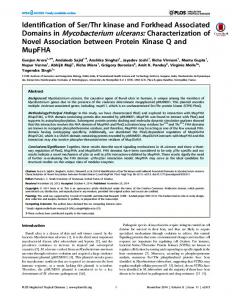
Figure 6. Part of an adsorption isotherm for 3H labeled CBHI CBD on BMCC ( ). In a dilution experiment several samples which had reached equilibrium were diluted and then filtered at different time points. In this way the disruption of equilibrium and the subsequent establishment of a new equilibrium could be followed (∇). This result shows that the binding is reversible and that the “ascending” isotherm is the same as the “descending” isotherm (Norde and Haynes, 1995). For understanding the mechanism of how cellulases work on the solid cellulose it is of course of fundamental importance to determine if they are reversibly or irreversibly adsorbed. In a simple experiment it was possible to show that by simply diluting the sample the CBD could be desorbed and would reach a new equilibrium position (Figure 6). In an attempt to measure the amount of bound CBD directly from the cellulose it was noted that during the wash step the CBD eluted so quickly that it was not possible to perform the analysis accurately in this way. The exchange rate of the CBD at the cellulose surface was calculated in an experiment where bound non-labeled CBD was competed with labeled CBD under conditions where equilibrium was maintained throughout the experiment. The same experiment was then conducted in the reverse order. The exchange rate was found to be temperature dependent, as would be expected. The exchange rate was similar to the hydrolysis rate for CBHI on cellotetraose. A comparison of the rates suggests that the CBD binding would not be rate limiting for the movement of a processive enzyme on the cellulose surface.
23
4 CONCLUSIONS CBDs form an essential part of cellulases in their hydrolysis of cellulose. Understanding their function is of fundamental imporance for the rational utilization of cellulases. The CBDs also form ideal affinity tags for immobilization of recombinant proteins to cellulose. The results described in this thesis provide a deeper understanding of the function of the fungal type CBDs than was previously possible. Determining the amino acids involved in the binding to cellulose is the first step in understanding the molecular details of the interaction. The results in this work indicate that the binding is specific for sites on the cellulose and dependent on the correct positioning of the involved side chains. The finding that CBDs of different origin have different affinities suggests that the affinity of a CBD is balanced for working together with the catalytic domain and / or with the whole mixture of secreted cellulases. It was also shown that in a protein with two domains linked together by an extended linker, the linking itself can markedly change the apparent behavior of the protein as compared to its individual parts. This is important to keep in mind when drawing conclusions of cellulase function based on observations of individual domains. Finally, the demonstration of reversibility and the calculation of exchange rates of CBD on cellulose is crucial for understanding how cellulases can move on the substrate surface. It allows for a model where CBHI can move processively along cellulose chains, contrary to what earlier has been suggested.
24
REFERENCES Bayer, E., Morag, E. and Lamed, R. (1994). The cellulosome – a treasure trove for biotecnology. Trends Biotechnol. 12, pp. 379 - 386. Béguin, P. and Aubert, J.P. (1994). The biological degradation of cellulose. FEMS Microbiol. Rev. 13, pp. 25 - 58. Beintema, J.J. (1994). Structural features of plant chitinases and chitin-binding proteins. FEBS Lett. 350, pp. 159 - 163. Beldman, G., Voragen, A.G.J. Rombouts, F.M., Searle-van Leeuwen, M.F. and Pilnik, W. (1987). Adsorption and kinetic behavior of purified endoglucanases from Trichoderma viride. Biotechnol. Bioeng. 30, pp. 251-257. Bhikhabhai, R., Johansson, G. and Pettersson, G. (1984) Cellobiohydrolase from Trichoderma reesei: internal homology and prediction of secondary structure. Int. J. Peptide Protein Res. 25, pp. 368 - 374. Blaak, H. and Schrempf, H. (1995). Binding and substrate specificities of a Streptomyces olivaceoviridis chitinase in comparison with its proteolytically processed form. Eur. J. Biochem. 229, pp. 132 - 139. Black, G., Hazlewood, G., Millward-Sadler, S., Laurie, J. and Gilbert, H. (1995) . A modular xylanase containing a novel non-catalytic xylan-specific binding domain. Biochem. J. 307, pp. 191 - 195. Brun, E., Gnas, P., Marion, D. and Barras, F. (1995). Overproduction, purification and characterization of the cellulose-binding domain of the Erwina chrysamthemi secreted endoglucanase EGZ. Eur. J. Biochem. 231, pp. 142 - 148. Coutinho, J.B. and Reilly, P. (1994). Structure-function relationships in the catalytic and starch binding domains of glucoamylase. Prot. Eng. 7, pp. 393 - 400. Denman, S., Xue, G.P. and Patel, B. (1996). Charachterization of a Neocallomastix patriciarum cellulase cDNA (CelA) homologous to Trichoderma reesei cellobiohydrolase II. Appl. Environ. Microbiol. 62, pp. 1889 - 1896. Din, N., Damude, H., Gilkes, N.R., Miller, J.C., Warren, R.A.J. and Kilburn, D.G. (1994). C1-Cx revisited: intramolecular synergism in a cellulase. Proc. Natl. Acad. Sci. USA 91, pp. 11383 - 11387.
25
Doi, R., Goldstein, M., Hashida, S., Park, J.S. and Takagi, M. (1994). The Clostridium cellulovorans cellulosome. Crit. Rev. Microbiol. 20, pp. 87 - 93. Fersht, A., Shi, J.P., Knill-Jones, J., Lowe, D., Wilkinson, A., Blow, D., Brick, P., Carter, P., Waye, M. and Winter, G. (1985). Hydrogen bonding and biological specificity analysed by protein engineering. Nature 314, pp. 235 - 238. Gilkes, N.R., Warren, R.A.J., Miller, R.C. and Kilburn, D.G. (1988). Precise excision of the cellulose binding domains from two Cellulomonas fimi cellulases by a homologous protease and the effect on catalysis. J. Biol. Chem. 263, pp. 10401 - 10407. Gilkes, N.R., Henrissat, B., Kilburn, D.G., Miller, R.C. and Warren, R.A.J. (1991). Domains in microbial β-1,4-glycanases: Sequence conservation, function, and enzyme families. Microbiol. Rev. 55, pp. 303 - 315. Gilkes, N.R., Jervist, E., Henrissat, B., Tekant, B., Miller, R.C., Warren, R.A.J. and Kilburn, D.G. (1992). The adsorption of a bacterial cellulase and its two isolated domains to crystalline cellulose. J. Biol. Chem. 267, pp. 6743 - 6749. Gilkes, N.R., Kilburn, D.G., Miller, R.C., Warren, R.A.J., Sugiyama, J., Chanzy, H. and Henrissat, B. (1993). Visualization of the adsorption of a endo-β-1,4glucanase and its isolated cellulose-binding domain to crystalline cellulose. Int. J. Biol. Macromol. 15, pp. 347 - 351. Henrissat, B. (1994). Cellulases and their interaction with cellulose. Cellulose 1, pp. 169 - 196. Hon, D. (1994). Cellulose: a random walk along its historical path. Cellulose 1, pp. 1 - 25. Irwin, D., Jung, E. and Wilson, D. (1994). Characterization of a Thermonospora fusca xylanase. Appl. Environ. Microbiol. 60, pp. 763 - 770. Iseli, B., Boller, T. and Neuhaus, J.M. (1993). The N-terminal cysteine rich domain of tobacco class I chitinase is essential for chitin binding but not for catalytic or antifungal activity. Plant Physiol. 103, pp. 221 - 226. Jencks, W.P. (1981). On the attribution and addititivity of binding energies. Proc. Natl. Acad. Sci. USA 78, pp. 4046 - 4050. Jentoft, N. and Dearborn, D.G. (1979). Labeling of proteins by reductive methylation using sodium cyanoborohydride. J. Biol. Chem. 254, pp. 4359 - 4365.
26
Johansson, G., Ståhlberg, J., Lindeberg, G., Engström, Å. and Pettersson, G. (1989). Isolated fungal cellulase terminal domains and a synthetic minimum analogue bind to cellulose. FEBS Lett. 243, pp. 389 - 393. Klyosov, A. (1990). Trends in biochemistry and enzymology of cellulose degradation. Biochemistry 29, pp. 10577 - 10585. Kraulis, P., Clore, M., Nilges, M., Jones, T.A., Pettersson, G., Knowles, J. and Gronenborn, A. (1989). Determination of the three-dimensional solution structure of the C-terminal domain of cellobiohydrolase I from Trichoderma reesei. Biochemistry 28, pp. 7241 - 7257. Kraulis, P. (1991). Molscript: a program to produce both detailed and schematic plots of protein structures. J. Appl. Cryst. 24, pp. 946 - 950. Lindeberg, G., Bennich, H. and Engström, Å. (1991). Purification of synthetic peptides: Immobilized metal ion affinity chromatography. Int. J. Pept. Protein Res. 38, pp. 253 - 259. Margolles-Clark, E., Tenkanen, M., Söderlund, H. and Penttilä, M. (1996). Acetyl xylan esterase from Trichoderma reesei contains an active-site serine residue and a cellulose-binding domain. Eur. J. Biochem. 237, pp. 553 - 560. Millward-Sadler, S.J., Poole, D., Henrissat, B., Hazlewood, G., Clarke, J.H. and Gilbert, H.J. (1994). Evidence for a general role for high-affinity non-catalytic domains in microbial plant cell wall hydrolases. Mol. Microbiol. 11, pp. 375 382. Nevalainen, H., and Penttilä, M. (1995). Molecular biology of cellulolytic fungi. In: Kuck, (Ed.). The Mycota II. Genetics and Biotechnology, Springer-Verlag, Berlin. Pp. 303 - 319. Nidetzky, B., Steiner, W., & Claeyssens, M. (1994). Cellulose hydrolysis by the cellulases from Trichoderma reesei: adsorptions of two cellobiohydrolases, two endocellulases and their core proteins on filter paper and their relation to hydrolysis. Biochem J. 303, pp. 817 - 823 Norde, W., and Haynes, C.A. (1995). Reversibility and the mechanism of protein adsorption. ACS Symp. Ser. 602, pp. 26 - 40. Ong, E., Greenwood, J., Gilkes, N., Kilburn, D.G., Miller, R.C. and Warren, R.A.J. (1989). The cellulose-binding domains of cellulases: tools for biotechnology. Trends. Biotech. 7, pp. 239 - 243. Penttilä, M., Lehtovaara, P., Nevalainen, H., Bhikhabhai, R. and Knowles, J. (1986). Homology between cellulase genes of Trichoderma reesei: complete nucleotide sequence of the endoglucanase I gene. Gene 45, pp. 253 - 263.
27
Quiocho, F. (1993). Probing the interactions between proteins and carbohydrates. Biochem. Soc. Trans. 21, pp. 442 - 448. Reese, E.T. (1982). Elution of cellulase from cellulose. Process Biochem. 17, pp. 2 - 6. Reinikainen, T., Ruohonen, L., Nevanen, T., Laaksonen, L., Kraulis, P., Jones, T.A., Knowles, J.K.C. and Teeri, T.T. (1992). Investigation of the function of mutated cellulose-binding domains of Trichoderma reesei cellobiohydrolase I. Proteins 14, pp. 475 - 482. Reinikainen, T., Teleman, O. and Teeri, T.T. (1995). Effects of pH and high ionic strength on the adsorption and activity of native and mutated cellobiohydrolase I from Trichoderma reesei. Proteins 22, pp. 392 - 403. Reinikainen, T., Takkinen, K. and Teeri, T.T (1996). Comparison of the adsorption properties of a single-chain antibody fragment fused to a fungal or a bacterial cellulose-binding domain. Enzyme Mictob. Technol. In press. Saloheimo, M., Lehtovaara, P., Penttilä, M., Teeri, T.T., Ståhlberg, J., Johansson, G., Pettersson, G., Clayssens, M., Tomme, P. and Knowles, J.K.C. (1988). EGIII, a new endoglucanase from Trichoderma reesei: characterization of both the gene and enzyme. Gene 63, pp. 11 - 21. Sambrook, J., Fritsch, E.F. and Maniatis, T. (1989). Molecular Cloning: A Laboratory Manual. 2nd edition. Cold Spring Harbor Laboratory, Cold Spring Harbor, NY. Shen, H., Schmuck, M., Pilz, I., Gilkes, N., Kilburn, D., Miller, R.C. and Warren, R.A.J. (1991). Deletion of the linker connecting the catalytic and cellulosebinding domains of endoglucanase A (CenA) of Cellulomonas fimi alters its conformation and catalytic activity. J. Biol. Chem. 17, pp. 11335 - 11340. Shoemaker, S., Schweickart, V., Lander, M., Gelfand, D., Kwok., S., Myambo, K. and Innis, M. (1983). Molecular cloning of exo-cellobiohydrolase I derived from Trichoderma reesei strain L27. Biotechnology 1, pp. 691 - 696. Sild, V., Ståhlberg, J., Pettersson, G. and Johansson, G. (1996). Effect of potential binding site overlap to binding of cellulase to cellulose: a two-dimensional simulation. FEBS Lett. 378, pp. 51 - 56. Srisodsuk, M., Reinikainen, T., Penttilä, M. and Teeri, T.T. (1993). Role of the interdomain linker peptide of Trichoderma reesei cellobiohydrolase I in its interaction with crystalline cellulose. J. Biol. Chem. 268, pp. 20756 - 20761. Srisodsuk, M. (1994). Mode of action of Trichoderma reesei cellobiohydrolase I on crystalline cellulose. VTT Publications 188. Technical Research Centre of Finland, Espoo, Finland. 107 p.
28
Ståhlberg, J., Johansson, G. and Pettersson, G. (1991). A new model for enzymatic hydrolysis of cellulose based on the two-domain structure of cellobiohydrolase I. Biotechnology 9, pp. 286 - 290. Teeri, T.T., Lehtovaara, P., Kauppinen, S., Salovuori, I. and Knowles, J.K.C. (1987). Homlogous domais in Trichoderma reesei cellulolytic enzymes: gene sequence and expression of dellobiohydrolase II. Gene 51, pp. 42 - 52. van Tilbeurgh, H., Tomme, P., Claeyssens, M., Bhikhabhai, R. and Pettersson, G. (1986). Limited proteolysis of the cellobioiohydrolase I from Trichoderma reesei. FEBS Lett. 204, pp. 223 - 227. Tomme, P., van Tilbeurgh, H., Pettersson, G., Van Damme, J., Vandekerckhove, J., Knowles, J., Teeri, T.T. and Clayssens, M. (1988). Studies on the cellulolytic system of Trichoderma reesei QM 9414. Eur. J. Biochem. 170, pp. 575 - 581. Tomme, P., Warren, R.A.J., Miller, R.C., Kilburn, D.G., Gilkes, N.R. (1995). Cellulose-binding domains: Classification ans properties. ACS Sym. Ser. 618, pp. 143 - 163. Vyas, N. (1991). Atomic features of protein-carbohydrate interactions. Curr. Opin. Struct. Biol. 1, pp. 737 - 740. Watanabe, T., Oyanagi, W., Suzuki, K., Ohnishi, K., and Tanaka, H. (1992). Structure of a gene encoding chitinase D of Bacillus circulans WL-12 and possible homology of the enzyme to other procaryotic chitinases and class III plant chitinases. J. Bacteriol. 174, pp. 408 - 414. Wilson, D., Spezio, M., Irwin, D., Karplus, A. and Taylor, J. (1995). Comparison of enzymes catalyzing the hydrolysis of insoluble polysaccharides. ACS Sym. Ser. 618, pp. 1 - 12. Xu, G.Y., Ong, E., Gilkes, N., Kilburn, D., Muhandiram, D.R., Harris-Brandts, M., Carver, J., Kay, L. and Harvey, T. (1995). Solution structure of a cellulosebinding domain from Cellulomonas fimi by nucleas magnetic resonance spectroscopy. Biochemistry 34, pp. 6993 - 7009.
Appendices of this publication are not included in the PDF version. Please order the printed version to get the complete publication (http://www.inf.vtt.fi/pdf/publications/1996/)
29