Materials Science, Vol. 20, No. 1, 2002
Structure, spectroscopy and dielectric properties of BaTiO3:Eu3+ nanocrystallites prepared by the sol–gel method* D. HRENIAK1, E. ŁUKOWIAK1, K. MARUSZEWSKI1, 2**, R. PĄZIK3, W. STRĘK1 1
Institute for Low Temperature and Structure Research, Polish Academy of Sciences, Wrocław, Poland 2
Mechanical Faculty, Institute of Materials Science and Applied Mechanics, Wrocław University of Technology, Wrocław, Poland 3
Chemistry Department, Opole University, Opole, Poland
Eu3+-doped BaTiO3 nanocrystalline powders have been obtained by sol–gel method. Their morpho logy, structure and dielectric properties have been investigated as a function of sintering temperature. The powders sintered at the temperatures below 800 °C demonstrate luminescence behaviour characteristic of the inversed symmetry Eu3+ sites, where only the 5D0 → 7F1 transitions are allowed. Above this temperature the system undergoes a phase transition characterized by lack of inversion symmetry enabling the dipole–electric transitions. Key words: barium titanate, europium, nanocrystallites, sol–gel processing, luminescence
1. Introduction Barium titanate (BaTiO 3) is a classical example of a ferroelectric crystalline material. The BaTiO 3 ceramics are widely applied in electronic devices as highpermittivity capacitors, infrared detectors or transducers. It is well known that ferroelectric and optical properties of ferroelectric ceramics depend on the grain size [1]. Recently, the sol–gel route has been suggested for preparation of transparent, nanostructured BaTiO3 monolithic xerogels [2]. The size-dependence of the ferroelectric phase transition of small BaTiO 3 particles has been a subject of many reports [3–10]. The ferroelectric crystal size of about 50 nm has been reported [7] as critical for ferroelectric properties. __________ *
The paper presented at the International Conference on Sol-Gel Materials, SGM 2001, Rokosowo, Poland. Corresponding author, e-mail:
[email protected].
**
PDF created with FinePrint pdfFactory trial version http://www.fineprint.com
44
D. HRENIAK et al.
In this paper, we present the preliminary studies of structural, optical and diele ctric properties of BaTiO 3 nanocrystallites (nc-BaTiO 3) doped with Eu3+ ions. They were prepared by the modified sol–gel method. It is well known that for crystal structure evaluation of crystalline materials X-ray powder diffraction (XRD) is used quite often. However, in the case of nc-BaTiO 3 it is not a sensitive enough technique to unravel subtle structure characteristics existing in the nanoscale range. It has been reported that XRD measurements were used only to probe global structure for BaTiO 3 crystallites [8]. However, there exist methods which are more suitable for investig ation of local structure and symmetry changes: combination of XRD and Raman scattering [7, 8] or infrared spectroscopies [11], measurements of the second harmonic-generation [8, 12] and photoluminescence studies [13]. We employed the europium(III) ion as an optical probe for structural investigations and detection of phase transitions in the nanocrystalline grains. The fluorescence properties of the europium(III) ion were investigated as a function of the sintering temperature. The basic dielectric properties of Eu 3+: BaTiO3 nanocrystalline powders were determined.
2. Experimental 2.1. Preparation The europium-doped BaTiO3 crystalline powders were prepared by the sol–gel method similar to that described by Tian et al. [14]. Barium acetate Ba(CH3COO)2 (Aldrich 99%), titanium butoxide Ti(OC4H9)4 (Alfa Aesar 99+%) and europium oxide Eu2O3 (Koch-Light Laboratories 99.99%) were used as starting materials. Acetyl acetone C5H8O2 (Aldrich 99+%) and acetic acid (POCh p.a.) were selected as solvents of titanium butoxide and barium acetate, respectively. Europium chloride EuCl 3⋅6H2O was obtained by reacting stoichiometric amount of europium oxide with hydrochl oride acid of analytical grade (POCh). Dissolved barium acetate was added dropwise to titanium butoxide solution under stirring. The obtained solutions were vigorously stirred at 50 °C for about 2 h. The europium salt was dissolved in small amount of water and added slowly to the obtained transparent, yellow sol with 1 molar percent of Eu vs. BaTiO3. The obtained solution was heated at approximately 100 °C for 24 h to form barium titanate (BT) gel powders. The samples of crushed gels were heated at 750 °C (A), 800 °C (B), 900 °C (C), 1100 °C (D) and 1200 °C (E). Pure, undoped samples of nc-BaTiO3 were obtained in a similar manner. 2.2. Apparatus Fluorescence spectra were recorded on an Ocean Optics SD 2000 spectrophotometer. Decay curves of luminescence were recorded with a Jobin-Yvon TRW 1000 spectrophotometer equipped with a photomultiplier (Hamamatsu R928). XRD diffractograms were measured using CuKα source radiation on a DRON-3 powder
PDF created with FinePrint pdfFactory trial version http://www.fineprint.com
BaTiO3:Eu3+ nanocrystallites prepared by the sol–gel method
45
diffractometer. The mean crystallite diameters were estimated applying the Scherrer formula [15] to the X-ray diffraction patterns. Electrical properties were measured using computer-based apparatus for dielectric measurements in the frequency range 100–1 MHz.
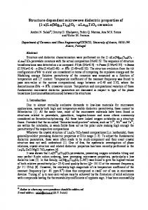
3. Results and discussion 3.1. XRD measurements The x-ray diffraction results of the undoped samples as a function of heating temperatures are shown in Fig. 1. The recorded patterns show that all the samples were well crystallized. The nanocrystalline particle sizes were estimated to be 24–30 nm for the samples A, B, C and about 43 nm for the samples D and E. Sharp peaks in all the recorded XRD patterns could be ascribed to pseudocubic paraelectric phases [16] which (probably) co-exist with the tetragonal ferroelectric BaTiO 3 phase [17] in the case of D and E.
tetragonal BaTiO3 (#JCPDS 05-0626) cubic BaTiO3 (#JCPDS 31-174)
E
normalized intensity
D C B A
30
35
40
45
50
55
60
2Θ(degree)
Fig. 1. XRD patterns of BaTiO3 heated at different temperatures
3.2. Luminescence The emission spectra of BaTiO 3:Eu3+ nanocrystallites prepared at different temperatures and measured at 300 K and 77 K are shown in Fig. 2. The spectra consist of characteristic bands attributed to the 5D0 → 7FJ (J = 0, 1–4) transitions. With increasing crystallite sizes the band with the maximum at 616 nm (the hypersensitive electric-dipole 5D0 → 7F2 transition) becomes much weaker as compared to the band with the maximum at 595 nm (assigned tentatively to the magnetic-dipole 5D0 → 7F1 transition). These changes are probably due to the variations in symmetry of the env i-
PDF created with FinePrint pdfFactory trial version http://www.fineprint.com
D. HRENIAK et al.
46
ronment of the Eu3+ ions and may be linked to the structural changes possible for the sample processed at temperatures above 1100 °C. λexc.=532 nm
Ba0.99Eu0.01TiO3
normalized intensity
5
300 K
a)
4 800 oC 900 oC 1100 oC
3 2 1 0 575
600
625
650
675
700
725
wavelength/nm
normalized intensity
2,0
b)
77 K 800 oC 900 oC 1100 oC
1,5 1,0 0,5 0,0 575
600
625
650
675
700
725
wavelength/nm Fig. 2. Emission spectra of Eu 3+-doped BaTiO3 powders prepared at different temperatures
Assuming the existence of the areas with the local tetragonal phase (what is in agreement with the XRD data) it can be assumed that the symmetry changes observed in the Eu3+ emission spectra are related to phase transitions occurring in those areas. The europium ions are positioned either inside of those spots. Changes in lumine scence spectra and stabilization of the tetragonal phase by Y [19] and Pr [20] ions have been reported for cubic ZrO 2. The emission spectra measured at room temperature are poorly resolved while the spectra measured at 77 K demonstrate a complex structure characteristic of the multi-site Eu 3+ spectra. 3.3. Decay time measurements We have measured the dependence of the emission decay times of the band attri buted to the 5D0 → 7F1 transition on the size of the BaTiO 3:Eu3+ grains. The examples of emission decays measured at 595 nm are shown in Fig. 3. As it can be seen the characteristic decay times increase systematically with increasing temperature. They
PDF created with FinePrint pdfFactory trial version http://www.fineprint.com
BaTiO3:Eu3+ nanocrystallites prepared by the sol–gel method
47
are shorter for BaTiO 3 sintered at lower temperatures and the emission decay profiles demonstrate clearly non-exponential character. The shorter decay times recorded for the samples heated at 800 °C and 900 °C may be due to the presence of small amounts of –OH groups in the crystallites leading to quenching of the Eu 3+ fluorescence. For the samples sintered at higher temperatures (1100 °C and 1200 °C) the emission decays are perfectly exponential and the estimated emission lifetimes are very long ( τ = 2.2 ms at 300 K and τ = 3.5 ms at 77 K).
normalized intensity
Ba0.99Eu0.01TiO3
λexc.= 532 nm, λrec.= 595 nm a)
τ = 2.2 ms
1100 oC 900 oC 800 oC
0.0 1.0m 2.0m 3.0m 4.0m 5.0m 6.0m 7.0m 8.0m 9.0m 10.0m
normalized intensity
time/s
b)
τ = 3.5 ms
77 K 0.0 1.0m 2.0m 3.0m 4.0m 5.0m 6.0m 7.0m 8.0m 9.0m 10.0m
time/s Fig. 3. Emission decay curves of Eu 3+–doped BaTiO3 powders prepared at different temperatures. The decays were obtained at 300 K (a) and at 77 K (b)
In our opinion, the characteristic non-exponential decay for samples treated at 800 °C and 900 °C is due to the aggregation of the Eu 3+ ions, which is more pronounced for smaller grains. Analogous results have been reported for Eu 3+-doped Lu2O3 nanocrystallites [18]. 3.4. Dielectric studies Dielectric studies have been performed in a wide temperature range. The electric permittivity εr' and dielectric loss tanδ have been measured at different frequencies for the undoped BaTiO3 powders sintered at 800 °C and 1100 °C. During the first
PDF created with FinePrint pdfFactory trial version http://www.fineprint.com
D. HRENIAK et al.
48
cycle of heating we have observed the maximum of εr' in the range 300–330 K. This maximum has not been observed in the second (Fig. 4) and successive cycles of heating. This effect results most likely from the material dehydration. There has been observed only a slight change of permittivity during further heating. During cooling of the sample we have observed monotonic decrease of permittivity. In this case we have not observed the maximum in the range of 300–330 K and only a weak temperature dependence of dielectric permittivity. Comparison between the room-temperature values of εr and tanδ at different frequencies is presented in Table 1. εr'
a) tanδ
0.06 b)
70
0.05 0.04
65
0.03 60
1 kHz 10 kHz 100 kHz 1 MHz
55
0.02 0.01 0.00
100 200 300 400 500 T/K
εr'
c)
0.14 d) tanδ
45
100 200 300 400 500 T/K
40
0.12 0.10 0.08
35
0.06 0.04 0.02
30 250 300 350 400 450 500 550 T/K
0.00 250 300 350 400 450 500 550 T/K
Fig. 4. Temperature-dependency of electric permittivity εr' and loss tangent tanδ of europium-doped BaTiO3 prepared at 800 °C (a, b) and at 1000 °C (c, d) measured at different frequencies
These results are distinctly different from those obtained for typical ferroelectric BaTiO3 which exhibits εr' in the range 103–104 [1]. Undoped samples demonstrated also the hysteresis loops characteristic of ferroelectric materials (Fig. 5), but the value of spontaneous polarization was estimated to be only 1.0⋅10–3 C⋅m–2. It is almost an order of magnitude smaller than that observed for tetragonal ferroelectric phase
PDF created with FinePrint pdfFactory trial version http://www.fineprint.com
BaTiO3:Eu3+ nanocrystallites prepared by the sol–gel method
49
BaTiO3 monocrystals (7.5⋅10–2 C⋅m–2) [1]. This fact indicates that the paraelectric phase dominates in undoped samples. Table 1. Room temperature values of εr' and tanδ at different frequencies Sample B (24 nm) εr' 68 67 66 65
Sample D (43 nm)
tanδ
εr'
tanδ
0.009 0.008 0.008 0.009
37 35 33 32
0.065 0.041 0.023 0.014
dielectric polarization/arb. units
1 10 100 1000
dielectric polarization
Frequency/kHz
415 K 420 K 425 K 430 K
electric field/arb. units
Fig. 5. Ferroelectric hysteresis loops of europium-doped BaTiO 3 obtained at 1100 °C measured at different temperatures
4. Conclusions The nc-BaTiO3 nanocrystals doped with Eu3+ ions have been obtained by the sol–gel technique. The structure and morphology of the BaTiO3 grains has been determined. The fluorescence properties of the Eu3+ ion were investigated as a function of thermal sintering of the nc-BaTiO3. It was found that with increasing temperature the intensity of the 5D0 → 7F2 transition decreased significantly as compared to the 5D0 → 7F1 transition. Above 1100 °C the second one dominates the spectrum. This suggests that above this
PDF created with FinePrint pdfFactory trial version http://www.fineprint.com
50
D. HRENIAK et al.
temperature a local phase transition occurs in the material influencing the symmetry of the Eu3+ ion sites, being probably rhombohedral at 77 K and tetragonal at 300 K. The synthesized nc-BaTiO 3 can well be described as pseudocubic, with some fraction of the particles retaining ferroelectricity. Acknowledgements The dielectric measurements have been performed in the laboratory of Prof. Z. Czapla at the Institute of Physics, University of Wrocław. This work was partially supported by a KBN grant No. PBZ-KBN013/T08/45.
References [1] NALWA H.S., Handbook of Low and High Dielectric Constant Materials and Their Applications , 1999, USA, Academic Press. [2] VASILJEV V.A., VOROTILOV K.A., YANOVSKAYA M.I., SOLOVIEVA L.I., SIGOV A.S., J. Sol–Gel Sci. Techn., 1998, 13, 877. [3] UCHINO K., SADANAGA E., HIROSE T., J. Am. Ceram. Soc., 1989, 72, 1555. [4] SAEGUSA K., RHINE W.E., BOWEN H.K., J. Am. Ceram. Soc., 1993, 76, 1505. [5] SHIH W.-H., LU Q., Ferroelectrics, 1994, 154, 71. [6] SHIH W.Y., SHIH W.-H., AKSAY I. A., Phys. Rev. B, 1994, 50, 575. [7] ZHANG M.-S., YU J., CHEN W., YIN Z., Progress Cryst. Growth Charact. Mat., 2000, 33. [8] FREY M.H., P AYNE D.A., Phys. Rev. B, 1996, 54, 3158. [9] MCNEAL M.P., JANG S.-J., NEWNHAM R.E., J. Appl. Phys., 1998, 83, 3288. [10] AYYUB P., PALKAR V.R., CHATTOPADHYAY S., MULTANI M., Phys. Rev. B, 1995, 51, 6135. [11] BUSCA G., BUSCAGLIA V., LEONI M., NANNI P., Chem. Mater., 1994, 6, 955. [12] LINES M.E., GLASS A.M., Principles and Applications of Ferroelectrics and Related Materials, 1977, Oxford, Clarendon. [13] ZHANG M.-S., YU J., CHEN W., ZHANG W., YIN Z., Solid St. Comm., 2001, 119, 659. [14] HU-YONG TIAN, Thermochimica Acta, 2000, 360, 57. [15] KLUG P., ALEXANDER L.E., X-ray Diffraction Procedure, 1954, New York, Wiley, Chap. 9. [16] Joint Committee for Powder Diffractions Standards, Card No. 31-174. [17] Joint Committee for Powder Diffractions Standards, Card No. 05-174. [18] ZYCH E., HRENIAK D., STRĘK W., J. Phys. Chem., accepted for publication. [19] PAJE S.E., LLOPIS J., J. Phys. Chem. Solids, 1994, 55, 671. [20] SAVOINI B., MUNOZ SANTIOUSE J.E., GONZALES R., Phys. Rev., 1997, B 56, 5856.
PDF created with FinePrint pdfFactory trial version http://www.fineprint.com