Studying Brain Morphometry using Conformal Equivalence Class Yalin Wang Dept of Neurology/Math UCLA
Wei Dai Dept of Math Zhejiang Univ
Yi-Yu Chou Dept of Neurology UCLA
[email protected]
[email protected]
[email protected]
Xianfeng Gu Dept of Comp Sci Stony Brook Univ
Tony F. Chan Dept of Math UCLA
Arthur W. Toga Dept of Neurology UCLA
Paul M. Thompson Dept of Neurology UCLA
[email protected]
[email protected]
[email protected]
[email protected]
Abstract
Two surfaces are conformally equivalent if there exists a bijective angle-preserving map between them. The Teichm¨uller space for surfaces with the same topology is a finite-dimensional manifold, where each point represents a conformal equivalence class, and the conformal map is homotopic to the identity map. In this paper, we propose a novel method to apply conformal equivalence based shape index to study brain morphometry. The shape index is defined based on Teichm¨uller space coordinates. It is intrinsic, and invariant under conformal transformations, rigid motions and scaling. It is also simple to compute; no registration of surfaces is needed. Using the Yamabe flow method, we can conformally map a genus-zero open boundary surface to the Poincar´e disk. The shape indices that we compute are the lengths of a special set of geodesics under hyperbolic metric. By computing and studying this shape index and its statistical behavior, we can analyze differences in anatomical morphometry due to disease or development. Study on twin lateral ventricular surface data shows it may help detect generic influence on lateral ventricular shapes. In leave-one-out validation tests, we achieved 100% accurate classification (versus only 68% accuracy for volume measures) in distinguishing 11 HIV/AIDS individuals from 8 healthy control subjects, based on Teichm¨uller coordinates for lateral ventricular surfaces extracted from their 3D MRI scans.Our conformal invariants, the Teichm¨uller coordinates, successfully classified all lateral ventricular surfaces, showing their promise for analyzing anatomical surface morphometry.
1. Introduction 3D Surface shape analysis is a key research topic in face recognition [12], anatomical modeling, statistical comparisons of anatomy and medical image registration. In research studies that analyze brain morphometry, many shape analysis methods have been proposed, such as spherical harmonic analysis (SPHARM) [4], medial representations (M-reps) [14], and minimum description length approaches [5], etc.; these methods may be applied to analyze shape changes or abnormalities in subcortical brain structures. Even so, a stable method to compute transformationinvariant shape descriptors would be highly advantageous in this research field. Here we propose a novel and intrinsic method to compute a Teichm¨uller space coordinate (shape indices) and we apply it to study brain morphometry in Alzheimers disease (AD), Williams syndrome (WS) and HIV/AIDS. Our Teichm¨uller space coordinates are based on the surface conformal structure and can be accurately computed using the Yamabe flow method. According to Klein’s Erlangen program, different geometries study the invariants under different transformation groups. Conformal geometry corresponds to the anglepreserving transformations. If there exists a conformal map between two surfaces, they are conformally equivalent. All surfaces can be classified by the conformal equivalence relation. For surfaces with the same topology, the Teichm¨uller space is a natural finite-dimensional manifold, where each point represents a conformal equivalence class and the distance between two shapes can be accurately measured. A shape index can be defined based on Teichm¨uller space coordinates. This shape index is intrinsic, and invariant under conformal transformations, rigid motions and scaling. It is simple to compute; no surface registration is needed. It is very general; it can handle all arbitrary topology surfaces
with negative Euler numbers. By computing and studying Teichm¨uller space coordinates and their statistical behavior, we can provide a promising approach to describe local changes or abnormalities in anatomical morphometry due to disease or development. In this work, we propose to study the Teichm¨uller space coordinate based shape index with genus-zero surfaces with three boundaries. With the discrete version of the surface Ricci flow method (also called the discrete Yamabe flow), we conformally projected the surfaces to the hyperbolic plane and isometrically embedded them in the Poincar´e disk. The proposed Teichm¨uller space coordinates are the lengths of a special set of geodesics under this special hyperbolic metric. For applications in brain morphometry research, we first converted a closed 3D surface model of the cerebral cortex into a multiple-boundary surface by cutting it along selected anatomical landmark curves. Secondly, we conformally parameterized each cortical surface using the Yamabe flow method. Next, we computed the Teichm¨uller space coordinates - the lengths of three boundaries (geodesics) on the hyperbolic space - as a 3 × 1 feature vector. This measure is invariant in the hyperbolic plane under conformal transformations of the original surface, and is the same for surfaces that differ at most by a rigid motion. We tested our algorithm on cortical and lateral ventricular surfaces extracted from 3D anatomical brain MRI scans. We applied our algorithm to analyze ventricular shapes in 3D volumetric MRI scans from 76 identical and 56 samesex fraternal twins. The proposed Teichm¨uller space coordinate features picked up stronger generic influence than volume measures. The proposed algorithm can map the profile of differences in surface morphometry between healthy controls and subjects with HIV/AIDS. Finally, we used a nearest-neighbor classifier together with our feature vector on the lateral ventricular surface data from a group of 11 HIV/AIDS individuals and a group of 8 matched healthy control subjects. Our classifier achieved a 100% accuracy rate and outperformed a nearest neighbor classifier based on total brain volume, which achieved an overall 68.42% accuracy rate on the same dataset. Our major contributions in this work include: a way to compute a new conformal equivalence based shape index, the Teichm¨uller space coordinate, on the Poincar´e disk in the parameter domain of a surface. Our proposed singularity-free Yamabe flow method preserves this invariant very well, so our method offers a stable way to calculate it in 2D parametric coordinates. To the best of our knowledge, it is the first work to apply the Teichm¨uller space coordinate to brain morphometry research. We treated the Teichm¨uller space coordinates as a random vector; by calculating the Mahalanobis distance from any vector to individual members of two groups of subjects, our method achieved a 100% accuracy rate in classifying the lateral ven-
tricular surfaces of HIV/AIDS individuals versus matched healthy control subjects. Our work may inspire more researchers to adopt conformal invariant based shape analysis in their own research.
1.1. Related Work In the computational analysis of brain anatomy, volumetric measures of structures identified on 3D MRI have been used to study group differences in brain structure and also to predict diagnosis [1]. Recent work has also used shape-based features [13], analyzing surface changes using pointwise displacements of surface meshes, local deformation tensors, or surface expansion factors, such as the Jacobian determinant of a surface-based mapping. For closed surfaces homotopic to a sphere, spherical harmonics have commonly been used for shape analysis, as have their generalizations, e.g., eigenfunctions of the Laplace-Beltrami operator in a system of spherical coordinates. These shape indices are also rotation invariant, i.e., their values do not depend on the orientation of the surface in space. Shape analysis based on spherical harmonic basis functions (SPHARM) is usually conducted in three steps, based on a pre-computed spherical parameterization of the surface: (1) estimating SH coefficients for the x, y and z-components with a leastsquares procedure, (2) normalizing the orientation of the first-order ellipsoid, and (3) reconstructing the surface at regularly spaced points on the sphere [16]. Chung et al. [4] proposed a weighted spherical harmonic representation. For a specific choice of weights, the weighted SPHARM is shown to be the least squares approximation to the solution of an anisotropic heat diffusion on the unit sphere. Davies et al. studied anatomical shape abnormalities in schizophrenia, using the minimal distance length approach to statistically align hippocampal parameterizations [5]. For classification, Linear Discriminant Analysis (LDA) or principal geodesic analysis can be used to find the best discriminant vector in the feature space for distinguishing diseased subjects from controls. Gorczowski [7] presented a framework for discriminant analysis of populations of 3D multi-object sets. In addition to a sampled medial mesh representation, m-rep [14], they also considered pose differences as an additional statistical feature to improve the shape classification results. With the Ricci flow method, Wang et al. [19] solved the Yamabe equation and conformally mapped the cortical surface of the brain to a Euclidean multi-hole punctured disk. Gu et al. [8] applied the surface Ricci flow method to study general 3D shape matching and registration. The hyperbolic Ricci flow has also been applied to study 3D face matching [20]. Recently, Jin et al. [11] introduced the Teichm¨uller shape space to index and compare general surfaces with various topologies, geometries and resolutions.
2. Theoretical Background and Definitions This section briefly introduces the theoretic background necessary for the current work. For details, we refer readers to [10] for algebraic topology and [9] for differential geometry. Surface Ricci curvature flow Let S be a surface embedded in R3 . S has a Riemannian metric induced from the Euclidean metric of R3 , denoted by g. Suppose u : S → R is a scalar function defined on S. It can be verified that ¯ = e2u g is also a Riemannian metric on S conformal to g the original one. The Gaussian curvatures will also be changed accordingly. The Gaussian curvature will become ¯ = e−2u (−∆g u + K), K where ∆g is the Laplacian-Beltrami operator under the original metric g. The above equation is called the Yamabe equation. By solving the Yamabe equation, one can design ¯ a conformal metric e2u g with a prescribed curvature K. The Yamabe equation can be solved using the Ricci flow method. The Ricci flow deforms the metric g(t) according to the Gaussian curvature K(t) (induced by itself), where t is the time parameter dgij (t) ¯ − K(t))gij (t). = 2(K dt The uniformization theorem [15] for surfaces says that any metric surface admits a Riemannian metric of constant Gaussian curvature, which is conformal to the original metric. Such metric is called the uniformization metric. Poincar´e disk model In this work, we use the Poincar´e disk to model the hyperbolic space H2 , which is the unit disk |z| < 1 in the complex plane with the metric ds2 = 4dzd¯ z obius transformation (1−z z¯)2 . The rigid motion is the M¨ z → eiθ
z − z0 , 1 − z¯0 z
where θ and z0 are parameters. The geodesics on the Poincar´e disk are arcs of Euclidean circles, which intersect the boundary of the the unit circle at right angles. Suppose S is a surface with a negative Euler number, and ˜ . Then its univerits hyperbolic uniformization metric is g ˜ g ˜ ) can be isometrically embedded in sal covering space (S, H2 . Any deck transformation of S˜ is a M¨obius transformation, which is a transformation from one universal covering space to another universal covering space and keeps projection invariant, and called a Fuchsian transformation. The deck transformation group is called the Fuchsian group of S.
Let φ be a Fuchsian transformation, let z ∈ H2 , the attractor and repulser of φ are limn→∞ φn (z) and limn→∞ φ−n (z) respectively. The axis of φ is the unique geodesic through its attractor and repulser. Teichmuller ¨ Space Let (S1 , g1 ) and (S2 , g2 ) be two metric surfaces, and let f : S1 → S2 be a differential map between them. If the pull-back metric induced by f satisfies the following condition: g1 = e2λ f ∗ g2 , then we say the map is conformal. Two metric surfaces are conformally equivalent, if there exists an invertible conformal map between them. All surfaces may be classified using this conformal equivalence relation. All conformal equivalence classes of surface with a fixed topology form a finite-dimensional manifold, the so-called Teichm¨uller space. Teichm¨uller space is a shape space, where a point represents a class of surfaces, and a curve in Teichm¨uller space represents a deformation process from one shape to the other. The coordinates of the surface in Teichm¨uller space can be explicitly computed. The Riemannian metric of The Teichm¨uller space is also well-defined. In this work, only genus-zero surfaces with three boundaries are considered, which are also called as topological pants. Let (S, g) be a pair of topological pants with a Riemannian metric g, with three boundaries ∂S = γ1 + γ2 + γ3 . Let ˜ g be the uniformization metric of S, such that the Gaussian curvature is equal to −1 at every interior point, and the boundaries are geodesics. If the length of the boundary γi is li under the uniformization metric, then (l1 , l2 , l3 ) are the Teichm¨uller coordinates of S in the Teichm¨uller space of all conformal classes of a pair of pants. Namely, if two surface share the same Teichm¨uller coordinates, they can be conformally mapped to each other. Figure 1 (a) illustrates a pair of pants with the hyperbolic metric, such that the three boundaries, γi , i = 1 − 3, are geodesics. The τi are the shortest geodesics connecting γj , γk , so τi is orthogonal to both γj and γk . The γi are divided to two segments with equal lengths by τj , τk . τ1 , τ2 and τ3 split the surface to two identical hyperbolic hexagons, with edge lengths γ21 , τ3 , γ22 , τ1 , γ23 , τ2 . Furthermore, all the internal angles are right angles. The lengths of τ1 , τ2 , τ3 are determined by γ1 , γ2 , γ3 . For the mapping in Figure 1 (a) to be made, the pair of pants can have any geometry, as long as it has the topology shown. It helps us to study general brain anatomical structures.
3. Computational Algorithms This section details the algorithms for computing the hyperbolic metric, and the Teichm¨uller coordinates.
3.1. Hyperbolic Ricci Flow Algorithm
where
In practice, most surfaces are approximated by discrete triangular meshes. Let M be a two-dimensional simplicial complex. We denote the set of vertices, edges and faces by V, E, F respectively. We call the ith vertex vi ; edge [vi , vj ] runs from vi to vj ; and the face [vi , vj , vk ] has its vertices sorted counter-clockwise. Figure 1 (b) shows the hyperbolic triangle, and its associated edge lengths li , yi , corner angles θi and conformal factors ui . A discrete metric is a function l : E → R+ , such that triangle inequality holds on every face, which represents the edge lengths. In this work, we assume all faces are hyperbolic triangles. The discrete curvature K : V → R is defined as the angle deficit, i.e., 2π minus the surrounding corner angles for an interior vertex, and π minus the surrounding corner angles for a boundary vertex. τ2 2
τ1 2
θ1 l3
τ1
τ2
l2
y2
y3
γ2 2
γ1 2
τ3
τ3 2
γ1
u1
γ3 2
γ3
u2
1 , sin(θk ) sinh(yi ) sinh(yj )
which is symmetric in i, j, so ∂Ki ∂uj
Suppose the mesh is embedded in R3 , so it has the induced 0 Euclidean metric. We use lij to denote the initial induced Euclidean metric on edge [vi , vj ]. Let u : V → R be the discrete conformal factor. The discrete conformal metric deformation is defined as lk yk ) = eui sinh( )euj . 2 2
(1)
The discrete Yamabe flow is defined as dui = −Ki , dt
(2)
where Ki is the curvature at the vertex vi . Let u = (u1 , u2 , · · · , un ) be the conformal factor vector, where n is the number of vertices, and u0 = (0, 0, · · · , 0). Then the discrete hyperbolic Yamabe energy is defined as Z uX n Ki dui . (3) E(u) = u0 i=1
Pn The differential 1-form ω = i=1 Ki dui is closed. We use ck to denote cosh(yk ). By direct computation, it can be shown that on each triangle, ∂θi ci + cj − ck − 1 =A , ∂uj ck + 1
∂θlj ∂θijk + i . ∂uj ∂uj
also for
3.1.1 Discrete conformal deformation
It is easy to
The Hessian matrix (hij ) of the hyperbolic Yamabe energy can be computed explicitly. Let [vi , vj ] be an edge, connecting two faces [vi , vj , vk ] and [vj , vi , vl ]. Then the edge weight is defined as hij =
Figure 1. (a). A pair of hyperbolic pants. (b) Discrete surface Yamabe flow.
∂θj ∂ui .
2ci cj ck − c2j − c2k + ci cj + ci ck − cj − ck ∂θi = −A ∂ui (cj + 1)(ck + 1)
y1
(b)
=
see that = which implies dω = 0. The discrete hyperbolic Yamabe energy is convex. The unique global minimum corresponds to the hyperbolic metric with zero vertex curvatures. This requires us to compute the Hessian matrix of the energy. The explicit form is given as follows:
u3
(a)
∂θi ∂uj
∂Kj ∂ui ,
θ3 θ2
l1
γ2
sinh(
A=
hii =
X ∂θjk i
j,k
∂ui
,
where the summation goes through all faces surrounding vi , [vi , vj , vk ]. The discrete hyperbolic energy can be directly optimized using Newton’s method. Because the energy is convex, the optimization process is stable. Given the mesh M , a conformal factor vector u is admissible if the deformed metric satisfies the triangle inequality on each face. The space of all admissible conformal factors is not convex. In practice, the step length in Newton’s method needs to be adjusted. Once the triangle inequality no longer holds on a face, then an edge swap needs to be performed.
¨ 3.2. Algorithm for Computing the Teichmuller Coordinates Figure 2 illustrates the major steps for computing the Teichm¨uller space coordinates. Frame (a) illustrates the input metric surface with three boundaries γ1 , γ2 , and γ3 . Frame (b) and (c) illustrate the curve τ1 , which connects γ2 , γ3 , and τ2 connecting γ1 , γ3 . Then we slice the surface S along ˜ By runτ1 and τ2 to obtain a simply connected surface, S. ning the hyperbolic Yamabe flow, the hyperbolic metric is obtained. S˜ then can be isometrically embedded onto the Poincar´e disk as shown in frame (d). The boundaries of the original surface γ1 , γ2 , γ3 map to geodesics. In frame (d), the M¨obius transformation φ1 that transforms τ1 to τ1−1
γ2
γ1 γ3 γ2 2 , τ2 , 2 , τ1 , 2 , τ3 .
γ1
γ1
γ2
4. Experimental Results
τ2
We applied our shape analysis to various anatomical surfaces extracted from 3D MRI scans of the brain. In this paper, the segmentations are regarded as given, and result from automated and manual segmentations detailed in other prior works, e.g. Chou et al. [3] and Thompson et al. [17].
γ3
γ3
(a)
(b)
γ2 γ1
γ3
4.1. Application to Studying Brain Surface Morphometry
γ1
τ1
γ2 γ3 γ3
(c)
(d) τ2−1
γ3
γ3
γ1
γ1
τ1−1 τ2 γ2
γ2
γ3
γ3
(e)
τ1
(f)
Figure 2. Computing Teichm¨uller coordinates.
can be directly computed. Similarly the M¨obius transformation φ2 can also be easily computed, which maps τ2 to τ2−1 . Then {φ1 , φ2 } are the generators of the Fuchsian group of S. In frame e, the embedding of S˜ in the hyperbolic disk is transformed by a Fuchsian transformation. Each color rep˜ transformed by a Fuchsian transforresents one copy of S, mation. Frame (f) shows the computation of the τi ’s, which are the shortest geodesics connecting the geodesic boundaries γj , γk .
τ2−1
γ3
γ1 τ1−1
τ3 τ2
γ2 γ3
τ1
Figure 3. A Poincar´e disk embedding for Teichm¨uller space coordinate computation.
The final result is shown in Figure 3. The original surface is separated by τ1 , τ2 , τ3 into two right-angled hyperbolic hexagons. The edge lengths of the hexagon are
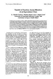
Figure 4 (a)-(b) illustrate the Teichm¨uller space coordinate computation on a left hemisphere cortical surface with 3 selected landmark curves: the Central Sulcus, Superior Temporal Sulcus, and Primary Intermediate Sulcus. After we cut a cortical surface open along the selected landmark curves, a cortical surface becomes topologically equivalent to a genus-zero surface with three open boundaries. The surface is topologically equivalent to the topological pant surface (Figure 1 (a)). We can compute its conformal parameterization to the Poincar´e disk with the hyperbolic Ricci flow and further compute its Teichm¨uller space coordinate. (a)-(b) illustrates a cortical surface and its embedding in the Poincar´e disk. The three boundaries are labeled as γi and two shortest geodesics that connect boundaries are labeled as τi . Lots of studies have associated accelerated dilation of the cerebral ventricles with risk for and progression of HIV/AIDS, dementia and MCI in elderly subjects [17, 2, 6, 3]. Ventricular changes reflect atrophy in surrounding structures, and ventricular measures and surface-based maps can provide sensitive assessments of tissue reduction that correlate with cognitive deterioration in illnesses. However, the concave shape, complex branching topology and narrowness of the inferior and posterior horns have made automatic analyses more difficult. To model the lateral ventricular surface, we automatically locate and introduce three cuts on each ventricle. The cuts are motivated by examining the topology of the lateral ventricles, in which several horns are joined together at the ventricular “atrium” or “trigone”. We call this topological model, creating a set of connected surfaces, a topology optimization operation. After modeling the topology in this way, a lateral ventricular surface, in each hemisphere, becomes an open boundary surface with 3 boundaries, a topological pant surface. After the topology optimization, a ventricular surface is topologically equivalent to a topological pant surface. We can then compute its Teichm¨uller space coordinate. Figure 4 illustrates how to compute Teichm¨uller space coordinates for a lateral ventricle. In the figure, γ1 , γ2 , and γ3 are labeled boundaries and τ1 and τ2 are the shortest geodesics between boundaries. Figure 4 (b) illustrates the surface with
Feature!based vs. Volume!based comparison for MZ twins (76 pairs)
160 150 140 130 120
# of Comparison Errors
110 100 90 80 70 60 50 40 30 20 10 0 0
10
20
30
40
50
10
# of feature!based errs
60
70
80
90
100
110
Index of MZ Twins # of volume!based errs
120
130
140
150
160
# of v!b errs ! f!b errs
(a) 120
Feature!based vs. Volume!based comparison for DZ twins (56 pairs)
110 100 90 80 70 60
the hyperbolic metric that is isometrically flattened onto the Poincar´e disk. When we make the topological change, we make sure each new boundary has the same Euclidean length across different surface. As a result, the lengths of each boundary under the Poincar´e disk metric are valid metrics for studying lateral ventricular surface morphometry.
4.2. Genetic Influences on Ventricular Structure in Twins We analyzed ventricular shapes in 3D volumetric MRI scans from 76 identical and 56 same-sex fraternal twins, scanned as part of a 5-year research project [3]. For each ventricular surface, we computed its Teichm¨uller space coordinates as a 3 × 1 vector. With the L2 norm, we hypothesized that our shape index could help find each subjects twin pair in the database, assuming it has a closer shape index than the rest of surfaces. In a preliminary research, we compared our shape index with volume measures. For a given ventricular surface, we computed and sorted the distances to all other ventricular surfaces. Then we counted how many “wrong” ventricular surface that has a shorter distance than the one to its twin peer. We called this number as the number of comparison errors associated with the ventricular surface. We define the error rate as the ratio of the error number and the number of
# off Comparison Errors
Figure 4. Computing Teichm¨uller space coordinates for a cortical surface and a ventricle surface. (a) is a left hemisphere cortical surface with 3 selected landmark curves. (b) shows the cortical surface is isometrically flattened to the Poincar´e disk. (c) and (d) illustrate a ventricular surface with labeled boundaries and its Poincar´e disk flattening result.
50 40 30 20 10 0 10 0
10
20
30
40
50
60
70
80
90
100
110
20 30 40
Index of DZ Twins
50 60 70 80
# of feature!based errs
# of volume!based errs
# of v!b errs ! f!b errs
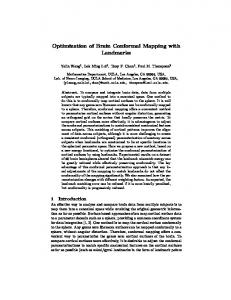
(b) Figure 5. Comparison of shape feature based algorithm and volume measure based algorithm to study genetic influence on ventricular shape in twins. (a) is the result on identical twin group and (b) is on same-sex fraternal twin group. The Teichm¨uller space based shape index captures reported a lower number of comparison errors. It may indicate that the Teichm¨uller space based shape index may be more useful to detect genetic effects than volume measures.
all surfaces compared. We computed the comparison errors for each ventricular surface with our shape index and volume measures. Figure 5 illustrates the number of comparison errors of two shape measures for all ventricular surfaces on 76 identical twins (MZ) and 56 same-sex fraternal twins (DZ). We also calculate the difference of number of comparison errors of volume measure and that of shape index measure. From the figure, we can learn that shape index feature did a better job than volume feature for MZ twin data because all volume feature error numbers are larger than the shape index error numbers. For 76 identical twins, the median error rate was 14.3% (versus 24.7% error rate for volume measures). For 56 same-sex fraternal twins, the median error rate was 22.3% (versus 26.4% error rate for volume measures). We also compared the number of comparison errors between each pair of twins and found that the median and mean of comparison errors between pairs of identical twins were consistently less than those of fraternal twins. This suggests that the Teichm¨uller space based shape
index may be more useful to detect genetic effects than volume measures.
4.3. Lateral Ventricle Shape Analysis of HIV/AIDS In another experiment, we compared ventricular surface models extracted from 3D brain MRI scans of 11 individuals with HIV/AIDS and 8 control subjects [17]. We automatically perform topology optimization on each ventricular surface and compute their lengths in the Poincar´e disk by the Yamabe flow method. For each pair of ventricular surfaces, we obtained a 6 × 1 vector, t = (t1 , t2 , ...t6 ), which consists of 3 boundary lengths for the left ventricular surface and 3 boundary lengths for right ventricular surface. Given this Teichm¨uller space coordinate based feature vector, we apply a nearest neighbor classifier based on the Mahalanobis distance, which is q d(t) = (t − µTc )T Σ−1 Tc (t − µTc ) q + (t − µTa )T Σ−1 Ta (t − µTa ) where µTc , µTa , ΣTc and ΣTa are the feature vector mean and covariance for the two groups, respectively. We classify t based on the sign of the distance of d(t), i.e., the subject that is closer to one group mean is classified into that group. For this data set, we performed a leave-one-out test. Our classifier successfully classified all 19 subjects to the correct group and achieved a 100% accuracy rate [18]. For comparison, we also tested a nearest neighbor classifier associated with a volume feature vector. For each pair of ventricular surface, we measure their volumes, (vl , vr ). We also use a nearest neighbor classifier based on the Mahalanobis distance, which is q d(v) = (v − µVc )T Σ−1 Vc (v − µVc ) q + (v − µVa )T Σ−1 Va (v − µVa ) where µVc , µVa , ΣVc and ΣVa are the feature vector mean and covariance two groups, respectively. We classify v based on the sign of the distance of d(v), i. e., the subject that is closer to one group mean is classified into that group. In the data set, we performed a leave-one-out test. The classifier based on the simple volume measurement successfully classified only 13 out of 19 subjects to the correct group and achieved a 68.42% accuracy rate. Figure 6 shows an interesting experimental result. Two pairs of lateral ventricular surfaces, are shown, from (a) a healthy control individual, and (b) from an individual with HIV/AIDS. Both of them have highly irregular shapes. Although generally HIV/AIDS makes the lateral ventricle dilate, here the ventricle from the control group has a bigger volume than the one from the HIV/AIDS group on both
sides. The volume differences between these two subjects are about 0.62% and 5.2% of the mean left and right side volumes in the whole data set. The volume-based classifier assigned all of these ventricular surfaces to the incorrect diagnostic groups while the Teichm¨uller space coordinate based classifier classified each of them correctly.
Figure 6. Lateral ventricular surfaces of a control subject (a) and an individual with HIV/AIDS. The volume of lateral ventricular surface of the control subject is greater than that of the HIV/AIDS individual. Our Teichm¨uller space coordinate feature successfully classified these surfaces, when entered into a nearest neighbor classifier. The volume measurement based nearest neighbor classifier achieved a 68.42% accuracy rate.
Studies of ventricular morphometry have also used 3D statistical maps to correlate anatomy with clinical measures, but automated ventricular analysis is still difficult because of their highly irregular branching surface shape. The new Teichm¨uller space shape descriptor requires more validation on other data sets, these experimental results suggest that (1) ventricular surface morphometry is altered in HIV/AIDS; (2) volume measures are not sufficient to distinguish HIV patients from controls; and (3) our Teichm¨uller space feature vector can be used to classify control and patient subjects. Our ongoing work is studying the correlation between the proposed feature vector and clinical measures (e.g., future decline) in an Alzheimer’s Disease data set.
5. Conclusion and Future Work In this paper, we propose a stable way to compute Teichm¨uller space coordinate for a surface with a certain type of branching topology. We applied it as a shape index to study brain morphometry for genetic shape analysis and HIV/AIDS. We included our shape index in a nearest neighbor classifier that correctly classified all lateral ventricle surfaces from 8 control and 11 HIV/AIDS individuals (versus only 68% accuracy rate for volume measure). Although our current work focuses on topological pant surfaces, for surfaces with more complicated topologies, their Teichm¨uller coordinates can still be computed using the hyperbolic metric. If the surface has Euler number χ, χ < 0, the surface can be decomposed to −χ number of pants, where the cutting curves are also geodesics under the hyperbolic metric.
Furthermore, two pants sharing a common cutting curve can be glued together with a specific twisting angle. The lengths of all cutting geodesics and the twisting angles associated with them form the Teichm¨uller coordinates of the surface. In the future, we will further explore and validate numerous applications of the Teichm¨uller shape space in neuroimaging and shape analysis research. Acknowledgement: This work was funded by National Institute of Health through the NIH Roadmap for Medical Research, Grant U54 RR021813 entitled Center for Computational Biology (CCB).
References [1] C. M. Botino, C. C. Castro, R. L. Gomes, C. A. Buchpiguel, R. L. Marchetti, and M. R. Neto. Volumetric MRI measurements can differentiate Alzheimer’s disease, mild cognitive impairment and nomral aging. International Psychogeriatrics, 14:59–72, 2002. [2] O. T. Carmichael, P. M. Thompson, R. A. Dutton, A. Lu, S. E. Lee, J. Y. Lee, L. H. Kuller, O. L. Lopez, H. J. Aizenstein, C. C. Meltzer, Y. Liu, A. W. Toga, and J. T. Becker. Mapping ventricular changes related to dementia and mild cognitive impairment in a large community-based cohort. Biomedical Imaging: Nano to Macro, 2006. 3rd IEEE International Symposium on, pages 315–318, April 2006. [3] Y. Chou, N. Lepor´e, M. Chiang, C. Avedissian, M. Barysheva, K. L. McMahon, G. I. de Zubicaray, M. Meredith, M. J. Wright, A. W. Toga, and P. M. Thompson. Mapping genetic influences on ventricular structure in twins. NeuroImage, 44(4):1312 – 1323, 2009. [4] M. Chung, K. Dalton, and R. Davidson. Tensor-based cortical surface morphometry via weighted spherical harmonic representation. IEEE Trans. Med. Imag., 27(8):1143–1151, Aug. 2008. [5] R. H. Davies, C. J. Twining, P. D. Allen, T. F. Cootes, and C. J. Taylor. Shape discrimination in the hippocampus using an MDL model. In Proc. Infor. Proc. Med. Imag. (IPMI), 2003. [6] L. Ferrarini, W. M. Palm, H. Olofsen, M. A. van Buchem, J. H. Reiber, and F. AdmiraalBehloul. Shape differences of the brain ventricles in Alzheimer’s disease. NeuroImage, 32(3):1060 – 1069, 2006. [7] K. Gorczowski, M. Styner, J.-Y. Jeong, J. Marron, J. Piven, H. Hazlett, S. Pizer, and G. Gerig. Statistical shape analysis of multi-object complexes. IEEE Conf. Comp. Vis. Patt. Recog. CVPR ’07, pages 1–8, June 2007.
[8] X. Gu, S. Wang, J. Kim, Y. Zeng, Y. Wang, H. Qin, and D. Samaras. Ricci flow for 3D shape analysis. Computer Vision, 2007. ICCV 2007. IEEE 11th International Conference on, pages 1–8, Oct. 2007. [9] H. W. Guggenheimer. Differential Geometry. Dover Publications, 1977. [10] A. Hatcher. Algebraic Topology. Camerbirdge University Press, 2006. [11] M. Jin, W. Zeng, F. Luo, and X. Gu. Computing Teichm¨uller shape space. IEEE Trans. Vis. Comput. Graphics, 15(3):504–517, 2009. [12] I. Kakadiaris, G. Passalis, G. Toderici, M. Murtuza, Y. Lu, N. Karampatziakis, and T. Theoharis. Threedimensional face recognition in the presence of facial expressions: An annotated deformable model approach. IEEE Trans. Pattern Anal. and Machine Intell., 29(4):640–649, April 2007. [13] S. Li, F. Shi, F. Pu, X. Li, T. Jiang, S. Xie, and Y. Wang. Hippocampal shape analysis of Alzheimer’s disease based on machine learning methods. American J. of Neuroradiology, 28:1339–45, 2007. [14] S. Pizer, D. Fritsch, P. Yushkevich, V. Johnson, and E. Chaney. Segmentation, registration, and measurement of shape variation via image object shape. IEEE Trans. Med. Imag., 18(10):851–865, Oct. 1999. [15] R. Schoen and S.-T. Yau. Lectures on Differential Geometry. International Press, 1994. [16] P. Thompson and A. Toga. A surface-based technique for warping 3-dimensional images of the brain. IEEE Trans. Med. Imag., 15(4):1–16, 1996. [17] P. M. Thompson, R. A. Dutton, K. M. Hayashi, A. Lu, S. E. Lee, J. Y. Lee, O. L. Lopez, H. J. Aizenstein, A. W. Toga, and J. T. Becker. 3D mapping of ventricular and corpus callosum abnormalities in HIV/AIDS. NeuroImage, 31(1):12–23, 2006. [18] Y. Wang, W. Dai, T. F. Chan, S.-T. Yau, A. W. Toga, and P. M. Thompson. Teichm¨uller shape space theory and its application to brain morphology. In Med. Image Comp. Comput.-Assist. Intervention, Proceedings, 2009. [19] Y. Wang, X. Gu, T. F. Chan, P. M. Thompson, and S.-T. Yau. Brain surface conformal parameterization with algebraic functions. Med. Image Comp. Comput.Assist. Intervention, Proceedings, Part II, pages 946– 954, 2006. LNCS 4191. [20] W. Zeng, X. Yin, Y. Zeng, Y. Lai, X. Gu, and D. Samaras. 3D face matching and registration based on hyperbolic Ricci flow. CVPR Workshop on 3D Face Processing 2008, pages 1–8, 23-28 June 2008.