© 2006 Nature Publishing Group http://www.nature.com/naturemethods
BRIEF COMMUNICATIONS
Sub-diffraction-limit imaging by stochastic optical reconstruction microscopy (STORM) Michael J Rust1,5, Mark Bates2,5 & Xiaowei Zhuang1,3,4 We have developed a high-resolution fluorescence microscopy method based on high-accuracy localization of photoswitchable fluorophores. In each imaging cycle, only a fraction of the fluorophores were turned on, allowing their positions to be determined with nanometer accuracy. The fluorophore positions obtained from a series of imaging cycles were used to reconstruct the overall image. We demonstrated an imaging resolution of 20 nm. This technique can, in principle, reach molecular-scale resolution.
Fluorescence microscopy is widely used in molecular and cell biology for noninvasive, time-resolved imaging with high biochemical specificity. Despite these advantages, standard fluorescence microscopy is not useful for ultra-structural imaging owing to a resolution limit set by the diffraction of light. Several approaches have been used to break this diffraction limit, including near-field scanning optical microscopy (NSOM)1, multiphoton fluorescence2, stimulated emission depletion (STED)3 and saturated structured-illumination microscopy (SSIM)4. NSOM, STED and SSIM have all achieved lateral resolution of tens of nanometers, substantially lower than the wavelength of light, but each with its own limitations. NSOM is difficult to operate in a noninvasive mode and has a low imaging depth. Multiphoton microscopy, STED and SSIM are based on nonlinear optical effects, and typically require the use of high-intensity pulsed lasers, which can induce sample damage. A continuing effort to develop existing4,5 and new techniques is needed to harness the benefits of fluorescence microscopy for ultraresolution biological imaging. Single-molecule detection offers new possibilities for obtaining sub-diffraction-limit spatial resolution. The position of a single emitter can be determined to almost arbitrarily high accuracy if a sufficient number of photons are collected6–9. Based on this principle, fluorescence imaging with one-nanometer accuracy (FIONA) has recently been demonstrated at the single-molecule level at room temperature9. Unfortunately, the localization accuracy of FIONA does not directly translate into imaging resolution as multiple emitters
in close proximity will still be difficult to resolve. This issue has been addressed in part by taking advantage of photobleaching of single dyes or photoblinking of quantum dots to distinguish their individual signals10–13. These approaches had been used to resolve 2–5 fluorophores within a diffraction-limited spot, but extending these techniques to more than a few fluorophores remains a challenge. In this work we report a new high-resolution imaging technique, stochastic optical reconstruction microscopy (STORM), in which a fluorescence image is constructed from high-accuracy localization of individual fluorescent molecules that are switched on and off using light of different colors. The STORM imaging process consists of a
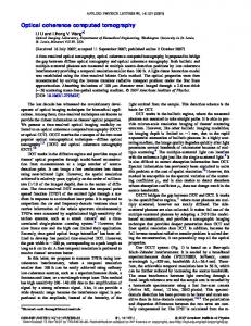
a
b
Figure 1 | STORM with photo-switchable fluorophores. (a) A STORM imaging sequence using a hypothetical hexameric object labeled with red fluorophores that can be switched between a fluorescent and a dark state by a red and green laser, respectively. All fluorophores are first switched to the dark state by a strong red laser pulse. In each imaging cycle, a green laser pulse is used to switch on only a fraction of the fluorophores to give an optically resolvable set of active fluorophores. Next, under red illumination, these molecules emit fluorescence until they are switched off, allowing their positions (white crosses) to be determined with high accuracy. The overall image is then reconstructed from the fluorophore positions obtained from multiple imaging cycles. (b) A single Cy5 switch on DNA can be turned on and off for hundreds of cycles before being permanently photobleached. A red laser (633 nm, 30 W/cm2) is used to excite fluorescence (black line) from Cy5 and to switch Cy5 to the dark state. A green laser (532 nm, 1 W/cm2) is used to return Cy5 to the fluorescent state. The alternating red and green line indicates the laser excitation pattern.
1Department of
Physics, 2Division of Engineering and Applied Sciences, 3Department of Chemistry and Chemical Biology, 4Howard Hughes Medical Institute, Harvard University, 12 Oxford Street, Cambridge, Massachusetts 02138, USA. 5These authors contributed equally to this work. Correspondence should be addressed to X.Z. (
[email protected]).
RECEIVED 7 JULY; ACCEPTED 31 JULY; PUBLISHED ONLINE 9 AUGUST 2006; DOI:10.1038/NMETH929
NATURE METHODS | VOL.3 NO.10 | OCTOBER 2006 | 793
BRIEF COMMUNICATIONS
Fluorescence (photons)
a 500 400 300 200 100 0 1,000
1,000 0
nm –500 )
–500 –1,000 –1,000
0
500
)
m y (n
d 12
Number of molecules
© 2006 Nature Publishing Group http://www.nature.com/naturemethods
500
x(
c
Figure 2 | The high localization accuracy of individual switches during each switching cycle defines the intrinsic resolution of STORM. (a) The point spread function (PSF) of the emission from a single switch on DNA during a single switching cycle. Fitting the PSF to a two-dimensional Gaussian (not shown) gives the centroid position of the PSF. (b,c) The centroid positions of an individual switch determined in 20 successive imaging cycles before (b) and after (c) correction for sample drift. Scale bars, 20 nm. (d) A histogram of the standard deviation of centroid positions. The standard deviation is determined as (σx + σy) / 2 for each switch using 20 imaging cycles, where σx and σy are the standard deviations of the centroid positions in the x and y dimensions. This histogram was constructed from 29 switches.
b
10 8 6 4 2 0 0
2
4
6
8
10
12
14
16
Standard deviation (nm)
series of imaging cycles (Fig. 1a). In each cycle, only a fraction of the fluorophores in the field of view are switched on, such that each of the active fluorophores is optically resolvable from the rest, that is, their images are not overlapping. This allows the position of these fluorophores to be determined with high accuracy. Repeating this process for multiple cycles, each causing a stochastically different subset of fluorophores to be turned on, allows the positions of many fluorophores to be determined and thus an overall image to be reconstructed. In the examples below we demonstrated an imaging resolution of approximately 20 nm using a simple total-internal-reflection fluorescence microscope, low-power continuous-wave lasers and a photoswitchable cyanine dye14,15. We have recently reported that a cyanine dye, Cy5, can be switched between a fluorescent and a dark state in a controlled and reversible manner by light of different wavelengths14. Red laser light that produces fluorescent emission from Cy5 can also switch the dye to a stable dark state. Exposure to green laser light converts Cy5 back to the fluorescent state, but the recovery rate depends critically on the close proximity of a secondary dye, Cy3 (ref. 14). Hereafter, we refer to the Cy3-Cy5 dye pair simply as a switch. Under illumination conditions allowing single-molecule detection, we found that such a switch, when attached to nucleic acids or proteins, can be cycled on and off hundreds of times before permanent photobleaching occurs (Fig. 1b and Supplementary Fig. 1 online). Here we demonstrate the concept of STORM using the cyanine switch, but in principle any suitable optically switched fluorophore could be used. The resolution of STORM is limited by the accuracy with which individual switches can be localized during a switching cycle. To determine the localization accuracy, we attached a switch to a short double-stranded DNA, which was surface-immobilized at low density so that single switches were clearly resolvable. These switches were periodically cycled on and off using green and red laser light; the red laser also served to excite fluorescence from Cy5. No fluorescence from Cy3 was recorded in this process. The fluorescence image from a single switch gave the point-spread function shown in Figure 2a. 794 | VOL.3 NO.10 | OCTOBER 2006 | NATURE METHODS
A Gaussian fit to this image was used to localize the position of the switch. The positions determined from multiple switching cycles showed a substantial spread (Fig. 2b), which was considerably reduced by correcting for sample drift over the course of the experiment (Fig. 2c and Supplementary Methods online). The standard deviation of the drift-corrected positions obtained from 20 imaging cycles is on average 8 nm for individual switches (Fig. 2d). This value gives a measure of the uncertainty in the localization of a single switch per imaging cycle. Correspondingly, the experimentally measured spread of switch positions follows a Gaussian distribution with a full-width half maximum of 18 nm, as expected from the 8 nm standard deviation (Supplementary Fig. 2 online). Therefore, under our present imaging conditions, two switches separated by 20 nm should be resolvable. To demonstrate the capability of STORM to resolve fluorescent molecules in close proximity, we constructed standard samples of linear, double-stranded DNA labeled with multiple switches separated by a well-defined number of base pairs. We also labeled the DNA strands with multiple biotins and attached them to a high-density streptavidin layer, increasing the likelihood of multiple attachments between the DNA and the surface, so that the DNA is immobilized in the plane. We first examined DNA strands containing two switches separated by 135 base pairs (Fig. 3a and Supplementary Fig. 3 online), corresponding to a length of 46 nm along the DNA contour. A STORM image obtained using the imaging procedure described above shows two clusters of measured switch positions, indicating that the two switches are well resolved (Fig. 3a). The distance between the centers of the two clouds had a mean value of 41 nm (Fig. 3b), in quantitative agreement with the theoretical mean (40 nm) determined using the known contour and persistence lengths of the DNA sample. We also imaged longer DNA samples labeled with four switches evenly separated by 46 nm along the contour. The STORM images indeed reveal four clusters of switch positions following a bent contour consistent with the persistence length of DNA (a measure of flexibility) and the engineered separation between the switches (Fig. 3c). These results indicate that STORM can image biological samples with sub-diffraction-limit resolution, and that features separated by 40 nm are well within our resolving power. A distinct advantage of STORM is its ability to localize a large number of switches within a diffraction-limited spot by cycling the switches on and off in a controlled manner, allowing this to be used as a general biological imaging technique. To demonstrate this capability, we prepared circular DNA plasmids coated with RecA protein and imaged them using indirect immunofluorescence with switchlabeled secondary antibody (Fig. 3d). STORM images of the RecA filaments reveal their circular structure with greatly increased resolution when compared with wide-field images. Parts of the filament appear to be unlabeled or kinked, possibly corresponding to regions of the plasmid that remain uncoated by RecA.
BRIEF COMMUNICATIONS b
0.7
Fraction of molecules
a
0.6 0.5 0.4 0.3 0.2 0.1 0.0 0
20
40
60
80
100
Inter-switch distance (nm)
© 2006 Nature Publishing Group http://www.nature.com/naturemethods
c
d
Figure 3 | STORM can resolve structures with sub-diffraction-limit resolution. (a) STORM cleanly resolves two switches separated by a contour length of 46 nm on dsDNA. The DNA construct is illustrated on the left; example STORM images of three individual DNA samples are shown on the right. The STORM images show two clearly separated clusters of measured switch positions (crosses), each corresponding to a single switch. The center-of-mass position of each cluster is marked by a red dot. The inter-switch distances are 46 nm, 44 nm and 34 nm for these three examples. Scale bars, 20 nm. (b) Comparison between the inter-switch distances measured using STORM (columns) and the predicted distance distribution considering the flexibility of DNA (dashed line). (c) STORM images of four switches attached to a dsDNA, pair-wise separated by a contour length of 46 nm. The measured switch positions are clustered by an automated algorithm and different clusters are indicated by different symbols. Scale bars, 20 nm. (d) STORM images of RecA-coated circular plasmid DNA. Indirect immunofluorescence images with switch-labeled secondary antibody taken by a total internal reflection microscope (top); the reconstructed STORM images of the same filaments (bottom). Scale bars, 300 nm.
In summary, we have demonstrated that STORM is capable of imaging biological structures with sub-diffraction-limit resolution. The resolution of the technique is limited in principle only by the number of photons emitted per switch cycle, but not the wavelength of light. For the cyanine switch, we have detected ~3,000 photons per switching cycle, independent of red-laser intensity (data not shown), predicting a theoretical localization accuracy of 4 nm8. The discrepancy between this theoretical prediction and our measured accuracy of 8 nm is most likely due to imperfect correction of stage drift and aberration owing to focus drift in our measurements. This measured localization accuracy corresponds to an imaging resolution of approximately 20 nm. Indeed, fluorescent switches separated by ~40 nm have been clearly resolved (Fig. 3). The cyanine switches can be turned on and off reliably for hundreds of cycles before photobleaching, allowing STORM to be used for resolving structures with many fluorophores in a potentially time-resolved manner. The circular structure of RecA filaments containing 10–20 switches can be resolved in a few minutes. The imaging speed may be improved by increasing the switching rate through stronger excitation or fluorophores with faster switching kinetics. We expect that STORM will be a valuable tool for high-resolution fluorescence in situ hybridization and immunofluorescence imaging. The STORM concept is also applicable to other photoswitchable fluorophores and fluorescent proteins, which will potentially allow high-resolution live-cell imaging.
Defense Advance Research Projects Agency and a Packard Science and Engineering Fellowship (to X.Z.). X.Z. is a Howard Hughes Medical Institute Investigator. AUTHOR CONTRIBUTIONS M.J.R. and M.B. conceived the STORM imaging concept. M.J.R., DNA imaging and data analysis; M.B., RecA imaging and data acquisition. X.Z. supervised the project. COMPETING INTERESTS STATEMENT The authors declare that they have no competing financial interests. Published online at http://www.nature.com/naturemethods/ Reprints and permissions information is available online at http://npg.nature.com/reprintsandpermissions/ 1. 2. 3. 4. 5. 6. 7. 8. 9. 10. 11. 12. 13.
Note: Supplementary information is available on the Nature Methods website. ACKNOWLEDGMENTS This work is supported by in part by the US National Institutes of Health, the
14. 15.
Pohl, D.W. & Courjon, D. Near Field Optics (Kluwer, Dordrecht, 1993). Zipfel, W.R., Williams, R.M. & Webb, W.W. Nat. Biotechnol. 21, 1368–1376 (2003). Hell, S.W. Nat. Biotechnol. 21, 1347–1355 (2003). Gustafsson, M.G.L. Proc. Natl. Acad. Sci. USA 102, 13081–13086 (2005). Hofmann, M., Eggeling, C., Jakobs, S. & Hell, S.W. Proc. Natl. Acad. Sci. USA 102, 17565–17569 (2005). Gelles, J., Schnapp, B.J. & Sheetz, M.P. Nature 331, 450–453 (1988). van Oijen, A.M., Kohler, J., Schmidt, J., Muller, M. & Brakenhoff, G.J. Chem. Phys. Lett. 292, 183–187 (1998). Thompson, R.E., Larson, D.R. & Webb, W.W. Biophys. J. 82, 2775–2783 (2002). Yildiz, A. et al.. Science 300, 2061–2065 (2003). Gordon, M.P., Ha, T. & Selvin, P.R. Proc. Natl. Acad. Sci. USA 101, 6462–6465 (2004). Qu, X., Wu, D., Mets, L. & Scherer, N.F. Proc. Natl. Acad. Sci. USA 101, 11298– 11303 (2004). Lidke, K.A., Rieger, B., Jovin, T.M. & Heintzmann, R. Opt. Express 13, 7052– 7062 (2005). Ram, S., Ward, E.S. & Ober, R.J. Proc. Natl. Acad. Sci. USA 103, 4457–4462 (2006). Bates, M., Blosser, T.R. & Zhuang, X. Phys. Rev. Lett. 94, 108101 (2005). Heilemann, M., Margeat, E., Kasper, R., Sauer, M. & Tinnefeld, P. J. Am. Chem. Soc. 127, 3801–3806 (2005).
NATURE METHODS | VOL.3 NO.10 | OCTOBER 2006 | 795