EJO ISSN 1120-6721
Eur J Ophthalmol 2017; 00 (00): 000-000 DOI: 10.5301/ejo.5000873
CASE REPORT
Sudden visual loss after cardiac resynchronization therapy device implantation Luigi A. De Vitis1, Alessandro Marchese1, Chiara Giuffrè1, Adriano Carnevali1,2, Lea Querques1, Livia Tomasso1, Giovanni Baldin1, Gisella Maestranzi1, Rosangela Lattanzio1, Giuseppe Querques1, Francesco Bandello1 Department of Ophthalmology, IRCCS Ospedale San Raffaele, University Vita-Salute, Milan - Italy Department of Ophthalmology, University of “Magna Graecia”, Catanzaro - Italy
1 2
Abstract Purpose: To report a case of sudden decrease in visual acuity possibly due to a cardiogenic embolism in a patient who underwent cardiac resynchronization therapy (CRT) device implantation. Methods: A 62-year-old man with severe left ventricular systolic dysfunction and a left bundle branch block was referred to our department because of a sudden decrease in visual acuity. Nine days earlier, he had undergone cardiac transapical implantation of a CRT device, which was followed, 2 days later, by an inflammatory reaction. The patient underwent several general and ophthalmologic examinations, including multimodal imaging. Results: At presentation, right eye (RE) best-corrected visual acuity (BCVA) was counting fingers and RE pupil was hyporeactive. Fundus examination revealed white-centered hemorrhagic dots suggestive of Roth spots. Fluorescein angiography showed delay in vascular perfusion during early stage, late hyperfluorescence of the macula and optic disk, and peripheral perivascular leakage. The first visual field test showed complete loss of vision RE and a normal left eye. Due to suspected giant cell arteritis, temporal artery biopsy was performed. Thirty minutes after the procedure, an ischemic stroke with right hemisyndrome and aphasia occurred. The RE BCVA worsened to hands motion. Four months later, RE BCVA did not improve, despite improvement in fluorescein angiography inflammatory sign. Conclusions: We report a possible cardiogenic embolism secondary to undiagnosed infective endocarditis causing monocular visual loss after CRT device implantation. It remains unclear how the embolus caused severe functional damage without altering the retinal anatomical structure. Keywords: Cardiac resynchronization therapy, Infective endocarditis, Retinal artery occlusion, Roth spot, Visual loss
Introduction Cardiogenic embolism can be a severe complication of infective endocarditis and may affect every vascular network, including ocular arteries (1). Infective endocarditis is a rare (3-10 per 100,000 people) but severe disease and is defined by infection of a native or prosthetic heart valve, the endocardial surface, or an indwelling cardiac device (2). Cardiac devices include permanent pacemakers, cardiac resynchronization therapy (CRT), and implantable cardioverter defibrillator (ICD); up to 2% of these devices become infected during the first 5 years after implantation. Accepted: August 27, 2016 Published online: February 22, 2017 Corresponding author: Prof. Giuseppe Querques Department of Ophthalmology University Vita-Salute IRCCS Ospedale San Raffaele Via Olgettina 60 20132 Milan, Italy
[email protected]
© 2017 Wichtig Publishing
We report a case of monocular acute visual decline in a patient who underwent cardiac surgery with implantation of a CRT device.
Case report A 62-year-old man was referred to our department because of a sudden decrease in right eye (RE) visual acuity. He had a smoking history and had hypertension; diabetes; peripheral artery disease, for which he underwent aortobifemoral bypass surgery; and ischemic dilated cardiomyopathy, characterized by left ventricular systolic dysfunction and left bundle branch block. He also underwent 4 coronary artery bypass graft surgeries in 2006 and right coronary artery drug-eluting stent implantation in 2013. In 2015, an ICD was placed because of a decrease in left ventricular ejection fraction (EF). However, the patient’s left ventricular function did not improve (EF 30%) and in January 2016 the ICD was upgraded to CRT. The surgical procedure consisted of transapical left ventricular electrocatheter implantation through left lateral thoracotomy; no complications occurred during the procedure and the patient was moved to the intensive care unit and later to the cardiology unit. Two days after CRT device implantation, the patient experienced the first signs of an inflammatory reaction (fever and
2
Visual loss after CRT device implantation
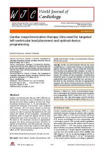
Fig. 1 - Right eye ultra-widefield retinal imaging shows white-centered hemorrhagic dots suggestive of Roth spots.
elevation of systemic inflammatory markers) and blood cultures, transesophageal echocardiography (TOE), and transthoracic echocardiography were performed. Although all these tests were negative, the patient began empiric therapy with piperacillin/tazobactam 4.5 g QID on suspicion of a fever of unknown origin. A week after the beginning of the inflammatory reaction, the patient referred a sudden decrease in RE visual acuity. Brain computed tomography (CT) and brain CT angiography (CTA), performed on suspicion of a stroke secondary to infective endocarditis, were negative. Blood cultures and TOE were repeated but they were still negative. A heart CT was executed but it was negative as well. Fluorodeoxyglucose positron emission tomography did not detect findings compatible with infective endocarditis or septic embolism. At the ophthalmologic examination, the left eye (LE) was normal while the RE best-corrected visual acuity (BCVA) was counting fingers and the RE pupil was hyporeactive. The RE fundus examination showed intraretinal hemorrhages, putatively Roth spots (Fig. 1), a pale optic disc with undefined borders, and altered macular reflex. The LE fundus examination only showed a tilted optic disk. We decided to perform fluorescein angiography (FA), which showed a delay in vascular perfusion of the RE and, in both eyes, peripheral perivascular leakage, ischemia, and late hyperfluorescence in optic disc and macular regions (Fig. 2). The LE was far
less altered than the RE on FA. Optical coherence tomography (OCT) and blue fundus autofluorescence were normal (Fig. 3). Visual field test detected a loss of vision in the RE while the LE was almost normal at the beginning and a few days later developed a quadrantanopia. Nevertheless, LE visual field improved 2 days later, probably owing to antibiotic therapy. Eight days after the patient’s visual acuity decline, a temporal artery biopsy was performed due to lack of improvement in the patient’s condition and on suspicion of giant cell arteritis (or Horton vasculitis), but the histopathologic analysis did not show any abnormalities. Thirty minutes after the biopsy, a stroke with right hemisyndrome and aphasia occurred. The diagnosis of stroke was confirmed by brain CT and brain CTA. At ophthalmologic examination, LE BCVA was normal while RE BCVA worsened to hand motion and RE pupil remained hyporeactive; RE and LE fundus did not change compared to the previous examination. We repeated FA and OCT but both did not show any change compared to the previous examinations. We also performed indocyanine green angiography, which was unremarkable. Visual field test was similar to previous ones. Considering that the patient was clinically stable, he was discharged 10 days after the temporal artery biopsy. Four months later, BCVA was not improved, despite improvement in FA inflammatory signs. © 2017 Wichtig Publishing
De Vitis et al
3
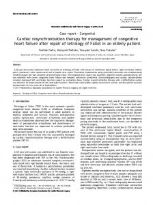
Fig. 2 - Right eye fluorescein angiography (FA) shows delayed perfusion (A), peripheral perivascular leakage (B), and late hyperfluorescence in optic disk and macular regions (C). Fig. 3 - Right eye infrared reflectance (A), fundus autofluorescence (B), and optical coherence tomography (C) show overall normal macula.
Discussion We report a case of sudden visual loss after CRT device implantation, probably due to cardiogenic embolism. Cardiogenic embolism is a severe complication of infective endocarditis. In high-income countries, major risk factors for endocarditis are degenerative valve disease, diabetes, cancer, intravenous drug use, congenital heart disease, and indwelling cardiac devices. Our patient had diabetes and underwent an invasive cardiac procedure to implant a CRT, which can act as an infection focus. Furthermore, surgical procedures increase the rate of infections by Staphylococcus aureus, the most frequently isolated microorganism associated with infective endocarditis in high-income countries (up to 30% of the cases). The clinical presentation of infective endocarditis is nonspecific, so it should be considered in © 2017 Wichtig Publishing
patients with fever of unknown origin in presence of characteristic risk factors. Infective endocarditis may also present directly with an embolic complication. Our patient presented with fever and subsequently with visual loss, putatively due to a systemic embolism, and both could be the presenting signs of endocarditis. During clinical examination, fever and cardiac murmur are the most common signs of endocarditis and are frequently associated with signs of its complications such as heart failure, stroke, or metastatic infection. Except for fever, none of these signs was present in our patient but at the ophthalmologic examination the presence of Roth spots was recorded, a rare sign of endocarditis defined as white-centered retinal hemorrhages (3). Our patient’s laboratory examinations, showing raised inflammatory markers, were compatible with endocarditis. According to modified Duke clinical criteria for diagnosis of infective endocarditis,
Visual loss after CRT device implantation
4
a diagnosis of infective endocarditis is possible (but not definite) in presence of 3 out of 5 minor criteria or 1 out of 2 major criteria associated with 1 out of 5 minor criteria. Major criteria (positive blood cultures and evidence of endocardial involvement by echocardiography) were both absent but our patient presented with (1) temperature >38°C, (2) vascular phenomena, and (3) immunologic phenomena (Roth spots), which are considered 3 minor criteria (4). In contrast with our hypothesis of an infective endocarditis, blood cultures were all negative and TOE was negative. However, it should be noted that about 10% of patients present with culturenegative endocarditis and although TOE sensitivity is very high (more than 90%) it is not 100% (2). It is also well-known that the sensitivity of Duke criteria is reduced in patients with suspected prosthetic valve endocarditis, right-sided infective endocarditis, and cardiac device infection (5). Retinal artery occlusion (RAO) is strongly associated with cardiac disease, and particularly with infective endocarditis: indeed, nearly half of patients with RAO will show echocardiographic abnormalities (6). Moreover, RAO often causes an acute and permanent decline in visual acuity, as happened to our patient. At fundus examination, the posterior pole usually appears whitened and swollen (opacified), with the presence of a so-called cherry-red spot in the foveolar region, but sometimes it can be almost normal, even if functionally damaged, as seen in our patient. Furthermore, the presence of Roth spots at fundus examination, along with Osler nodes and Janeway lesions, is a rare but specific sign of endocarditis. Detection on FA of delay in vascular perfusion during early stage, peripheral perivascular leakage and ischemia, and late hyperfluorescence in optic disc and macular regions were compatible with RAO (1, 7). Furthermore, RE complete loss of vision and LE quadrantanopia on visual field test may suggest a bilateral cardiogenic embolism.
In conclusion, we report the first case of a possible cardiogenic embolism secondary to an infective endocarditis causing RE complete visual loss and LE clinically silent quadrantanopia after CRT device implantation. It remains unclear how the embolus caused such severe functional damage without altering the retinal anatomical structure in a more consistent way.
Disclosures Financial support: F. Bandello: advisory board member: Allergan, Inc.; Novartis Pharmaceuticals Corporation; Farmila-Thea; Bayer Schering Pharma; Pfizer, Inc.; Alcon, Inc.; Bausch and Lomb; Genentech, Inc.; Alimera Sciences, Inc.; Thrombogenics, Inc. Conflict of interest: None of the authors has conflict of interest with this submission.
References 1. 2. 3. 4. 5. 6.
7.
Recchia FM, Brown GC. Systemic disorders associated with retinal vascular occlusion. Curr Opin Ophthalmol. 2000;11(6): 462-467. Cahill TJ, Prendergast BD. Infective endocarditis. Lancet. 2016; 387(10021):882-893. Mahroo OA, Graham EM. Images in clinical medicine. Roth spots in infective endocarditis. N Engl J Med. 2014;370(25):e38. Li JS, Sexton DJ, Mick N, et al. Proposed modifications to the Duke criteria for the diagnosis of infective endocarditis. Clin Infect Dis. 2000;30(4):633-638. Prendergast BD. Diagnostic criteria and problems in infective endocarditis. Heart. 2004;90(6):611-613. Sharma S, Naqvi A, Sharma SM, Cruess AF, Brown GC; Retinal Emboli of Cardiac Origin Group. Transthoracic echocardiographic findings in patients with acute retinal arterial obstruction. A retrospective review. Arch Ophthalmol. 1996;114(10):1189-1192. Hayreh SS. Acute retinal arterial occlusive disorders. Prog Retin Eye Res. 2011;30(5):359-394.
© 2017 Wichtig Publishing