ICTON 2012
Tu.A6.5
Super-Resolution Imaging by High-Index Microspheres Immersed in a Liquid Arash Darafsheh, Michael A. Fiddy, and Vasily N. Astratov* Department of Physics and Optical Science, Center for Optoelectronics and Optical Communications, University of North Carolina at Charlotte, 9201 University City Blvd., Charlotte, NC 28223-0001, USA * Tel: 1 (704) 687 8131, Fax: 1 (704) 687 8197 E-mail:
[email protected], http://maxwell.uncc.edu/astratov/astratov.htm ABSTRACT We demonstrate that the super-resolution imaging of a commercial Blu-ray® disk containing a one-dimensional array of 200 nm width stripes separated by 100 nm width grooves can be achieved using high-index microspheres in a liquid environment. By using barium titanate glass microspheres (2 – 20 µm diameters) with index around 1.9 totally immersed in isopropanol with index 1.37 we demonstrate better than λ/6 far field resolution, where λ= 600 nm is the peak wavelength of the white-light illumination system. The results of this work can be used in biomedical microscopy, microfluidics and nanophotonics applications for imaging individual cells and/or nanoparticles in a liquid environment. Keywords: super-resolution, subdiffraction resolution, imaging, microscopy, barium titanate, microsphere. 1. INTRODUCTION In recent years super-resolution imaging emerged as a very broad area of research based on using near field probes [1], solid immersion lenses [2], novel metamaterials [3,4], superlenses [5], and various modifications of fluorescence-based techniques such as stimulated emission depletion (STED) [6]. An ideal imaging device should be able to magnify sub-diffraction limited object features and project them into the far field. These processes of collection of near-field spatial harmonics and their conversion into propagating fields are usually realized in structures with a sophisticated engineering design. Previously they have been realized using surface polariton plasmon gratings [7] and hyperbolic metamaterials [3,4], however these structures can operate only in a narrow range of wavelengths and they are very difficult to manufacture. In this context, super-resolution imaging using microspheres emerged as a surprisingly simple way of achieving subdiffraction resolution [8]. In this method, the surface of the object can be considered as a collection of nanoscale dipole antennas which contain information about its structure in the optical near field. It has been demonstrated that if a microsphere with index (n) around 1.46 and size from 2 to 9 µm is placed in a contact position, its near contact region can convert the high spatial frequency structural information encoded in the evanescent field into propagating modes. This opens up a simple approach to super-resolution imaging of these high frequency components by looking “through the microsphere” at the virtual image produced by the microsphere at a certain depth below the surface of the structure. An additional advantage of this technique is that it can be performed using a white light illumination. Both a far-field resolution between λ/8 and λ/14 and a magnification between ×4 and ×8.2 have been demonstrated by such white-light nanoscopy [8]. In addition, it has been shown that the super-resolution capability of this technique can be reinforced by semi-immersing the corresponding microspheres in a liquid droplet, producing a sharper contrast with a comparatively smaller magnification factor [9]. It should be noted that imaging in the presence of the liquid has a great potential for developing applications of this technique in biological microscopy since most of the cell cultures and, more generally, biological samples are in a liquid environment. However, imaging using semi-immersed spheres is technically complicated due to dynamical process of the droplet’s evaporation which leads to gradually varying resolution and magnification of the optical setup. Much more stable imaging might be obtained with the spheres totally submerged in a liquid; however, in the previous studies it has been argued [9] that “this effect cannot be expected when the microsphere is absolutely submerged by the liquid layer”. In this work, we experimentally demonstrate that super-resolution imaging by microspheres which are totally immersed in the liquid is possible if the index of spheres is sufficiently high. By using barium titanate glass (BTG) microspheres (2 – 20 µm diameters) with index around 1.9, totally immersed in isopropanol with index 1.37, we resolved a one-dimensional array of 200 nm width stripes separated by 100nm width grooves, i.e. a structure on a commercial Blu-ray® disk, which cannot be resolved using conventional microscopy. 2. EXPERIMENTAL For experimental testing of super-resolution we used a commercial Blu-ray® disk with nominal track pitch size of 300 nm consisting of 200 nm width stripes separated by 100 nm width grooves as shown in Fig. 1(a). The 100-μm-thick transparent protection layer of the disk was peeled off before using the microsphere. The BTG microspheres (n ~ 1.9) were positioned on the surface of the samples either individually using an optical tapered
978-1-4673-2227-0/12/$31.00 ©2012 IEEE
1
ICTON 2012
Tu.A6.5
fiber controlled by a hydraulic micromanipulator or in groups using self-assembly in a liquid. The isopropanol with index n ~ 1.37 was poured on the surface of the samples by a micropipette to fully cover the microspheres. Both the Blu-ray® disk and microspheres had hydrophilic surfaces that resulted in small contact angles and in a good liquid infiltration.
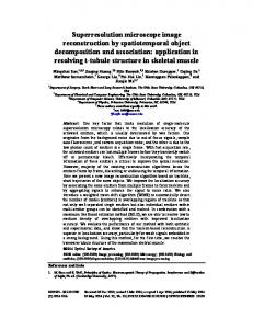
Figure 1: (a) Scanning electron microscope (SEM) image of a commercial Blu-ray® disk with 200 nm width stripes separated by 100 nm width grooves; (b) BTG microspheres with diameter sizes ~2 – 20 µm immersed in isopropanol used to resolve the sub-diffraction features of the sample (a); (c) imaging with an optical microscope with 20× and NA=0.4 objective lens through the microspheres. An FS70 Mitutoyo microscope in reflection illumination mode with 20× (NA = 0.4) microscope objectives was used for imaging. The white light illumination was provided by a halogen lamp with 600 nm peak wavelength. The subdiffraction features of the Blu-ray® disk cannot be resolved using conventional microscopy, as has been demonstrated previously [8,9]. Use of microspheres clearly increases the resolution dramatically. We showed that the super-resolution imaging of the Blu-ray® disk without liquid can be achieved using microspheres with moderate index of refraction such as borosilicate glass (n ~ 1.47), soda lime glass (n ~ 1.51), polystyrene (n ~ 1.59), and sapphire (n ~ 1.77). However, all these microspheres were found to lose their imaging capability, if they are completely covered with a layer of isopropanol. These results were found to be generally consistent with the previous studies performed for silica microspheres with n~1.46 [8,9]. High index (n ~ 1.9) BTG spheres showed very different behaviour in these experiments. Without liquid infiltration they did not produce any images. However, they provided super-resolution imaging in cases when they were totally covered with liquid, as illustrated in Fig. 1(c). The depth of focusing in Fig. 1(c) is below the surface of the structure, as can be seen by comparison with the case of near-surface focusing presented for the same liquid-infiltrated structures in Fig. 1(b). It is seen that the far field virtual images of individual 100 nm width grooves (dark stripes) are perfectly resolved in Fig. 1(c), which means that the resolution on the order of λ/6, well in excess of the Rayleigh resolution limit for the line objects (r = 0.5λ / NA), was demonstrated in this work. 3. CONCLUSIONS Super-resolution imaging by microspheres has emerged as a new paradigm in microphotonics [8,9]. Such imaging is connected with other extraordinary properties of microspheres such as photonic nanojets [10], nanojet-induced modes [11,12], polarization-transmission properties [13] and various other applications [14-15]. In this work, we experimentally demonstrated that the super-resolution imaging by microspheres totally immersed in the liquid is possible if the index of spheres is sufficiently high. By using BTG microspheres (2 – 20 µm diameters) with index around 1.9 totally immersed in isopropanol with index 1.37 we demonstrate better than λ/6 far field resolution. Similar results were obtained for various liquids including water. These results can be used in biomedical microscopy, microfluidics and nanophotonics applications for imaging individual cells and/or nanoparticles in a liquid environment. 4. ACKNOWLEDGMENTS This work was supported by the U.S. Army Research Office (ARO) through Dr. J. T. Prater under Contract No. W911NF-09-1-0450 and from the National Science Foundation (NSF) under grant ECCS-0824067.
2
ICTON 2012
Tu.A6.5
REFERENCES [1] B. Hecht, et al.: Scanning near-field optical microscopy with aperture probes: Fundamentals and applications, J. Chem. Phys., vol. 112, pp. 7761-7774, May 2000. [2] S.M. Mansfied and G.S. Kino: Solid immersion microscope, Appl. Phys. Lett., vol. 57, pp. 2615-2616, Dec. 1990. [3] Z. Jacob, L.V. Alekseyev, E. Narimanov: Optical hyperlens: Far-field imaging beyond the diffraction limit. Opt. Express, vol. 14, pp. 8247-8256, Sept. 2006. [4] Z. W. Liu, et al.: Far-field optical hyperlens magnifying sub-diffraction-limited objects, Science, vol. 315, p. 1686, Mar. 2007. [5] J.Y. Lee, et al.: Near-field focusing and magnification through self-assembled nanoscale spherical lenses, Nature, vol. 460, pp. 498-501, Jul. 2009. [6] S. W. Hell: Far-field optical nanoscopy, Science, vol. 316, pp. 1153-1158, May 2007. [7] I.I. Smolyaninov, Y.J. Hung, C.C. Davis: Magnifying superlens in the visible frequency range, Science, vol. 315, pp. 1699-1701, Mar- 2007. [8] Z. Wang, et al.: Optical virtual imaging at 50 nm lateral resolution with a white-light nanoscope, Nature Commun., vol. 2, pp. 1-6, Mar. 2011. [9] X. Hao, et al.: Microsphere based microscope with optical super-resolution capability, Appl. Phys. Lett. vol. 99, 203102, Nov. 2011. [10] Z. Chen, A. Taflove, V. Backman: Photonic nanojet enhancement of backscattering of light by nanoparticles: A potential novel visible-light ultramicroscopy technique, Opt. Express, vol. 12, pp. 12141220, Apr. 2004. [11] A.M. Kapitonov and V.N. Astratov: Observation of nanojet-induced modes with small propagation losses in chains of coupled spherical cavities, Opt. Lett., vol. 32, pp. 409-411, Feb. 2007. [12] S. Yang and V.N. Astratov: Photonic nanojet-induced modes in chains of size-disordered microspheres with an attenuation of only 0.08 dB per sphere, Appl. Phys. Lett., vol. 93, 261111, Jul. 2008. [13] A. Darafsheh, V.N. Astratov: Periodically focused modes in chains of dielectric spheres, Appl. Phys. Lett, vol. 100, 061123, Feb. 2012. [14] V.N. Astratov: Fundamentals and Applications of Microsphere Resonator Circuits, in Photonic Microresonator Research and Applications, I. Chremmos, O. Schwelb, and N. Uzunoglu, Eds., New York: Springer Series in Optical Sciences, vol. 156, 2010, pp. 423-457, ISBN: 978-1-4419-1743-0. [15] A. Darafsheh, et al.: Contact focusing multimodal microprobes for ultraprecise laser tissue surgery, Opt. Express, vol. 19, pp. 3440-3448, Feb. 2011.
3