D. BODZNICK, J. C. MONTGOMERY AND D. J. BRADLEY that radiate outwards and open to the exterior via skin pores. The receptor epithelium and the canal ...
J. exp. Biol. 171, 107-125 (1992) Printed in Great Britain © The Company of Biologists Limited 1992
107
SUPPRESSION OF COMMON MODE SIGNALS WITHIN THE ELECTROSENSORY SYSTEM OF THE LITTLE SKATE RAJA ERINACEA BY DAVID BODZNICK*, JOHN C. MONTGOMERY! AND DAVID J. BRADLEY Marine Biological Laboratory, Woods Hole, MA 02543, USA Accepted 23 June 1992 Summary The electroreceptors of elasmobranchs are strongly modulated by thefish'sown ventilation but this source of potential interference is suppressed within the medulla. The mechanism for the suppression is thought to be based on the common mode nature of the ventilatory noise, i.e. it is of the same amplitude and phase for all of the electroreceptors, compared with environmental electric fields which affect the receptors differentially. Evidence for the common mode suppression hypothesis is provided here in skates by the observation that the response to an artificial common mode stimulus that is independent of ventilation and delivered through an electrode inserted into the animal's gut is also suppressed by the medullary neurons; the extent to which a particular neuron suppresses the responses to the gut stimulus and to ventilation is similar. In addition, a potential modulation of 5-150/iV is measured between the skate's interior and the sea water during ventilation and this appears to be responsible for the self-stimulation. By passing d.c. or sinusoidal currents through the gut electrode it is demonstrated that this ventilatory potential is due to the variable shunting of a standing d.c. potential across the fish's skin by the opening and closing of the mouth and gill slits during ventilation. Osmoregulatory ion-pumping appears to contribute to the production of the d.c. potential.
Introduction Elasmobranch fishes possess an acutely sensitive electrosensory system with electoreceptor organs known as ampullae of Lorenzini. These ampullary organs are arranged in a series of 4-6 clusters on each side of the body. At each cluster, the receptor cells are located in subdermal alveoli at the blind endings of canals * Present address: Department of Biology, Wesleyan University, Middletown, CT 06457, USA. t Present address: Department of Zoology, University of Auckland, Auckland, New Zealand. Key words: elasmobranch, electroreception, noise suppression, reafference, medulla, Raja erinacea.
108
D. BODZNICK, J. C. MONTGOMERY AND D. J. BRADLEY
that radiate outwards and open to the exterior via skin pores. The receptor epithelium and the canal walls have a very high resistance in comparison with the conductivity along the canals themselves. With this arrangement, afferent firing rate effectively measures changes in potential difference between the sea water at the skin opening and the basal surface of the receptor cells in the interior of the animal. The best-documented behavioral role of the elasmobranch electroreceptors is in prey detection, where the system is used to detect the d.c. and lowfrequency bioelectric fields of other animals in the aquatic environment. However, it is also possible that uniform electric fields such as those induced by the motion of the animal itself or of ocean currents within the earth's magnetic field provide useful information for orientation and navigation. The anatomy, physiology and behavioral uses of the elasmobranch electrosense are reviewed in Bodznick and Boord (1986), Kalmijn (1988) and Montgomery (1988). The d.c. bioelectric fields of prey animals appear to result from differences in the electrical properties among the various body surfaces the animals have in contact with the surrounding water (Kalmijn, 1988) and in many cases probably include a major contribution from the potentials produced across ion-exchange surfaces. In fishes these d.c. potentials are thought to be modulated at low frequencies by changes in resistance along the current pathway caused by the opening and closing of the mouth and gill slits during ventilation. One problem inherent in the electrosensory system is that the animal must detect extremely weak bioelectric fields produced by other animals on top of its own very similar bioelectric fields. The electric fields produced by elasmobranchs are typically of lower amplitude than those produced by bony fishes (Kalmijn, 1974) but, even so, recordings from primary afferent fibers of the electrosensory system in the thornback ray, Platyrhinoldis triseriata, show afferent input to be strongly modulated by the animal's own ventilation (Montgomery, 1984). An interesting observation is that, in the secondary neurons of the electrosensory system in Platyrhinoidis, this ventilatory modulation is virtually absent. The animals are able to distinguish between afferent activity due to their own ventilation (reafference) and that due to extrinsic fields and they can effectively suppress the ventilatory reafference in the brain. What is the mechanism for this noise suppression? Montgomery (1984) showed that ventilatory modulation is of similar amplitude and phase in all afferent fibers from one ampullary cluster; in other words, ventilatory modulation is common mode. Afferents from canals of opposite orientation respond differentially to extrinsic fields, but show a very similar response to ventilation. This indicates that the ventilatory reafference is primarily driven by a potential modulation between the animal's interior and the surrounding water rather than by the small potential differences measured in the water around the fish during ventilation. These observations support an earlier suggestion by Kalmijn (1974) that the grouping of ampullae into clusters sharing a common internal reference may permit the elimination of unwanted noise by a mechanism of common mode suppression. New and Bodznick (1990) have recently shown that suppression of ventilatory
Electrosensory common mode signal suppression
109
reafference is also present in the little skate (Raja erinacea), though apparently to a lesser degree than that shown in Platyrhinoidis. They found that in Raja afferent modulation during ventilation is effectively common mode within all the electrosensory afferents, even those from opposite sides of the body, and direct evidence was obtained for a contribution of contralateral input to the noise suppression mechanism. The demonstration that ventilatory modulation is common mode shows that noise suppression could operate via a common mode suppression mechanism, and this is further supported by the importance of the contralateral input for the suppression (New and Bodznick, 1990). However, other possibilities exist. For example, in the weakly electric fish Gnathonemuspetersii, ampullary electroreceptors respond to the animal's own electric organ discharge. This unwanted reafference is removed by an elegant mechanism of modifiable efference copy (for a review, see Bell, 1986). Although no evidence was found for this mechanism in Raja (New and Bodznick, 1990), one aim of the present study was to provide a more direct test of the common mode suppression hypothesis versus other mechanisms by determining whether an experimentally imposed common mode electrosensory input, independent of ventilation, would also be suppressed within the central nervous system. In addition, we provide new information on the characteristics and origins of the bioelectric fields associated with ventilation in elasmobranchs. Materials and methods Experiments were performed on 20 specimens of the little skate Raja erinacea Mitchill at the Marine Biological Laboratory in Woods Hole, MA. Animals were caught in short trawl tows in Vineyard Sound and kept in cooled sea water until their return to the laboratory, where they were maintained in holding tanks at a temperature of 14°C. For experimentation, the animals were anesthetized by immersion in tricaine methanesulfonate (approximately 0.02%), the cranium was opened to expose the brain, which was then decerebrated by a transection at the optic chiasm. Some animals were then paralyzed by intravenous injection of tubocurarine chloride (3 mg kg" 1 ). In others, the spinal cord was transected about 1 cm behind the brain to permit normal ventilatory movements while immobilizing the animal's trunk and tail. A salt-bridge electrode of PE90 tubing filled with 1.5% agar in sea water was inserted into the gut via the anus. An additional Ag/AgCl electrode, made from 0.2mm diameter silver wire coated with Teflon except near the tip, was implanted through a small skin incision into the interior of the animal in the region between the hyoid and buccal ampullary clusters on the head. The skin incision was then sealed with tissue adhesive (Histoacryl, Trihawk). The animals were positioned on a Plexiglas head holder to stabilize the brain for microelectrode recording. A Plexiglas plate inserted through the mouth provided support for the cranium, but also meant that the mouth was held partly open. In the case of paralyzed animals, a stream of oxygenated sea water directed into the mouth provided for respiration. The rays were immersed in sea water up
110
D . B O D Z N I C K , J. C. MONTGOMERY AND D . J. BRADLEY
to the level of the cranial opening, and the water temperature in the experimental bath was regulated at 8-10°C. These procedures followed NIH guidelines for care and use of experimental animals and were approved by the Institutional Animal Care and Use Committees of the Marine Biological Laboratory and Wesleyan University. Electrosensory afferent activity was recorded with glass micropipettes (4 mol 1~' NaCl; 25 MQ) in the anterior lateral line nerve within the cranium. Platinumblack-tipped indium electrodes (2-5 [xm tip diameter, 2-7 MQ) were used to make recordings from neurons within the dorsal octavolateralis nucleus, where ascending efferent neurons (AENs), the principal output neurons, were identified by antidromic stimulation from the lateral mesencephalic nucleus. Single unit activity was recorded during ventilation, during stimulation from the gut electrode and during stimulation by uniform extrinsic electric fields. Spike data were analyzed from poststimulus time histograms of 25-50 ventilatory cycles or stimulus presentations. The response was measured as the peak-to-peak change in firing rate produced by the stimulus or, in the case of a zero-firing rate nonlinearity, the increase above spontaneous firing rate was measured and this valued was doubled to obtain the effective peak-to-peak modulation. Ventilation was monitored with the piezoelectric crystal of a phonograph cartridge coupled to movements of the branchial chamber. This signal was used to trigger the histogram program at the beginning of expiration. The stimulus delivered through the gut electrode was a continuous 1 or 2 Hz sine wave centered about zero and applied between the gut electrode and salt bridges (1.5 % agar in sea water) located along all four sides of the experimental aquarium. The amplitude was adjusted to give a 20 juV peak-to-peak signal measured between the interior of the animal and an indifferent electrode placed in the seawater bath. These amplitude and frequency values were chosen to approximate normal ventilatory potential modulations recorded from skates. Longitudinal and transverse uniform fields were applied through salt bridges as a continuous 2 Hz sine wave, typically at a peak-to-peak amplitude of 2juVcm~'. This intensity is within the linear range of the intensity response functions of afferents and dorsal nucleus neurons. The response of an electrosensory unit to a uniform field depends on the orientation of its canal with respect to the field. For this reason, the overall response of units to uniform fields was taken as the vector addition of their responses to the two fields presented at right angles. The receptive field of units was located by a small (5 mm) roving dipole electrode. Potential modulations measured between the Ag/AgCl electrodes inside the body and those in the water during ventilation were amplified by a PAR model 113 d.c. differential preamplifier with half-amplitude low-frequency cut-off at 0.03 Hz. In four animals, this so-called ventilatory potential was monitored after the intravenous injection of l-4mmol of NaCl (1 or 2 ml injections into the caudal vein) to salt-load the animal. This was done in order to stimulate increased osmoregulatory ion transport and thereby to assess its contribution to the potential. In two other cases, the internal electrode was implanted in an otherwise
Electrosensory
common
mode signal suppression
111
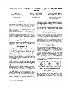
intact animal to record the size of the ventilatory potential and to monitor changes induced by placing the animal in diluted sea water. Results Ventilatory potential With a Ag/AgCI electrode inserted beneath the skin of a skate, an electrical potential modulation is measured between the fish's interior and the surrounding sea water coincident with ventilatory movements. This ventilatory potential lasts 1-3 s but can vary greatly in both waveform and amplitude among fish (Fig. 1A) and in a single fish at different times. Most often it takes the form of a biphasic wave, which is inside-negative during expiration and then inside-positive during inspiration, but in other cases the biphasic potential is reversed in polarity. More rarely the ventilatory potential is monophasic, triphasic or even more complex. In two unrestrained and unoperated animals the ventilatory potential amplitude was 5-27 fxV. The ventilatory potential in operated animals set up for physiological recordings ranged in peak-to-peak amplitude from less than 5/iV to more than 150 fiV, but was usually 15-25 fiV. Gradual changes in amplitude were often seen in the hour immediately after surgery. Cycle by cycle changes in the intensity of the normal breathing movements affected the ventilatory potential amplitude (see bottom record, Fig. 1A), and occasional 'coughs', when water was vigorously ejected through the spiracle, generally caused brief, large inside-positive ventilatory potentials. The pattern of firing of afferent electrosensory fibers during ventilation was as one would predict given their known excitatory response to a cathodal stimulus located at the skin pore. That is, when the internal potential became positive relative to the sea water, the firing rate in the afferents was accelerated; negative inside potential suppressed afferent firing (Fig. 1A). As noted previously (Montgomery, 1984; New and Bodznick, 1990), this is true regardless of the location of the skin pore of a particular receptor on the body surface. The degree of afferent modulation was also directly proportional to the amplitude of the co-occurring ventilatory potential (Fig. IB). The ventilatory potential was investigated experimentally. Placing a skate in 70 % sea water resulted in a threefold increase in the ventilatory potential over 30min; it returned to normal soon after the fish was returned to full sea water. Salt-loading animals by intravenous injection of l-4mmol of NaCl in order to stimulate enhanced osmoregulatory ion transport caused a relatively small or no immediate change in the potential, but after a lag of 5-20 h the potential increased dramatically to a peak of 230-350 fiV and then returned to more normal levels after 72-96 h (Fig. 2). The origin of the ventilatory potential was studied further with the use of a seawater-agar bridge electrode inserted through the anus into the fish's gut. Current from a constant-current isolator, passed between the gut electrode and salt-bridge electrodes on the four sides of the tank, created an electrical potential
112
D. BODZNICK, J. C. MONTGOMERY AND D. J. BRADLEY
80£
E
/
I
706050403
-a 30o E
l
E
l
E
I
20-
J
10-
a a
a
0 0
10
20 30 40 50 60 Ventilatory potential (/u,V)
70
Fig. 1. Modulation of electrosensory afferent firing closely follows the waveform and amplitude of a potential modulation recorded between the skate's interior and the surrounding sea water during ventilation. (A) Three examples from different fish show simultaneous recordings of primary afferent firing (top trace) and the ventilatory potential (bottom trace) recorded with a subdermal Ag/AgCI electrode relative to a second electrode in the water. Upward deflections in the ventilatory potential records represent increased positivity inside the fish relative to the sea water. Periods of expiration (E) and inspiration (I) are indicated under recordings. Calibration: horizontal, 0.5 s for all traces; vertical, traces 1, 2 and 5, 50 j.iV cm~\ traces 3, 4 and 6, 100 ^V cm"1. (B) Peak-to-peak afferent modulation is shown as a function of the ventilatory potential amplitude recorded simultaneously. Filled triangle indicates the mean response of afferents to a 20^V gut stimulus.
between the inside of the fish near the ampullary clusters in the head and the surrounding sea water that, as detailed below, appeared to be nearly the same over all areas of the skin surface that were under water. A 3-15 Hz sinusoidal potential produced by a current stimulus (200-300 ^iA) through the gut electrode and monitored between the internal Ag/AgCI electrode and the indifferent electrode
80
Electrosensory common mode signal suppression 350-
113
2mmolNaCl ——- 1 mmol NaCl
300250-
g 200 H Er 150 o
1
100
o
50
0
10
20
30 40 50 60 70 80 Time after NaCl injection (h)
90
100 110
Fig. 2. The effect of intravenous injection of either 1 or 2 mmol of NaCl on the amplitude of the ventilatory potential modulation in two skates.
in the sea water showed an amplitude modulation during ventilation due to apparent changes in the resistive pathway between the animal's interior and the sea water outside. The peak-to-peak amplitude of the potential created across the body surface decreased during expiration and increased during inspiration (Fig. 3A), coinciding with the separate phases of the usual bipolar ventilatory potential. In cases where the waveform of the ventilatory potential was nearly monophasic, the resistance changes measured in this way were similarly unidirectional and coincident with the ventilatory potential (Fig. 3C). In animals in which the normal ventilatory potential was otherwise quite small, a d.c. potential created by passing d.c. current of 100-250fiA through the gut electrode resulted in a large normal-looking ventilatory potential modulation measured with the subdermal Ag/AgCl electrode (Fig. 3B). Reversing the polarity of the gut current reversed the polarity of each phase of the ventilatory potential. In animals with larger ventilatory potentials, a d.c. gut stimulus of the correct polarity and amplitude canceled and even reversed the polarity of the normal ventilatory potential (Fig. 3C). These results support the hypothesis that the ventilatory potential is a modulation of a standing d.c. potential that exists across the fish's body surface. The d.c. gut stimulus required to cancel the normal ventilatory potential provides an estimate of this standing d.c. potential. The d.c. potential change created by a gut stimulus just sufficient to suppress the normal ventilatory potential ranged from 20-450 yiV, and in almost all cases the required polarity of the gut stimulus was negative. In two animals exhibiting a ventilatory potential of reversed phase, i.e. inside-positive on expiration, negative on inspiration, a positive d.c. gut stimulus suppressed the ventilatory potential and a negative potential enhanced it.
114
D. BODZNICK, J. C. MONTGOMERY AND D. J. BRADLEY
A
VP
\h^^\^
-150 ixA
E l
E l
E l
E
1
E
I
+ 125 fiA
J
-125 /iA £
/
E I
Fig. 3. (A) Resistance changes between the skate's interior and the surrounding sea water during ventilation are indicated (lower trace) by the varying amplitude of the potential created when a sinusoidal current stimulus (12 Hz) is delivered between a salt-bridge electrode in the fish's gut and similar electrodes on the sides of the aquarium. Potential was measured in this and all records of the figure with a subdermal Ag/AgCl electrode relative to a second electrode in the sea water; positive is upwards in all traces of the figure. Periods of expiration (E) and inspiration (/) are indicated beneath the traces. The upper trace is the normal ventilatory potential (VP) modulation recorded from the animal at approximately the same time. Note the first periods of the ventilatory cycle in the two traces are accurately aligned for comparison. Calibration marks at lower right indicate: horizontal, 0.8 s; vertical, upper trace 25 ftV, lower trace lOOjtiV. (B) Effects of passing d.c. current between the gut electrode and sea water on the ventilatory potential. The top trace shows that the normal ventilatory potential is nearly undetectable in this skate. The lower traces show normal-looking biphasic ventilatory potentials recorded while delivering the indicated d.c. currents throughout the trace. At onset, 125 fiA current caused a d.c. potential shift of about 200fiV between the skate's interior and sea water. Calibrations: horizontal, 0.8s; vertical, 50,uV. (C) As in B, but in this skate the ventilatory potential (top), which is a simple monophasic positive wave, is canceled (middle) and then reversed in polarity (bottom) by the d.c. currents indicated. The onset of — 150/uA current created a d.c. potential shift of 200 ;uV, indicating that the normal standing d.c. potential in thisfishis 200/xV inside-positive. In the bottom record the increased amplitude of the transcutaneous potential created by sinusoidal current (3.3 Hz) from the gut electrode (as in A) indicates a simple monophasic resistance increase between the skate's interior and the sea water, which is coincident with the ventilatory potential. Calibrations: horizontal, 0.4s; vertical, top three traces 50juV, bottom trace 125^V.
Electrosensory common mode signal suppression
115
Central suppression of common mode signals The gut electrode was used to create a common mode stimulus, that is one of nearly the same amplitude and phase, for all of the electroreceptors as a means of testing the hypothesis of a central common mode rejection mechanism for suppressing ventilatory reafference. The potentials created by the gut electrode stimulus were measured with the subdermal Ag/AgCl electrode in the region of the ampullary clusters and an indifferent electrode, which was moved to a series of locations around the skate, in the bath. The records in Fig. 4 show that the gut electrode produced a good common mode stimulus with a nearly identical potential modulation for virtually all of the electrosensory canals with skin pores beneath the water surface, though the field in the region of the most caudal hyoid pores in some animals was slightly phase-shifted and smaller in amplitude than at other locations (Fig. 4, position E). The significant exception to this was found in the case of the dorsal and medial hyoid pore group. The skin pores of these receptors were located above the water surface in the experimental situation, and
50 ixV 0.1s
Fig. 4. The gut stimulus is nearly identical for all electroreceptors in the water. On the left are shown the potentials recorded between the subdermal Ag/AgCl electrode (asterisk on skate drawing) and a similar electrode positioned in the sea water at the locations shown on the right. An exception is the reversed-phase potential recorded on the skin above the water at F. Dashed line around F indicates water level.
116
D. BODZNICK, J. C. MONTGOMERY AND D. J. BRADLEY
the potential on the surface of the skin in this region was phase-reversed in comparison with the potentials recorded in the sea water. The common mode nature of the gut stimulus was confirmed in recordings from more than 200 primary electroreceptive afferents. Afferents from canals of different orientations and locations on the body responded differentially to uniform electric field stimuli, but gave nearly identical responses to the gut stimulus (Fig. 5). The ampullary organs of skates in a uniform electric field are sensitive to the potential drop across the skin plus the drop within the animal's body along the length of the ampullary canal. Thus, as illustrated in Fig. 5, afferents from electroreceptor organs with longer canals appear to be more sensitive to uniform fields than those from organs with shorter canals. However, the amplitude of the receptors' responses to the gut stimulus was independent of canal length, indicating that the skate's interior, at least in the region of the ampullary clusters in the head, is nearly isopotential with respect to the gut stimulus and that the effective voltage drop is across the skin. The amplitude of the response to the gut stimulus was also similar to the afferent modulation produced by a ventilatory potential of the same magnitude (Fig. IB). The common mode nature of the response to the gut stimulus was evident in all afferents with a receptive field located in the water. In several instances afferents were found whose receptive fields could not be located. These were presumed to be in the dorsomedial hyoid group and their responses were phase-reversed, consistent with the polarity of the field produced by the gut stimulus in that location. The responses of 62 AENs and 85 primary afferents to the gut stimulus and to uniform electric fields in the water were compared. A signal to noise ratio (S/N) for each electroreceptive unit studied was defined as the overall response of the unit to uniform fields of 2 /iV cm"' (signal) divided by its response to the 20 //V gut stimulus (noise). The S/N in primary electroreceptor afferents was relatively homogeneous and averaged 0.96 (S.D. 0.7, N=85) (Fig. 6). In contrast, the S/N for AENs was quite variable, even within a single animal. Approximately onethird had S/N values similar to that of primary afferents and the remaining twothirds were larger, indicating suppression of the response of these AENs to the gut stimulus compared with that to extrinsic fields (Figs 6, 7). The mean S/N for all AENs recorded was 4.6 (S.D. 9.1, N-62), a value 4.8 times higher than the average for primary afferents. A subset of cells in Fig. 6 was recorded from five animals that were not paralyzed by curare injection but instead had spinal transections and were ventilating naturally. For both primary afferents and AENs in these animals the ventilatory modulation was also measured and an S/N value was determined where the signal was again the response to uniform fields (2 Hz sinusoidal stimuli; 2jitVcm~l) but the noise was the peak-to-peak ventilatory modulation. The uniform field stimuli were presented without a fixed phase relationship to the ventilatory cycle, which had a variable 1.5-3 s period. For primary afferents the mean S/N was 1.2 (S.D. 0.7, N=47) and, as with the gut stimulus, the S/N for AENs was quite variable but averaged 4.8 (S.D. 6.2, N=28), four times higher than
Gut
Is
4
0
1
0
- 1s Is
1 "s
~
~
-7-7 1s
40
-
s
7
'
1s
'.m
1s
lw .v
1s
;":-""1 40 1
40
1s
--
Fig. 5 . The common mode nature of the gut stimulus is indicated by poststimulus time histograms of responses from four primary afferents (with receptive fields shown on the central diagram) to the gut stimulus and to longitudinal and transverse uniform fields of 2 p ~ c m - ' . Responses to uniform fields vary depending on the length and orientation of the ampullary canal of the particular receptor, but responses of all four units to the gut stimulus are nearly identical. Each histogram is of 30 trials of 1s duration; maximum on vertical axis is 40 counts per 32 ms bin.
---I
Transverse
rw 1s 4
'"r"l -
Longitudinal
1 s P2
~ --r=-+d i ~;j
40.
118
D. BODZNICK, J. C. MONTGOMERY AND D. J. BRADLEY 70 T Q Primary afferent neurons • Ascending efferent neurons
60-
£ 40 o |
30-|
1
20 10-
0
— O
cN —'
m (N
t 5 n ci
•*