The stomatogastric nervous system of Homarus gammarus. A, Lateral view of the stomach showing the in situ location of nerves and ganglia on the dorsal part of ...
The Journal
Suppression of Oscillatory Activity Implication of GABAergic Inputs J. R. Cazalets, Laboratoire
I. Cournil,
de Neurobiologie
M. Geffard,”
of Neuroscience,
Crustacean
September
1987,
7(9):
2884-2893
Neurons:
and M. Moulins
et Physiologie ComparBes,
CNRS and Universite
Generation of rhythmic pyloric motor output in the crustacean stomatogastric ganglion results from synaptic connections and cellular properties of a 14-cell network of pyloric neurons. These cellular properties are under the influences of modulatory inputs, which act, for the most part, in an activating mode, i.e., they enhance the bursting properties of the pyloric neurons and/or their ability to express their regenerative properties. Here we attempt to demonstrate that the pyloric motor output is also under the control of suppressive afferent inputs that are able to stop the pyloric rhythm in a long-lasting manner. lmmunohistochemistry, using GABA antibodies, indicates that GABAergic-like fibers are present in both the stomatogastric ganglion and its afferent nerve. Bath-applied GABA suppresses spontaneous pyloric rhythmic activity. This is due to an inability of the pyloric pacemakers to express their bursting properties. The suppressive effect of GABA is blocked by picrotoxin and mimicked by muscimol. Isolating the pyloric neurons from all descending spiking influences with tetrodotoxin demonstrates that exogenously applied GABA acts directly on the pyloric neurons. To confirm the existence of a physiological suppressive system for the pyloric motor pattern, we show that the stimulation of an afferent nerve, known to contain GABA-like fibers, also causes the cessation of rhythmic activity and the inability of the pyloric neurons to express their bursting properties.
It is increasinglyevident that central rhythmic motor networks are under the control of modulatory inputs (Selverston and Moulins, 1985;Harris-Warrick, 1987).Theseinputs do not control the pattern of the output step by step, ascan proprioceptive feedback, but have, instead, long-lasting instructive effectsthat shapethe final motor output. One of the best-known examplesof such modulatory control is the 14- neuron network of the crustaceanstomatogastricganglion, which drives the alternating dilations and constrictions of the pyloric chamber(Moulins and Nagy, 1985;Marder, 1987). All the neurons involved in this network have the ability to
Received Nov. 17, 1986; revised Mar. 6, 1987; accepted Mar. 9, 1987. We wish to thank E. Marder, S. L. Hooper, F. Nagy, and J. Simmers for critical comments on the manuscript and help in preparing the English version of the text. This work was supported by MRT Grant 85-C- 1152. Correspondence should be addressed to Jean-Rent Cazalets, Laboratoire de Neurobiologie et Physiologie ComparCes, CNRS, Universite de Bordeaux I, Place du Dr. Peyneau, F-33 120 Arcachon, France. a Present address: Laboratoire de Neuroimmunologie, Institut de Biologie Cellulaire et Neurochimie, CNRS, Domaine de Carreire, rue Camiile Saint-Saens, F-33077, Bordeaux, France. Copyright 0 1987 Society for Neuroscience 0270-6474/87/092884-10$02.00/0
de Bordeaux
I, F-33120 Arcachon,
France
produce bursting pacemaker potentials (BPPs; Miller, 1987), i.e., regenerative depolarizations and hyperpolarizations that underlie their bursting discharge.The voltage-dependent conductancesresponsiblefor theseregenerativepropertiesareunder the control of modulatory inputs, sincetheir removal decreases or turns off the production of pyloric BPPs, and hencerhythmic output (Moulins and Coumil, 1982; Nagy and Miller, 1987). Only a few modulatory input neurons to the pyloric network have been identified (Nagy et al., 1981; Russelland Hartline, 1981; Dickinson and Nagy, 1983; Nagy and Dickinson, 1983), but there is pharmacological evidence (Beltz et al., 1984; Claiborne and Selverston, 1984; Marder and Eisen, 1984; Marder, 1987) that suggeststhat this network is the target of a palette of such inputs. To date all the identified modulatory inputs (Russelland Hartline, 1981; Dickinson and Nagy, 1983; Nagy and Dickinson, 1983) and most of the putative neuromodulatory substancesapplied to the network have permissive effects on the pyloric output (Beltz et al., 1984; Hooper and Marder, 1984;Marder and Eisen, 1984; Harris-Wanick, 1987).They are all able to start the pyloric rhythm in a quiescentganglion, or to enhanceongoingpyloric activity, even if in somecasesthere is a diminution in the dischargeof someof the target neurons (Flamm and Harris-Warrick, 1986a, b). The presentstudy wasundertaken to determineif this network also receives modulatory “suppressive inputs,” that is, inputs that reduce and finally suppressits rhythmic activity. Marder and Paupardin-Tritsch (1978) have shown that iontophoretic application of GABA resultsin hyperpolarization and conductance increasein several pyloric neurons. We proceeded from this observation to determine if (1) the pyloric network receives GABAergic-like inputs, (2) exogenously applied GABA suppressesthe ability of pyloric neuronsto produce BPPs, and (3) some of the neuronal inputs to the pyloric network have suppressive effects on the pyloric output. A brief report of this work has appearedelsewhere(Cazalets et al., 1985).
Materials
and Methods
Experimentswere performedon the EuropeanlobsterHomarus gammurus andthe capelobsterJusus lakzndii; althoughsimilarresultswere obtainedfrom both species, all resultsreportedherearefromHomarus. The animalswerekeptin tanksof aeratedandcirculatingseawater. Two experimentalsalineswereused:artificial seawater (MoulinsandVedel, 1977),400 mMNaCl, 9.8 mM KCl, 10.1mMCad&, 52.6 mMMgCl,, 27.8mMNa,SO,,2.5mMNaHCO,,0.6mMNaBr;andHEPES-buffered saline(Miller and Selverston,1982).479 mM N&l, 12.74mM KCl. 13.7rn& CaCl,, 10mM MgSO,, 3.9 mM Na,SO,,-5&M HEPES.The pH of the salineswasadjustedto 7.45with HCl or NaOH. GABA immunohistochemist~. The anti-GABA antibodiesusedfor immunohistochemistry werepurified,by affinity chromatography, from
The Journal
of Neuroscience,
September
1987,
7(9)
2885
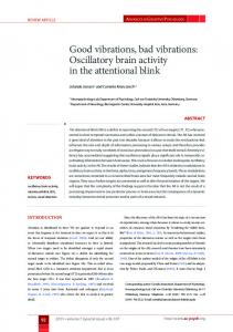
Figure 1. The stomatogastric nervoussystem of Homarusgammarus. A, Lateral view of the stomach showing the in situ location of nerves and ganglia on the dorsal part of the stomach and around the esophagus. B, The isolated nervous system. The circle around the STG represents the Vaseline pool through which drugs were superfused. C, Simplified wiring diagram of the pyloric motor network. l -, Inhibitory synapse, --+.+ electrotonic junction. Abbreviations: AB, anterior burster intemeuron; B, brain; C,, early constrictor muscle; C,, late constrictor muscle; COG, commissural ganglion; CS, cardiac sac; D, dilator muscle; dvn, dorsal ventricular nerve; GM, gastric mill; ZC, inferior cardiac motoneuron; ion, inferior esophageal nerve; LP, lateral pyloric motoneuron innervating C,; lvn, lateral ventricular nerve; M, mouth, OE, esophagus; OG, esophageal ganglion; on, esophageal nerve; P, pylorus; PD, pyloric dilator motoneuron; PY, pyloric motoneuron innervating C2; son, superior esophageal nerve; STG, stomatogastric ganglion; stn, stomatogastric nerve; VD, ventricular dilator motoneuron.
rabbit anti-GABA antiserum obtained by Seguela et al. (1984). The anti-GABA antiserum does not cross-react with glutamate, aspartate, or other related molecules, such as alanine, glycine, or taurine (Seguela et al., 1984). The stomatogastric nervous system and the second and third abdominal ganglia were dissected in artificial seawater (adjusted to pH 6.2 with HCl) and fixed with 5% glutaraldehyde diluted in 0.4 M cacodylate buffer (pH 7.75) for 90 min. The nervous tissue was first reacted with 1% lysine for 1 hr, then with 0.1% sodium borohydrate for 1 hr (to reduce the autofluorescence of the fixed tissue), and postwashed overnight at 4°C in PBS (0.0 1 M phosphate buffer containing 0.15 M NaCl), pH 7.45, to which was added 10% sucrose. Frozen sections (18 p) were incubated for 18 hr with the anti-GABA antibodies diluted l/200 in PBS containing 0.3% Triton X- 100 and 10% BSA (in order to prevent nonspecific binding). After a 1 hr wash in fresh PBS, the reaction of the anti-GABA antibodies was visualized by incubating the sections for 1 hr with sheep anti-rabbit IgG linked to fluorescein isothiocyanate (FITC; Miles) diluted l/100 in PBS. After washing, the sections were mounted in a 1:3 mixture of PBS-glycerol and observed with a Leitz fluorescence microscope (excitation filter, 450-490 nm; emission filter, 520 nm). Absorption controls were carried out to test the specificity of antiGABA antibodies used in this procedure. The anti-GABA antibodies were diluted according to the normal procedure and preincubated for 24 hr with or without the immunogen (GABA-glutaraldehyde-BSA). The 2 solutions were then centrifuged and the supematants collected. Successive sections of the ganglia were treated alternately with the absorbed and nonabsorbed antibodies and subsequently observed and compared under fluorescence as above. Electrophysiological techniques. After removing the stomatogastric nervous system from the dorsal part of the stomach (Fig. lA), the isolated nervous system was pinned out in a Sylgard-lined petri dish (Fig. 1B) and maintained at 15°C. Isolated preparations, corresponding to the “combined preparation” of Selverston et al. (1976) and consisting of the stomatogastric ganglion (STG), the paired commissural ganglia (COGS), and the esophageal ganglion (OG), were used. Intrasomatic recordings of pyloric neurons were made with glass microelectrodes filled with 2 M potassium acetate (r = 15-20 MQ). Neurons were identified as described for Homarusby Robertson and Moulins (198 1) and for Jasus by Nagy (198 1). For intracellular current injections, cell bodies were always penetrated with 2 microelectrodes, one for membrane potential recording and the other for current injection. Neuronal activity was recorded on a MP552 1 Schlumberger tape recorder and visualized either on a 5 113 Tektronix oscilloscope or a Gould ES 1000 electrostatic recorder. The effects of GABA and other drugs were studied by superfusing (1 ml/min) the solutions through a Vaseline well (2 ml vol) built around the STG. GABA, picrotoxin (PTX), tetrodotoxin (TTX), and oxotremorine were obtained from Sigma; muscimol from Serva. Axonal conduction in nerves could be reversibly blocked by superfusion of an isotonic sucrose solution (750 mM) (adjusted to pH 7.45 with NH,OH)
through a Vaseline pool built around a desheathed portion of the appropriate nerve.
Results GABA-like immunoreactive projections to the STG The lobster STG consists of a dense neuropil surrounded by neuronal somata. The axons of pyloric motoneurons project from the STG in several motor nerves, the principal one being the dorsal ventricular nerve (dvn) (Fig. 1B). The STG is connected to the anterior ganglia (COGS and OG) by a single nerve tract, the stomatogastric nerve (stn, Fig. 1, A, B), which carries numerous input fibers to the STG. With anti-GABA antibodies we visualized immunoreactive fibers in the neuropil of the STG (Fig. 2, A, D), but none of the cell bodies in this ganglion were labeled. GABA-like immunoreactive fibers were also detected in the input nerve (stn, Fig. 2B), but not in the main output motor nerve (dvn, Fig. 2C). Although the results shown here are from Homarus, identical results were obtained with Jasus. Since none of the cell bodies stained in the STG, it is likely that the immunoreactive neuropilar structures are the terminal processes of neurons whose somata are located in other ganglia and project to the STG via the stn. In Homarus, some of the immunoreactive fibers projecting to the STG probably originate in the OG, in which we saw at least 3 strongly immunoreactive cell bodies, and/or the COGS, which also contain some small immunoreactive cell bodies. The nerves that interconnect these ganglia (see Fig. 1A) also contain GABA-like immunoreactive fibers. These results depend critically on the specificity of the antibodies. Although several tests have been carried out previously to establish this specificity (Seguela et al., 1984), we have performed 2 further controls. First, we demonstrated that the antibodies recognize identified GABAergic neurons in the lobster abdominal ganglion (Otsuka et al., 1967). Figure 3B shows that after treatment with the anti-GABA antibodies, the somata of these neurons, 12, and 13, were strongly immunoreactive. Several small-diameter cell bodies, probably including 11 of Otsuka et al. (1967), were also labeled, but numerous other neurons of the ganglion were not. Thus, the antibodies used in the present study do seem to recognize neuronal structures known to be
2888 Cazalets
et al.
l
GABAergic
Inputs to Crustacean
Pyloric Neurons
Figure 2. GABA-like immunoreactivity in and near the stomatogastric ganglion ofhlomarus. A, Longitudinal section of the stomatogastric ganglion after incubation with GABA antibodies (the stn is to the left, the dvn to the right). Note immunoreactive fibers in the neuropil and the absence of labeled cell bodies (arrow.) B, C, Immunoreactive fibers in a transverse section of the input nerve (stn; B) but not in a transverse section of the main motor nerve (dvn; C). D, E, Absorption control on serial transverse sections of the stomatogastric ganglion: positive reaction observed in the neuropil of a section incubated with the nonabsorbed antibodies (0) disappeared in the subsequent section when incubated with the absorbed antibodies (E). Calibration lines; 50 pm (A); 25 pm (B-E).
The Journal of Neuroscience,
September
1987, 7(9) 2987
Figure 3. Cell bodies of known GABAergic inhibitory motoneurons in the abdominal ventral cord of the lobster Homamslabeled with GABA antibodies used in the present study. A, Map of the identified cells in the third abdominal ganglion. The cell bodies of inhibitory motoneurons are marked in white (12, 13) (from Otsuka et al., 1967). B, Longitudinal horizontal section of the third abdominal ganglion after antibody treatment; 12 and probably 13 neurons are immunoreactive. Calibration lines, 100 pm. GABAergic. Second, after absorption by the antigen, the antibodiesare unableto stain either identified GABAergic structures or the structures in the STG that were labeled prior to absorption. Absorption controls (see Materials and Methods) were performed on both the GABAergic neurons of the ventral nervous chain (data not shown)and the immunoreactive structures of the STG (Fig. 2, D, E). In all cases,the immunological reactions disappearedwhen the sectionswere treated with antibodies that had been incubated with the immunogen. Thus it
A
CONTROL
B 5x 10⁻⁴M GABA
seemsthat the antibodies specifically recognize GABA when coupled to proteins by glutaraldehyde, and we conclude that the structureslabeled in the stn and STG contain GABA or at least a GABA-like compound. Suppressionof the pyloric output by GABA application The activity of the pyloric neurons can be recorded intracellularly from in vitro preparations (Fig. 1B). The pyloric output consistsof the sequential activity of 6 types of neurons (Fig.
C WASH
AB
PD
LP
VD
v
I/
VI
Figure4. Bath-application of GABA on the STG causes cessation of pyloric rhythmic activity. Simultaneous intracellular recordings from the 6 types of pyloric cells (see Fig. 1C). A, Control. Z?,Superfusion over the STG of saline containing 5 x 1O-4 M GABA stops the bursting discharges of the 6 neurons, although they continue to fire tonically at low frequency. C, Restoration of the rhythm 10 min after wash with normal saline. Horizontal bar, 0.5 set; vertical bars, 5 mV.
2888
Cazalets
et al. * GABAergic
Inputs
to Crustacean
Pyloric
Neurons
tions indicate that the cessationof oscillationscausedby GABA is not due simply to a hyperpolarizing effect. Suppressionof the oscillatory propertiesof the pyloric pacemakersby GABA application The generation of pyloric motor activity depends largely on endogenousneuronal properties (Russell and Hartline, 1978) that are expressedonly under the influence of modulatory inputs arising from“anterior centers” (Miller and Selverston, 1982; Moulins and Coumil, 1982; Russelland Hartline, 1982).These propertiescan be testedby (1) sustainedartificial depolarization, which evokes oscillatory behavior in a nonoscillating neuron n I r i (see Fig. 9A), or an increase in the frequency of an already oscillating neuron (Fig. 5A, 1); or by (2) a brief depolarization, which triggersa full regenerativedepolarizing responsethat outBI 2.5~1o’~bl GABA B2 lasts the activating depolarizing pulse (Fig. 5A, 2). These tests LlluliJw were performed on the pyloric pacemakerneurons in the presence of GABA. Sustaineddepolarization (even at high current levels) did not evoke cycling, but only increasedthe tonic firing of the stimulated neurons (Fig. 5B, l), while brief, strong depolarizations failed completely to trigger oscillatory potentials (Fig. 5B, 2). I i ----A The following resultsfurther reinforce the notion that, in the Figure 5. GABA appliedto the STG causes the pyloric pacemaker presenceof GABA, the pyloric neurons are no longer able to express their bursting properties. Extracellular stimulation of neurons(PD and AB) to be unableto fire rhythmic burstsof action potentials.A, Control.BeforeGABA application,the2 pacemakers were the stn is known to activate input fibers that promote the burstoscillatingspontaneously. Sustained depolarization(bycurrentinjection ing potential of pyloric neurons;in a a systemwhere the pyloric into PD) increasedthe frequencyof oscillation(A,), while a brief deneuronsare not cycling, a brief stimulation of the stomatogastric polarizationprovokedan immediate,full oscillation(A,). B, During nerve can initiate and sustain the oscillatory activity of the GABA application(2.5 x 1O-4M), a long-durationdepolarizationdid not induceanyoscillations(B,) and a brief depolarizationwasunable pyloric neurons (Fig. 6A; note the long-term activating effect of to provokea regenerativeresponse (B,). Horizontalbars,1.25set (A,, this stimulation). In the presenceof 2.5 x lo-“ M GABA, howB,); 0.5 set (Ar, Bd).Vertical bars, 12mV (PD); 15mV (AB); and 30 ever, a longer stimulation at the samefrequency had no such nA for injectedcurrent(i). activating effect on the bursting potential of the PD and AB neurons (Fig. 6B). Poststimulus injection of depolarizing current, which would be expected to counteract a simple GABA4A). The 2 pyloric dilator motoneurons (PD) and the anterior mediated membrane hyperpolarization, was also ineffective in burster interneuron (AB) are electrically coupled, have endogtriggering oscillations. enousoscillatory properties, and act as pacemakersfor the pyTheseexperiments thus show that the inability of the pyloric loric network (Miller, 1987). They rhythmically inhibit all the neurons to burst in the presenceof GABA is not due only to a other pyloric neurons, the synaptic interactions of which (Fig. simplemembranehyperpolarization; rather, GABA must cause lc) determine the timing of their bursts of action potentials either the inactivation of a conductance directly involved in and hence the structure of the pattern. The motor pattern reBPP production or an increasein a steady hyperpolarizing concorded from isolated stomatogastric nervous systemsis comductance that shuntsthose currents that generatebursts. parable to that recorded by electromyography in the intact anPharmacological characterization of the GABA effect imal (Rezer and Moulins, 1983). In most isolatedpreparations, the pyloric neurons discharge rhythmically for hours at freTo eliminate the possiblity of a nonspecific action of GABA on quenciesvarying from 1 to 0.2 Hz, depending on the preparathe pyloric neurons, and to begin characterization of the memtion. In preparations in which the pyloric neurons oscillate at brane receptors involved in its suppressiveeffect, several an1 Hz (Fig. 4A), complete cessationof all rhythmic activity was tagonistsand agonistsof GABA were tested. PTX is known to obtained by superfusingthe STG with saline containing 5 x block the action of GABA in many preparations (Takeuchi, 1O-4M GABA (Fig. 4B). With preparations in which the spon1976). In a STG in which the PD neuronswere cycling strongly taneouspyloric rhythm wasinitially slower, the sameresult was and spontaneously(Fig. 7A), the application of salinecontaining obtained using 1O-4M GABA. The effectsof GABA application 3.5 x 1O-4M GABA silencedthe PD neuron (Fig. 7B). However, are rapidly reversible, oscillatory pyloric activity being comsaline containing the sameconcentration of GABA applied in pletely restoredafter 10 min of washingwith normal saline(Fig. the presenceof PTX was without effect on PD bursting activity 4c). In an earlier study, Marder and Paupardin-Tritsch (1978) (Fig. 7c), aswasbath-applied PTX alone(Fig. 70). Bicucculine, found that GABA causeshyperpolarization and a conductance a well-known GABA antagonist (Curtis et al., 1970) was effecincreasein pyloric neurons. Moreover, the membraneconductive only at high concentrations (1O-3 M), and its effect wasless tances that underlie the slow-wave oscillations are voltage-dereproducible than that of PTX. This is consistentwith the results pendent and can be inactivated by steady hyperpolarization of Marder and Paupardin-Tritsch (1978), who found that 1O-4 (Russelland Hartline, 1982). However, the following observaM PTX partially suppressedthe hyperpolarizing responseof
The Journal
A
of Neuroscience,
September
1987,
7(9)
2889
NORMAL SALINE
PD
.... -72mV
i
-
Figure 6. In the presence of GABA, the bursting properties of the pacemaker (A& PD) neurons can no longer be unmasked by stimulation of the stn. A, In a preparationthat wasnot cycling,stnstimulationinducedlong-termoscillatoryactivity in thepenetrated neurons.B, Duringsuperfusion of the ganglionwith 2.5 x 1O-4M GABA, strongerstimulation(stim)of the stn wasunableto induceneuronaloscillations,and a sustained depolarization
of PD produced only tonic firing. Horizontal
bar, 1 set; vertical bars, 10 mV, 10 nA.
STG neurons to iontophoretically applied GABA and that bicucculine (1O-4 M) had no effect. Muscimol, a potent agonistof GABA (Krogsgaard-Larsenand Arm, 1979), had actions similar to those of GABA, but was effective at lower concentrations. Bath-applied muscimol(7.5 x 1O-6 M) reduced the rhythmic activity of an oscillating PD neuron (cf. Fig. 7, E, F). At higher muscimol concentrations (5.5 x 10-SM), the oscillatory activity of the PD neuron stopped completely (Fig. 7G). As for GABA, this effect was reversible; a few minutesafter the beginning of washingwith normal saline, the PD oscillations were completely restored (Fig. 7H). The suppressiveeffectsof GABA are TTX-resistant All the above resultswere obtained from preparations in which the STG remainedconnected to the premotor centers(Fig. 1B). It has previously been shown that the pyloric neurons receive extrinsic neuromodulatory inputs that have an activating effect on their oscillatory behavior. Thus it remains possiblethat the inactivating effects of GABA seenhere may be due to presynaptic inhibition (Kirk and Wine, 1984) of these activating inputs. To test this possibility, the pyloric neurons were isolated from all spiking synaptic inputs by superfusingthe STG with salinecontaining 2 x lo-’ M TTX. Under theseconditions, the pyloric pacemakersstoppedoscillating (Fig. 8B), but it waspos-
sibleto reinducenonspikingpyloric oscillatory behavior by bathapplication of a muscarinic agonist, oxotremorine (Fig. SC), which mimics the activating effects of an identified cholinergic input (Nagy et al., 1985). The critical experimental observation was that, although completely deafferented, oxotremorine-activated pacemakerneuronswere still sensitiveto GABA. Again, bath-application of salinecontaining lo+ M GABA causedcomplete cessationof oscillatory activity (Fig. 80). At this GABA concentration, a brief current pulse injected into a PD neuron still triggered a reduced oscillation in the PD neuron, aswell as in the electrically coupled AB interneuron. A single (reduced) oscillation wasalsoobtained by steady depolarization of the PD neuron (Fig. 8D). However, with a slightly increased GABA concentration (2.5 x 1O-4 M), the pacemaker neurons completely lost their regenerative ability. Even large-amplitudecurrent pulsesor strong, sustaineddepolarizations failed to trigger oscillations (Fig. 8E). After several minutes of washwith saline containing TTX and oxotremorine alone, the cycling activity of theseneuronswasrestored(Fig. 80. Theseresultsshowthat, for oxotremorine-induced BPPs in the presenceof TTX, the suppressiveeffectsof GABA do not occur via mechanismspresynaptic to the target neurons. We have no reason to believe that spontaneousBPPs are qualitatively different from BPPs induced by oxotremorine, and thus it is likely that, in normal
2990
B
Cazalets
et al. - GABAergic
3.5~10-~M
GASA 1
C
D
Inputs
,I
to Crustacean
Pyloric
Neurons
F
7.5x 10⁶M
MUSCIMOL
G
5.5~10~
MUSCIMOL
i
3.5x 10-94 GASA 10-4M PTX
lo-“M
PTX
Figure 7. The action of GABA is antagonized by PTX and mimicked by muscimol. A-D, The oscillatory behavior of a PD neuron (A) was suppressed by bath-application of GABA containing saline (B). However, the same concentration of GABA was ineffectke in the presence of PTX (0. Bath-aDnlication of PTX alone did not modifv the cvcline of the PD neuron (0). E-H, Bath-applied muscimol diminished @) 0; suppressed (G) the oscillatory activity of a PD neuron (compare to E). This effect was reversible; 8 min after the beginning of the wash with normal saline, the oscillatory activity was completely restored (IQ. Horizontal bars, 0.5 set (A-D); 1 set (E-H). Vertical bars, 10 mV.
saline as well, GABA’s suppressiveeffects are due to direct action on the pyloric pacemakerneurons.
Comparable suppressive effects can be obtained by stimulating STG input fibers To demonstrate that the pyloric pacemaker neurons receive inactivating GABAergic inputs requiresselective stimulation of identified GABAergic fibers and evidence that theseinputs have a suppressiveeffect on the oscillatory behavior of the pyloric pacemakers.To date, such input neurons have not been identified. However, it has been possible to show that electrical stimulation of multifiber input nerve to the STG can causethe cessationof pyloric rhythmic activity in a way similar to that of bath-applied GABA. Stimulation of the stn itself cannot be used becausethis input nerve contains many activating fibers and its stimulation normally initiates or enhancesthe oscillatory activity of the pyloric neurons(seeFig. 54). We therefore used the esophagealnerve (on), which we know by immunohistochemistry contains GABA-like fibers (see above), but which, unlike the stn, carries relatively few activating elements that descendto the STG. In the experiment shown in Figure 9A, we recorded from a PD neuron that wasquiescent,probaby owing to hyperpolarization, sincesustaineddepolarization could evoke oscillation at a constant frequency (Fig. 9, upper trace). However, a similar sustaineddepolarization applied 2 set after a 25 Hz, 10 set stimulation of the on failed to produce any oscillatory
activity (secondtrace, Fig. 9A). Thus, following stimulation of the on, the PD neuron was unable to express its oscillatory properties. This is directly comparableto the experiment shown in Figure 9B, in which the oscillatory behavior of a PD neuron is suppressedby on stimulation. This experiment also shows that on stimulation producesnot only poststimulushyperpolarization of PD, but also the loss of its oscillatory properties; a sustaineddepolarization immediately after the stimulation is totally unable to produce oscillations. Sincethe on clearly contains fibers that have direct excitatory effects on the PD neuron (ascan be seenduring on stimulation in Fig. 9) the subsequentinactivation of the PD neuron could be due to post-tetanic depressionrather than to the stimulation of suppressivefibers. Although this possiblity cannot be completely ruled out, PD neurons never exhibit long-term posttetanic depressionin responseto high-frequency firing induced either by depolarizing current injection or by stimulation of known excitatory synaptic inputs. Moreover, the fact that strong inactivation of the PD neuron alsofollows a brief, low-frequency stimulation of the on (data not shown) is difficult to reconcile with an effect resulting from post-tetanic depression. These2 experiments also show that on stimulation haslonglasting suppressiveeffects on the PD neuron. In the experiment shown in Figure 9A, the ability to produce BPPs was not completely recovered until 45 set after on stimulation, while in that shown in Figure 9B, although BPPs could be obtained by depolarization some 35 set after stimulation, recovery of spontaneous oscillatory activity took as long as 2 min. Again, it is difficult to explain suchlong-term inactivation in terms of posttetanic depression;rather, the time course of these effects suggestsinactivation via stimulation of modulatory suppressive fibers. Finally, it is important to note that the PD neuron inactivation seenin Figure 9B was obtained from a preparation in which the COGS were removed. This suggeststhat these effects were the result of the activation of a direct pathway that originated in or passedvia the OG en route to the STG. Discussion The present study showsthat the singleinput nerve (stn) to the stomatogastricganglion in at least 2 decapod crustaceanscontains GABA-like immunoreactive fibersthat ramify in the STG. Moreover, bath-applied GABA suppressesthe ongoing rhythmic activity of the pyloric network located in the STG and this appearsto be due to a direct action of GABA on pyloric neurons. Consistent with these observations is that similar inactivating effectscan be obtained by direct electrical stimulation of the on which contains GABA-like fibers and runs into the stn. These results, therefore, suggestthat, in addition to the modulatory activating inputs to the pyloric neuronsthat are responsiblefor their ability to produce BPPs (Marder, 1987) there exist modulatory inactivating inputs that are able to suppressthe ability of pyloric neuronsto produce BPPs. An important role in this suppressionappearsto be played by neuronscontaining GABA.
GABAergic neurons in the crustacean stomatogastric nervous system The immunohistochemicalresultspresentedherewere obtained with antibodies whose selectivity had been tested previously (Seguelaet al., 1984). Moreover we have shown that theseantibodies recognize identified neurons in the Homarus ventral nerve cord known to be GABAergic (Otsuka et al., 1967).These
The Journal
of Neuroscience,
September
1987,
7(9)
2891
C 2xlQ’M
TTX+1fJ5M
OXOTREMORINE
CONTROL
PD
ti’:”
Ay+-y
--j-m’V’/‘/”
Figure 8. GABA suppresses pharmacologically induced oscillatory properties of “isolated” pyloric pacemaker cells. A, In normal saline the AB and PD neurons oscillated with a burst of spikes on the crest of each oscillation. B, Superfusion on the STG of saline containing 2 x lo-’ M TTX blocked spiking in the PD and AB neurons, which also stopped cycling. C, Cycling (without spikes) was then induced by the addition of 10m5 M oxotremorine in the saline. D, Subsequent addition of lO-4 M GABA gradually suppressed the oscillatory properties of the neurons. However, with this GABA concentration, an oscillation could still be triggered either by a short depolarizing pulse or with a prolonged depolarization. E, After application of GABA at a higher concentration (2.5 x 1O-4 M), injection of depolarizing current completely failed to trigger oscillations. F, Washing the preparation with saline containing TTX and oxotremorine alone reversed the effects of GABA. Horizontal bar, 1 set; vertical bars, 10 mV (AB neuron) and 3 nA.
A PD
i
I
i to
1i
I t35
-L
-1
9. Direct stimulation of STG input fibers can suppress the oscillatory properties of the pyloric pacemaker PD neuron. A, Sustained depolarizations (10 set) induced by current injection (i) into the cell body of a quiescent PD neuron evoked oscillations at a constant frequency (upper recording). After a 10 set stimulation of the esophageal nerve at 25 Hz (horizontal bar, second recording), the same depolarization of the PD neuron was unable to induce oscillations. The expression of oscillatory properties of the PD neuron returned slowly, but recovery was still not complete 14 set after the end of the stimulation (third depolarization). B, Similar stimulation of the on (horizontal bar) suppressed the oscillatory behavior of a previously active PD neuron. This effect was long-lasting, as subsequent sustained depolarization of PD did not evoke oscillations until 35 set (t35) after the on stimulation (to). Note, in B, that the COGS had been removed and the PD neuron oscillations were induced by superfusion on the STG of saline containing 5 x 10⁻⁶ M oxotremorine. Horizontal bar, 1.75 set; vertical bar, 15 mV.
Figure
2892
Cazalets
et al. * GABAergic
Inputs
to Crustacean
Pyloric
Neurons
immunohistochemical results are consistent with biochemical analyses that show that glutamic acid decarboxylase (GAD) activity can be detected in those regions of the stomatogastric nervous system where immunoreactive structures have been identified (I. Cournil, unpublished observations). It is our conelusion, therefore, that GABAergic neurons are present in this nervous system, and that they probably originate in the OG and/or COGS and project to the STG via the stn. By contrast, none of the cell bodies in the STG and none of their axons in the STG output nerve exhibit any immunoreactivity. This negative result is important for two reasons. First, it is well established that all chemical synapses within the pyloric network in the STG are inhibitory (Selverston et al., 1976). Several of these synapses, where the AB, lateral pyloric (LP), pyloric (PY), and inferior cardiac (IC) neurons are presynaptic (for details, see Fig. l), are blocked by the GABA antagonist PTX (Bidaut, 1980; Eisen and Marder, 1982) and for this reason they were thought to be possibly GABAergic. However, Eisen and Marder (1982) have shown that the IPSPs evoked centrally in the pyloric network by these neurons are similar, in terms of reversal potential and ionic dependence, to glutamate responses, which are also blocked by PTX (Marder and Paupardin-Tritsch, 1978). In addition, most of these neurons are motoneurons and there is strong evidence that at their neuromuscular junctions they release glutamate (Lingle, 1980). Thus, their conclusion was that these neurons used glutamate at both their central and peripheral synapses. Our results are consistent with this conelusion, since none of the STG neurons exhibited GABA-like immunoreactivity. Second, it was important, in the present study, to know whether any synaptic connections made by pyloric neurons within the STG were mediated by GABA. Our rationale for using exogenously applied GABA was to mimic the effects of putative GABAergic inputs extrinsic to the pyloric network. This appreach would not have been possible if GABA was implicated in the synaptic relationships within the network. Inactivation of pyloric rhythmicity Numerous studies on the pyloric network have reaffirmed the notion that modulatory control is exerted by superior centers on the pyloric network. Russell (1979) initially observed that sucrose blockade of the unique afferent nerve (stn) to the STG produced a considerable decrease in activity of the pyloric neurons. Subsequent reports (Miller and Selverston, 1982; Moulins and Cournil, 1982; Nagy and Miller, 1987) have shown that suppression of afferent extrinsic inputs to the network caused the loss of the regenerative properties of the pyloric neurons, i.e., their ability to produce regenerative bursts of action potentials. Moreover, bath-application experiments have established that numerous putative neuromodulators, such as acetylcholine, dopamine, serotonin, octopamine, and peptides (proctolin, FMRFamide, substance P), have an activating effect on the pyloric neural network (for a review, see Marder, 1987). Parallel with this, an identified cholinergic neuronal input (Dickinson and Nagy, 1983; Nagy and Dickinson, 1983) that is able to induce or enhance, in a long-term manner, the BPP ability of the pyloric neurons has been described. Thus, all evidence to date has shown that the higher centers exert activating control of pyloric neurons. However, in vivo, the pyloric muscles do not always operate at the relatively high and regular cycle frequencies observed in
vitro (Rezer and Moulins, 1983) but instead exhibit activities with a wide range of low and irregular frequencies. It is possible that, in the intact animal, diminished pyloric frequency could be due to a corresponding decrease in the activating modulatory inputs. Here we suggest that a decrease in pyloric rhythmicity may be due not only to a reduction of activating systems, but to the existence of an inactivating system operating antagonistically. A strong argument for the latter is the presence in the stomatogastric nervous system of fibers whose stimulation leads to a loss of bursting properties in the pyloric pacemaker neurons (Fig. 9). A strong candidate as mediator of this suppressive role is GABA. Our data show that GABA can exert a suppressive action on the BPP production of pyloric neurons (Figs. 5, 6), and that this effect is direct (Fig. 8). Moreover, the existence of GABA-like fibers in the stn, as determined by immunohistofluorescence (Fig. 2), reinforces the idea of a suppressive role for this molecule on pyloric neurons of the stomatogastric ganglion. Although we do not yet know whether GABA’s effect on the pyloric pacemaker neurons is due to “attacks” on conductances directly involved in the generation of BPPs or whether it causes a shunt by conductance increase, it is clear that, functionally, the pyloric pacemakers lose their bursting ability in its presence. We also do not yet know if the inactivation of oscillations in the other pyloric neurons is due to a direct action of GABA or is the result, in part at least, of the loss of the drive by the PDAB neuron pacemaker group. One possible problem stems from the relatively high GABA concentrations needed to obtain a complete cessation of the pyloric rhythm. We have found that, depending on the preparation, although GABA begins to act at 10m4M, a concentration of 5 x 1O-4 M is generally required to stop cycling. Even though a dose-response relation (frequency of cycling versus GABA concentration) is seen (data not shown), the range of concentration over which this can be observed is too small to establish a dose-response curve. It is possible that the bath-application technique used in our experiments could be responsible for the apparently high concentrations of GABA needed. GABA applied with this technique on the well-studied GABA neuromuscular junction of the crayfish starts to induce conductance increases only at 1O-5 M, and maximal effects are reached at 5 x 1O-4 M (Takeuchi, 1976). As has been proposed by other authors (Deisz et al., 1984), the existence in crustaceans of a highly efficient glial uptake system for GABA could limit the access of GABA to postsynaptic receptors when it is bath-applied. The observation that muscimol, a poor substrate for uptake, acts at concentrations 10 times lower than GABA supports this idea. The role of such an inactivating system in the overall control of the pyloric network could be multiple. First, it could operate in a dynamic equilibrium with permissive influences, so as to finely adjust the membrane properties and level of activity of the pyloric neurons. Second, by acting in a more drastic manner, this suppressive system could cause cessation of pyloric activity, as happens during “nonfeeding” periods in the intact animal (Rezer and Moulins, 1983). A third role of such inactivation, and one we have observed occurring spontaneously in some in vitro preparations (unpublished data), could be to “liberate” certain pyloric neurons from the pyloric network to allow participation in other rhythmic activities (for example, cardiac activity; Moulins and Vedel, 1977).
The Journal
In conclusion, suppressive control of the pylot-ic network of the lobster can be exerted by modifying the ability of the pyloric pacemakers to produce BPPs. Experiments are now in progress to show if this control extends as well to the pyloric constrictor
neurons. We also intend to define the conductancesimplicated in this GABAergic suppressionof BPP ability and to characterize the afferent inputs responsiblefor this inactivation of the pyloric
output.
References Beltz, B., J. S. Eisen, R. Flamm, R. M. Harris-Warrick, S. L. Hooper, and E. Marder (1984) Serotoneraic innervation and modulation of the stomatogastric ganglion of three decapod crustaceans (Panulirus interruptus, Homarus americanus and Cancer irroratus). J. Exp. Biol. 109: 35-54. Bidaut, M. (1980) Pharmacological dissection of pyloric network of the lobster stomatogastric ganglion using picrotoxin. J. Neurophysiol. 44: 1089-l 101. Cazalets, J. R., I. Coumil, and M. Moulins (1985) GABAergic inactivation of burst generating oscillations in lobster pyloric neurons. Sot. Neurosci. Abstr. I I: 1022. Claibome, B. J., and A. I. Selverston (1984) Histamine as a neurotransmitter in the stomatogastric nervous system of the spiny lobster. J. Neurosci. 4: 708-721. Curtis, D. R., A. W. Duggan, D. Felix, and G. A. Johnston (1970) GABA, bicucculine and central nervous inhibition. Nature 226: 12221224. Deisz, R. A., M. Dose, and H. D. Lux (1984) The time course of GABA action on the crayfish stretch receptor: Evidence for a saturable GABA uptake. Neurosci. Lett. 47: 245-520. Dickinson, P. S., and F. Nagy (1983) Control of a central pattern generator by an identified modulatory intemeurone in crustacea. II. Induction and modification of plateau properties in pyloric neurons. J. Exp. Biol. 105: 59-82. Eisen, J. S., and E. Marder (1982) Mechanisms underlying pattern generation in lobster stomatogastric ganglion as determined by selective inactivation of identified neurons. III. Synaptic connections of electrically coupled pyloric neurons. J. Neurophysiol. 48: 1392-14 15. Flamm, R. E., and R. M. Harris-Watrick (1986a) Amine& modulation in lobster stomatogastric ganglion. I. Effects on motor pattern and activity of neurons within the pyloric circuit. J. Neurophysiol. 55: 847-865. Flamm, R. E., and R. M. Harris-Warrick (1986b) Aminergic modulation in lobster stomatogastric ganglion. II. Targets neurons of dopamine, octopamine, and serotonin within the pyloric circuit. J. Neurophysiol. 55: 866-88 1. Harris-Wanick, R. M. (1987) Chemical modulation of central pattern generators. In Neural Control ofRhythmic Movements, A. H. Cohen, S. Rossignol, and S. Grillner, eds., Wiley, New York (in press). Hooper, S. L., and E. Marder (1984) Modulation of a central generator by two neuropeptides, proctolin and FMRFamide. Brain Res. 305: 186-191. Kirk, M. I)., and J. J. Wine (1984) Identified interneurons produce both primary afferent depolarization and presynaptic inhibition. Science 225: 854-856. Krogsgaard-Larsen, P., and J. Amt (1979) GABA receptor agonists: Relationship between structure and biological activitv in vivo and in vitro. In GABA Biochemistry and CNS F&ctions, P.Mandel and F. V. de Feudis, eds., pp. 303-321, Plenum, New York, London. Lingle, C. (1980) The sensitivity of decapod foregut muscles to acetylcholine and glutamate. J. Comp. Physiol. 138: 187-l 99. Marder, E. (1987) Neurotransmitters and neuromodulators. In The Crustacean Stomatogastric System: A Modelfor the Study of Central Nervous Systems, A. I. Selverston and M. Moulins, eds., pp. 263306, Springer-Verlag, Berlin, Heidelberg, New York. Marder, E., and J. S. Eisen (1984) Electrically coupled pacemaker neurons respond differently to the same physiological inputs and neurotransmitters. J. Neurophysiol. 51: 1362-1374.
of Neuroscience,
September
1987,
7(9)
2893
Marder, E., and D. Paupardin-Tritsch (1978) The pharmacological properties of some crustacean neuronal acetylcholine, y-aminobutyric acid and L-glutamate responses. J. Physiol. (Lond.) 280: 2 13-236. Miller, J. P. (1987) Pyloric mechanisms. In The Crustacean Stomatogastric System: A Model for the Study of Central Nervous Systems, A. I. Selverston and M. Moulins, eds., pp. 109-136, Springer-Verlag, Berlin, Heidelberg, New York. Miller, J. P., and A. I. Selverston (1982) Mechanisms underlying pattern generation in lobster stomatogastric ganglion as determined by selective inactivation of identified neurons. II. Oscillatory properties of pvloric neurons. J. Neuroohvsiol. 48: 1378-l 39 1. Mouiins, M., and I. Coumil (1982) All-or-none control of the bursting properties of the pacemaker neurons of the lobster pyloric pattern generator. J. Neurobiol. 13: 447-458. Moulins, M., and F. Nagy (1985) Extrinsic inputs and flexibility in the motor output of the lobster pyloric neural network. In Model Neural Networks and Behavior, A. I. Selverston, ed., pp. 49-67, Plenum, New York, London. Moulins, M., and J. P. Vedel (1977) Programmation centrale de l’activitt motrice rythmique du tube digestif anterieur chez les crustacts decapodes. J. Phvsiol. (Paris) 73: 47 l-5 10. Nagy, F. (198 1) Etude de l’expression d’activites matrices rythmiques organisees par des generateurs paucineuroniques du systeme nerveux stomatogastrique des crustactes decapodes. Doctoral thesis, Universite de Bordeaux I. Nagy, F., and P. S. Dickinson (1983) Control of a central pattern generator by an identified modulatory intemeurone in crustacea. I. Modulation of the pyloric motor output. J. Exp. Biol. 105: 35-58. Nagy, F., and J. P.Miller (1987) Pyloric pattern generation in Panulirus interruptus is terminated by blockade of activity through the stomatogastric nerve. In The Crustacean Stomatogastric System: A Model for the Study of Central Nervous Systems, A. I. Selverston and M. Moulins, eds., pp. 136-l 39, Springer-Verlag, Berlin, Heidelberg, New York. Nagy, F., P. S. Dickinson, and M. Moulins (198 1) Modulatory effects of a single neuron on the activity of the pyloric pattern generator in crustacea. Neurosci. Lett. 23: 167-173. Nagy, F., J. A. Benson, and M. Moulins (1985) Cholinergic inputs reduce a steady outward K+ current allowing activation of a Ca++ conductance which underlies the burst-neneratine oscillations in lobster pyloric neurons. Sot. Neurosci. Abitr. II: 1522. Otsuka, M., E. A. Kravitz, and D. D. Potter (1967) Physiological and chemical architecture of a lobster aandion with oarticular reference to gamma-amino-butyrate and gluramite. J. Neurophysiol. 30: 725752. Rezer, E., and M. Moulins (1983) Expression of the crustacean pyloric pattern generator in the intact animal. J. Comp. Physiol. 153: 17-28. Robertson, R. M., and M. Moulins (198 1) Oscillatory command input to the motor pattern generators of the crustacean stomatogastric ganglion. I. The pyloric rhythm. J. Comp. Physiol. 143: 453-463. Russell, D. F. (1979) CNS control of pattern generators in the lobster stomatogastric ganglion. Brain Res. 177: 598-602. Russell, D. F., and D. K. Hartline (1978) Bursting neural networks: A reexamination. Science 200: 453-456. Russell, D. F., and D. K. Hartline (198 1) A multiaction synapse evoking both EPSPs and enhancement of endogenous bursting. Brain Res. 223: 19-38 Russell, D. F., and D. K. Hartline (1982) Slow active potentials and bursting motor patterns in pyloric network of the lobster, Pam&us interruptus. J. Neurophysiol. 48: 9 14-937. Seguela, P., M. Geffard, R. M. Buijs, and M. Le Moal (1984) Antibodies against y-aminobutyric acid: Specificity studies and immunocytochemical results. Proc. Natl. Acad. Sci. USA 81: 3888-3892. Selverston, A. I., and M. Moulins (1985) Oscillatory neural network. Annu. Rev. Physiol. 47: 29-48. Selverston, A. I., D. F. Russell, J. P. Miller, and D. G. King (1976) The stomatogastric nervous system: Structure and function of a small neural network. Prog. Neurobiol. 7: 2 15-290. Takeuchi, A. (1976) Studies of inhibitory effects of GABA in invertebrate nervous systems. In GABA in Nervous System Function, E. Roberts, T. N. Chase, and B. Tower, eds., pp. 255-267, Raven, New York.