in the parietal cortex. Exp Brain Res 22:8%96. Stafstrom CE, Schwindt PC, Flatman JA, Crill WE (1984). Properties of subthreshold response and action potential.
Experimental BrainResearch
Exp Brain Res (1988) 70:463-469
9
Springer-Verlag 1988
Sustained excitatory synaptic input to motor cortex neurons in awake animals revealed by intracellular recording of membrane potentials M. Matsumura 1, T. Cope 2, and E . E . Fetz Department of Physiologyand Biophysics,and RegionalPrimate Research Center, Universityof Washington, Seattle, WA 98195, USA
Summary. 1. Most of the intracellular electrophysiological data on cortical neurons has been obtained in anesthetized or reduced preparations, and differs from observations in awake, intact animals. To determine whether these differences are due to experimental techniques or physiological factors, we recorded membrane potentials intracellularly from motor cortex neurons in chronically prepared cats and monkeys under Nembutal-anesthetized, Halothane-anesthetized, and unanesthetized conditions, or during transitions between anesthetized and awake conditions. 2. Resting membrane potentials were found to depend on the anesthetic state of the animal. Membrane potentials of neurons recorded in awake animals were more depolarized than those recorded in the anesthetized state. In the awake state membrane potentials were all less than -65 mV. 3. The input resistance of neurons recorded in awake animals were significantly smaller than those measured in the anesthetized state. Action potentials recorded in awake animals typically showed an undershoot (i.e. negative values at peak), implying that voltage-dependent conductances may be altered. Undershoot of the action potential was more prominent in pyramidal tract neurons (PTNs) than nonPTNs. 4. These data suggested that in awake animals motor cortex neurons, especially PTNs, receive sustained excitatory synaptic input or neuro-modulatory activities. Key words: Motor cortex - Sustained excitation IntraceUular recording - Membrane potential
Present addresses: ~ Department of Neurophysiology, Primate Research Institute, Kyoto University, Inuyama, Aichi 484, Japan 2 Departmentof Cell Biologyand Anatomy, Universityof Texas, Health Science Center at Dallas, Dallas, TX 75235, USA Offprint requests to: E.E. Fetz (address see above)
Introduction Membrane potentials recorded from neurons in the CNS of higher mammals appear to be significantly different for anesthetized and unanesthetized animals. Levels of anesthesia have been suggested to affect neuronal firing threshold as well as membrane potentials (Sasaki and Otani t962). In deeply anesthetized cats the membrane potentials were reported to be -70 mV or more in spinal motoneurons (Eccles 1964) and in Betz cells of motor cortex (Phillips 1956). Recording in chronic awake animals, experimenters have rarely encountered such large membrane potentials, either in cortical neurons (Brons and Woody 1980; Woody and Gruen 1978), or in motoneurons (Chase et al. 1980, Glenn and Dement 1981, Matsumura and Woody 1986). These observations could indicate that in awake animals neurons receive sustained excitatory or disinhibitory input from presynaptic sources; alternatively, the tonic conductance of specific ionic channels may be affected by anesthetics. However, membrane potentials recorded in these different studies could not be compared directly because different investigators have used different types of electrodes and/or different acceptance criteria for successful intracellular recording. To eliminate these sources of variance, we compared membrane potentials in the same population of motor cortex neurons with identical recording conditions and acceptance criteria, under different anesthetic states. Preliminary results were reported elsewhere (Matsumura 1981).
Material and methods Relevant data was obtained in two different series of experiments, in cats and monkeys.
464
Experiment I Seven cats (weighing 2.8-4.0 kg) were used for a comparative study of membrane potential of motor cortex neurons in different states. In preparation for chronic recording sessions cats were anesthetized with Nembutal (sodium-pentobarbital, 35 mg/kg, i.p.) and craniotomized. A concentric bipolar stimulating electrode was implanted into the medullary pyramidal tract (PT) to idenfify PT-neurons. Two tubes for head fixation were cemented to the skull. The bone covering motor cortex (area 4 gamma) was exposed by opening the frontal sinus, and covered by dental acrylic cement or bone wax. Details of this surgery has been reported previously (Woody and Black-Cleworth 1973). After ample recovery time from the surgery (usually 7 days), the cat was restrained for recording by enclosing its body, except for the head, in a linen towel. The head was attached to a stereotaxic frame (David Kopf) by four rods attached to the implanted tubes. Under Halothane anesthesia, the bone wax or dental cement covering the bone over motor cortex was removed. A small hole was drilled in the skull, and the dura was incised by a fine hyperdermic needle. Glass microelectrodes were made from 1.0 mm omega-tubing (W.P.I.) with a vertical pipette puller (David Kopf). The tips of the electrodes were beveled until the electrode resistance dropped to 12-25 Mf~ with 3 M KC1. The recorded signal was led to a high input impedance amplifier (WPI) and stored on an FM tape recorder (0-3 KHz). A ten millisecond constant-current pulse was injected intraceUularly to monitor the input resistance of the recorded neurons. Recording sessions consisted of two or three successive stages of anesthesia and waking. In three cases recording was started with the Halothane anesthetized state (1% Halothane + 1 1/min nitrous oxide + 2 l/rain oxygen). After 2 h of intracellular recording of cortical neurons, Halothane administration was stopped and the cat was allowed to recover from anesthesia for at least 30 rain, and recording continued. In the remaining four cats, recording was started with the cat awake. After 2-3 h of intracellular recording, the cats were deeply anesthetized with Nembutal (35 mg/kg, i.p.), and additional cells recorded in the same cortical area. Criteria for intracellular recording were identical for all recording sessions, as follows; 1. abrupt DC shift of more than 40 mV on penetration, 2. discharge without broadening of action potential, 3. stable resting potential for at least 30 s. Procedures usually applied in acute experiments to maintain intracellular recording for long times, e.g., pneumothorax and artificial ventilation, were precluded in this experiment. All surgical procedures were performed under full anesthesia; restrains and recordings in awake cats were painless, and cats showed no signs of stress or discomfort. Animal treatment was supervised by professional veterinarians under the "Guide for the Care and Use of Laboratory Animals" by NIH (1978), and were in accordance in the current guidelines.
Experiment H In two macaque monkeys (weighing 4.2 and 5.2 kg), intracellular recordings were obtained from precentral cortex neurons during transient periods of anesthesia; the animals were briefly anesthetized with doses of ultra-short acting barbiturate (Brevital) or Halothane gas. Preceding the experiment, monkeys were trained to perform a behavioral task (alternate flexion and extension of the wrist), and to tolerate sitting in a primate chair. After recovery time of at least a week following surgery to implant a head fixation device, intracellular recording were made. All surgical procedures for recording were similar to Experiment I.
Awake
/B l,,iill;ii
Nembutal
|
i
100 msec
_ll
25
~v!
II
II L
9
16 mse(~
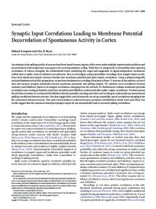
Fig. 1. Comparison of intracellular recordings from motor cortex neurons in awake (upper) and Nembutal anesthetized (lower) cat. Records show the moment of penetration (left) and membrane potential 30 s later (right). Note that in the awake condition the membrane potential is more depolarized and the action potentials are smaller. Depolarizing current pulses of 1 nA, 10 ms duration were applied every 100 ms. (Note: in these records the bridge was not balanced before the penetration)
After intracellular recordings were obtained in the awake condition, the monkeys were anesthetized with Halothane gas (1-1.5%), mixed with Nitrous oxide (1 l/min) and oxygen gas (3 1/ min), to investigate the alteration of the membrane electrical properties during induction of anesthesia. In other cases, intracellular recordings were first obtained under Halothane anesthesia, and then Halothane was discontinued to document the changes during the recovery from anesthesia. In several cases, Brevital (50 mg/kg, i.p.) was administered instead of Halothane. Recording conditions and criteria for intracellular recording were the same as Experiment I.
Results
Experiment I: cat Membrane potentials under different anesthetic conditions. A total of 115 neurons (35 in awake, 21 in Halothane anesthetized and 59 Nembutal anesthetized conditions) which met the criteria for intracellular recording were obtained from 10 hemispheres of 7 cats. Figure 1 shows representative examples of intracellular recording from the same cat, when awake (top) and anesthetized with Nembutal (bottom). Typical records at the moment of cell penetration, at left, show an abrupt DC potential shift, followed by steady initial discharge for up to 30 s. The right records show subsequent action potentials on a faster sweep. These records illustrate the two major differences observed under these conditions. 1) The neuron recorded in the awake state had a smaller (i.e. more depolarized) membrane poten-
465
&
lo
AWAKE 5
I
HALOTHANE ~g
|
I
: PT Neurons
I
I
5 L/
, ,I--~,
I
,
lo
NEMBUTAL 5
40
5O
60
70
80
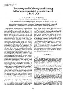
Membrane Potential (mV) Fig. 2. Comparison of resting membrane potentials of motor cortex cells under three different anesthetic conditions: awake (upper), Halothane anesthetized (middle) and Nembutal anesthetized (lower). PT-neurons are indicated by hatched bars. In all cases membrane potential was measured within 30 s after penetration. Note absence of neurons with membrane potentials above 65 mV in the awake group
tial than the neuron recorded under Nembutal anesthesia (in this case, -51 mV vs. -57 mV). 2) The neuron in the awake condition exhibited smaller action potentials than that recorded under Nembutal anesthesia (-6 mV vs. +11 mV). Some neurons recorded under Halothane or Nembutal anesthesia showed further increases in resting membrane potential (becoming more hyper-
polarized) after stable recording over several minutes, whereas membrane potentials observed in neurons penetrated in the awake condition stayed at the same level. This increase of membrane potential might be attributed to recovery from an injured state, or other factors that could not be controlled in this experiment. However, to compare the membrane potential of neurons recorded in different anesthetic conditions under the same circumstances, and avoid any bias from such long term changes, the values of membrane potential were measured during the same period of recording, approximately 30 s after the moment of penetration, just after initial discharges had ceased. Figure 2 compares the magnitudes of membrane potentials obtained under these 3 different anesthetic conditions. The difference of the mean membrane potentials during awake and Nembutal anesthetized conditions (8 mV) was statistically significant (t = 4.18, p < 0.001). The difference between awake and Halothane anesthetized conditions (4 mV) was also significant (t = 2.63, p