ated from the hydrolysis of membrane-bound phospha- tidylinositol 4,5 ... cell, including through ligand-gated ion channels, which would provide routes for Li influx ..... Hille, B. (1992) Ion Channels of Excitable Membranes, Second Ed. Sinauer ...
SYNAPSE 28:271–279 (1998)
Synapse-Specific Accumulation of Lithium in Intracellular Microdomains: A Model for Uncoupling Coincidence Detection in the Brain ANATOLII Y. KABAKOV,1,4 NIKOLAS B. KARKANIAS,3,4 ROBERT H. LENOX,1,2,3 AND ROGER L. PAPKE1,3 1Department of Pharmacology and Therapeutics, University of Florida Medical College, J.H. Miller Health Center, Gainesville, Florida 2Department of Psychiatry, University of Florida Medical College, J.H. Miller Health Center, Gainesville, Florida 3Department of Neuroscience, University of Florida Medical College, J.H. Miller Health Center, Gainesville, Florida 4The first and second authors contributed equally to this work.
KEY WORDS
bipolar disorder; glutamate receptor; dendritic spine; ion diffusion
ABSTRACT Lithium’s therapeutic specificity for the treatment of bipolar disorder may be attributable in part to an ability to target sites where there are high levels of synaptic activity. We show that glutamate receptors expressed in oocytes are highly permeable to lithium. Mathematical simulations of Li1 diffusion in mature dendritic spines suggest that in the presence of 1 mM extracellular lithium one synaptic current can increase Li1 concentration in the spine head by 4 mM with a decay time constant of about 15–20 ms. Two or more current spikes will produce oscillations between 6 and 8 mM or potentially higher. These results predict that the local intracellular lithium in dendritic spines can rise to high enough levels to uncouple second messenger mechanisms of coincidence detection. Synapse 28:271–279, 1998. r 1998 Wiley-Liss, Inc. INTRODUCTION Bipolar disorder, formerly known as manic-depression, affects approximately 1% of the US population and represents a serious public health problem (Keller and Baker, 1991). Treatment with lithium salts has proven the most effective therapy for bipolar disorder (Lenox and Manji, 1995; Lenox et al., 1997). Lithium has many effects within the cell, and while the precise mechanisms that underlie the therapeutic actions of lithium are still unclear, lithium is known to affect several G-protein-coupled signal transduction systems (Lenox and Manji, 1995; Manji et al., 1995), such as the second messengers derived from phosphoinositide (PI) signaling, specifically IP3 and DAG, which are generated from the hydrolysis of membrane-bound phosphatidylinositol 4,5, bisphosphate (PIP2 ). At submillimolar concentrations, lithium (but not other monovalent cations) is known to uncompetitively inhibit myo-inositol1-phosphatase (EC50 5 0.8 mM) (Hallcher and Sherman, 1980), a key enzyme involved in recycling inositol into the membrane for the regeneration of PIP2. Higher concentrations of lithium (approx. 5 mM) will also inhibit receptor mediated signaling within the adenylate and guanylate cyclase systems and subsequent generation of cyclic AMP and cyclic GMP (Kanba et al., 1991; Ebstein et al., 1980). The homeostatic regulation of intracellular lithium concentration is a balance of active and passive pror 1998 WILEY-LISS, INC.
cesses which parallel and interact with the systems that regulate other intracellular ions such as sodium, potassium, calcium, and bicarbonate. The extracellular (i.e., serum) concentration of lithium attained during the therapeutic use of lithium salts can be easily measured, and is in the range of 1 mM (Lenox and Manji, 1995; Lam and Christensen, 1992; Gani et al., 1993). However, intracellular concentrations of lithium are crucial for predicting physiological effects. Intracellular concentrations in the brain are relatively difficult to evaluate, but averaged over large samples of tissue have been estimated to also be in the range of 0.5–1.0 mM (Lam and Christensen, 1992; Gani et al., 1993). The passive electrochemical handling of lithium disposition across the membrane would predict tenfold higher intracellular concentrations due to the cell’s resting negativity. Therefore, it is clear that lithium concentrations in the brain are not at equilibrium. There are several paths by which lithium may enter a cell, including through ligand-gated ion channels, which would provide routes for Li1influx in a brain regionspecific manner, based on the selective expression of neurotransmitter receptor subtypes. Influx through these pathways would not only be rapid, but synchro-
*Correspondence to: Roger L. Papke, Ph.D., Department of Pharmacology & Therapeutics, P.O. Box 100267, University of Florida, Gainesville, FL 32610. Received 30 January 1997; accepted 31 July 1997.
272
A.Y. KABAKOV ET AL.
nized both temporally and spatially to synaptic activity. Such activity-dependent local heterogeneity of lithium concentrations is difficult to measure directly, but we can calculate the lithium diffusion from the synapse area to the dendrite body by using experimentally determined values for lithium permeability through glutamate receptor subtypes and applying those data to a simulation of diffusion within a synaptic spine. The concentration of lithium averaged over the entire neuron might be modest, while local concentrations in the dendritic spines become significantly elevated and thereby alter the relative balance of second messenger systems that are critical to triggering long-term events associated with neuronal plasticity. In this study, we estimated the lithium component of glutamate-activated current through ionotropic glutamate receptors using heterologous expression and two electrode voltage clamp techniques in Xenopus oocytes. Then we applied these data to a mathematical simulation of lithium diffusion in the dendritic spine by using published parameters of spine geometry (Barbour et al., 1994; Spacek and Harris, 1997) and synaptic current (Vyklicky et al., 1991). Our results confirm that 1 mM extracellular Li1 can lead to intracellular levels sufficient to profoundly alter the second messengermediated coincidence detection in dendritic spines associated with the activation of glutamate receptors under physiological conditions. Preliminary results have been published in abstract form (Karkanias et al., 1995; Kabakov et al., 1997). RESULTS Electrophysiology We obtained data on all three classes of glutamate receptors, AMPA-selective, kainate-selective, and NMDA-selective. In all cases, lithium appears to be approximately equally permeable as sodium; however, the macroscopic conduction differs among the subtypes. AMPA-selective receptors: GluR1, GluR1 1GluR2, and GluR3 The permeability and conductance of lithium through various AMPA receptor subtypes were examined by applying voltage ramps during the plateau phase of an agonist response. Current–voltage relationships (Fig. 1) were obtained in both control Ringer (115 mM sodium) and lithium-Ringer (115 mM lithium substituted for sodium). From these results we see that subunit composition plays a key role in determining lithium conduction, though not its relative permeability. The I–V relationship for the GluR1 (Fig. 1A) and GluR1 1 R2 (Fig. 1C) receptors displayed no shift in reversal potential, indicating that (compared to potassium) the relative permeability of lithium is equal to sodium for these receptor types. In these experiments, equilibration times with
the extracellular lithium-Ringer were relatively short (2 min), so that little lithium uptake by the oocyte should occur and the equilibrium potential for lithium remained high, comparable to that for sodium. While both GluR1 and GluR1 1 R2 receptor subtypes show equal permeability for lithium (compared to sodium), they show striking differences in the conductance or slope of the I–V relations. Specifically, while for GluR1 sodium and lithium appear to have both similar permeability and conductance, conductance increases in lithium when the GluR2 subunit is present. This result suggests that the expression of GluR2 results in receptors that may effectively target a synapse for local increases in lithium, which is likely to be important for the synaptic receptors of specific neuronal subpopulations in the hippocampus. The GluR3 channel resembles GluR1 in calcium permeability and inward rectification. However, the macroscopic conductance of GluR3 homomeric receptors in the presence of lithium was greatly increased compared to conduction in sodium. The currents for GluR3 were approximately 2.8 times larger in the presence of high lithium (Fig. 1B). Kainate-selective receptors: GluR6(R) and GluR6(Q) The permeability and conductance of lithium through concanavilin A treated GluR6(Q) and GluR6(R) receptors were also evaluated and showed somewhat smaller macroscopic conductance in lithium compared to sodium (see Fig. 1D for representative GluR6(Q) trace). Although macroscopic conductance was reduced in high extracellular lithium, these receptors were equally permeant to lithium (compared to sodium), as indicated by the lack of a reversal potential shift when the primary extracellular ion was exchanged (Fig. 1D). NMDA-selective receptors: NMDAR1 1 NMDAR2a and NMDAR1 1 NMDAR2b The NMDAR1 1 NMDAR2a and NMDAR1 1 NMDAR2b subunit combinations were evaluated for their relative permeability and macroscopic lithium conduction. While for both receptor subtypes lithium permeability was comparable to that of sodium, the macroscopic conduction was less for both. A representative current–voltage relationship for NMDAR1 1 NMDAR2a is shown in Figure 1E. Macroscopic conduction and permeability These experiments measure macroscopic conduction (g), where g 5 NPopen g (N 5 total number of channels, Popen 5 proportion of channels open, and g 5 conductance of a single channel). For any given cell over the duration of the experiment, N may be considered a constant, and so a change in conductance must represent either a change in Popen or in g. Note that for the
LITHIUM DISTRIBUTION IN DENDRITIC SPINE
Fig. 1. Glutamate receptor current–voltage relationships in sodium and lithium. Shown are the current–voltage relationships for five different glutamate receptor subtypes generated from voltage ramps applied during periods of sustained agonist application. To calculate the receptor-specific current–voltage relationships, we subtracted out passive membrane responses to the same voltage ramps applied in the absence of agonist. The current–voltage relationships obtained in normal (sodium) Ringer are plotted as black lines, and those obtained in lithium-Ringer are plotted as gray lines. In panel F the data plotted by the gray line represents a whole oocyte response obtained in 5 mM lithium-Ringer. (A) The I–V relationship of GluR1
273
homomeric receptors. (B) The I–V relationship of GluR3 homomeric receptors. (C) The I–V relationship of GluR1 1 GluR2 heteromeric receptors. (D) The I–V relationship of GluR6(Q) homomeric receptors. (E) The I–V relationship of NMDAR11 NMDAR2a heteromeric receptors. (F) Representative traces illustrating the effect of 5 mM lithium on the macroscopic response of GluR3 expressing oocytes to 100 µM kainate. Control responses (black line) were obtained in normal (115 mM Na1 ) Ringer, and then the extracellular solution was exchanged for Ringer containing 5 mM Li1 (plus 110 mM Na1, gray line). The scale bar is 100 nA and the duration of each trace is 2 min. The period of agonist application is indicated by the bar below the traces.
274
A.Y. KABAKOV ET AL.
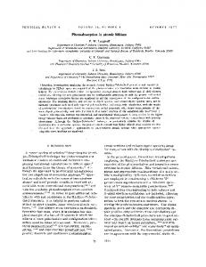
Fig. 2
LITHIUM DISTRIBUTION IN DENDRITIC SPINE
GluR1 1 GluR2 subunit combination, the I–V relationship has an increased slope in the presence of lithium for both the inward and the outward arms of the relationship. However, under these experimental conditions outward currents should be carried by potassium (as in the control condition), not lithium, so the single channel conductance should not differ in this voltage range. This suggests that the increase in macroscopic current may then be due to a change in channel activation probability rather than single channel conductance. While it will be of interest to study the effects of low lithium concentrations on channel activation in detail (Karkanias and Papke, in preparation), of most relevance to the present analysis is the observation that lithium will permeate the glutamate receptors equally well as sodium. Extrapolations between conditions of high and low extracellular lithium Although complete substitution of lithium for sodium represents a strictly experimental condition, this method provides the most sensitive assessment of the essential biophysical properties of the channel, and this assessment is applicable for channel function in vivo under physiological conditions. Our preliminary studies with low extracellular lithium (Karkanias and Papke, in preparation) suggest that lithium components of synaptic currents should roughly scale as the mole fraction of lithium in the extracellular solution. Specifically, glutamate receptors that have reduced macroscopic conduction in high extracellular lithium (e.g., GlurR6 and NMDAR1) showed no anomalous reductions in current when extracellular lithium was reduced to the range of 1–5 mM. However, interestingly, a significant (P , 0.05) potentiation of GluR3 responses remained when the extracellular lithium was reduced to 5 mM (Fig. 1F). After 5 min in the presence of 5 mM Li1 and 110 mM Na1, responses to 100 µM Kainic acid were 25 6 5%
Fig. 2. Lithium concentration dynamics determined by spine geometry. Computer simulations of lithium distribution in the dendritic spines during synaptic stimulation are represented for three different spine geometries. The lithium concentration is displayed in threedimensional plots as functions of the distance from the synapse and time. Calculations were based on the separation of the space into seven segments of 0.2 µm length each. The lithium component of one current pulse was assumed to be a 10 pA spike with a 4 ms decay constant corresponding to 1% of the total synaptic current in the presence of 1 mM extracellular lithium. Intracellular lithium was assumed to be 0.5 mM in the resting state. These simulations were run for a series of seven input pulses occurring at the apical site indicated by the arrows. (A) If the spine is modeled as a simple cylinder, the lithium concentration in the head oscillates between 0.6 and 1.2 mM and follows the kinetics of the input currents. (B) If a prototypical spine (10) is considered, then lithium concentrations reach a plateau elevation several times larger than is predicted for the simple shaft model and the kinetics of decline are slower. (C) A constriction of the neck is equivalent to the diffusion barriers that may be present in mature spines. In this case, elevations in intracellular lithium are predicted that would profoundly inhibit a wide range of secondmessenger mediated signals.
275
larger than those of cells retained in 115 mM Na1 (n 5 10 in each group). Computer simulations of local lithium concentration domains Alterations of lithium concentration in a dendritic spine head is a result of lithium currents through the post-synaptic membrane and lithium diffusion in intracellular space. The most significant factors for our model are the lithium component of the synaptic current and lithium diffusion from the spine to the dendrite body. Note that in our simulations we assumed that the local lithium concentration domains are small and the temporal windows relatively brief. These assumptions permitted us to ignore the extrusion rates of lithium. Estimation of the lithium component of synaptic current We may make a first approximation of the contribution of any particular extracellular cation in the synaptic inward current as being proportional to its extracellular mole fraction, the electrochemical driving force, and the conductivity and Popen of activated channels for this cation. In our case, the synaptic current is carried mostly by the main extracellular cation, sodium, the concentration of which is