REVIEWS
Synaptic tagging during memory allocation Thomas Rogerson, Denise J. Cai, Adam Frank, Yoshitake Sano, Justin Shobe, Manuel F. Lopez-Aranda and Alcino J. Silva
Abstract | There is now compelling evidence that the allocation of memory to specific neurons (neuronal allocation) and synapses (synaptic allocation) in a neurocircuit is not random and that instead specific mechanisms, such as increases in neuronal excitability and synaptic tagging and capture, determine the exact sites where memories are stored. We propose an integrated view of these processes, such that neuronal allocation, synaptic tagging and capture, spine clustering and metaplasticity reflect related aspects of memory allocation mechanisms. Importantly, the properties of these mechanisms suggest a set of rules that profoundly affect how memories are stored and recalled.
Departments of Neurobiology, Psychiatry & Biobehavioral Sciences, and Psychology, Integrative Center for Learning and Memory, Brain Research Institute, University of California, Los Angeles, California 90095–1761, USA. Correspondence to A.J.S. e-mail:
[email protected] doi:10.1038/nrn3667 Published online 5 February 2014
The molecular and cellular mechanisms underlying the acquisition, consolidation, reconsolidation, extinction and recall of memory have attracted a great deal of attention1–7. By comparison, little is known about memory allocation8, the process that determines which specific neurons and synapses in a neural network will store a given memory. We propose that memory allocation is a phase of memory formation that encompasses those processes that determine the exact sites where memories are stored and that has specific interactions with other more traditional phases of memory, including acquisition and consolidation (see below). The significance of having mechanisms that determine the allocation of information to particular neurons and synapses within a neural network is theoretically crucial for the efficient storage and recall of that information. Inefficient allocation of information leads to suboptimal use of storage space, whether hard disks or synaptic sites are involved. For example, theoretical studies suggest that there is a balance between the stability and size of a memory representation and the maximum amount of information that can be stored: larger representations can be more stable, but storage space could be wasted; small representations save storage space, but memories are more easily disrupted9–12. By directing related information to overlapping populations of neurons, memory allocation mechanisms could link these memories, place them within a common context, save storage space and perhaps alter memory strength and stability 8. Memory allocation mechanisms may also organize the storage of information into component elements that encode features that are shared
across related experiences, thereby linking the storage of these experiences13,14. Thus, memory allocation includes mechanisms that ‘file’ and ‘cross-reference’ information in brain circuits. This Review presents research detailing the mechanisms of memory allocation at both the synaptic (synaptic allocation) and neuronal (neuronal allocation) scale. More importantly, it attempts to integrate these previously separate areas of memory research into a unified view of how brain circuits regulate which neurons and synapses are committed to storing a given memory. Hopefully, this will facilitate the development of hypotheses, experiments and theories that elucidate why specific neurons and synapses are committed to storing a given memory as opposed to other neurons and synapses that receive similar input.
Neuronal allocation Neuronal allocation is a newly discovered phenomenon of memory formation that accounts for how specific neurons in a network, and not others that receive similar input, are committed to storing a specific memory. For example, previous studies have shown that changes in neuronal excitability that are triggered by the transcription factor cyclic AMP-responsive element-binding protein (CREB) modulate the probability that a given neuron will be involved in storing a specific memory. We propose that neuronal allocation mechanisms work closely with synaptic allocation mechanisms (that is, synaptic tagging and capture, spine clustering, and so on) that determine how information is parcelled to specific synapses. Although neuronal and synaptic allocation mechanisms
NATURE REVIEWS | NEUROSCIENCE
VOLUME 15 | MARCH 2014 | 157 © 2014 Macmillan Publishers Limited. All rights reserved
REVIEWS most probably work seamlessly during memory formation, their distinction is useful for designing, interpreting and describing allocation studies. Molecular and cellular studies of neuronal allocation. Most studies of neuronal allocation to date used the amygdala as a model circuit. For example, previous studies suggested that changes in neuronal excitability triggered by CREB modulate the probability that a given lateral amygdala neuron will be involved in memory 15,16. As many of the memory mechanisms studied to date are conserved across different brain regions, it is possible that the mechanisms of memory allocation found in the amygdala will also be present throughout the brain. The amygdala has a key role in the modulation and storage of fear memories17. Circuits in the lateral amygdala are thought to store the association between the conditioned stimulus (for example, a tone) and the unconditioned stimulus (for example, a footshock) in fear conditioning 17. More than 70% of all lateral amygdala neurons receive information regarding the auditory conditioned stimulus18 or the unconditioned stimulus19. However, only a smaller subset of these neurons goes on to encode the memory 20. Accordingly, only a subset of lateral amygdala neurons undergoes plasticity after auditory fear conditioning. Studies of modified AMPA receptors that can electrophysiologically tag synapses that are involved in learning indicated that only one-third of recorded amygdala neurons showed synaptic changes after fear conditioning 21. Other studies confirm that only a fraction of lateral amygdala neurons actually encode memory for auditory fear conditioning 22. This suggests that specific mechanisms govern the allocation of fear memories to specific neurons in the amygdala, with further studies suggesting that CREB plays an important part in this process8. Initial memory allocation studies used viral vectors to demonstrate that changing the levels of CREB within a specific subpopulation of lateral amygdala neurons could affect the probability of these neurons being recruited into an auditory fear memory: increasing the levels of CREB within a subset of lateral amygdala neurons increases the probability that these neurons are involved in fear conditioning, whereas decreasing the levels of this transcription factor has the opposite effect 15,16,23. Three main strategies were used to demonstrate the role of CREB in neuronal allocation. First, studies using immediate-early genes as markers for a memory trace showed that lateral amygdala neurons with higher levels of virus-encoded CREB were approximately three times more likely to be recruited to the auditory fear memory trace than their neighbouring neurons. A number of control experiments showed that if learning was blocked, the memory trace was no longer biased to the neurons with higher CREB levels15, showing that learning is needed for biasing immediate-early gene expression to the cells with high CREB levels. In addition, manipulations that interfered with CREB functioning in specific lateral amygdala neurons decreased the probability of these neurons being recruited to the memory trace15.
Second, strategies that either inactivated16 or deleted23 the lateral amygdala neurons that expressed virusencoded CREB also suggested that the memory was disproportionally represented in these neurons. For example, inactivation of lateral amygdala neurons expressing virus-encoded CREB with the allatostatin system24 triggered temporary amnesia for auditory fear conditioning, whereas inactivating a similar number of neurons with normal levels of CREB did not16. Related results were also obtained with conditioned taste aversion, another form of memory that involves amygdala circuits16,25. Last, electrophysiological studies showed that after training in auditory fear conditioning, the lateral amygdala neurons transfected with the virus-encoded CREB showed greater synaptic strength than non-transfected neurons, a result that is consistent with the idea that memory is encoded in these neurons as increases in synaptic strength16. Importantly, additional results supported the hypothesis that CREB modulates neuronal allocation by controlling neuronal excitability 16: neurons with higher CREB levels are more excitable and therefore more likely to fire in response to sensory input, more likely to be involved in synaptic changes underlying memory and thus are more likely to be over-represented in the memory trace (FIG. 1). As mentioned, findings from other brain regions have generally paralleled those from memory allocation studies in the amygdala. Studies in the cortex have suggested that the population of neurons encoding a given memory is a subset of the population that was initially activated during learning 26. The barrel cortex receives somatosensory information from facial whiskers, and therefore it has been used to study cortical plasticity during fear conditioning in which whisker stimulation is paired with a footshock27. Whisker stimulation has also been used for a whisker-signalled trace eyeblink-conditioning task; in this case, repeatedly pairing whisker stimulation with a mild periorbital shock28 (or air puff 29; see below), following a stimulus-free trace interval, triggered trace conditioning in mice. These results were extended by recent work using two-photon in vivo calcium imaging that showed that pairing whisker stimulation with a footshock led to a decrease in the number of neurons in the barrel cortex that responded to whisker stimulation29, suggesting refinement of the memory trace. Importantly, similar findings were also obtained in rabbits with the whisker-signalled trace eyeblinkconditioning approach30, suggesting that there are processes that determine which neurons in the barrel cortex are involved in conditioning responses. We propose that these processes are not random and that instead there are mechanisms that determine not only which cells respond initially but also shape the sparser and possibly more efficient memory trace. The results reviewed above strongly support the role of CREB in neuronal memory allocation and suggest that similar mechanisms may also be present in other brain regions. Accordingly, there is indirect evidence that CREB is involved in regulating neuronal allocation in the hippocampus. First, transduction of CA1 neurons with virus-encoded CREB before training in
158 | MARCH 2014 | VOLUME 15
www.nature.com/reviews/neuro © 2014 Macmillan Publishers Limited. All rights reserved
REVIEWS a
c
Presynaptic action potentials
Gradient of locally enhanced dendritic excitability and locally diffusible plasticity-related proteins
Synapse-specific potentiation and L-LTP
d
b Increased CREB levels and excitability
Increased CREB levels and over-representation in memory trace
Postsynaptic action potentials Ensemble 1
Memory-positive neuron
Ensemble 1
Ensemble 2
e
Ensemble 2
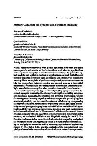
Figure 1 | Integrating neuronal and synaptic allocation. a | Neurons with increased cyclic AMP-responsive element-binding protein (CREB) levels (green ‘halo’ in ensemble 2) are more excitable and therefore more likely to fire Nature postsynaptic action potentials in response to presynaptic action potentials and are more likely to beReviews involved| Neuroscience in synaptic changes underlying memory. b | Thus, these these neurons are more likely to be over-represented in the memory trace (‘memory-positive’ neuron). c | Synapse-specific potentiation results in the local diffusion of plasticity-related proteins (for example, RAS and RHOA) from an activated synapse and a local enhancement in excitability. d | This increased excitability promotes plasticity in nearby synapses for a brief period of time. e | This results in clustering of potentiated synapses in close proximity to previously activated synapses. L-LTP, late long-term potentiation.
the Morris water maze31 or in contextual conditioning 32 enhances these hippocampus-dependent memories. In these experiments, the virus only transduced approximately 25% of neurons in CA1 (REF. 32), resulting in a subset of neurons expressing increased levels of CREB; these neurons probably recruited a higher proportion of the memory trace in order for the memory to be strengthened by CREB. Importantly, viral transduction had to occur before training for the behavioural enhancement, as posttraining injection of virus-encoded CREB without any other manipulation did not alter memory performance31. This is consistent with the idea that the enhancement observed was cell-specific and due to recruitment of the memory trace by high levels of CREB in a subset of neurons as opposed to some other effect of CREB. As in the amygdala, there is evidence that an increase in excitability may also affect memory allocation in the hippocampus. For example, transgenic mice expressing a constitutively active form of CREB showed reduced afterhyperpolarization currents in hippocampal CA1 pyramidal neurons that led to increased excitability and reduced thresholds for long-term potentiation (LTP)33. Whole-cell recordings in behaving rats showed that hippocampal CA1 neurons that were recruited into encoding a given place (place cells) showed lower spike thresholds. In addition, these place cells showed peaked versus flat subthreshold membrane potentials that were sensitive to an animal’s location34. Interestingly, this
increase in excitability seemed to precede place-cell formation during spatial exploration34, as if prior events set the stage for the allocation of place information to a subset of neurons in the hippocampus. Furthermore, increasing excitability by depolarizing the somatic membrane potential of a silent neuron (that is, a neuron that previously did not fire to a spatially tuned location) during spatial exploration led to the emergence of a spatially tuned place cell35. CREB also seems to regulate neuronal excitability in other structures that are required for memory, in which it may again affect neuronal allocation36. The aforementioned hypothesis stating that prior events determine which place cells will encode a given environment is supported by additional evidence from studies of a phenomenon termed ‘preplay’ (REF. 37). Preplay is complementary to the better-known phenomenon of replay, the recapitulation of place-cell sequences experienced during previous spatial explorations (usually while the animal is in quiet rest or sleep). Preplay takes place before (not after) exploration of novel environments. Remarkably, the preplay studies suggested that not only are there mechanisms that allocate which specific hippocampal place cells will encode a given place but that these neuronal allocation mechanisms may also determine the sequence in which these future place cells are activated during spatial exploration. It is conceivable that spontaneous firing events that occur before actual spatial
NATURE REVIEWS | NEUROSCIENCE
VOLUME 15 | MARCH 2014 | 159 © 2014 Macmillan Publishers Limited. All rights reserved
REVIEWS exploration engage neurons with the highest excitability more often than they engage neurons with comparatively lower excitability, thus leading to the statistical regularities reflected in preplay. Consequently, during preplay, and then spatial exploration, neurons with the lowest spike thresholds would on average be recruited first, followed by others with the next higher spike thresholds, and so on. This process would attach spatial attributes to a sequence of place cells with the highest excitability. From the perspective of memory allocation, these studies provide another piece of evidence that memory allocation is not random and that instead there are mechanisms that determine, ahead of time, which neurons may be involved in a given memory. Studies in the piriform cortex also support the idea that increases in excitability have a crucial role in determining the neuronal ensemble encoding a given memory 38. The stimulation of a random subpopulation of piriform cortex neurons by activation of channel rhodopsin 2, paired with either an aversive or an appetitive stimulus, is sufficient to allocate the storage of that information to these activated neurons. This suggests that in the piriform cortex, as in the amygdala and perhaps the hippocampus, increasing the probability that a neuron will fire during learning (in this case, by activation of channelrhodopsin 2) is sufficient to bias the allocation of both appetitive and aversive memories to specific subpopulations of neurons. The aforementioned studies, in the amygdala, hippocampus and piriform cortex, all suggest that increased neuronal excitability has a profound role in memory allocation. Owing to this convergent evidence, it is highly likely that increased neuronal excitability is a determinant of memory allocation. Beyond CREB, there are many other mechanisms that modulate neuronal excitability in specific neurons of a circuit. It is very likely that some or many of these mechanisms may also affect neuronal allocation. The hypothesis proposed here is that the mechanisms involved in the consolidation of one memory may also trigger changes in the excitability of the neurons engaged in storing that memory, so that for a time they are more likely to be involved in the storage of subsequent memories. Any mechanism that affects excitability within these parameters could very well affect the probability that a given neuron responds and stores stimuli that trigger memory formation. Considering the fundamental evolutionary importance of recalling the right set of memories at a critical time, we suspect that there are many mechanisms of memory allocation, including multiple strategies to shape the excitability of neurons after memory formation. Indeed, abnormalities in mechanisms that affect neuronal excitability are thought to contribute to a range of cognitive disorders39. Nevertheless, it is important to note that there are other strategies (in addition to increases in neuronal excitability) that determine which neurons are involved in storing a given memory. For example, extensive elegant experiments in the sensory and motor cortices have shown that the very neurons and networks that are active in processing motor and sensory information are the ones that are engaged in storing pertinent related information40–42.
A key function of memory allocation mechanisms. One of the proposed roles of memory allocation is to link memories that are formed within a defined temporal window 8. The idea is that the first memory-creating event activates CREB in a subpopulation of neurons; this activation leads to an increase in excitability in these neurons that then biases the storage of memory for a second event to many of the same neurons that stored the first event. Because of the overlap between the memory traces for the two events, recall of one event may also lead to the recall of the other. The result would be the coordinated storage and retrieval of related memories (FIG. 2). Although this hypothesis has not been directly tested, the evidence reviewed next is consistent with its predictions. Recent studies that used transgenic mice that express DREADDs (designer receptors exclusively activated by designer drugs) addressed the important question of how the brain could link two separate memories43. The artificial ligand clozapine-N-oxide (CNO) binds to transgenic DREADD and triggers strong depolarization and spiking. In these studies, DREADD was expressed under the control of the activity-dependent Fos promoter and the tetracycline-inducible system, so that DREADD could be expressed in an inducible and activity-dependent manner. The results showed that in a novel environment (context A), these transgenic mice expressed DREADDs from the Fos promoter in activated neurons. Later, this ensemble of neurons expressing DREADDs was reactivated by CNO while the mice were fear conditioned in a different context (context B). To recall the memory for context B, both populations of neurons (the DREADD-expressing neurons activated in context A and those neurons activated during exposure to context B) needed to be simultaneously activated. The results suggested that the transgenic mice formed a memory representation that integrated or linked contexts A and B. It is likely that CNO-driven activation of the representation of context A biased the allocation of the memory for context B to many of the same neurons that stored context A, thus closely integrating the memories for both contexts. A subsequent study44, which specifically manipulated the dentate gyrus using channelrhodopsin 2, further substantiated the claims of Garner et al.43. Together, these findings suggest that memory allocation mechanisms could be one of the reasons why recalling one memory while encoding another can result in the linking or integration of the two memories45. In addition to increases in neuronal excitability as a mechanism for memory integration, findings from a study using calcium imaging and electrophysiological stimulation in rat hippocampal slices suggest that synaptic plasticity could contribute to possible post-training shifts in neuronal allocation46. The results indicated that initially distinct neuronal ensembles (that is, possibly representing distinct memories) can become more similar after co‑activation of these two neuronal ensembles using stimulation protocols that are designed to trigger synaptic plasticity. The authors stimulated a set of hippocampal Schaffer collateral inputs and visualized the activated CA1 pyramidal neurons using calcium imaging.
160 | MARCH 2014 | VOLUME 15
www.nature.com/reviews/neuro © 2014 Macmillan Publishers Limited. All rights reserved
REVIEWS a Acquisition
b Recall Episode A
Episode A-positive neuron
Episode B
Episode B-positive neuron
Episode B
Episode A
Neuron responsive to episodes A and B
Figure 2 | Coordinated storage and retrieval of temporally related memories. a | During acquisition, neurons in a Nature Reviews Neuroscience neural circuit (grey circles) are recruited into encoding episode A (blue). This increases their excitability so|that shortly thereafter, they are also very likely to be involved in encoding episode B (purple). b | With time, the increase in excitability wanes and sequent episodes are no longer stored in the same neurons. A consequence of this pattern of storage is that recall of episode B will also result in the recall of episode A (and vice versa), whereas recall of subsequent episodes will be unaffected. Figure is adapted, with permission, from REF. 8 © (2009) American Association for the Advancement of Science.
This was then repeated for a distinct set of Schaffer collateral inputs. Using stimuli that were expected to produce synaptic plasticity, the authors paired stimulation of the two Schaffer collateral inputs and found that the overlap between the ensembles of activated CA1 pyramidal neurons increased significantly. This suggests a mechanism for neuronal allocation in which the neuronal ensembles encoding two distinct memory traces can change and become linked (that is, there is greater overlap in the neurons engaged by each memory) owing to their coordinated activation (that is, possibly recall) in the hippocampus. The examples from the amygdala, hippocampus and cortex described above demonstrate that there are allocation mechanisms that determine which neurons store a given memory in a neurocircuit. As we previously proposed, these mechanisms may function to link memories and modulate their storage and retrieval8. Next, we summarize the evidence for synaptic allocation mechanisms. Although neuronal and synaptic allocation mechanisms have a different history and have been studied separately, we propose that they are seamlessly integrated during memory formation.
Synaptic allocation Synaptic allocation encompasses any mechanism that governs how specific synapses come to store a given memory. Inherent in the idea of synaptic allocation is the concept that multiple synapses could be activated by a given set of inputs, but specific mechanisms determine which synapses actually go on to encode the memory. For example, there is evidence that synapses do not always respond identically to a given stimulation pattern47 and that a synapse’s history of activation can affect its responses, a phenomenon referred to as metaplasticity 48,49. In addition, there is also extensive evidence that the stable potentiation of a given set of synapses can, under certain circumstances, affect how other synapses in the same neuron respond to plasticity-inducing stimuli, a phenomenon that reflects mechanisms of synaptic tagging and capture50,51. All of these mechanisms may shape how synapses are recruited to encode a given
memory, how memories become linked in neurocircuits and whether they will be remembered. It is important to note that there is a natural relation between synaptic and neuronal allocation and that these two processes work seamlessly during memory encoding and storage: for example, the excitability of a cell, which determines neuronal allocation, is also a key determinant of whether any given synapse will undergo plasticity during encoding. Higher excitability increases, whereas lower excitability decreases, the probability of synaptic plasticity occurring at any one engaged spine. Nevertheless, there are useful distinctions between these two processes, and this section focuses specifically on synaptic allocation mechanisms. Molecular and cellular studies of synaptic allocation. Synaptic tagging and capture mechanisms provide a compelling example of memory allocation at the synaptic scale. The idea of synaptic tagging was developed to explain how input specificity is achieved during LTP50,51. A mechanism was needed to account for the fact that many plasticity-related proteins (PRPs), which are essential for the maintenance of LTP and long-term memory, are generated in the cell body, but only specific synapses52,53 are potentiated. The synaptic tagging and capture hypothesis proposes that the synapses activated during LTP induction become tagged in a protein synthesis-independent manner. These tagged synapses then capture PRPs, which are needed for the maintenance of LTP and, by extrapolation, long-term memory 50,51 (FIG. 3). The initial experimental evidence54 in support of the synaptic tagging and capture hypothesis came from a series of elegant in vitro electrophysiological studies in which two stimulating electrodes (S1 and S2) were placed in independent pathways that innervate the same population of rodent hippocampal CA1 neurons. In agreement with previous experiments demonstrating the specificity of LTP52,53, repeated strong tetanization of the S1 pathway could elicit lasting protein synthesis-dependent LTP (late LTP (L‑LTP)) in the S1 but not S2 pathway. Surprisingly, after induction of L‑LTP in the S1 pathway, repeated tetanization of the S2 pathway was able to induce L‑LTP even in the presence of protein synthesis
NATURE REVIEWS | NEUROSCIENCE
VOLUME 15 | MARCH 2014 | 161 © 2014 Macmillan Publishers Limited. All rights reserved
REVIEWS a
b
c
Synaptic tag Synaptic tag formation and E-LTP
Synapsespecific potentiation and L-LTP
d
e
Presynaptic action potentials
Ensemble 1
Increased CREB levels
Plasticityrelated proteins
Increased CREB levels
Ensemble 1
Memory A- and B-positive neuron
Memory Apositive neuron
Ensemble 1
Ensemble 1
Ensemble 1
Ensemble 2
Ensemble 2
Figure 3 | Synaptic tagging and capture. a | The synaptic tagging and capture hypothesis proposes that the Reviewsof | Neuroscience synapses activated during early long-term potentiation (E-LTP) induction (as depicted byNature the presence presynaptic action potentials) become tagged in a protein synthesis-independent manner that involves calcium/calmodulindependent protein kinase II (CaMKII) and actin (not shown). b | These tagged synapses then capture plasticity-related proteins (PRPs) downstream of the CaMKII–CaMKIV–cyclic AMP-responsive element-binding protein (CREB) pathway, which is needed for the maintenance of LTP and, by extrapolation, long-term memory linked to ensemble 1. c | The formation of the strong memory A (indicated by turquoise shading) induces late LTP (L‑LTP) in a subset of synapses in neuronal ensemble 1 (depicted as a single neuron for clarity) but not in neuronal ensemble 2 (not shown). For at least 1 hour after strong training, neuronal ensemble 1 is able to share plasticity-related proteins that can convert a weak memory into a strong one (‘synapse-specific potentiation’). d | An arriving action potential (memory B) at the top right synapse of ensembles 1 and 2 sets a new synaptic tag. e | Subsequently, the weak memory B (depicted in part d) is able to elicit L‑LTP in neuronal ensemble 1 (owing to the presence of plasticity-related proteins) but not in neuronal ensemble 2. Ensemble 1 therefore becomes positive for memory A and memory B (indicated by dual blue and purple shading). This is an example of how synaptic tagging and capture can determine which neurons (not just synapses) would encode a given memory.
inhibitors. Perhaps the proteins needed for the maintenance of L‑LTP, which were synthesized during the repeated tetanization of the S1 pathway, could be shared by synapses tagged during S2 tetanization and therefore support L-LTP in this second pathway. Accordingly, a weak tetanization of the S2 pathway, which could only elicit a transient potentiation (early LTP (E‑LTP)), if preceded 1 hour earlier by L‑LTP induction in the S1 pathway, was capable of inducing L‑LTP in the S2 pathway 54. Perhaps, E‑LTP was sufficient to tag the S2 set of synapses that were then capable of capturing the proteins needed for the maintenance of L‑LTP that was generated by repeated tetanization of the S1 pathway. Later studies55 using similar techniques confirmed that the subthreshold E‑LTP-inducing tetanization could precede the repeated L‑LTP tetanization by up to 1 hour and still be converted to L‑LTP. This suggested that the tag set during E‑LTP can be maintained for up to 1 hour. Remarkably, long-term depression (LTD) also seems to be capable of taking advantage of this synaptic tagging and capture mechanism56: short-lived LTD in one set of synapses can be converted into long-lasting or late LTD by L‑LTP at another set of synapses of the same neurons.
Converging evidence for the synaptic tagging and capture hypothesis came from pioneering studies conducted on cultured Aplysia spp. neurons57. These studies used a neuronal culture system in which a single Aplysia spp. sensory neuron makes synaptic connections with two physically separate motor neurons. In this elegant co‑culture system, five pulses of serotonin adjacent to the synapses between the sensory and the motor neurons trigger long-term facilitation (LTF; the Aplysia spp. equivalent of L‑LTP) of synaptic transmission, whereas a single pulse of serotonin only generates short-term facilitation (STF; the Aplysia spp. equivalent of E‑LTP). In agreement with the synaptic tagging findings in rodents, STF induced in a set of synapses of the sensory neuron can be converted into LTF by inducing LTF in another set of synapses of the same sensory neuron. Presumably, STF generates tags that can then be used to capture PRPs generated by LTF in other synapses of the same sensory neuron. Subsequent studies established that the temporal properties of the tag were similar to those of the tag in the rodent hippocampus and that synaptic tagging and capture required protein kinase A activity and CREB function58.
162 | MARCH 2014 | VOLUME 15
www.nature.com/reviews/neuro © 2014 Macmillan Publishers Limited. All rights reserved
REVIEWS Recently, pharmacological studies further explored the molecular underpinnings of synaptic tagging 59,60. These studies suggested that calcium/calmodulindependent protein kinase II (CaMKII) and actin remodelling are important for setting the tag and that the CaMKII–CaMKIV–CREB pathway is important for the synthesis of the PRPs that are presumably shared between tagged synapses (FIGS 3,4). These proteins could include activity-regulated cytoskeleton-associated protein (ARC), GluR1, HOMER1A and protein kinase Mζ. Interestingly, low-frequency stimulation in conjunction with the application of dopamine D1 and D5 receptor agonists, brain-derived neurotrophic factor (BDNF) and carbachol can induce L‑LTP, suggesting that these molecules may mediate the effects of repeated tetanization and trigger the production of proteins that are needed for the maintenance of L‑LTP61–63. Structural studies of synaptic crosstalk. Recent studies have also probed synaptic tagging and capture-like phenomena at individual dendritic spines. Findings from these and related studies64–66 have also revealed other molecules that are likely to be involved in this process. Specifically, two-photon glutamate uncaging and fluorescence lifetime imaging were used to show that induction of LTP at one spine (which is reflected by increases in spine size) can
Presynaptic neuron 1 Presynaptic neuron 1
affect the probability of LTP being induced at a nearby spine in response to subthreshold stimulation64. The probability of this synaptic ‘crosstalk’ is inversely related to both distance between spines and time between inducing stimuli. This and another related study64,65 found that, after LTP induction, calcium-dependent RAS activity increases for ~5 minutes within the activated spine and then diffuses ~10 μm into adjacent spines. This spread of RAS signalling affects the threshold for LTP induction locally, perhaps via its ability to briefly (~1 minute) increase AMPA receptor exocytosis, leading to synaptic strengthening within and around the stimulated spine64,65. Furthermore, activated RHOA, a RAS homologue, is able to briefly (~5 minutes) diffuse up to 5 μm from stimulated spines and could also be another mechanism for local synaptic crosstalk66. Further research using two-photon imaging has provided additional functional insights into synaptic tagging and capture-like processes that are localized to neighbouring dendritic spines. These ground-breaking experiments used both imaging and electrophysiological recordings to study synaptic tagging and capture at the level of single spines67. The results indicated that E‑LTP at one spine can be converted to L‑LTP when L‑LTP has previously been induced at a nearby spine. LTP was measured as an increase in spine volume using two-photon microscopy and was validated using perforated-patch
Axon
Actin and CaMKII ‘tag’
Presynaptic neuron 2 RAS and RHOA gradient
Presynaptic neuron 2
Axon
Postsynaptic neuron
Postsynaptic neuron
PRPs
Neurotransmitter release
Axon
Dendrite
Figure 4 | Molecular mechanisms for synaptic clustering and synaptic tagging and capture. Neuron 1 is Nature Reviews | Neuroscience strongly activated (depicted by multiple red action potential traces), which leads to the formation of synaptic tags involving calcium/calmodulin-dependent protein kinase II (CaMKII) and actin in stimulated synapses. Diffusion of RAS and RHOA (indicated by green shading) from the activated synapses promotes plasticity in nearby synapses (~10 μm) for a brief period of time (