International Orthopaedics (SICOT) (2008) 32:223–227 DOI 10.1007/s00264-006-0310-9
ORIGINAL PAPER
Synchronisation of tibial rotational alignment with femoral component in total knee arthroplasty Dong-Hoon Lee & Jai-Gon Seo & Young-Wan Moon
Received: 3 October 2006 / Revised: 17 November 2006 / Accepted: 27 November 2006 / Published online: 12 January 2007 # Springer-Verlag 2007
Abstract The rotational axis of the tibial component in total knee arthroplasty described by Insall is generally accepted, but rotational mismatch between the femoral and the tibial components can occur because the alignment of each component is determined separately. We developed a connecting instrument to synchronise the axis of the tibia to the axis of the femur. We compared the rotational axis of the tibial component using our method and medial one third of tibial tuberosity (Insall’s reference) in 70 consecutive TKAs. The rotational axis of the tibial component from the femoro-tibial synchronisation was rotated internally 13.8°± 5.8° (range, 2° – 24°) more than the axis of Insall’s reference. Eighty three percent of patellae tracked centrally and the patellae tilt measured 2.2° on average. More attention should be given to the rotational congruency between the femorotibial components, because the recent prosthetic design has more conforming articular surfaces. Résumé La rotation axiale du composant tibial dans la prothèse totale du genou décrite par Insall est généralement acceptée mais elle peut poser problème lorsqu’il existe un trouble de rotation sur le composant fémoral et le composant tibial du fait d’une détermination séparée de la position de chaque composant au moment de l’implantation. Nous avons pour cela développé un instrument qui permet de synchroniser l’axe du tibia et l’axe du fémur. Nous avons comparé la rotation axiale du composant tibial en utilisant notre méthode à propos de 70 prothèses totales D.-H. Lee : J.-G. Seo (*) : Y.-W. Moon Department of Orthopedic Surgery, Samsung Medical Center, Sungkyunkwan University School of Medicine, 50 Ilwan-Dong Seoul 135-710, Kangnam-Ku, South Korea e-mail:
[email protected]
consécutives du genou. L’axe de rotation après synchronisation des deux composants a été noté en rotation interne. Cette rotation interne est plus importante que celle prévue par l’axe de référence d’Insall qui est représenté par le tiers interne de la tubérosité tibio-intérieure. 83% des rotules avaient une course normale et un accrochage rotulien a été noté dans 2,2% en moyenne. Une attention particulière doit être portée à la congruence en rotation des éléments fémoro-tibiaux pendant l’implantation d’une PTG d’autant que cette congruence est plus importante dans les nouveaux dessins de prothèses totales du genou récentes.
Introduction Correct rotational alignment of femoral and tibial components is one of the difficult and controversial aspects in total knee arthroplasty (TKA), but as of yet, there is no ‘golden rule’ for this subject [1, 10, 13, 14, 16, 23, 25]. Numerous studies have been undertaken to establish the rotational axis of the femur whereby the transepicondylar axis is regarded as more reliable [4, 7]. For the rotational axis of the tibia, medial 1/3 of the tibial tuberosity advocated by Insall (Insall’s reference) is approved by most surgeons [14]. But determination of the rotation by fixed anatomical landmarks can lead to rotational mismatch between the femoral and the tibial components. Incorrect kinematics caused by femorotibial rotational mismatch can cause several problems such as femoro-tibial subluxation, accelerated tibial polyethylene (PE) wear and patellofemoral dysfunction [2, 4, 20]. In this study, we describe a method to determine the rotational axis of the tibial component with reference to the rotation of the femoral component to achieve rotational congruency and compare it with Insall’s reference.
224
International Orthopaedics (SICOT) (2008) 32:223–227
Patients and methods We conducted a prospective, observational study of 70 consecutive primary posterior cruciate ligament substituting TKAs (Scorpio, Osteonics, Stryker, New Jersey, USA) in 70 patients. There were three men and 67 women enrolled in the study. The mean age of the patients at the time of surgery was 69±6.6 years (range, 54–82 years). Patients had a confirmed diagnosis of degenerative osteoarthritis in 69, and osteonecrosis of distal femur in one. Two kinds of rotational axes of the tibial component were measured intraoperatively. The first was the axis of the tibial component using a customised instrument (Linker) developed for the rotational congruency between the femorotibial components. The second was the line connecting the medial 1/3 of the tibial tuberosity and the centre of the posterior cruciate ligament, described by Insall [14]. We postulated that the rotational congruency between the femoral and the tibial component in full extension could be achieved by the author’s method using Linker. Surgical technique All procedures were performed by a single senior surgeon. A standard midline skin and capsular incision that divides the quadriceps tendon in its medial 1/3 and peels the quadriceps expansion from patella was performed. Soft tissue balancing was performed by release of medial soft tissues, both cruciate ligaments and cheilectomy. These procedures were progressively performed to correct a fixed deformity, bringing the limb alignment into an approximate mechanical axis. Coronal and sagittal alignment of the femoral component were determined using the intramedullary alignment system and rotational alignment was accessed with the transepicondylar axis (TEA) and Whiteside’s line. After anterior skim cut, the distal femoral cutting block was assembled and adjusted to match the thickness of the femoral component (for example, 8 mm for Scorpio). Once in place, the distal femoral cutting block represented coronal, saggital and rotational axes of the femoral component. Next, we used a connecting instrument (Linker) to place the proximal tibial cutting block. After a headless pin was inserted just below the tibial plateau on the line of the mechanical axis of the tibia, the knee was fully extended. While maintaining the knee in full extension, a specially designed instrument (Linker) to synchronise the three dimensional axes between the femoral and tibial components was connected with the cutting slot of the distal femoral cutting block over the headless pin of the proximal tibia (Fig. 1a). With this procedure, the direction of the tail of Linker represented the mechanical axis of the femur. Additional mediolateral soft tissue balancing was per-
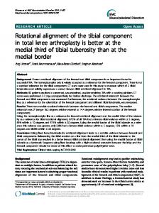
Fig. 1 a The tail of the connecting instrument (Linker) represents the mechanical axis of the femur. b Mechanical axis of the femur and the tibia could be parallel after meticulous balancing of the soft tissues
formed until the mechanical axis of the tibia could be parallel to the tail of Linker (Fig. 1b). The goal in this procedure was to gain proper limb alignment with equal tension on the medial and lateral collateral ligaments. During tensioning of both collateral ligaments, varus or valgus stresses were applied to identify any remaining laxity; in addition, the alignment of the second toe was confirmed to point anteriorly because a degree of rotational movement might be possible after removing both cruciate ligaments. Keeping the mechanical axes between femur and tibia parallel, a pair of headless pins were inserted into the holes of Linker and the direction of these two pins indicated the rotation of the tibial component (Fig. 2a). Using this
Fig. 2 a The rotational axis of the tibial component is synchronised with the rotation of the femoral component by using Linker. Two headless pins (arrow) were inserted into the holes of Linker. b A line parallel to the headless pins indicates the rotational axis of the tibial component
International Orthopaedics (SICOT) (2008) 32:223–227
procedure, we could determine the three dimensional axes of the tibial component including the rotational axis that is synchronised with the axis of the femoral component. After resection of the proximal tibia, the rotational axis of the tibial component was marked with methylene blue on the cut surface of the tibia (Fig. 2b). The medial and lateral borders of tibial tuberosity were palpated and marked at both medial 1/3 of the tubercle and at the centre of the tibial insertion area of the posterior cruciate ligament with methylene blue. Then the axis advocated by Insall was also marked on the cut surface of the tibia. The angle between the two axes was measured with a protractor. Postoperatively, the tilt of the patella was measured by the methods described by Gomes et al. [12]. Statistical analysis was performed with the use of SPSS statistical software system version 11.5 (SPSS Inc., Chicago, Illinois). The t-test for paired samples was used to compare the rotation of the tibial component between the authors’ method and Insall’s reference. The relationship between lateral patellar tilting and the angular deviation between the authors’ method and Insall’s reference was analysed with Spearman correlation analysis. P values of