INTERNATIONAL JOURNAL OF ONCOLOGY 44: 685-692, 2014
Synergistic cell growth inhibition by the combination of amrubicin and Akt-suppressing agents in K-ras mutation-harboring lung adenocarcinoma cells: Implication of EGFR tyrosine kinase inhibitors SHIZUKA ITO, TADASHI IGISHI, MIYAKO TAKATA, YASUTO UEDA, SHINGO MATSUMOTO, MASAHIRO KODANI, KENICHI TAKEDA, HIROKI IZUMI, TOMOHIRO SAKAMOTO, KOSUKE YAMAGUCHI, HARUHIKO MAKINO, HIROKAZU TOUGE, HIROKI CHIKUMI and EIJI SHIMIZU Division of Medical Oncology and Molecular Respirology, Faculty of Medicine, Tottori University, Yonago 683-8504, Japan Received October 7, 2013; Accepted November 26, 2013 DOI: 10.3892/ijo.2014.2249 Abstract. Previously we showed that Akt-suppressing agents, combined with amrubicin, synergistically inhibited the growth of small cell lung cancer cells. The combined effects of chemotherapeutic agents and Akt-suppressing agents, including epidermal growth factor receptor (EGFR) tyrosine kinase inhibitors, were evaluated in A549 lung adenocarcinoma cells harboring K-ras mutation and wild-type EGFR. Only amrubicin and not other chemotherapeutics (cisplatin, pemetrexed and paclitaxel) synergistically inhibited cell growth when combined with an Akt inhibitor, LY294002. The combination of amrubicin and LY294002 enhanced Annexin V binding to cells. A non-specific tyrosine kinase inhibitor, genistein, suppressed Akt and showed synergistic interaction in combination with amrubicin. Two EGFR tyrosine kinase inhibitors (EGFR-TKIs), gefitinib and erlotinib, suppressed Akt activity at clinically achievable concentrations and demonstrated synergism when combined with amrubicin. The suppression of K-ras expression by siRNA interfered with this synergism and inhibited both EGFR and Akt activity in A549 cells. In Ma10 cells, which harbor wild-type EGFR and K-ras, EGFR-TKIs neither suppressed Akt activity nor exhibited such synergism when combined with amrubicin. We concluded that the synergism by the combination of EGFR-TKI and amrubicin is attributable, at least partially, to K-ras mutation in A549 cells. The combina-
Correspondence to: Dr Tadashi Igishi, Division of Medical
Oncology and Molecular Respirology, Faculty of Medicine, Tottori University, Yonago 683-8504, Japan E-mail:
[email protected]
Key words: Akt, amrubicin, epidermal growth factor receptor tyrosine kinase inhibitor, lung adenocarcinoma, v-Ki-ras2 Kirsten rat sarcoma viral oncogene homolog
tion of EGFR-TKI and amrubicin may be a promising treatment for lung cancer with wild-type EGFR and K-ras mutation. Introduction Lung cancer is the leading cause of cancer-related death worldwide. More than 80% of lung cancers are non-small cell lung carcinoma (NSCLC) with lung adenocarcinoma being the most common subtype. Over half of patients with NSCLC have advanced or metastatic disease at the time of diagnosis. Systemic chemotherapy is the standard treatment for such patients with advanced NSCLC. Despite recent improvements in diagnosis and first-line treatment, the prognosis remains poor, with an overall 5-year survival probability of only about 15% (1). Over the last decade, the molecular heterogeneity of NSCLC has become better understood, and it is now clear that some tumors are characterized by ‘driver’ oncogene mutations. The most prevalent mutated oncogenes identified in lung adenocarcinoma are epidermal growth factor receptor (EGFR), v-Ki-ras2 Kirsten rat sarcoma viral oncogene homolog (K-ras), and anaplastic lymphoma kinase (ALK, where translocation and not mutation is present) (2,3). Several randomized trials demonstrated that EGFR tyrosine kinase inhibitors (EGFR-TKIs), erlotinib and gefitinib, are more effective for patients harboring activating EGFR mutations than standard platinum-based chemotherapy, at least in terms of response rate, progression-free survival, toxicity profile and quality of life (4-6). A multi-targeted tyrosine kinase inhibitor, crizotinib, has also been reported to exhibit clinical activity against ALK-translocated NSCLC (7). Although K-ras mutation is a major driver mutation, there is no effective treatment that targets the active form of the K-ras protein. Several alterations in intracellular signaling are involved in the development of cancer and tumor progression (8). The phosphatidylinositol 3-kinase (PI3K)/Akt (also known as protein kinase B) pathway is believed to be a potential target cancer therapy (9). As a biological function, the proliferative and anti-apoptotic effects of Akt-mediated signaling have been
686
ITO et al: SYNERGISM OF AMRUBICIN AND Akt-SUPPRESSING AGENTS IN LUNG ADENOCARCINOMA
established through extensive studies (10,11). Phosphorylated Akt was detected in 70% of the tumor specimens from NSCLC patients (12), suggesting a high incidence of PI3K/Akt pathway activation in NSCLC cells. Activated Akt is also proposed to contribute to increased resistance to chemotherapy in NSCLC (13). Accordingly, if Akt also is activated in the K-ras mutation-harboring cancer, suppression of Akt may be a strategy for sensitizing cancer cells against chemotherapeutic agents and to improve treatment outcomes. Amrubicin (AMR) is a totally synthetic anthracycline anticancer drug based on doxorubicin, whose hydroxyl group at position 9 is replaced by an amino group in AMR to enhance efficacy (14). In recent years, AMR monotherapy and combination therapy have been actively studied and shown promise for the treatment of small-cell lung cancer (SCLC) (15). In addition, clinical activity of AMR has been proposed for the treatment of NSCLC (16). We previously showed that Akt-suppressing agents synergistically inhibit cell growth when combined with AMR or sensitize cancer cells to AMR in SCLC cells (17). Therefore, it is hypothesized that the combination of Akt-suppressing agent and AMR can be an effective treatment strategy for NSCLC. We report here that the combination of AMR and Akt-suppressing agents, including EGFR-TKIs, show synergistic cell growth inhibition and that this synergism by the combination of EGFR-TKIs and AMR may be involved with the K-ras mutation itself in A549 lung adenocarcinoma cells that have wild-type EGFR and mutant K-ras genes. Materials and methods Chemicals and reagents. AMR (a gift from Dainippon Sumitomo Pharama, Tokyo, Japan) and pemetrexed (PEM) (a gift from Eli Lilly, Indianapolis, IN, USA) were dissolved in distilled water and stored at -20˚C. A stock solution of cisplatin (CDDP) (a gift from Nippon Kayaku, Tokyo, Japan) was reconstituted with water, diluted in 0.9% sodium chloride solution, and stored at -20˚C. Gefitinib (a gift from AstraZeneca, Chestitre, UK), erlotinib (a gift from F. Hoffmann-La Roche, Basel, Switzerland), paclitaxel (PTX) (a gift from Brisol-Meyers-Squibb, Tokyo, Japan), 2-(4-morpholinyl)-8-phenyl-4H-l-benzopiran-4-one (LY294002) (Wako Pure Chemical Industries, Osaka, Japan), and 4', 5,7-trihydroxy-isoflavone (genistein) (Wako Pure Chemical Industries) were dissolved in dimethyl-sulfoxide and stored at -20˚C. 3-(4,5-Dimethyl-thiasol-2-yl)-2,5-diphenyltetrazolium bromide (MTT) (Wako Pure Chemical Industries) was dissolved in phosphate-buffered saline (PBS) and stored at -20˚C. Cells. The sources of A549, Ma10 and PC9 cells, all of which were lung adenocarcinoma cell lines, were described previously (18). A549 and Ma10 cells were maintained in Dulbecco's modified Eagle's medium (DMEM) supplemented with 10% fetal bovine serum (FBS) and antibiotics (100 U/ml penicillin and 100 µg/ml streptomycin). PC9 cells were also maintained in RPMI-1640 medium supplemented with 10% FBS and antibiotics. These cells were grown in a humidified atmosphere of 5% CO2 - 95% air at 37˚C. MTT assay. MTT assay was performed to evaluate cell proliferation inhibition. Cells were counted with a hematocytometer,
and 104 cells were incubated in 100 µl of medium containing the indicated drugs for 72 h using 96-well flat-bottom multi‑plates (Nalge Nunc, Penfield, NY, USA). After 72 h, 10 µg of MTT in 10 µl PBS was added to each well and incubation was continued for an additional 4 h. Thereafter, 100 µl of 0.04 N HCl in 2-propanol was added and the multi-plates were incubated overnight to solubilize the MTT formazan crystal. The absorbance of each well was measured at a 570-nm wavelength (reference 650 nm) using a Sunrise scanning multi-well spectrometer (TECAN Japan, Kanagawa, Japan). Each experiment was performed in duplicate or triplicate for each drug concentration and was independently performed two or three times. Flow cytometry with Annexin V. Apoptosis rates were determined by flow cytometry analysis using an Annexin V-FITC (Beckman Coulter, Fullerton, CA, USA). A549 cells were plated at a density of 106 cells per well in 6-well plates and then treated with LY294002 (25 µM) and/or AMR (0.1 µM) for 24 h. Staining was performed according to the manufacturer's instructions. Fluorescence-activated cell sorting (FACS) analysis was performed immediately after staining using a FACSCalibur flow cytometer (Becton‑Dickinson, Franklin Lakes, NJ, USA). Western blot analysis. Cells were seeded in 6-well tissue culture plates. Twenty-four hours after cell seeding, cells were washed with ice-cold PBS and lysed in lysis buffer [20 mM HEPES, 10 mM EGTA (pH 8), 1% Triton X-100, 40 mM β-glyserophosphate, 2.5 mM MgCl2, 2 mM Na3VO4] including 1 mM PMSF, 1 mM DTT, 10 mg/ml leupeptin, 20 µg/ml aprotinin, and a phosphatase inhibitor cocktail (Nacalai Tesque, Kyoto, Japan). After 5 min on ice, lysates were centrifuged at 13,000 x g for 10 min at 4˚C and the supernatant was collected. Protein was measured by using the Bio-Rad Protein assay reagent (Bio-Rad Laboratories, Hercules, CA, USA), and protein lysates containing 20 µg of total cellular protein were subjected to discontinuous SDS-polyacrylamide gel electrophoresis. Proteins were electrotransferred to a polyvinylidene fluoride (PVDF) membrane (GE Healthcare Japan, Tokyo, Japan) for 40 min at 4˚C at 200 V. Non‑specific binding was blocked by incubation with 5% non‑fat milk in tris-buffered saline containing 0.1% Tween‑20 (TBST) for 1 h at room temperature. The following primary antibodies were probed (1:100 unless otherwise indicated): anti-c-K-ras clone Ab-1 (Calbiochem, San Diego, CA, USA), anti-Akt, anti-phospho‑Akt (Ser473), anti-EGFR, anti-phospho-EGFR (Y1068) (Cell Signaling Technology, Beverly, MA, USA), and anti-β -actin antibody (Sigma-Aldrich Japan, Tokyo, Japan), overnight and washed twice with TBST. After washing, proteins were detected by incubation with horseradish peroxidase-labeled secondary antibodies (GE Healthcare Japan). Finally, each protein was detected using an enhanced chemiluminescence detection system (ECL prime) (GE Healthcare Japan) and captured with an ImageQuant LAS400 (GE Healthcare Japan). Transfection with siRNA. Either 5x10⁴ cells or 5x10³ cells were seeded in 6- or 96-well tissue culture plates. Twenty-four hours after cell seeding, transient small interfering RNA (siRNA) directed against specific K-ras (ON-TARGETplus siRNA SMART pool; Thermo Fischer Scientific, Rockford, IL, USA) or control siRNA-A (sc-37007; Santa Cruz Biotechnology,
INTERNATIONAL JOURNAL OF ONCOLOGY 44: 685-692, 2014
687
Santa Cruz, CA, USA) was transfected to A549 cells according to the manufacturer's instructions. Briefly, A549 cells were treated with the indicated concentration of siRNA using 5 µl Lipofectamine 2000 transfection reagent in Opti-MEM I reduced serum medium (both from Invitrogen, Carlsbad, CA, USA) for 6 h. The medium was removed and replaced with fresh DMEM supplemented with 10% fetal calf serum and antibiotics. Cells were used 24 h after transfection for western blot analysis or MTT assay. Assessment of combination effect. To assess the combination effect of the indicated agents qualitatively, isobologram analysis was utilized as described previously (19). The percentage of cell proliferation was calculated as: [(mean absorbance of drug-treated wells - mean absorbance of cell-free wells)/(mean absorbance of vehicle cells - mean absorbance of cell‑free wells)] x 100. We used the concentration producing 50% inhibition of cell growth (IC50) to evaluate dose-response interactions. A combination index (CI) was used to compare the combination effect of the two drugs quantitatively between control and treated cells. The CI quantitatively depicts synergism (CI 1). The CI for each fraction-affected value representing the percentage of proliferation inhibited by a drug was calculated using the Chou and Talalay method (20). The fraction-affected value (Fa)/CI plots were constructed in Excel 2007. Results Effects of LY294002 on Akt activity and interactions with chemotherapeutic agents in A549 cells. We tested the interaction between an Akt inhibitor, LY294002 and representative chemotherapeutic agents including AMR in A549 cells. A549 cells were treated with the indicated concentration of LY294002 for 1 h. LY294002 at 25 µM effectively suppressed Akt phosphorylation (Fig. 1A). We previously reported that the combination of LY294002 and AMR synergistically inhibited the growth of N417 cells, derived from SCLC. In A549 cells, the combination of LY294002 and AMR also synergistically inhibited cell growth, whereas only additive interactions were observed in the combination of LY294002 with CDDP and PEM. In the combination of LY294002 and PTX, only antagonistic effects were observed, as judged by isobologram analysis (Fig. 1B). To evaluate whether the synergism observed in the combination of AMR and LY294002 is attributable to an enhancement of apoptotic cell death, the binding of Annexin V to cells was measured by flow cytometry after treatment with either AMR (0.1 µM), LY294002 (25 µM) or the combination. Although Annexin V binding did not differ remarkably after treatment with the single agent compared to untreated cells, a clear increase in Annexin V binding was observed after the simultaneous combination of LY294002 and AMR (Fig. 1C). Effects of genistein on the activity of Akt and synergistic cell growth inhibition by the combination of AMR. Because Akt works downstream of tyrosine kinases (21), we tested whether genistein, a non‑specific tyrosine kinase inhibitor, suppresses Akt activity and synergistically inhibits cell growth in combi-
Figure 1. Combination effects of LY294002 and chemotherapeutic agents in A549 cells. (A) A549 cells were treated with the indicated concentrations of LY294002 for 1 h. Total cellular protein (20 µg) from A549 cells lysate was subjected to western blot analysis with anti-phospho-Akt (upper panel) and anti-Akt antibody (lower panel). (B) A549 cells were treated with a combination of LY294002 and cisplatin (CDDP), amrubicin (AMR), pemetrexed (PEM) or paclitaxel (PTX) for 72 h. The combination effects were evaluated by isobologram analysis. The envelopes of additivity are defined by three isoeffect lines constructed from the dose-response curves of the single agents. The concentration producing 50% cell growth inhibition (IC50) of LY294002, CDDP, AMR, PEM or PTX alone is expressed as one on the ordinate and the abscissa. The plotted data points show the relative values of the concentrations producing IC50. (C) A549 cells were treated with the indicated concentrations of LY294002 and/or AMR for 24 h, and the binding of Annexin V to cells was measured by a flow cytometer.
nation with AMR. A549 cells were treated with the indicated concentration of genistein for 6 h. As expected, genistein concentration-dependently suppressed Akt activity in the
688
ITO et al: SYNERGISM OF AMRUBICIN AND Akt-SUPPRESSING AGENTS IN LUNG ADENOCARCINOMA
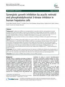
Figure 2. Effect of genistein on Akt activity and drug interaction with amrubicin (AMR) in A549 cells. (A) A549 cells were treated with the indicated concentrations of genistein for 6 h. Total cellular protein (20 µg) from the cell lysate was subjected to western blot analysis with anti-phospho-Akt (upper panel) and anti-Akt antibody (lower panel). (B) A549 cells were treated with a combination of genistein and AMR for 72 h. Dose-response curves were plotted on the basis of the data derived from the MTT assay. The survival cell fraction was expressed as the percentage of optical density (%OD) in reference to OD of the untreated cells. The combination effects were evaluated with isobologram analysis. The envelopes of additivity are defined by three isoeffect lines constructed from the dose-response curves of the single agents. The concentration producing 50% cell growth inhibition (IC50) of AMR or genistein alone is expressed as one on the ordinate and the abscissa. The plotted data points show the relative values of the concentrations producing IC50.
concentration range