492424
research-article2013
PSSXXX10.1177/0956797613492424Banissy et al.Motion and Color Perception in Synesthesia
Research Article
Synesthesia for Color Is Linked to Improved Color Perception but Reduced Motion Perception
Psychological Science 24(12) 2390–2397 © The Author(s) 2013 Reprints and permissions: sagepub.com/journalsPermissions.nav DOI: 10.1177/0956797613492424 pss.sagepub.com
Michael J. Banissy1,2, Victoria Tester1, Neil G. Muggleton2,3,4, Agnieszka B. Janik1, Aimee Davenport5, Anna Franklin5, Vincent Walsh2, and Jamie Ward5 1
Department of Psychology, Goldsmiths College, University of London; 2Institute of Cognitive Neuroscience, University College London; 3Institute of Cognitive Neuroscience, National Central University; 4Laboratories for Cognitive Neuroscience, National Yang-Ming University; and 5School of Psychology and Sackler Centre for Consciousness Science, University of Sussex
Abstract Synesthesia is a rare condition in which one property of a stimulus (e.g., shape) triggers a secondary percept (e.g., color) not typically associated with the first. Work on synesthesia has predominantly focused on confirming the authenticity of synesthetic experience, but much less research has been conducted to examine the extent to which synesthesia is linked to broader perceptual differences. In the research reported here, we examined whether synesthesia is associated with differences in color and motion processing by comparing these abilities in synesthetes who experience color as their evoked sensation with nonsynesthetic participants. We show that synesthesia for color is linked to facilitated color sensitivity but decreased motion sensitivity. These findings are discussed in relation to the neurocognitive mechanisms of synesthesia and interactions between color and motion processing in typical adults. Keywords color perception, perception, motion perception Received 10/11/12; Revision accepted 5/6/13
Introduction Synesthesia is a rare experience in which one property of a stimulus (the inducer) triggers a secondary percept (the concurrent) that is not typically associated with the first. For example, in grapheme-color synesthesia, an achromatic grapheme triggers a secondary percept of color. Approximately 4% of adults experience some form of synesthesia, with the most common concurrent being color (Simner et al., 2006). The authenticity of synesthetic experiences has been well established, and there is now a growing body of work documenting the neurocognitive mechanisms involved (see Bargary & Mitchell, 2008; Rouw, Scholte, & Colizoli, 2011; and Simner, 2012, for review). For example, a variety of studies have indicated distributed differences in cortical structure (see, e.g., Rouw et al., 2011, for review) and function (e.g., differences in early visual
cortex excitability at rest; Terhune, Tai, Cowey, Popescu, & Cohen Kadosh, 2011) in synesthesia in which color is the evoked sensation. Despite this, our understanding of broader perceptual differences in synesthetes remains limited, and few studies have examined the extent to which synesthetes show distributed perceptual differences that extend beyond their synesthetic experience. This is, however, an important question to answer, not only to constrain accounts of synesthesia but also to inform us about how alterations in cortical development can impact perceptual processing more generally. Moreover, perception is an inherently multisensory
Corresponding Author: Michael J. Banissy, Department of Psychology, Goldsmiths College, University of London, New Cross, London SE14 6NW, England E-mail:
[email protected]
Motion and Color Perception in Synesthesia 2391 process, and thus synesthesia offers a unique opportunity to examine how alterations in typical multisensory development can impact basic perceptual processing. Studies examining the perceptual processing of stimuli that do not evoke synesthesia (in synesthetes) have yielded increasing evidence to suggest that synesthesia is linked to facilitated sensory processing of the concurrent modality. For example, Yaro and Ward (2007) reported that synesthetes who experience color as their evoked sensation show superior color-perception abilities. Furthermore, we recently showed that synesthetes who experience color as their evoked sensation show facilitated color processing, whereas synesthetes who experience a tactile concurrent show facilitated tactile processing (Banissy, Walsh, & Ward, 2009). However, the extent to which these benefits extend across all aspects of perceptual space remains to be determined. For example, in the case of enhanced color perception, studies to date have examined only the perception of hue, but the perceptual experience of color also varies in terms of luminance and saturation. Whether synesthetes who experience color as their evoked sensation show differences across all perceptual dimensions of color remains to be determined. At the neural level, there is also evidence to suggest that synesthetes are likely to show perceptual differences that go beyond the presence of synesthetic experiences. Barnett and colleges (2008) reported that linguistic-color synesthetes show increased cortical responsiveness to high-spatial-frequency Gabor patches and luminancecontrast stimuli relative to nonsynesthetes. These stimuli do not evoke synesthesia, but they are processed by parvocellular pathways of the visual system that are sensitive to high spatial frequency and chromatic processing (Derrington & Lennie, 1984; Kaplan, 1991). In this regard, increased parvocellular responsiveness may contribute to the facilitated perceptual processing of color reported in synesthetes who experience color as their evoked sensation (Banissy et al., 2009; Yaro & Ward, 2007). A further intriguing feature of the study by Barnett and colleagues (2008) was that synesthetes also tended to display reduced cortical responsiveness relative to controls when shown low-spatial-frequency and low-contrast stimuli that are processed by magnocellular pathways of the visual system (Derrington & Lennie, 1984; Kaplan, 1991). This finding is in line with our own recent neuroanatomical findings showing that, relative to nonsynesthetes, synesthetes who experience color as their evoked sensation show increased brain volume in regions of the fusiform gyrus that have been related to color processing, but a reduction in brain volume in the middle temporal visual area (area MT/V5, which is related to motion processing and receives input from magnocellular pathways; Banissy et al., 2012). We interpreted these findings in
relation to data from nonsynesthetes indicating that neural regions involved in color and motion processing typically inhibit one another (i.e., they compete for resources; Ellison, Battelli, Cowey, & Walsh, 2003; Morland, Ogilvie, Ruddock, & Wright, 1996; Walsh, Ellison, Battelli, & Cowey, 1998), and suggested that in synesthesia for color,1 the additional percepts of color may lead to a prioritizing of areas involved in color and form processing (e.g., facilitated V4 activity), leading to an inhibition of and concomitant decrease in motion-selective brainregion sensitivity. Given these findings, it is feasible that synesthetes who experience color as their evoked sensations may show a reduction in their motion-perception abilities. To address this, we conducted two experiments to examine color and motion processing in individuals who experienced color as their evoked synesthetic sensations. In Experiment 1, we examined the relationship between motion processing and the presence of color synesthesia by comparing the motion-coherence thresholds of synesthetes who experienced color as their evoked sensations with those of nonsynesthetic control participants. In Experiment 2, we reexamined the relationship between color processing and the presence of color synesthesia using a novel color-perception task that examined colorperception abilities linked to hue, saturation, and luminance.
Experiment 1: Motion Perception In this experiment, we examined motion-perception abilities in groups of synesthetes who experienced color as their evoked sensation and nonsynesthetes by using random-dot kinematograms (in which a dense pattern of random dots are displaced coherently in one direction or the opposite) to establish motion coherence thresholds. Building on neuroanatomical findings showing that synesthetes who experience color as their evoked sensation show reduced brain volume in MT/V5 (Banissy et al., 2012) and findings from Barnett et al. (2008) showing a trend for reduced magnocellular responsiveness in linguistic-color synesthesia, we predicted that synesthetes would show elevated motion-coherence thresholds relative to nonsynesthetes.
Method Participants. Ten female synesthetes (mean age = 34.7 years, SE = 5.53) who experienced color as their evoked sensation and 10 female controls (mean age = 28.5 years, SE = 3.12) took part in this experiment. Both groups had normal or corrected-to-normal vision and did not significantly differ in age (p = .34). Synesthete status among
Banissy et al.
2392
Materials. The test was presented on a 17-in. cathoderay-tube screen with a resolution of 1,024 × 768 pixels and a 100-Hz refresh rate. A chin rest was used to keep participants’ distance from the screen (57 cm) constant during the motion-coherence test. Procedure. Testing was carried out for all participants in normal lighting conditions. To examine motioncoherence thresholds, we used a previously published random-dot-kinematogram paradigm (Kalla et al., 2011; Snowden & Kavanagh, 2006) programmed using the Cogent toolbox (http://www.vislab.ucl.ac.uk/cogent .php) for MATLAB (The MathWorks, Natick, MA). Following a practice block of 20 trials, five blocks of 80 trials were completed. On each trial, participants were asked to focus on a central fixation cross. This was followed by the presentation of a display consisting of a 30° × 30° area in the middle of the computer screen containing 1,320 white dots (4 × 4 pixels; approximately 0.12° × 0.12°) on a gray background (35.8 cd/m−2), such that there were approximately 1.47 dots per square degree; these dots faded linearly to the background color over the first and last 6° of the axis of coherent motion. These dots were faded in over the course of 100 ms, remained for 400 ms, and then faded out for 100 ms. A proportion of the dots moved in the same (i.e., coherent) direction, whereas the remainder moved in random directions. Coherent moving dots always moved to either the left or the right of the screen at a speed of 0.5° per second (noise dots also moved at this speed), and the participants’ task was to indicate the direction of the coherent motion by making forced-choice responses. The initial display contained dots in a 1-to-1 signal-to-noise ratio. Following correct judgments, the degree of coherent motion was decreased by 1 dB. Incorrect judgments led to a 3-dB increase in the degree of coherent motion. No feedback on errors was given. To calculate a coherence threshold, we calculated the proportion of correct responses for each coherence level presented across all blocks (i.e., 400 trials). A probit analysis was used to find the coherence level at which the participants would be expected to perform with 75% accuracy.
.50
Motion-Coherence Threshold
participants in the first group was confirmed by either using the Eagleman Battery (scores less than 1 indicate the presence of synesthesia; Eagleman, Kagan, Nelson, Sagaram, & Sarma, 2007) or on tests of consistency over time (the synesthetes showed a level of consistency greater than 85% in their inducer-concurrent mappings over at least a 3-month period). Basic visual acuity was assessed in both groups using the Freiburg Visual Acuity Test (Bach, 1996); levels of visual acuity did not significantly differ between groups (p = .60).
.40 .30 .20 .10 .00
Synesthetes
Nonsynesthetes
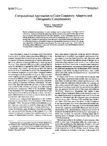
Fig. 1. Results from Experiment 1: mean motion-coherence thresholds for synesthetes and nonsynesthetes. Error bars represent standard errors of the mean.
Results The group results are summarized in Figure 1, and individual participants’ data are provided in Table 1. A higher motion-coherence threshold was seen for synesthetes relative to nonsynesthetic participants. An independentsamples t test revealed a significant difference between the groups, t(18) = 2.96, p = .008, d = 1.40, with synesthetes
Table 1. Motion-Coherence Thresholds for Each Synesthete and Control Participant Subject Synesthete Synesthete Synesthete Synesthete Synesthete Synesthete Synesthete Synesthete Synesthete Synesthete Control 1 Control 2 Control 3 Control 4 Control 5 Control 6 Control 7 Control 8 Control 9 Control 10
Motion-coherence threshold 1 2 3 4 5 6 7 8 9 10
.296 .375 .487 .400 .335 .436 .626 .554 .528 .278 .279 .245 .225 .210 .198 .425 .306 .323 .393 .385
Note: Thresholds relate to the level of coherence (expressed as proportions) in the motion of signal dots at which participants could correctly identify the direction of motion.
Motion and Color Perception in Synesthesia 2393 showing reduced performance (i.e., elevated thresholds) on the task.
Experiment 2: Color Perception Previously, we reported that synesthesia for color is linked to improved color perception (Banissy et al., 2009; Yaro & Ward, 2007). In our previous work, we used the Farnsworth-Munsell 100 Hue Test (Farnsworth, 1943), which requires the ordering of a series of finely spaced hues and is a common test of color discrimination (e.g., Franklin, Snowden, et al., 2010). However, color varies not only in hue but also along the perceptual dimensions of colorfulness and brightness.2 Importantly, the colors of synesthetic associations also vary along these three perceptual dimensions (Beeli, Esslen, & Jäncke, 2007; Simner et al., 2005). Therefore, the question arises whether any synesthetic advantage in color processing is restricted to hue, or whether there are also benefits for the other dimensions of color. Additionally, it is important to establish whether there is a synesthetic advantage for color processing on a task that taps perceptual processes more purely than the Farnsworth-Munsell 100 Hue Test and in which discriminations are more rapid and automatic. In the present experiment, we addressed these issues using a visual search task (e.g., Gilbert, Regier, Kay, & Ivry, 2006) to probe the color-perception abilities of synesthetes and nonsynesthetes. Stimuli on the task trials were the chromatic target and distractors that varied along one color dimension at a time: hue, saturation, or luminance (i.e., the task had a 3 × 2 design contrasting color dimension against stimulus group). We also administered a control visual-search task that was identical except it used achromatic line stimuli differing in orientation (Franklin, Catherwood, Alvarez, & Axelsson, 2010).
Method Participants. Thirty new participants took part in the study (age range = 18–21 years). Twenty of these were
control participants (4 males, 16 females), and the remaining 10 participants were synesthetes who experienced color as their evoked sensation (1 male, 9 females). All participants had normal or corrected-to-normal vision and completed two color-vision tests prior to testing (Ishihara, 1993; Lanthony, 1998). Synesthete status was verified using the Eagleman Battery (Eagleman et al., 2007). Materials. The visual-search task was presented on a calibrated 21-in. cathode-ray-tube monitor (Sony Trinitron GDM-F520). Target-distractor color pairs were chosen that varied along only one color dimension (hue, lightness or saturation). The colors were chosen from four regions of color space that roughly corresponded to the end points of the two cone-opponent processes (resulting in colors that were cherry, teal, violet, or chartreuse in appearance; see Jameson & D’Andrade, 1997). For each region, a midpoint color was chosen, and color pairs were selected around this midpoint so that the colors varied in luminance, hue, or saturation (as defined by the International Commission on Illumination 1976 L*, u*, v* color space) whereas the other two dimensions of color were kept constant. Color pairs were also chosen so that the perceptual difference between colors would be roughly equivalent along all three dimensions (stimulus pairs were initially equated in euclidean distance in CIE color space, yet pilot testing revealed the luminance difference to be more easily detected than hue or saturation during the search task, so the luminance difference was reduced). The CIE chromaticity coordinates of the stimuli and the gray background (which had same luminance as the midpoint), as verified by a colorimeter (ColorCAL; Cambridge Research Systems, Kent, England), are given in Table 2. Procedure. Participants were seated 57 cm from the monitor and were tested in a dark room, with the only light being that emitted from the monitor. They were informed that a clocklike arrangement of 12 colored discs
Table 2. International Commission on Illumination (CIE) Color-Space Coordinates, Hue Angle, and Saturation for the Four Midpoint Colors in Color Stimuli CIE 1931 color-space coordinates Color Chartreuse Teal Purple Cherry
CIE 1976 color-space coordinates
x
y
Y
L*
u*
v*
CIE hue angle
CIE saturation
0.388 0.274 0.301 0.371
0.382 0.325 0.273 0.311
25 25 25 25
68.25 68.25 68.25 68.25
16.36 –32.81 2.37 33.91
29.78 –9.71 –34.07 –3.68
61 197 274 354
0.498 0.498 0.498 0.498
Note: The coordinates of the gray background were x = .33, y = .33, Y = 25, and the coordinates of the white point used for xyY to L*u*v* conversions were x = .33, y = .33, Y = 62.5 cd/m2. Stimulus pairs were created around the midpoint by varying hue (±7ΔE), saturation (±0.183 CIE saturation), or lightness (±1Y).
Banissy et al.
2394 would be flashed very briefly on the screen and that one of the discs would differ very slightly from the others in color (see Fig. 2). Prior to trial onset, the screen displayed the gray background with a white fixation cross in the center of the screen. On each trial, the search array consisted of 14 notional circles (diameter = 30 mm) evenly spaced around a larger notional circle (diameter = 110 mm). Discs appeared in 12 of the circles—each circle except for the top and bottom circles on the midline, resulting in 6 colored discs in each hemifield with a gap between the two sets of discs. Participants were required to indicate which side (left or right) the target color (i.e., the color of the larger notional circle) was presented on by making a speeded button press. After 10 practice trials, 192 trials were presented in a random order. Each of the 12 color pairs (4 color regions × 3 color dimensions) was shown 16 times, with the side of presentation of the target color fully counterbalanced. Each stimulus in each pair appeared equally often as a target and as a distractor. Stimuli were displayed for 200 ms, after which there was an interstimulus interval of 3,000 ms, during which the response was recorded. No feedback was given. The control test used the same procedure, but the stimuli consisted of black diagonal lines (3 mm thick and
Results and discussion The results are summarized in Figure 2. In terms of accuracy in the color task itself, a 2 (group) × 3 (color dimension) analysis of variance (ANOVA) revealed a main effect of group, F(1, 28) = 17.13, p < .001, ηp2 = .16, but no Group × Dimension interaction, F(2, 56) = 2.24, n.s., ηp2 = .02. That is, the superior performance of synesthetes extended across all color dimensions and was not limited to hue. There was also a main effect of color dimension, F(2, 56) = 44.10, p < .001, ηp2 = .35, which was due to the saturation differences’ being generally harder to perceive. Although synesthetes were more accurate at color search, they did not differ from controls in their response times: A 2 × 3 ANOVA on response time revealed no main effect of group, F(1, 28) = 0.33, ηp2 = .01, or Group × Dimension interaction, F(2, 56) = 1.90, ηp2 < .01. The main effect of color dimension was significant, an effect due to the fact that reactions times to the saturation stimuli were slower, F(2, 56) = 34.94, p < .001, ηp2 = .05. On the line-orientation control task, the synesthetes did not differ from controls in terms of either accuracy, t(28) = 0.67, n.s., d = 0.25, or response time, t(28) = 0.307, n.s., d = 0.11. That is, their selective advantage for color was not due to a general performance advantage on perceptual visual-search tasks.
General Discussion
b 100
Percentage of Correct Responses
45 mm long), one of which had a slightly different orientation from the others. There were 48 trials incorporating of 6 different stimulus pairs, and each stimulus in each pair appeared equally often as a target and as a distractor. The control test was conducted after the color test.
Controls Synesthetes
95 90 85 80 75 70
Orientation
Task
Color
Luminance Chroma
Hue
Dimension
Fig. 2. Stimuli and results from Experiment 2. Panel (a) shows example color and orientation search arrays. The graph in (b) shows mean visual-search accuracy as a function of participant group and task (left side of graph) or dimension (right side of graph). Error bars represent standard errors of the mean.
We examined whether synesthesia for color is linked to differences in perceptual sensitivity that extend beyond the synesthetic experience itself. We found that synesthesia for color is linked to an increase in the perception of color differences but a decrease in the perception of motion. These findings imply that synesthesia for color is associated with wider differences in visual perception, including beneficial and negative consequences for perceptual sensitivity. A prevailing view of synesthesia is that the condition is linked to excess cortical connectivity between inducer and concurrent brain regions (see Bargary & Mitchell, 2008; Rouw et al., 2011, for review). Our data suggest that even if this is the case, such increased connectivity may come at a cost to other visual regions, leading to reductions in motion-coherence perception. This is line with our previous neuroanatomical findings showing that synesthesia is linked to reduced gray-matter volume in MT/V5 (Banissy et al., 2012) and those of Barnett et al. (2008) showing a trend for a reduction in magnocellular
Motion and Color Perception in Synesthesia 2395 responsiveness in linguistic-color synesthesia (although some caution is warranted on this point; see below). Collectively, these results imply that synesthesia for color may be linked to more distributed differences in neural responsiveness than simply excess connectivity between inducer and concurrent brain areas, and that these differences may be manifested behaviorally as reductions in some aspects of perception. One factor that is likely to contribute to the reduced motion-coherence perception displayed by synesthetes is how interactions between color and motion in nonsynesthetes are governed. In nonsynesthetes, a number of studies have pointed to a mutual inhibition of neural regions involved in color and form processing and motion processing, with motion processing modulating color and form processing and vice versa (Ellison et al., 2003; Morland et al., 1996; Walsh et al., 1998). For example, the suppression of neural regions involved in motion processing leads to a disruption of motion perception but a concomitant facilitation of color and form processing (Ellison et al., 2003; Walsh et al., 1998). This pattern has often been interpreted as evidence for a competition for resources between motion and color areas in the visual system (Ellison et al., 2003; Walsh et al., 1998), and it is feasible that in color synesthesia, the recurrent perception of color in the absence of chromatic stimuli may result in a prioritizing of neural representations involving color, leading to a reduction in activity in motion-selective brain regions and parallel behavioral manifestations (i.e., reduced motion perception). One might also suggest that our findings of reduced motion perception in synesthesia for color indicate that synesthetes show a selective loss of magnocellularpathway cells. We, however, would urge caution in this interpretation at this stage for two reasons. First, it will be important for future studies to examine the group differences reported here across different motion speeds. In the present study, we identified group differences between synesthetes and controls at a slow motion speed, but to make a stronger claim for deficits in magnocellular responsiveness, it will be necessary for future studies to verify that these differences extend across other motion speeds. Second, one should note that there is some debate over whether losses in lowerspeed motion perception reflect a selective loss of magnocellular-pathway cells (Lee, Wehrhahn, Westheimer, & Kremers, 1993; Snowden & Kavanagh, 2006). In view of this, although there is a clear pattern of disruption in motion-coherence processing in color synesthetes, we would urge caution in assuming that this is reflective of a selective loss of magnocellular-pathway cells. Our finding that color synesthetes show superior color perception is in line with our previous findings highlighting that color synesthetes show a perceptual advantage
in color sorting on the Farnsworth-Munsell 100 Hue Test (Banissy et al., 2009; Yaro & Ward, 2007). We have extended these findings by demonstrating that this perceptual advantage is present not only for hue (as reported previously) but also for luminance and saturation, and that this advantage is present even on a speeded task in which perceptual discrimination is faster and more automatic. The findings also suggest that the enhancements in color perception displayed by synesthetes are not purely a function of increased parvocellular responsiveness (Barnett et al., 2008) because synesthetes show perceptual benefits for dimensions of color that do not rely solely upon parvocellular projections (i.e., luminance; Lee, Pokorny, Smith, Martin, & Valberg, 1990). Several factors may contribute to the enhanced color processing shown by synesthetes. First, the additional experience of color in the perception of nonchromatic stimuli may lead to enhanced perceptual experience with colors and, therefore, superior color perception. Indeed, in other visual domains, perceptual learning and experience has been shown to facilitate perception in typical adults (e.g., Fahle & Poggio, 2002). Second, the presence of synesthesia may increase the number of color labels that the synesthete possesses, leading to changes in the internal structure of color space, as has been observed in cross-cultural studies (Robertson, Davies, & Davidoff, 2000; Yaro & Ward, 2007); however, this is only likely to aid perception across the boundaries for those terms and does not explain enhanced color perception that is distributed evenly throughout color space. Third, enhanced color processing in synesthesia may reflect differences in brain development due to changes in cortical excitation (Cohen Kadosh, Henik, Catena, & Walsh, 2009) or cortical connectivity (Bargary & Mitchell, 2008). It is also interesting to consider whether other types of synesthesia (i.e., those not involving color) might be linked to a perceptual profile similar to that observed in our study with synesthetes who experienced color as their evoked experience (i.e., increased color perception but reduced motion perception). Although our lack of a synesthetic control group means that we cannot definitively answer this question, in our prior work we have shown that synesthetes who experience touch as their evoked sensation do not show enhanced color perception, whereas synesthetes who experience color do (Banissy et al., 2009). We therefore feel that it is unlikely that other types of synesthetes (e.g., vision-touch synesthetes; Ward, Banissy, & Jonas, 2008) would show a similar pattern. This assumption does rest, however, on our view that the most parsimonious explanation for our data is that the color-motion relationship is mediated by interactions between neural systems involved in processing these attributes in typical adults (e.g. Ellison et al.,
2396 2003; Morland et al., 1996; Walsh et al., 1998). It will be interesting to address this question with future studies. In summary, we report that synesthetes who experience color as their evoked sensation show broader differences in perceptual sensitivity that extend beyond their synesthetic experiences. The findings of enhanced color processing but reduced motion processing are in line with principles that modulate interactions between color and motion processing in healthy adults, and suggest that synesthesia for color may be linked with more distributed differences in neural responsiveness that can be both positive and negative for perceptual processing. Author Contributions M. J. Banissy, V. Walsh, N. G. Muggleton, J. Ward, and A. Franklin developed the study concept. All authors contributed to the study design. Testing and data collection were performed by V. Tester, A. B. Janik, and A. Davenport. M. J. Banissy and J. Ward analyzed and interpreted the data. M. J. Banissy, N. G. Muggleton, A. Franklin, and J. Ward drafted the manuscript, and all authors approved the final version of the manuscript for submission.
Declaration of Conflicting Interests The authors declared that they had no conflicts of interest with respect to their authorship or the publication of this article.
Funding The British Academy (M. J. Banissy; Grant pf100123), the European Research Council (A. Franklin; Grant 283605), and the Medical Research Council (V. Walsh; Grant G0700929) supported this work.
Notes 1. Note that we expected this to be the case only in synesthesia in which color was the evoked sensation, because there is a known relationship between color and motion processing in typical adults and because previously we had shown that synesthetes who experienced touch as their evoked sensation did not differ from nonsynesthetes in their color-perception abilities (Banissy et al., 2009). 2. Hue is the perceptual property of color that corresponds to the physical dominant wavelength; colloquially, this property is called “color.” Brightness is the perceived amount of light exhibited by an area (perceived luminance—lightness is luminance relative to other stimuli). Colorfulness is the intensity of a color (chroma is the intensity relative to the brightness of a white; saturation is the intensity relative to the brightness of a color; see also Hunt & Pointer, 2011).
References Bach, M. (1996). The Freiburg Visual Acuity Test—Automatic measurement of visual acuity. Optometry and Vision Science, 73, 49–53. Banissy, M. J., Stewart, L., Griffiths, T., Muggleton, N. G., Walsh, V., Ward, J., & Kanai, R. (2012). Grapheme-color
Banissy et al. and tone-color synaesthesia is associated with structural brain changes in visual regions implicated in color, form and motion. Cognitive Neuroscience, 3, 29–35. Banissy, M. J., Walsh, V., & Ward, J. (2009). Enhanced sensory perception in synaesthesia. Experimental Brain Research, 196, 565–571. Bargary, G., & Mitchell, K. J. (2008). Synaesthesia and cortical connectivity. Trends in Neurosciences, 31, 335–342. Barnett, K. J., Fox, J. J., Molholm, S., Kelly, S. P., Shalgi, S., Mitchell, K. J., & Newell, F. N. (2008). Differences in early sensory-perceptual processing in synaesthesia: A visual evoked potential study. NeuroImage, 43, 605–613. Beeli, G., Esslen, M., & Jäncke, L. (2007). Frequency correlates in grapheme-color synaesthesia. Psychological Science, 18, 788–792. Cohen Kadosh, R., Henik, A., Catena, A., & Walsh, V. (2009). Induced cross-modal synesthetic experience without abnormal neuronal connections. Psychological Science, 20, 258–265. Derrington, A. M., & Lennie, P. (1984). Spatial and temporal contrast sensitivities of neurones in the lateral geniculate nucleus of macaque. Journal of Physiology, 357, 219–240. Eagleman, D. M., Kagan, A. D., Nelson, S. S., Sagaram, D., & Sarma, K. (2007). A standardized test battery for the study of synesthesia. Journal of Neuroscience Methods, 159, 139– 145. Ellison, A., Battelli, L., Cowey, A., & Walsh, V. (2003). The effects of expectation on facilitation of colour/form conjunction tasks by TMS over area V5. Neuropsychologia, 41, 1794–1801. Fahle, M., & Poggio, T. (2002). Perceptual learning. Cambridge, MA: MIT Press. Farnsworth, D. (1943). The Farnsworth Munsell 100-hue dichotomous tests for color vision. Journal of the Optical Society of America, 33, 568–576. Franklin, A., Catherwood, D., Alvarez, J., & Axelsson, E. (2010). Hemispheric asymmetries in categorical perception of orientation in infants and adults. Neuropsychologia, 48, 2648– 2657. Franklin, A., Sowden, P., Notman, L., Gonzales-Dixon, M., West, D., Alexander, I., . . . White, A. (2010). Reduced chromatic discrimination in children with autism spectrum disorders. Developmental Science, 13, 188–200. Gilbert, A. L., Regier, T., Kay, P., & Ivry, R. B. (2006). Whorf hypothesis is supported in the right visual field but not the left. Proceedings of the National Academy of Sciences, USA, 103, 489–494. Hunt, R. W. G., & Pointer, M. R. (2011). Measuring colour. Chichester, England: Wiley. Ishihara, S. (1993). Ishihara tests for colour-blindness. Tokyo, Japan: Kanehara. Jameson, K., & D’Andrade, R. G. (1997). It’s not really red, green, yellow, blue: An inquiry into perceptual color space. In C. L. Hardin & L. Maffi (Eds.), Color categories in thought and language (pp. 295–318). Cambridge, England: Cambridge University Press. Kalla, R., Muggleton, N., Spiegel, R., Bueti, D., Claassen, J., Walsh, V., & Bronstein, A. (2011). Adaptive motion processing in bilateral vestibular failure. Journal of Neurology, Neurosurgery & Psychiatry, 82, 1212–1216.
Motion and Color Perception in Synesthesia 2397 Kaplan, E. (1991). The receptive field of retinal ganglion cells in cat and monkey. In A. G. Leventhal (Ed.), Vision and visual dysfunction (pp. 10–40). Boston, MA: CRC Press. Lanthony, P. (1998). Lanthony tritan album. Paris, France: Luneau Ophtalmologie. Lee, B. B., Pokorny, J., Smith, V. C., Martin, P. R., & Valberg, A. (1990). Luminance and chromatic modulation sensitivity of macaque ganglion cells and human observers. Journal of the Optical Society of America A, 7, 2223–2236. Lee, B. B., Wehrhahn, C., Westheimer, G., & Kremers, J. (1993). Macaque ganglion cell responses to stimuli that elicit hyperacuity in man: Detection of small displacements. Journal of Neuroscience, 13, 1001–1009. Morland, A. B., Ogilvie, J. A., Ruddock, K. H., & Wright, J. R. (1996). A new abnormality of human vision provides evidence of interactions between cortical mechanisms sensitive to movement and those sensitive to colour. Proceedings of the Royal Society B: Biological Sciences, 263, 1087–1094. Robertson, D., Davies, I., & Davidoff, J. (2000). Color categories are not universal: Replications and new evidence from a stone-age culture. Journal of Experimental Psychology, 129, 369–398. Rouw, R., Scholte, H. S., & Colizoli, O. (2011). Brain areas involved in synaesthesia: A review. Journal of Neuropsychology, 5, 214–242. Simner, J. (2012). Defining synaesthesia. British Journal of Psychology, 103, 1–15.
Simner, J., Mulvenna, C., Sagiv, N., Tsakanikos, E., Witherby, S., Fraser, C., . . . Ward, J. (2006). Synaesthesia: The prevalence of atypical cross-modal experiences. Perception, 35, 1024–1033. Simner, J., Ward, J., Lanz, M., Jansari, A., Noonan, K., Glover, L., & Oakley, D. A. (2005). Non-random associations of graphemes to colours in synaesthetic and non-synaesthetic populations. Cognitive Neuropsychology, 22, 1069–1085. Snowden, R. J., & Kavanagh, E. (2006). Motion perception in the ageing visual system: Minimum motion, motion coherence, and speed discrimination thresholds. Perception, 35, 9–24. Terhune, D. B., Tai, S., Cowey, A., Popescu, T., & Cohen Kadosh, R. (2011). Enhanced cortical excitability in grapheme-color synesthesia and its modulation. Current Biology, 21, 2006–2009. Walsh, V., Ellison, A., Battelli, L., & Cowey, A. (1998). Taskspecific impairments and enhancements by magnetic stimulation of human visual area V5. Proceedings of the Royal Society B: Biological Sciences, 265, 537–543. Ward, J., Banissy, M. J., & Jonas, C. (2008). Haptic perception in synaesthesia. In M. Grunwald (Ed.), Human haptic perception: Basics and applications (pp. 259–265). Basel, Switzerland: Birkhäuser. Yaro, C., & Ward, J. (2007). Searching for Shereshevski: What is superior about the memory of synaesthetes? Quarterly Journal of Experimental Psychology, 60, 681–695.