Abstract. This paper reports a systematic investigation of the growth and attachment of small gold nanoparticles to the functionalized surface of larger silica ...
PRAMANA — journal of
c Indian Academy of Sciences �
physics
Vol. 69, No. 2 August 2007 pp. 277–283
Synthesis and characterization of silica–gold core-shell (SiO2 @Au) nanoparticles DEEPIKA KANDPAL1,∗ , SUCHITA KALELE2 and S K KULKARNI2 1
Department of Physics, G.B. Pant University of Agriculture & Technology, Pantnagar 263 145, India 2 Department of Physics, University of Pune, Pune 411 007, India ∗ Corresponding author. E-mail: d kandpal77@rediffmail.com MS received 9 December 2006; revised 9 March 2007; accepted 11 April 2007 Abstract. This paper reports a systematic investigation of the growth and attachment of small gold nanoparticles to the functionalized surface of larger silica nanoparticles by three different methods. Nearly monodispersed silica particles and gold nanoparticles were prepared by sol–gel method. The size of the particle could be altered by changing the concentration of reactants, temperature and the time for which they react. The nanocoreshell particles prepared by three different methods were studied using scanning electron microscopy (SEM), UV–vis spectroscopy and Fourier transform infrared spectroscopy. We have found that the third method (c), a combination of the first two methods (a) and (b), has given better results. Keywords. Core-shell particles; silica particles; gold particles. PACS Nos 78.66.Jg; 78.66.Sq
1. Introduction Nanoparticles have been extensively investigated during the last few decades because of their unique properties and application potential. Nanoparticles can be considered as intermediate between molecules and solids. However, both molecules and solids have definite dimension and well-defined properties and the properties are not size-dependent. Nanoparticles, on the other hand, are characterized by size-dependent properties. For nanoparticles, both size and surface effects are important. By controlling these, it is possible in principle to design materials of required optical, magnetic, elastic, chemical etc. properties [1]. There is increasing interest in the design and synthesis of topological structures composed of monocrystals of various size and shape. Such materials may have unusual optical properties as a result of increasing topological complexity. Core-shell structures are of central interest in this context. Core-shell particles constitute a novel class of materials with potential application in chemically stabilizing colloidal particles, catalysis,
277
Deepika Kandpal, Suchita Kalele and S K Kulkarni fluorescent diagnostics, photonic band-gap materials, preparation of biconjugates etc. [2]. Solid metallic nanoparticles are well-known for their attractive optical properties, strong optical resonance and a large and fast nonlinear optical polarizability associated with the plasmon frequency of the conduction electron in the particle [3–5]. Colloidal metal shells on the other hand can have resonance that can be tuned over a wide range as a function of the core-to-shell ratio. The greatly enhanced ability to manipulate the boundary conditions of the resonating conduction electrons makes it possible to cover theoretically the ultraviolet, visible and infrared parts of the spectrum. Westcott et al [6] have shown in several papers how such shells composed of gold can be grown around silica colloids and how the single particle properties can be exploited. Silica colloids provide for a convenient dielectric core as they can be grown with a small polydispersity. We are motivated not only by the single particle properties but also by the prospect of using the collective response of metallodielectric colloids if they are arranged on a regular three-dimensional (3D) lattice. For instance, it has been shown recently that regular 3D structures with feature sizes on the order of the wavelength under consideration, also called photonic crystals, can have a full photonic band gap in the visible region, if they are made from metallodielectric spheres [7–9]. We have synthesized the silica–gold core-shell (SiO2 @Au) nanoparticles using an approach described in experimental section. The presence of the shell is analysed by UV–vis absorption spectroscopy, scanning electron microscopy (SEM) and Fourier transform infrared spectroscopy (FTIR).
2. Experimental section Silica and gold particles were separately synthesized by sol–gel method and silica particles are coated with respective shell materials (Au) in a multistep process. Details of the synthesis and analysis techniques are given below. 2.1 Synthesis of gold-coated silica particles Silica particles were synthesized using St¨ ober method by hydrolysis and condensation of TEOS (tetraethylorthosilicate Si(OC2 H5 )4 ) [10]. By changing the concentration of water, ethanol, NH4 OH and TEOS we can vary the size of the monodispersed silica particles. Silica nanoparticles were functionalized to attach the gold particles on it by using the bi-functional organic solvent 3-aminopropyltriethoxysilane (APS) [3]. The gold metal has very little affinity for silica, and it does not form the passivating oxide film in solution. So silane coupling agent is used as surface primer. The strong chemical affinity of primary amines for gold drives chemisorptions of the colloids in the case of 3-aminopropyltriethoxysilane (APS). We have used three different methods to form silica–gold core-shell particles by varying the parameters to get fine results. (a) Citrate method: Here we have used the gold prepared by reducing 0.002 M HAuCl4 solution by 0.01 M trisodium citrate Na3 (C6 H5 O7 ) for coating as well as for gold nucleation [11].
278
Pramana – J. Phys., Vol. 69, No. 2, August 2007
Silica–gold core-shell nanoparticles
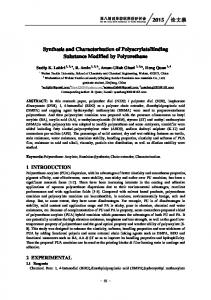
Figure 1. Flow chart showing the experimental procedure.
(b) Borohydride method: Here, we have used the gold prepared by reducing 0.002 M HAuCl4 /K2 CO3 solution by 0.001 M sodium borohydride (NaBH4 ) for coating as well as for gold nucleation [12]. (c) This method is a combination of the above two methods in which, gold prepared by varying the concentration of reducing agent 0.01 M sodium borohydride (NaBH4 ) is used for coating and gold solution prepared by citrate method is used for gold nucleation. We find coloured silica–gold core-shell particles (SiO2 @Au) by all the three different methods. The whole synthesis is described in figure 1. The characterization is done by UV–vis absorption spectroscopy, scanning electron microscopy (SEM) and Fourier transform infrared spectroscopy (FTIR).
Pramana – J. Phys., Vol. 69, No. 2, August 2007
279
Deepika Kandpal, Suchita Kalele and S K Kulkarni
Figure 2. SEM picture of silica particles (∼350 nm).
Figure 3. UV–visible spectrum of pure gold particles.
3. Result and discussion The size of monodispersed silica nanoparticles prepared by St¨ ober method was studied using scanning electron microscopy (SEM). The SEM picture of silica particles is shown in figure 2. Functionalized and gold nuclide silica particles in solution were treated many times with gold nanoparticles and centrifuged. Gold particles were found to be attached to silica core. It could be observed that the originally white particles of silica turned into coloured ones. Optical absorption was recorded at various stages of addition of gold nanoparticles in every method using UV–visible absorption spectroscopy. Optical absorption of pure gold solution was found at 525 nm as shown in figure 3. In silica–gold core-shell particles prepared by method (a) we did not find any measurable plasmon peak shift in pure gold. UV–visible absorp280
Pramana – J. Phys., Vol. 69, No. 2, August 2007
Silica–gold core-shell nanoparticles
Figure 4. UV–visible spectra of SiO2 @Au core-shell particles prepared by method (b).
Figure 5. UV–visible spectra of SiO2 @Au core-shell particles prepared by method (c).
tion spectrum for methods (b) and (c) are shown in figures 4 and 5 respectively. In figure 4, we have seen from the UV–visible spectra of silica–gold core-shell particles that the plasmon peak of pure gold shows slight shift but the peak is not so sharp and the gold particles prepared by using the reducing agent sodium borohydride (NaBH4 ) is not too much stable. The gold particles prepared by citrate method are more stable but its reduction rate is very slow. In nucleation process silica react with separately prepared gold nanoparticles so the gold particles should be stable, but in coating the shell of gold nanoparticles were prepared directly on the surface of functionalized and gold nuclide silica nanoparticles. These gold particles
Pramana – J. Phys., Vol. 69, No. 2, August 2007
281
Deepika Kandpal, Suchita Kalele and S K Kulkarni
Figure 6. SEM pictures of silica–gold core-shell nanoparticles after the third coating method (c).
Figure 7. FTIR spectra of silica and silica@Au particles prepared by method (c).
directly attach with silica core and now they behave like stable core-shell particles. So for coating reducing reagent (NaBH4 ) is more useful because of its high reduction rate. We have seen from the UV–visible spectra of silica–gold core-shell particles that after two coatings (SiO2 @Au1, SiO2 @Au2) the peak was red-shifted to 528 nm, 531 nm respectively (see figure 5). But after third coating (SiO2 @Au3) the peak was blue-shifted to 529 nm. Although figure 5 shows a slight blue shift in the peak, it depends on the number of coatings. Initially, less number of gold nanoparticles were attached with silica core and the no. of coatings increases the gold particles which smoothly cover the whole surface of silica, which shows large measurable blue shift in peak. There are many reports where optical properties of core-shell particles of silica and gold are studied [2,11,12]. Halas group [14] has reported the results of SiO2 @Au composite. Initially for very small thickness the absorption peak is red-shifted with respect to the original position of pure gold. As the shell thickness increased, the optical absorption peak started shifting towards lower wavelength. The formation of silica–gold core-shell particles prepared by
282
Pramana – J. Phys., Vol. 69, No. 2, August 2007
Silica–gold core-shell nanoparticles method (c) was also studied by scanning electron microscopy (SEM). The results are shown in figure 6. The size of the core (SiO2 ) nanoparticles is found to be ∼260 nm, the size of shell (Au) nanopatricles is ∼20 nm and core-shell ratio is ∼13 : 1. The structure study, i.e. bonding of these core–shell particles was also done using Fourier transform infra-red spectroscopy (FTIR). The results are shown in figure 7. This figure shows the FTIR spectrum of silica, functionalized silica and silica–gold core-shell particles. For SiO2 and SiO2 @Au particles peak 1103, 80 cm−1 can be assigned to Si–O–Si bond and peak 947 cm−1 can be attributed to Si–OH bond and for functionalized silica particles peaks at 3215 cm−1 is assigned to NH bond. After coating these particles with gold, the intensity of Si–O–Si and Si–OH peaks has been reduced significantly. This indicates the presence of gold shell in silica particles. Silica@gold core-shell particles were synthesized. It was observed that the plasmon resonance peak position of gold depends upon the shell thickness. Thus, by changing shell thickness, it is possible to design a material with desired optical properties. The present method, the combination of citrate and borohydride method, gives much better results and it works at room temperature. The presence of gold coating was confirmed by FTIR spectroscopy. The morphology of these particles was studied using SEM. Acknowledgement One of the authors (Deepika Kandpal) would like to thank the Indian Academy of Sciences, Bangalore for providing two months summer fellowship for this project work. References [1] A P Alivisatos, J. Phys. Chem. 100, 13226 (1996) [2] L M Liz-Marzan, M A Correa-Duarte, I Pastoriza-Santos, P Mulvaney, T Ung, M Giersig and N A Kotov, Handbook of surfaces and interfaces of materials edited by H S Nalwa (Academic Press, 2001), vol. 3, Chapter 5, p. 189 [3] F Hache, D Richard, C Flytzanis and U Kribig, Appl. Phys. 47, 347 (1988) [4] J Y Bigot, J C Merle, O Cregut and A Daunois, Phys. Rev. Lett. 75, 4702 (1995) [5] M Perener, P Bost, U Lemmer, G Von Plessem, J Feldmann, U Becker, M Menig, M Schmitt and H Schmidt, Phys. Rev. Lett. 78, 2192 (1997) [6] S L Westcott, S J Oldenburg, T R Lee and N J Halas, Langmuir 14, 5396 (1998) [7] A Moroz, Europhys. Lett. 50, 466 (2000) [8] A Moroz, Phys. Rev. Lett. 83, 5274 (1999) [9] W Y Zhang, X Y Lei, Z L Wang, D G Zheng, W Y Tam, C T Chang and P Sheng, Phys. Rev. Lett. 84, 2853 (2000) [10] W Stober, A Fink and E Bohn, J. Coll. Int. Sci. 62, 26 (1968) [11] J Turkenvich, P C Stevenson and J Hillier, Discuss. Faraday Soc. 11, 55 (1951) [12] C Graf and A V Blaaderen, Langmuir 18, 524 (2002) [13] S J Oldenburg, D Averitt, S L Westcott and N J Halas, Chem. Phys. Lett. 288, 243 (1998) [14] S J Oldenburg, J B Jackson, S L Westcott and N J Halas, Appl. Phys. Lett. 75, 2897 (1999) Pramana – J. Phys., Vol. 69, No. 2, August 2007
283