R. C. Singh, O. Singh, M. P. Singh and P. S. Chandi (2008). Sensors & Actuators. B. 135. 352. 5. Z. K. Tang, G. K. L. Wang, P. Yu, M. Kawasaki, A. Ohtomo, ...
Synthesis, Characterization and Clustering Phenomenon of Zinc Oxide Nanocrystals H. S. Virk & Poonam Sharma Nanotechnology Laboratory, DAV Institute of Engg. & Technology, Kabir Nagar, Jalandhar-144008
Zinc oxide (ZnO) nanocrystals were prepared by quenching method, i.e; mixing sintered ZnO powder with ice cold ethanol. Addition of ethylenediamine to this mixture produced nanorods of ZnO. XRD spectrum reveals that ZnO nanocrystals display hexagonal wurtzite structure. Clustering phenomenon is recorded in HRTEM and SEM photographs. Key Words: Zinc Oxide, Ethanol, Nanocrystals, Nanorods, Clustering
INTRODUCTION Zinc oxide (ZnO) is one of the most important semiconducting materials, having a wide range of potential applications [1, 2]. ZnO is an important electronic and photonic material because of its wide direct based gap of 3.37 eV. In recent years, ZnO nanocrystals have been used for solar cell applications [3], gas sensors [4] and ultraviolet lasing action at room temperature [5, 6]. Nanocrystals of ZnO have been prepared using both physical and chemical methods [7-10]. Among them sol-gel chemistry, spray pyrolysis, microemulsion, precipitation solvothermal and hydrothermal methods are being extensively used [11-15]. Generally, most of these methods of synthesis require relatively high temperatures or involve the use of expensive chemicals or apparatus. It is therefore advisable to find simple methods to produce ZnO nanocrystals using commonly available chemicals. A detailed study of the interaction of alcohols with Zn metal has revealed that the C-O bond of the alcohol is readily cleaved on Zn metal surfaces giving hydrocarbons and the oxidic species on the metal surface [16, 17]. Using this simple technique, we found that ZnO nanocrystals are readily produced by the reaction of zinc metal with ethanol. Addition of ethylenediamine (EDA) to the reaction mixure produces ZnO nanorods, giving evidence of clustering phenomenon. EXPERIMENTAL 5mg of zinc powder (CDH grade) was heated in an oven for 6 hours in crucible at a temperature of 8000C. 5ml of ethanol was kept in a refrigerator to cool it to 00C. Ice cold ethanol was poured into heated zinc powder. ZnO nanocrystals are produced by quenching process. The resulting suspension is centrifuged to retrieve the product, washed with alcohol and dried. ZnO nanorods were prepared by addition of 1ml of ethylenediamine to mixture of 5mg of zinc powder and 5ml of ethanol at the room temperature. The other part was kept for characterization of ZnO nanocrystals. Scanning Electron Microscope (Jeol, JSM-6100) was used for recording the morphology of nanorods prepared by adding ethylenediamine. X-ray Diffraction studies were carried out at Sophisticated Analytical Instruments Facility (SAIF) set up by Punjab University,
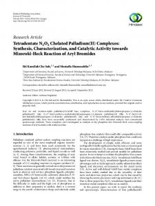
Chandigarh using X' Pert PRO (PANanalytical, Netherlands). High Resolution Transmission Electron Microscope (HRTEM, Hitachi H 7500) at SAIF was used for measurements of nanocrystal diameters. RESULT AND DISCUSSION XRD spectrum of ZnO nanocrystals, using a Cu-Kα radiation source of λ=1.5406A0, is shown in Fig. 1. The spectrum shows some prominent peaks with most prominent peak at 36.2682 corresponding to hkl (101). The XRD pattern of ZnO nanocrystals could be indexed to the hexagonal wurtzite structure (space group: P63mc (186), cell constants a = 0.3254 nm and c = 0.5209nm). Table1 summarizes the XRD spectrum results of ZnO nanocrystals which almost perfectly match with those calculated for ZnO using the standard ICSD Card # : 067849 [18].
Fig.1. XRD spectrum of ZnO nanocrystals showing some prominent peaks Table.1 Assignment of Miller indices to ZnO nanocrystals Sr. No. 1. 2. 3. 4. 5.
2 θ degree 31.7737 34.4373 36.2682 47.5728 56.6385
d-spacing (nm) 0.2816 0.2604 0.2477 0.1911 0.1625
hkl 100 002 101 102 110
6. 7. 8.
62.9179 68.0153 69.1571
0.1477 0.1378 0.1358
103 110 201
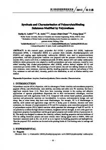
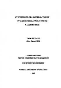
The morphology ZnO nanocrystals is revealed in HRTEM images shown in Fig. 2 (a,b). The images show clustered appearance of ZnO nanocrystals (Fig.2a). The diameter of nanocrystals varies from 7.73 to 8.86 nm. Addition of EDA to reaction mixure of ZnO and ethanol produced nanoroads as revealed by SEM image (Fig. 3). It is a clear evidence of clustering phenomenon of nanocrystals. Another evidence of clustering phenomenon has been observed by Nath et al. [19] after heavy ion irradiation of ZnO nanocrystals using Pelletron beam. Virk and Sharma [20] have also reported clustering phenomenon on irradiation of CdO quantum dots using 90 MeV Carbon ion beam recently.
Fig. 2(a) HRTEM micrograph of ZnO nanocrystals showing clustering
Fig. 2(b) HRTEM images of ZnO nanocrystals showing diameter measurement
Fig.3. SEM image of ZnO nanorod prepared by adding EDA to ZnO nanocrystals
CONCLUSIONS ZnO nanocrystals can be prepared at room temperature by simple quenching method. The ethanol reacts with Zn metal to produce ZnO by cleavage of C-O bond on metal surface. EDA acts as a shape directing agent and produces nanorods of ZnO. HRTEM and SEM characterization reveals clustering phenomenon in ZnO nanoparticles.
ACKNOWLEDGEMENTS The authors wish to express their gratitude to the Principal DAV Institute of Engineering & Technology, Jalandhar and DAV College Managing Committee, New Delhi for providing research grants for setting up Research Centre and Nanotechnology Laboratory in Jalandhar. They are thankful to staff of SAIF, Punjab University, Chandigarh.for providing instrumental analysis facilities on priority. REFERENCES 1. Z. L. Wang (2004). Mater. Today 7. 26. 2. Z. L. Wang, X. Y. Kong, Y. Ding, P. Gao, W. L. Hughes, R. Yang and Y. Zhang (2004). Adv. Funct. Mater. 14. 943. 3. Q. Li, V. Kumar, Y. Li, H. Zhang, T. J. Marks and R. P. H. Chang (2005). J. Am. Chem. Soc. 17. 1001. 4. R. C. Singh, O. Singh, M. P. Singh and P. S. Chandi (2008). Sensors & Actuators B. 135. 352. 5. Z. K. Tang, G. K. L. Wang, P. Yu, M. Kawasaki, A. Ohtomo, H. Koinmua and Y. Segawa (1998). Appl. Phys. Lett. 72. 3270. 6. M. H. Huang, S. Mao, H. Feick, H. Yan, Y. Wu, H. Kind, E. Webber, R. Russo and P. Yang (2001). Science 292.1897. 7. M. Ghosh (2004). J. Nanosci. Nanotech. 4. 136. 8. K. Koch (1985). Chem. Phys. Lett. 122. 507. 9. M. D. Mustafa (2006). J. Mater. Chem. 16. 2940. 10. J. Buha (2007). Crystal Growth & Design 7. 113. 11. L. Guo, Y. L. Ji, H. Xu, P. Simon, Z. Wu (2002). J. Am. Chem. Soc. 124. 14864. 12. C. Xu, G. Xu, Y. Liu and G. Wang (2000). Solid State Commun. 122. 175. 13. S. Chakarbarti and G. Chaudhari (2004). Mater. Chem. Phys. 87. 196.
14. B. Liu and H. C. Zeng (2003). J. Am. Chem. Soc. 125. 4430. 15. N. Verghese, L. S. Panchakarla, M. Hanapi, A. Govindaraj and C. N. R. Rao (2007). 42. 2117. 16. L. S. Pahchakarla, A. Govindaraj and C. N. R. Rao (2007). J. Cluster Sci. 18. 660. 17. K. R. Harikumar (1999). J. Phys. Chem. B. 103. 2445. 18. O. Garcia-Martinez et al. (1997). Solid State Ionics 63. 442. 19. S. S. Nath, D. Chakdar, G. Gope and D. K. Avasthi (2007). Nano Trends 3. 1-10. 20. H. S. Virk and Poonam Sharma (2009). J. Nano Sci. (Communicated)