[7] H.R. Shioya, T. Agatruna, H. Furukawa, S. Naruto, & Y. Sugano, Biorg. Med. Chem., 2004, 14, 455. [8] T. Oka, T. Yasusa, T. Ando, M. Watanabe, F. Yoneda, ...
Available online at www.derpharmachemica.com
ISSN 0975-413X CODEN (USA): PCHHAX
Der Pharma Chemica, 2016, 8(7):46-54
(http://derpharmachemica.com/archive.html)
Synthesis, Characterization, Crystal structure and DFT calculations of 1-benzofuran-2-carboxylic acid G. Krishnaswamy1, Nivedita R. Desai1, Krishna Murthy Potla1, P. A. Suchetan2 and Arunakumar D. B.*1 1
Department of Studies and Research in Chemistry, Tumkur University, Tumkur-572 103, India Department of Chemistry, University College of Science, Tumkur University, Tumkur-572 103, India _____________________________________________________________________________________________ 2
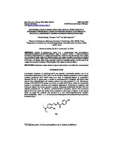
ABSTRACT In this study, 1-benzofuran-2-carboxylic acid was synthesized and characterized by FT-IR, 1HNMR and single crystal XRD analysis. The compound crystallizes in monoclinic system with space group P21/n and Z = 4. Its vibrational frequencies and optimized geometric parameters has been calculated using DFT method with UB3LYP/6-31G (d, p) basis sets by Gaussian 03 software. The calculated vibrational frequencies and optimized geometric parameters (bond lengths and bond angles) were compared with experimentally measured values and found to be in good agreement with each other. Additionally, molecular electrostatic potential maps, density of states, frontier molecular orbitals and the other related molecular energy values have been evaluated using the same theoretical calculations. Keywords: 1-benzofuran-2-carboxylic acid, DOS, FT-IR spectrum, Density functional theory, X-ray diffraction. _____________________________________________________________________________________________ INTRODUCTION Benzofurans are important heterocyclic compounds, which are naturally occurring and exhibit not only remarkable biological activities but also acts as useful building blocks in the synthesis of natural products [1, 2]. Many of the natural benzofurans have physiological, pharmacological and toxic properties [3].These derivatives have displayed wide range of biological activities such as antibacterial [4], analgesic, anti-inflammatory [5,6], anticancer [7] and cardiovascular [8]. In addition, benzofurans are used in cosmetic formulations [9] and optical brighteners [10, 11].To the best of our knowledge and literature survey reports reveal that results based on the X-ray study, IR and HOMO-LUMO analysis of the title compound have not been reported. Hence, the present study was taken up to synthesize 1-benzofuran-2-carboxylic acid and establish the chemical structure by IR, 1HNMR and single crystal xray diffraction. The vibrational frequencies and optimized geometric parameters were calculated using the DFT method with 6-31G (d, p) basis sets level. MATERIALS AND METHODS Chemistry The purity and progress of reaction was assessed by thin layer chromatography on precoated silica gel plates. Infrared spectrum was obtained with a JASCO FTIR-4100 spectrometer using KBr pellets in the range of 4000 –
46
Arunakumar D. B. et al Der Pharma Chemica, 2016, 8 (7):46-54 _____________________________________________________________________________ 400 cm-1 and results was expressed in wave numbers (cm-1). 1HNMR spectrum was recorded on a JEOL-400 MHz NMR instrument using DMSO-d6 as solvent with chemical shifts being reported as δ units relative to TMS. Synthesis of 1-benzofuran-2-carboxylic acid (2) Ethyl-1-benzofuran-2-carboxylate (1) (0.20 g, 0.0010 mmol) was taken in a round bottom flask containing ethanol: water (4:1, 5 mL), to this sodium hydroxide (0.21 g, 0.0052 mmol) was added and refluxed for 4-5 hrs as shown in Scheme-1. The reaction was monitored by thin layer chromatography. After completion of the reaction, the reaction mixture was poured into ice cold water and extracted to ethyl acetate layer. Then the organic layer was washed with water and dried over anhydrous sodium sulphate. The organic layer was evaporated under vacuum to get pale yellow crystalline compound (2). IR (KBr, cm-1): 3445 (OH str), 3062-2922 (CH str), 1684 (C=O str), 1087-1102 (C-O-C); 1 H NMR (400 MHz, DMSO-d6): δ 13.5 (s, 1H, OH), 7.80-7.78 (d, 1H, Ar-H, J = 8.0 Hz), 7.71-7.67 (t, 2H, Ar-H, J = 8.0 Hz ), 7.52-7.48 (t, 1H, Ar-H, J = 16 Hz ), 7.37-7.34 (t, 1H, Ar-H, J = 12 Hz ).
O
O
NaOH
O
Ethanol:H2O, ∆
1
O O
OH
2
Scheme-1: Synthesis of 1-benzofuran-2-carboxylic acid
X-ray Crystallography The crystals suitable for X-ray diffraction were developed by slow evaporation of ethyl acetate at room temperature. 1-Benzofuran-2-carboxylic acid was subjected to single crystal X-ray diffraction at 298 K on an Oxford Diffraction X caliber Gemini single crystal X-ray diffractometer using graphite monochromated Mo Kα radiation (λ= 0.71073 Å). The CrysAlisPro software [12] was used for data collection, reduction and absorption correction. The structure of the compound was solved by direct methods and refined on F2 by full-matrix least-squares procedures using SHELX-97 [13] within the WinG-X suite [14]. The parameter for data collection and structure refinement of the title compound are listed in table 1. All non-hydrogen atoms were refined using anisotropic thermal parameters. Hydrogen atoms bonded to carbon were included in the structure factor in calculated positions using a riding model. The MERCURY [15] packages were used for molecular and packing diagrams. Computational details For computation, the initial atomic coordinates of the compound were obtained from the experimental structure. The vibrational frequencies were calculated at the same level of theory for the optimized structure using DFT/B3LYP and DFT/HF methods with 6-31G (d, p) basis sets with Gaussian 03 [16] package and Gauss view molecular visualization programs on the personal computer. To investigate the reactive regions of the compound, the molecular electrostatic potential maps were calculated using the same method. Density of States calculations were performed by using Gauss Sum 3.0 [17] package. RESULTS AND DISCUSSION Structure description From single crystal XRD studies, it has been found that the title compound belongs to monoclinic crystal system and possess P21/n space group with cell dimensions a = 4.7512(6) Å, b = 22.133(2) Å, c = 7.1740(8) Å, β = 96.337(7)o and V = 749.79(15) Å3. The molecular structure of the compound with displacement ellipsoids drawn at 50% probability is shown in figure 1a. The molecule is planar as shown in figure 1b and the benzofuran oxygen is anti to the carboxylic –OH group across the C-C bond with root mean squared deviation (r.m.s.d) being 0.013 Å and the O18-C7-C14-O15 torsion being 179.49o. The O15–H15···O16 strong hydrogen bonds forming R22(8) rings are shown in table 2 and figure 2a. The π…π interactions along ‘a’ axis (centroid-centroid distance being 3.7160 Å) helps in forming a one dimensional zig-zag architecture as displayed in figure 2b.
47
Arunakumar D. B. et al Der Pharma Chemica, 2016, 8 (7):46-54 _____________________________________________________________________________
a)
b)
Figure 1: a) ORTEP view of the title compound with atom labeling in color. Atoms are shown as 50% thermal ellipsoids. b) View of the molecule displaying its planarity
Optimized geometry The calculated geometric parameters by DFT method and experimental structure parameters (bond lengths and bond angles) were listed in table 3 and figure 3 shows the optimized geometry of the title compound. In carboxylic acid group, the C14-O15, C14-O16 and C14-C7 bond lengths were observed as 1.252(19), 1.278(18) and 1.457(2) Å respectively. These bond lengths have calculated as 1.215 (B3LYP)/1.217(HF), 1.351 (B3LYP)/1.351 (HF) and 1.466 (B3LYP)/1.476 (HF) respectively. In furan ring, the C7-O18, C1-O18 and C7-C8 were calculated as 1.377 (B3LYP)/1.371(HF), 1.364 (B3LYP)/1.368 (HF) and 1.364 (B3LYP)/1.379 (HF) respectively. These bond lengths were observed as 1.387, 1.373 and 1.337 Å respectively. We can see that the C1-C2, C2-C3, C3-C4, C5-C6 and C6-C1 bond lengths in the benzene ring have been calculated from 1.410 to 1.391 Å at B3LYP and from 1.387 to 1.401 Å at HF levels. These bond length ranges from 1.387 to 1.388 Å in X-ray study. The bond angles have been calculated for C1-O18-C7, O18-C7-C14, O15-C14-O16 and O15-C14-O16 as 105.9 (B3LYP)/105.5 (HF), 119.4 (B3LYP)/119.1 (HF), 123.5(B3LYP)/124.5 (HF) and 125.8(B3LYP)/125.5 (HF)o respectively and observed as 104.8, 116.8, 124.4 and 124.4o respectively. Table 1: Crystallographic data and structure refinement of title compound CCDC reference no CCDC 1032498 Empirical formula C9H6O3 Formula weight 162.14 Wavelength(Å) 0.71073 Crystal system Monoclinic a (Å) 4.7512(6) b (Å) 22.133(2) c (Å) 7.1740(8) α (°) 90 β (°) 96.337(7) γ (°) 90 V (Å3) 749.79(15) Space group P21/n Z 4 T (K) 293(2) Crystal size (mm) 0.36 x 0.20 x 0.16 ρcalcd (g cm-3) 1.436 -1 µ (mm ) 0.109 θ range (°) 1.84 – 30.53 h / k / l indices –6 / 6, –30/ 31, –9 /10 Reflections collected 13374 Unique reflection, Rint 2271 [R(int) = 0.0479] GooF 1.028 R1[I > 2σ(I)] 0.0492 wR2[all data] 0.1430 ∆ρmax, ∆ρmin (e Å-3) 0.25 and -0.20
48
Arunakumar D. B. et al Der Pharma Chemica, 2016, 8 (7):46-54 _____________________________________________________________________________ Table 2: Hydrogen bonding parameters for the title compound D–H…A (Å) D–H (o) H…A (o) D…A (o) D–H…A (Å) O15–H15···O16 0.82 (2) 1.82 (2) 2.6239 (2) 166 (2) Cg1…Cg2* 3.7160 Symmetry codes: (i) 2-x, -y, -z; (ii) 1+x, y, z *Cg1 and Cg2 are the centroids of the benzene and the furan ring respectively. Table 3: Optimized and experimental geometries of the title compound. B3LYP/ HF/ Expt. Values 6-31G(d,p) CC-PVDZ Bond length (Ao) C14-O15 1.2522(19) 1.21565 1.21706 C14-O16 1.2784(18) 1.35154 1.35166 C14-C7 1.457(2) 1.46661 1.47687 C7-O18 1.3870(17) 1.37730 1.37114 C1-O18 1.373(2) 1.36446 1.36851 C7-C8 1.337(2) 1.36402 1.37925 C2-C8 1.430(2) 1.43479 1.43833 C1-C2 1.387(2) 1.41086 1.41895 C2-C3 1.397(2) 1.40582 1.41553 C3-C4 1.380(3) 1.38865 1.39791 C4-C5 1.388(3) 1.41037 1.42022 C5-C6 1.372(3) 1.39213 1.40047 C6-C1 1.388(2) 1.39122 1.40116 Bond angle (o) C1-O18-C7 104.89(12) 105.90906 105.56370 O18-C7-C14 116.87(13) 119.42958 119.16683 O15-C14-O16 124.46(15) 123.58589 124.55885 C14-C7-C8 131.25(14) 128.58804 128.15692 O18-C1-C2 110.94(13) 110.60204 110.95958 C1-C2-C3 119.01(15) 118.97543 119.02904 C1-O18-C7 104.89(12) 105.90906 105.56370 O16-C7-C14 120.56(13) 121.57711 121.39791 O15-C14-O16 124.46(15) 125.84218 125.46657 O18-C1-C6 125.48(14) 119.42958 119.16683 C4-C5-C6 122.36(17) 123.58589 124.55885
Figure 2: Optimized geometry of title compound
Electrostatic potential The electrostatic potential is well established as an effective tool for interpreting and predicting molecular reactive behavior toward electrophiles [18-20]. The calculated electrostatic potential and magnetic contour map of title compound is depicted in figure 4a & b respectively. It shows that the negative potential is localized above and below the plane of furan and oxygen molecules. This result indicates the effective π conjugation among these rings. So, the molecular electrostatic potential map of compound confirms the existence of intramolecular interaction observed in the solid state.
49
Arunakumar D. B. et al Der Pharma Chemica, 2016, 8 (7):46-54 _____________________________________________________________________________
(a)
(b)
Figure 4: (a) Electrostatic potential map (b) Magnetic contour map of title compound
HOMO-LUMO Analysis The highest occupied molecular orbital (HOMO) and lowest unoccupied molecular orbital (LUMO) are named as frontier molecular orbitals (FMO). The FMO play an important role in optical and electrical properties as well as in quantum and UV-Vis spectra [21-23]. HOMO-LUMO orbitals are the main orbitals involved in the chemical stability of molecule. The HOMO represents donor the ability to donate electrons, while LUMO as an electron acceptor. The distributions and HOMO-LUMO orbitals energy values have been calculated at the UB3LYP/6-31G (d, p) level and are shown in figure 5. The Frontier molecular orbital energy gap relates the stability of compound to hardness. The large HOMO-LUMO gap means hard compound and small energy gap means soft compound, the compound with small energy gap is more polarizable with high chemical reactivity [24]. The energy of separation between the HOMO and LUMO reflects the chemical activity and explains the charge transfer interaction within the molecule, which in turn influences the biological activity. In fact, the compound has the lowest energy gap of 4.735 eV, show the highest activity. The +ve phase represented in red color and –ve phase is represented in green color. By using HOMO and LUMO energy values, η = (I − A)/2 (chemical hardness), ζ =1/η (chemical softness), µ = 1/2 (ELUMO + EHOMO) (chemical potential) and the Global electrophilicity index ω = µ 2/2 η were also calculated and the results are tabulated in table 4.
Figure 5: Distribution and energy levels of HOMO and LUMO of title compound
50
Arunakumar D. B. et al Der Pharma Chemica, 2016, 8 (7):46-54 _____________________________________________________________________________ Table 4: The calculated frontier orbital energies and other molecular properties of compound Molecular property EHOMO (eV) ELUMO (eV) Energy gap (eV) Ionization potential (I) Electron affinity (A) Chemical potential (µ) Global hardness (η) Softness (ζ) Electrophilicity (ω)
6-31G (d, p) -6.367 -1.632 4.735 6.367 1.632 3.999 2.367 11.49 3.374
Vibrational Assignment The calculated vibrational frequencies and experimentally measured values are presented in table 5. The calculated FT-IR along with experimental infrared spectra is shown in figure 6a & b respectively. C-H vibrations: The C-H stretching modes were normally observed at 3100-3000 cm-1 [25-27]. In the title compound, these modes were observed at 3062-2844 cm-1 in the FT-IR spectrum and were calculated as 3064 cm-1. The aromatic C-H in-plane bending vibration bands appear in the region 1300-1000 cm-1. As seen from the spectrum of the title compound these bands are observed at 1298-1112 cm-1 and calculated as 1305-1136 cm-1. The C-H outof-plane bending modes occur in the lower frequency region at 900-675 cm-1 [28-29]. In the title compound, these modes are observed at 884-613 cm-1 and calculated as 904-616 cm-1. O-H stretching: Carboxylic acid dimer display very broad intense O-H stretching in the region 3300-2500 cm-1 and there will be superimposition of weak C-H stretching bands on the broad O-H bands. Hydrogen bonding if present in five or six member ring system would reduce the O-H stretching band to 3200-3550 cm−1region [30]. In the title compound, the O-H stretching is assigned to 3445 cm-1 in the FT-IR spectrum and calculated as 3768 cm-1. The O– H in-plane bending vibration appears in the range of 1440–1395 cm-1. Krishna kumar and Mathammal [31] observed the in-plane bending vibration at 1463 cm-1 in the FT-IR. These in-plane bending is observed at 1477 cm-1, calculated as 1454 cm-1 in the title compound. One of the characteristic band appear in the dimeric carboxylic acid spectra is out-of-plane bending of the bonded O-H at 920 cm-1 , these band appear in the spectra of the title compound at 944 cm-1 and calculated as 930 cm-1. C=O vibrations: The C=O stretching bands are more intense and carboxylic acid dimer has a center of symmetry hence only asymmetric C=O stretching absorbs the IR in the region of 1720-1706 cm-1 and internal hydrogen bonding reduces frequency of carbonyl stretching absorption to a greater extent30.Hence, in the title compound the C=O stretching is observed at 1684 cm-1 and calculated as 1696 cm-1. Ring vibrations: There is great mixing of the ring vibrational and substituent modes. Especially, in-plane and out-ofplane modes are the most difficult to assign due to mixing of ring and also substituent modes. Skeletal vibrations, involving C=C stretching vibration absorb in the 1600-1585 and 1500-1400 cm-1 regions [32]. In the present case, these bands are observed at 1613, 1586, 1577 and 1477 cm-1, were calculated as 1616, 1592, 1584 and 1454 cm-1 respectively. The ring carbon–carbon stretching vibrations appear in the region 1400–1650cm−1 in benzene derivatives [30]. In general, the bands of variable intensity are observed at 1625–1590, 1590–1575, 1540– 1470, 1465–1430 and 1380–1280cm−1 from the wavenumber ranges by Varsanyi [33-34].
51
Arunakumar D. B. et al Der Pharma Chemica, 2016, 8 (7):46-54 _____________________________________________________________________________
(a)
(b) Figure 6: (a) Calculated and (b) experimental FT-IR spectra of title compound
52
Arunakumar D. B. et al Der Pharma Chemica, 2016, 8 (7):46-54 _____________________________________________________________________________ Table 5: Vibration wavenumbers obtained for the title compound using UB3LYP/6-31G (d, p) Modes No Experimental Calculated Vibrational assignments ν1 3445 3768 νOH ν2 3062 3064 νCH ν3 3030 νCH ν4 3013 νCH ν5 2844 νCH ν6 1684 1696 νCO ν7 1613 1616 νCC ring + νCO ν8 1586 1592 νCC ring ν9 1576 1584 νCC ring ν10 1477 1454 νCC + δCOH + νCO ν11 1446 ν12 1341 1367 νCOH + νCC + νCO + δCCO ν13 1331 1311 νCC ring ν14 1298 1305 νCO + νCC ν15 1258 1248 νCC ring + νCO + ν16 1223 1205 δCCH + νCC + δCOH ν17 1187 1186 δCOH + δCCH + δCO ν18 1143 1160 δCOH + δCCH + νCO ν19 1112 1136 δCCH + δCCC ν20 1087 1060 νCC + δCCH ν21 1102 1011 νCC + δCCH + νCO + δCCC ν22 944 930 γCH ν23 884 904 νCC + νC-COOH ν24 863 840 ν25 844 832 γCH ν26 815 800 ν27 767 768 δCCC + νCO ν28 747 760 rCCO + γOH ν29 613 616 δOCC + δOCO + δCCC ν30 586 584 γOH + γCCCO ν31 571 576 δCCO + δCCC + δOCO ν- Stretching, δ- in-plane bending, γ- out-of-plane bending.
CONCLUSION In the present study, the title compound was synthesized and its structure has been confirmed by IR, 1HNMR and single X-ray diffraction studies. The single crystal studies shows that the BA crystallizes in monoclinic crystal system with space group P21/n and are interlinked by O15–H15···O16 strong hydrogen bonds forming a one dimensional zig-zag architecture. The optimized geometric parameters (bond length and bond angles) and vibrational frequencies were compared with experimental data are in good agreement. The Molecular electrostatic potential map shows that –ve potential regions are mainly localized on benzofuran and oxygen atoms. The calculated HOMO-LUMO energy gap corresponds to 4.735 eV and their plot has been presented to understand charge transfer interactions. Additionally, the density of states and the other molecular properties such as Ionization Potential (I), Electron affinity (A), Global Hardness (η), Chemical potential (µ) and Global Electrophilicity (ω) were calculated and tabulated respectively. Since the title compound is an important intermediate in organic synthesis the obtained results will provide helpful information for the further group transformation and derivatization. Funding Authors are thankful to the Department of Science and Technology, New Delhi, Government of India for providing financial assistance and Tumkur University to carry out the project under the DST FAST TRACK [SR/FT/CS81/2010 (G)] scheme. Supplementary Material CIF file containing complete crystallographic data for the structure reported in this paper was deposited with Cambridge Crystallographic Data Centre, CCDC No. 1032498 and data can be obtained free of charge from the Cambridge Crystallographic Data Centre via www.ccdc.cam.ac.uk/datarequest/cif. REFERENCES [1] T. J. Simpson, 1985, Thomson RH the Chemistry of Natural Products Blackie, London.
53
Arunakumar D. B. et al Der Pharma Chemica, 2016, 8 (7):46-54 _____________________________________________________________________________ [2] C.W. Bird & G.W.H. Cheeseman, In Comprehensive Heterocyclic Chemistry, Pergamon Press, Oxford, 1984, 4, 89. [3] A. R. Katrizky, C. N. Fali, & J.Q. Li, J. Org. Chem., 1997, 62, 8205. [4] T. Fukail, O.Hano, & S. Terada, Planta. Med., 2004, 70, 685. [5] H. Stanislv, U. Petri, V. Jitka, K.P. Ivan, & Collect. Czech. Chem. Commun., 2000, 65, 280. [6] R.B. Patil, & A. R. Prasad, Ind. J. Het. Chem., 2009, 18, 235. [7] H.R. Shioya, T. Agatruna, H. Furukawa, S. Naruto, & Y. Sugano, Biorg. Med. Chem., 2004, 14, 455. [8] T. Oka, T. Yasusa, T. Ando, M. Watanabe, F. Yoneda, T. Lashida, & J. Knoll, Biorg. Med. Chem., 2001, 9, 1213. [9] A.Y. Leung, & S. Foster, 1996, Encyclopedia of Common Natural Ingredients Used in Food, Drugs, and Cosmetics. Wiley, New York. [10] B. Elvers, S. Hawkins, & G. Schulz, Ulmann’s Encyclopedia of Industrial Chemistry, Optical Brighteners, fifth ed, VCH, Weinheim, 1999, A18, 153. [11] O. Oter, K. Ertekin, C. Kirilmis, M. Koca, & M. Ahmedzade, Sens. Act. B. Chem., 2007, 122, 450-456. [12] Agilent, Crys Alis PRO. 2011, Agilent Technologies UK Ltd, Yarnton, England. [13] G. M. Sheldrick, Acta. Cryst A., 2008, 64, 112–122. [14] L. J. Farrugia, J. Appl. Cryst., 2012, 45, 849-854. [15] C. F. Macrae, I. J. Bruno, J. A. Chisholm, P. R. Edgington, P. McCabe, E. Pidcock, L. Rodriguez-Monge, R. Taylor, J. van de Streek, & P. A. Wood, J. Appl. Cryst., 2008, 41, 466–470. [16] M. J. Frisch, et al. 2004, Gaussian 03, Revision C.02. Wallingford CT: Gaussian Inc. [17] N. M. O’Boyle, A. L. Tenderholt, & K. M. Langner, J. Comp. Chem., 2008, 29, 839-845. [18] P. Politzer, & D. G. Truhlar, 1981, Eds. Chemical Applications of Atomic and Molecular Electrostaric Potentials, Plenum Press, New York. [19] E. Scrocco, Tomasi, & J. Adu, (). Quantum. Chem., 1978, 11, 115. [20] P. Politzer, & K. C. Daiker, In the Force Concept in Chemistry, Deb BM, Ed., Van Nostrand Reinhold., New York, 1981, 294. [21] M. Maczka, W. Zierkiewicz, D. Michalska, & Hanuza, Spectrochim. Acta. Part A., 2014, 128, 674–680. [22] S. Ramachandran, & G. Velraj, Rom. Journ. Phys., 2013, 58, 305–317. [23] K. Chaitanya, Spectrochim. Acta A., 2012, 86, 159–173. [24] Y. Wang, S. Saebo, & C. U. Jr Pittman, Journal. Molecular. Structure., 1993, 281, 91–98. [25] I. Fleming, 1976, Frontier Orbitals and Organic Chemical Reactions, John Wiley & Sons, London, UK. [26] A. M. Asiri, M. Karabacak, M. Kurt, & K. A. Alamry, Spectrochim. Acta A., 2011, 82, 444–455. [27] Y. Akkaya, & S. Aky¨uz, Vib. Spectrosc., 2006, 42, 292. [28] J. Baker, A. A. Jarzecki, & P. Pulay, J. Phys. Chem. A., 1998, 102, 1412. [29] E. Koglin, E. G. Witte, & R. J. Meier, Vib. Spectros., 2003, 33, 49–61. [30] M. Silverstein, G. C. Basseler, & C. Morill, 1981, Spectrometric Identification of Organic Compounds, Wiley, New York. [31] V. Krishnakumar, & R. Mathammal, J. Raman. Spectrosc., 2009, 40, 264. [32] G. Socrates, 2001, Infrared and Raman Characteristic Group Frequencies-Tables and Charts 3rd ed, Wiley, New York. [33] G. Varsanyi, & Adam Hilger, 1974, London, UK. [34] D. Lin–Vien, N. B. Colthup, W. G. Fateley, & J. G. Grasselli, 1991, Handbook of Infrared and Raman Characteristic Frequencies of Organic Molecules, Academic Press, Boston.
54