assistance with the NMR techniques, Jos van Brussel, John A. P. P. van Dijk and Jopie ... 6 J. M. Rademaker-Lakhai, D. van den Bongard, D. Pluim, J. H. Beijnen.
View Article Online / Journal Homepage / Table of Contents for this issue
PAPER
www.rsc.org/dalton | Dalton Transactions
Synthesis, single-crystal and solution structure analysis and in vitro cytotoxic activity of two novel complexes of ruthenium(II) with in situ formed flavanone-based ligands.† Justyn Ochocki,*a Maria Kasprzak,a Lilianna Ch˛eci´nska,b Andrea Erxleben,c El˙zbieta Zyner,a Leszek Szmigiero,d Ariadna Garza-Ortize and Jan Reedijke
Published on 07 September 2010. Downloaded on 18/11/2013 17:21:56.
Received 24th May 2010, Accepted 12th July 2010 DOI: 10.1039/c0dt00535e Synthesis, structure and properties of two new flavanone complexes of Ru(II) are described. The new complexes form during the reaction of ruthenium(III) chloride with 3-aminoflavone (3-af) dissolved in an aliphatic alcohol. The formed products depend on the alcohol used and were found to be: cis-dichloridobis(3-imino-2-methoxyflavanone)ruthenium(II)·3H2 O (1) from a methanolic solution and cis-dichloridobis(3-imino-2-ethoxyflavanone)ruthenium(II)·2H2 O (2) from an ethanolic solution, in which the original ligand 3-af had been converted by dehydrogenative alcoholysis to an entirely new ligand. This paper presents the X-ray structure and detailed 1 H-NMR analysis of both new compounds, as well as the study of their antiproliferative activity. The coordination of Ru(II) is octahedral with [RuCl2 N2 O2 ] chromophores, having trans chlorides and common Ru–L distances. Both 1 and 2 are highly cytotoxic towards the cisplatin resistant EJ and L1210 cell lines, and both complexes are as active as cisplatin in the sensitive cell lines. They display the ability to overcome cisplatin resistance in the drug resistant sub-lines EJcisR and L1210R. The present evidence suggests that the mechanism of biological activity may be different for these ruthenium compounds compared to cisplatin.
Introduction Complexes of ruthenium(III) and ruthenium(II) have been found in recent years as interesting groups of compounds with potential anticancer activity. In many in vitro and in vivo studies they showed cytotoxic, pro-apoptotic and antimetastatic properties1–3 often also able to overcome cisplatinresistance.4 Up till now only two compounds, namely NAMI-A, ( trans - imidazolium [ tetrachlorido ( dimethylsulfoxide ) imidazoleruthenate( III )]) and KP 1019 (trans-indazolium[tetrachloridobis(indazole)ruthenate(III)]) are currently being clinically tested.5,6 Ruthenium compounds often produce low systemic toxicity and good selectivity towards cancer cells.7 Some evidence suggests that their affinity to transferrin enhances the selectivity, as cancer cells usually have more transferrin receptors (higher iron need) than normal cells.8 Ru(II) complexes are more cytotoxic than those of Ru(III), and the latter may be reduced in tumour cells— a Department of Bioinorganic Chemistry, Medical University, Muszynskiego 1, 90-151, Ł´od´z, Poland b University of Ł´od´z, Department of Crystallography and Crystal Chemistry, Pomorska 149/153, Ł´od´z, 90-236, Poland c School of Chemistry, National University of Ireland, Galway, University Road, Galway, Ireland d Medical University of Ł´od´z, Department of Nucleic Acids Biochemistry, Mazowiecka 6/8, Ł´od´z, 92-215, Poland e Leiden Institute of Chemistry, Leiden University, PO Box 9502, 2300 RA, Leiden, The Netherlands † Electronic supplementary information (ESI) available: Experimental details. CCDC reference numbers 779229 and 779230. For ESI and crystallographic data in CIF or other electronic format see DOI: 10.1039/c0dt00535e
This journal is © The Royal Society of Chemistry 2010
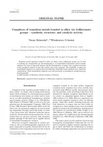
the hypothesis is known as “activation by reduction” (solid tumours create a hypoxic and a more acidic environment than the surrounding tissues, due to a poorer blood supply and higher metabolism).9 Depending on the ligands used in syntheses, the compounds may also act as topoisomerase poisons, or as other enzyme inhibitors.10 The ruthenium central ions may coordinate DNA molecules (like guanine base), increase free radicals concentration and act even as photoactive agents.10–13 A suitable new ligand appeared to be 3-aminoflavone (3-af),14 as it has the possibility to chelate via N and O to transition metal ions. The crystal structures of free 3-aminoflavone (3-af) and its coordination compound of formula [Cu(3-af)2 (NO3 )2 ] were recently determined.15,16 In the latter compound the copper ion is octahedrally surrounded by two amino nitrogen atoms and two carbonyl oxygen atoms. Two chelating 3-af ligands form the short bonds, and two coordinated nitrate ions occupy the axial positions. It appeared interesting to check if this ligand would chelate to ruthenium in a similar way and to see whether the formed compound would display anticancer activity. The present paper deals with the synthesis, properties and biological activity of ruthenium(II) compounds formed from 3aminoflavone (3-af) and Ru(III) ruthenium(III) chloride. Most surprisingly, and depending on the used alcoholic solvent (MeOH, EtOH), the flavone ligand was oxidized, while solvolysing to form a 3-imino-2-alkoxyflavanone as a chelating ligand to ruthenium (see Scheme 1 below); meanwhile the ruthenium had been reduced to divalent ruthenium. The synthesis, spectral properties, 3D structure and antiproliferative (in vitro) activity of the 2 new compounds are described below. Dalton Trans., 2010, 39, 9711–9718 | 9711
View Article Online
Spectral analysis NMR (1 H NMR, 2D 1 H COSY) experiments were carried out on a Bruker 300 DPX spectrometer at Leiden Institute of Chemistry, Leiden University, and Avance DRX500 spectrometer in the Center of Molecular and Macromolecular Research, Polish Academy of Science. IR spectra were carried out on a Spectrometer ATI Mattson Infinity Series FTIRTM . Scheme 1 Schematic representation of the dehydrogenative alcoholysis observed for 3-af in the presence of Ru(III).
Experimental section
Published on 07 September 2010. Downloaded on 18/11/2013 17:21:56.
Chemicals 3-Hydroxyiminoflavanone, 3-aminoflavone, and the two Ru complexes were synthesized as described below. Other reagents were purchased from Sigma-Aldrich, Alfa Aesar and POCh (Poland). Synthesis of 3-aminoflavone (3-af) The synthesis followed the procedure as has been described elsewhere 14 Synthesis of cis-dichloridobis(3-imino-2-methoxyflavanone)ruthenium(II)·3H2 O (1) 118 mg (0.05 mmol) of 3-aminoflavone and 61 mg (0.025 mmol) of dihydrated ruthenium(III) chloride (Aldrich) were dissolved in 25 ml of methanol and left stirring for 24 h at room temperature, protected from light. Then the mixture was filtered and the solution was left to evaporate slowly to let the complex crystallize. The dark crystalline precipitate was then filtered off and dried. Yield (110 mg, 60%); mp >300 ◦ C, decomp. Elemental analysis: found (calculated for C32 H32 Cl2 N2 O9 Ru) C 50.31% (50.55%), H 4.10% (4.21%), N 3.60% (3.68%), Cl 9.22% (9.32%). The amount of Cl was determined by means of mercurometric titration. The analysis indicates a purity of 95%. 1 H-NMR, CD2 Cl2 (d, ppm): 3.35 (OCH3 ), 7.15 (H8 ), 7.24 (H6 ), 7.4–7.7 (H32 –H36 ), 7.25 (H7 ), 8.45 (H5 ), 14.25 (H1 ); IR (KBr pellet, cm-1 ): 1646, 1607, 1580, 1554, 1372, 976, 523. Every new synthesized sample of the compound was identified on the basis of the fingerprint region of IR spectrum. Synthesis of cis-dichloridobis(3-imino-2-ethoxyflavanone)ruthenium(II)·2H2 O (2) 118 mg (0.05 mmol) of 3-aminoflavone and 61 mg (0.025 mmol) of dihydrated ruthenium(III) chloride (Aldrich) were dissolved in 25 ml of anhydrous ethanol and left stirring for 24 h at room temperature, protected from light. Then the mixture was filtered and the solution was left to evaporate slowly for the complex to crystallize. The dark crystalline precipitate was then filtered off, washed with cold water, and dried over anhydrous calcium chloride. Yield (80 mg, 52%); mp. >300 ◦ C, decomp. Elemental analysis: found (calculated C34 H34 Cl2 N2 O8 Ru) C 52.12% (52.99%), H 4.32% (4.45%), N 3.75% (3.64%), Cl 9.10% (9.20%). The amount of Cl was determined by means of mercurometric titration. The analysis confirms the purity to be at least 95% 1 H-NMR, CDCl3 (d, ppm): 1.12 (CH3 ), 3.64 (CH2 ), 7.15 (H8 and H6 ), 7.43 (H32 –H36 ), 7.69 (H7 ), 8.49 (H5 ), 14.55 (H1 ) ; IR (KBr pellet, cm-1 ): 1606, 1575, 1549, 1372, 946, 502. 9712 | Dalton Trans., 2010, 39, 9711–9718
X-ray crystallographic analysis The black single crystal of 1 was used for measurements on an AFC5S Rigaku diffractometer.17 X-ray intensities were collected using graphite-monochromated Mo-Ka radiation and the w scan technique. After each group of 150 reflections the three standard reflections were monitored and insignificant intensity fluctuation was observed. All data were corrected for Lorentz and polarization effects.18 The analytical absorption correction 19 was also used, T min = 0.750, T max = 0.842. The structure of 1 was solved by direct methods (SHELXS97 20 ) and refined on F 2 by the full-matrix leastsquares technique (SHELXL97 21 ). All non-hydrogen atoms were refined anisotropically. The nitrogen and carbon-bonded H-atoms ˚ C–H = were included in the calculated positions (N–H = 0.86 A; ˚ 0.93–0.96 A) and constrained to ride on their parent atoms with isotropic displacement parameters equal to 1.2 U eq (N,C) (or 1.5 U eq for the methyl C atoms). The oxygen-bonded H-atom (H1 W in the water molecule) was located from the difference map and refined isotropically (data not shown). Crystal data for 2 were collected at room temperature on an Enraf-Nonius-KappaCCD diffractometer 22 using graphite˚ For data monochromated Mo-Ka radiation (l = 0.71069 A). reduction and cell refinement the programs DENZO and SCALEPACK were applied.23 The structure was solved by direct methods and subsequent Fourier syntheses and refined by fullmatrix least squares on F 2 using the SHELXS-97 and SHELXL97 programs.20,21 The scattering factors were those given in the SHELXL program. The methyl group of the ligand was found to be disordered over two positions with site occupancy factors of 0.7 and 0.3. Non-hydrogen atoms except for the disordered methyl carbons were refined with anisotropic displacement factors. Hydrogen atoms except those for the water molecules of crystallization were generated geometrically and refined as riding atoms with isotropic displacement factors equivalent to 1.2 times those of the atom to which they were attached (1.5 for methyl groups). Selected crystal data and details of the structure refinements for 1 and 2 are collected in Table 1. Geometrical calculations and drawings were performed with PLATON 24 and SHELXTL PLUS.25 Cell cultures Human bladder carcinoma cells, EJ, were a generous gift from the Institute of Oncology in Gliwice, Poland.26 An EJ-derived sub-line EJcisR or EJ-CPR (cisplatin resistant) was obtained in our laboratory by culturing the parental line with cisplatin (0.5–6 mM) during ten months. Mouse leukemia cells L1210 and L1210R (sub-line resistant to cisplatin) cells were provided by the Department of Molecular Pharmacology. All cell lines were cultured in RPMI medium (Sigma) supplemented with 10% FBS (Gibco) and 50 mg ml-1 gentamycin, in 5% CO2 atmosphere at 37 ◦ C. This journal is © The Royal Society of Chemistry 2010
View Article Online
Published on 07 September 2010. Downloaded on 18/11/2013 17:21:56.
Table 1 Crystallographic data and experimental details of the X-ray studies of 1 and 2
Empirical Formula Formula weight Crystal colour and habit Crystal size/mm Crystal system Space group a/A˚ b/A˚ c/A˚ b (◦ ) V /A˚ 3 Z Dc /g cm-3 m(Mo-Ka)/mm-1 F(000) q range/◦ No. of measured reflections No. of independent reflections (Rint ) No. of observed reflections No. of parameters Final R1 , wR2 (I > 2s(I))a a
1
2
C32 H26 Cl2 N2 O6 Ru·3H2 O 760.57 Black prism 0.50 ¥ 0.40 ¥ 0.30 Monoclinic C 2/c (15) 22.434(4) 8.694(8) 17.630(10) 93.56(3) 3432(4) 4 1.472 0.666 1552 2.74–27.50 7803 3938 (7.4%) 2828 (I > 2s(I)) 213 R1 = 4.38%, wR2 = 11.14%
C34 H30 Cl2 N2 O6 Ru·2H2 O 770.31 Dark red block 0.30 ¥ 0.20 ¥ 0.20 Monoclinic C2/c (15) 22.482(4) 9.126(2) 18.052(4) 93.55(3) 3696.6(3) 4 1.385 0.618 1576 4.6–25.0 47659 3123 (9.6%) 1357 (I > 2s(I)) 217 R1 = 5.3%, wR2 = 10.9%
R1 = R �F o |-|F c �/R |F o |; wR2 = [R w(F o 2 -F c 2 )2 /R w(F o 2 )2 ]1/2 ; w-1 = s2 (F o 2 ) + (aP)2 ; P = (F o 2 + 2F c 2 )/3.
Human lymphocytes were isolated from peripheral blood of healthy donors. The blood was purchased from Regional Blood Bank of Lodz, Poland, with permission of the local ethical committee. Blood was collected in MonovetteTM tubes with sodium citrate and processed within 3 h. Mononuclear cells fraction R 1077(Sigma), using standard was isolated with Histopaque� procedures and then cultured in RPMI medium supplemented with 10% foetal bovine serum, phytohemagluttinin-M (Biological Industries) and gentamycin, in a humidified atmosphere with 5% CO2 . 0.3 ml of full blood was used for 1 ml of lymphocyte culture. MTT [3-(4,5-dimethylthiazol-2-yl)-2,5-diphenyltetrazolium bromide] assay In 24-well plates the EJ cells were inoculated in the number of 2 ¥ 104 per 1 ml of medium (5 ¥ 104 for EJ-CPR line), L1210 and L1210R cells were inoculated in the number of 2 ¥ 103 per ml of medium. After 24 h the tested compounds were added and the plates were incubated for another 24 and 72 h. The tested compounds were dissolved in N¢,N¢-dimethylformamide (DMF). Concentration of DMF in cell cultures was 0.2%. Then 0.05 ml (for EJ cells) or 0.1 ml (for EJ-CPR, and L1210/L1210R cells) of MTT solution in PBS (5 mg ml-1 ) was added and incubated until the cells were apparently coloured. After removing medium, the coloured cells were dissolved in DMSO and the absorbance was measured at l = 540 nm, on an Ultrospec III UV/VIS spectrophotometer. The results were transformed into relative percentage values, assuming the control (with DMF) result as 100%, and then compared. Every experiment was repeated at least three times. The lymphocytes were cultured in 24 well plates, as mentioned above. After 24 h from inoculation the tested compounds were added and the plates were incubated for another 72 h. Concentration of DMF in cell cultures was 0.2%. Then 0.1 ml of MTT solution in PBS (5 mg ml-1 ) was added and incubated until the cells were apparently coloured (approx. 4 h). After removing This journal is © The Royal Society of Chemistry 2010
medium, the coloured cells were dissolved in DMSO and the absorbance was measured at l = 540 nm, on an Ultrospec III UV/VIS spectrophotometer. The results were transformed into relative percentage values, assuming the control (with DMF) result as 100%, and then compared. The experiment was repeated three times.
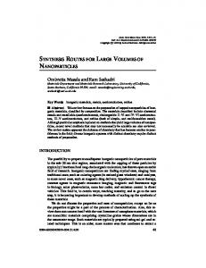
Results and discussion General observations The two newly formed compounds have been isolated and subsequently characterized by elemental analysis and routine IR, which showed a clear ligand transformation from the original 3-af, to the new derived ligand. The newly formed ligands do not exist as such and were not isolated, but were studied only as a part of the complexes. To understand the changed ligands, both single crystal X-ray diffraction, and advanced 2D proton NMR analyses in solution were performed. Finally the anticancer activity has been studied using different cell lines. X-ray analysis Crystallographic data and details of refinement for both compounds are reported in Table 1 and selected bond lengths and angles are reported in Table 2. Fig. 1 and 2 describe the projection of each structure; a full discussion is given only for compound 2. Description of the crystal structures The X-ray analysis of 2 clearly proves the conversion of the starting ligand 3-af into the iminoethoxy derivative, 3-ief. Fig. 1 presents a view of the molecular structure of 2, while selected bond lengths and angles are listed in Table 2. The structure is composed of one Ru atom, two 3-ief ligands in their imino-oxo form, two chloride ligands and two water molecules (one of which is disordered over Dalton Trans., 2010, 39, 9711–9718 | 9713
View Article Online ˚ bond angles [◦ ] for 2 and 1 Table 2 Relevant bond lengths [A],
Ru(1)–O(1) Ru(1)–Cl(1) C(2)–N(1) Cl(1)–Ru(1)–O(1) Cl(1)–Ru(1)–Cl(1a)a O(1)–Ru(1)–N(1a)a
Published on 07 September 2010. Downloaded on 18/11/2013 17:21:56.
a
1
2
2.175(2) 2.3351(12) 1.292(4) 85.81(7) 169.44(6) 175.00(9)
2.171(4) 2.326(2) 1.302(6) 86.3(1) 168.98(9) 175.1(2)
1
2
Ru(1)–N(1)
1.914(3)
1.910(4)
C(1)–O(1) Cl(1)–Ru(1)–N(1) O(1)–Ru(1)–N(1) Cl(1)–Ru(1)–O(1a)a
1.261(4) 92.82(8) 77.37(11) 87.96(7)
1.250(6) 93.4(1) 77.4(2) 87.2(1)
symmetry operation -x, y, 1/2 -z
Fig. 1 View of the molecular structure of the coordination entity in compound 2 with the atom numbering scheme. Thermal ellipsoids are drawn at the 50% level. For clarity, only one position of the disordered methyl group is shown. Lattice water is not shown.
2 positions) of lattice crystallization. The two 3-ief ligands chelate the Ru atom through their the imine nitrogen and carbonyl oxygen with the imino groups (carbonyl groups) being mutually cis to each other. The four ligand donor atoms are co-planar, the octahedral coordination geometry is completed by two chloride ions trans to each other. The Ru–N, Ru–O and Ru–Cl bond lengths are in the normal range. C–O and C–N bond distances of 1.250(6) A˚ and 1.302(6) A˚ are in line with C O and C N double bonds. The five atoms forming the chelate ring (O1/C1/C2/N1/Ru1) do not deviate significantly from planarity (maximal deviation ˚ The Ru coordination moves from the mean plane, 0.017 A). the exocyclic nitrogen and oxygen slightly out of the ring plane (distance to the best-weighted plane 0.256(8) A˚ for oxygen and 0.153(8) A˚ for nitrogen). With the twist angle between the phenyl group and the heterocycle being 86.2(2)◦ , the phenyl ring is almost
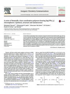
Fig. 2 The molecular structure of the coordination entity 1 with the atom-numbering scheme. Displacement ellipsoids are drawn at the 50% probability level and H atoms are shown as small spheres of arbitrary radii. Lattice water is not shown.
perpendicular to the heterocyclic ring plane. In the crystal packing the molecules are arranged in stacks along the y-axis. Hydrogen bonds are formed between the imine nitrogens and water molecules of crystallization and typical parameters have been included in Table 3, together with those of the other compound 1 (Fig. 3). ˚ and the angle (N(1)– The values for (N(1) ◊ ◊ ◊ O(1w) = 2.936(7) A, H ◊ ◊ ◊ O(1w) = 153◦ are indicative for medium-strong hydrogen bonds. The O(1 W)–O(2 W) distance (2 positions) is considered as a rather strong hydrogen bond (see Table 3). Given the solvolysis of the ligand by ethanol, a similar reaction was performed in MeOH, and indeed, a closely related product, 1 was found, with the EtO group replaced by the MeO group, and its structure is described below. Fig. 2 shows a displacement ellipsoid plot of compound 1 with the atom numbering scheme. As in the structure of 2, the central ruthenium atom is located on a twofold rotation axis and adopts
Table 3 Hydrogen-bonding geometry details for 1 and 2 1
D–H/A˚
H ◊ ◊ ◊ A/A˚
D ◊ ◊ ◊ A/A˚
D–H ◊ ◊ ◊ A [◦ ]
N(1)–H(1A) ◊ ◊ ◊ O(1 W) O(1 W)–H(1 W) ◊ ◊ ◊ O(2 W)b O(2 W) . . . Cl(1)c
0.86 0.98(5) A
2.08 1.71(5) A
2.876(6) 2.644(9) 3.212 (8)
153 158(4) A
2
˚ D–H [A]
˚ H ◊ ◊ ◊ A [A]
˚ D ◊ ◊ ◊ A [A]
D–H ◊ ◊ ◊ A [◦ ]
N(1)–H ◊ ◊ ◊ O(1w) O(1 W) ◊ ◊ ◊ O(2 W)
0.900 A
2.102 A
2.933(7) 2.528(8)
153.2 A
a
H atoms not located; symmetry operations: b x, y - 1, z; c - x, y, 1/2 -z.
9714 | Dalton Trans., 2010, 39, 9711–9718
This journal is © The Royal Society of Chemistry 2010
Published on 07 September 2010. Downloaded on 18/11/2013 17:21:56.
View Article Online
a distorted octahedral environment (see Table 2 for geometric information). The equatorial plane, defined by O1, C1, C2, N1, Ru1 atoms determining a 5-membered chelate ring, is essentially planar, however atoms deviate more (maximum deviation is 0.033 ˚ from the best plane which has been observed in structure 2. A) Since the bond lengths and angles for 1 agree well with reported values, for 2 no detailed discussion is needed. It is worth mentioning that in 1, the conformation of a 6membered heterocyclic ring formed by O2, C3, C2, C1, C9, C4 atoms, is between a half-chair and a half-boat. Asymmetry parameters indicate that the twofold axis (passing through the O2–C3 bond) exists as well as the mirror plane (passing through the C3 atom). Both pseudo-symmetry parameters C 2 and C s are ˚ q= equal to 10.5(3)◦ . The puckering parameters27 Q = 0.342(3) A, ◦ ◦ 116.5(5) and f = 225.7(6) confirm the above mentioned results. The crystal-packing arrangement in 1 is influenced by solvent molecules, each molecule of ruthenium(II) complex is surrounded by three water molecules. The O(1 W) atom is an bifurcated acceptor of two hydrogen atoms from the NH groups, and simultaneously it donates two hydrogen atoms from another two water O(2 W) molecules. Moreover, each of the two O(2 W) molecules acts as a donor to a chloride ligand; the hydrogen atoms of O(2 W) were, however, not resolved. Finally, the molecules of complex 1 and water are linked into infinite chains running along twofold rotation axes. NMR analysis To provide additional evidence for the chemical structure of new Ru(II) compounds in solution, proton NMR analyses were performed. Fig. 4 presents the 1 H NMR spectra of 1 in deuterated
Fig. 3 Part of crystal structure of (1), showing a chain of molecules linked by hydrogen bonds (dashed lines).
This journal is © The Royal Society of Chemistry 2010
Fig. 4 1 H NMR spectra of 1 recorded in deuterated CH2 Cl2 at 294 K, using solvent residual peaks as a reference. After addition of D2 O, the spectrum was recorded again (upper spectrum).
CH2 Cl2 , where the presence of the fast exchangeable imino moiety (H1 ) was confirmed by addition of some drops of deuterated water (Fig. 4, upper spectrum). This imino resonance peak is localized downfield (14.25 ppm) as expected. The splitting pattern in 1 can be clearly identified and 2D 1 H COSY spectrum shows the correlations between the resonance peaks. In the COSY spectrum (Fig. 5) an intense cross peak from H5 resonance (doublet, assigned on accounts fir the 3 J coupling and also based on the splitting pattern) with the signal at 7.24 ppm can be assigned to H6 resonance while the less intense cross peak corresponds to H7 (7.75 ppm). Finally the cross peak from H7 could be undoubtedly assigned to H8 resonance peak (7.15 ppm). This connectivity and splitting pattern matches accurately with the flavone aromatic ring structure. The set of peaks with less definition and increased broadness were assigned to the protons H32 –H36 in the phenyl moiety and the strong coupling pattern could be observed by the cross peaks between them. These results are in agreement with the theoretical information and structural analysis.
Fig. 5 2D 1 H COSY spectrum of 1 recorded in CD2 Cl2 at 294 K, using TMS as internal standard. Aromatic region only.
Dalton Trans., 2010, 39, 9711–9718 | 9715
Published on 07 September 2010. Downloaded on 18/11/2013 17:21:56.
View Article Online
The broadness observed in some 1 H NMR resonance peaks is presumably the result of fluxional behaviour or conformational fluctuations by the phenyl and methoxyl rotation. To confirm this possibility, the influence of temperature was studied and the changes registered by 1 H NMR (ESI†). When the temperature was lowered (293 K to 233 K) some signals became increasingly sharper (see Fig. S1†). Stability studies confirmed the presence of the original compound even after five days in dichloromethane, chloroform and DMF. In DMSO, solvolysis was completed after 48 h (data not shown). Comparative results were obtained in 1 H-NMR spectra of 2 which were carried out in deuterated CDCl3 (Fig. 6). Even though in this solvent overlapping of aromatic signals is observed, the aliphatic proton signals are clearly indicating the presence of the ethyl structure, which can be assigned to the ethoxyl moiety of the ligand (Fig. 6 and structural scheme inserted). The resonance peak at 3.64 ppm corresponds to the methylene moiety, while the resonance peak at 1.12 ppm corresponds to the methyl moiety. The integration values are in agreement with the structure proposed. Further assignment of the resonance peaks was supported by the COSY studies and by comparison with results obtained from 1. Spectra recorded in DMF and DMSO showed important solvolytic substitution for conclusive assignment and correct description of the chemical structure (spectra not shown).
Fig. 6 1 H NMR spectra of 2 recorded in deuterated CHCl3 at 294 K, using TMS as a reference.
As for 1, the 1 H NMR spectrum of 2 in deuterated CHCl3 shows the presence of a fast exchangeable imino moiety (H1 ), localized downfield (14.55 ppm) as expected and further confirmed by addition of D2 O (Fig. 7, upper spectra) and bidimensional studies (COSY studies, see Fig. S2†). The small high-field shift of this imino hydrogen, when comparing 2 (14.55 ppm) with 1 (14.73 ppm in CDCl3 ) could be explained as generated by the bigger electron-donor effect of the ethoxyl moiety. The immediate exchange of deuterium by this imino moiety in 1 and the less rapid exchange for 2 confirms the last observation.
Fig. 7 1 H NMR spectra of 2 recorded in deuterated CHCl3 at 294 K, using TMS as reference. After addition of D2 O, the spectrum was recorded again (upper spectra, after 30 and 120 min).
chemotherapeutic agent—used here as a reference compound.29 For comparison, cisplatin resistant sub-lines were tested derived from EJ and L1210, respectively: EJcisR and L1210R. Human lymphocytes are used as a model of normal cells to assess toxicity of tested compounds towards healthy tissues. It is worth noticing that both 1 and 2 have the ability to overcome resistance to cisplatin in the resistant cell lines which suggests alternative mechanisms of activity than the ones described for cisplatin. IC50 values and resistance factors for the cell lines (incubation time 72 h) are shown in Table 4. The ruthenium compounds can also act in a shorter time than cisplatin, as their cytotoxicity after 24 h is almost the same as after 72 h of incubation. On the contrary, cisplatin treatment requires 72 h to reach maximum cisplatin toxicity (see Fig. S3 in ESI†). The ruthenium compounds are less toxic for normal lymphocytes than cisplatin. For both 1 and 2 IC50 values for lymphocytes are above their IC50 values for cancer cells. Cisplatin is equally or even more toxic for lymphocytes than for cancer cells (see Table 4 and Fig. S4 in ESI†). The result is very promising, as potential anticancer agents should be safe for healthy cells and toxic towards cancer cells. Even though both Ru compounds develop an important cytotoxic activity, 2 is the most promising antitumour active compound, in particular due to the significant activity against EJcisR and L1210R cell lines. Table 4 IC50 values (in mM) of the tested compounds and cisplatin in four cell lines. The resistance factors are placed in parentheses (RF). Incubation time: 72 h
MTT antitumor assay The cell lines used for the cytotoxic experiments were human bladder carcinoma EJ cells (also known as MGH-U1 or T24)28 and mouse leukemia cells L1210. Bladder carcinomas are often treated with cisplatin—the earliest and best known metal based 9716 | Dalton Trans., 2010, 39, 9711–9718
Cisplatin 1 2
Lymphocytes
EJ
EJcisR
L1210
0.8 ± 0.2 4.7 ± 1.1 4.8 ± 1.1
1.6 ± 0.5 2.7 ± 0.6 2.2 ± 1.2
11.4 ± 4 (7.1) 0.5 3.5 ± 0.5 (1.3) 0.7 2.3 ± 0.8 (1) 0.3
L1210R 2.3(4.6) 1.7(2.4) 0.5(1.7)
This journal is © The Royal Society of Chemistry 2010
Published on 07 September 2010. Downloaded on 18/11/2013 17:21:56.
View Article Online
Concluding remarks
Acknowledgements
Ruthenium(III) complexes often have good anticancer properties in vitro and in vivo. Many of them are cytotoxic in vitro, but their cytotoxicity does not necessarily correlate with their potential therapeutic effectiveness in vivo.30 Some ruthenium complexes despite their low toxicity in vitro have good pro-apoptotic or antimetastatic properties in animal models with comparatively low general toxicity for the organism. Nevertheless, ruthenium(II) complexes are often more cytotoxic than those of ruthenium(III). They are considered as the actual cytostatic agents per se or after “activation by reduction” of Ru(III) compounds.9 This evidence suggests that the mechanism of biological activity for ruthenium compounds is quite different from the cisplatin mechanism and requires further investigation. Our results are also conformable with findings of other authors, that many ruthenium(II) compounds are cytotoxic in vitro and may overcome drug resistance. They are usually also less toxic for normal cells and tissues than platinum compounds. It is a very important property, as drug resistance of cancer cells and high general toxicity of anticancer drugs are the major problems in chemotherapy. This is a very important property, as drug resistance of cancer cells is one of the major problems in chemotherapy. To the best of our knowledge, only one study is known, describing the flavonoid complex of ruthenium(II).31 3Hydroxyflavonoid complexes of ruthenium(II) displayed potent cytotoxicity in vitro and good physiological acceptability for mice. To date, we have been unable to find any other information about ruthenium compounds with flavonoid ligands. Although the area of flavonoid-ruthenium compounds is not extensively explored, we decided to broaden it with a new approach. Bearing in mind that many flavonoids, including methylated ones, have chemopreventive and antiproliferative properties,32 we synthesized and tested two novel ruthenium compounds with flavanone-based ligands, and obtained very promising results. For comparison, the most popular class of currently investigated ruthenium(II) complexes is the class of organometallic arene complexes.2 Cytotoxicity of several ruthenium(II) arene compounds, measured by means of IC50 value in vitro, differs from below 1 mM to over 100 mM. Many tested compounds display their IC50 towards different cell lines in the range of 20–80 mM,33 and towards ovarian cancer cells A2780 in the range of 0.5–50 mM.4 Compounds 1 and 2 seem therefore to have really high antiproliferative activity towards human bladder carcinoma and murine leukemia cells. That fact shows that ruthenium flavonoid complexes may represent an interesting group of compounds for potential anticancer application.
This work was supported partly by Polish Ministry of Science and Higher Education grant No. 1823/B/P01/2008/35, the Medical University of Lodz grant statute 503-3016-2 and UE European Social Funds grant No. 505-07-050/WFARM/RNSD/09. This work was supported in part (AGO, JR)) by the Council for the Chemical Sciences of the Netherlands Organization for Scientific Research (CW–NWO) and in part by CONACYT (National Council of Science and Technology) as a doctoral fellowship to AGO. JO acknowledges gratefully a fellowship from the Deutcher Akademischer Austauschdienst. We thank A. W. M. Lefeber for assistance with the NMR techniques, Jos van Brussel, John A. P. P. van Dijk and Jopie A. Erkelens-Duijndam for technical assistance with syntheses and analyses.
List of abbreviations: 3-af 3-imf 3-ief CDDP MTT 1 2
3-aminoflavone 3-imino-2-methoxyflavanone 3-imino-2-ethoxyflavanone cis-diamminedichloridoplatinum(II), or cisplatin 3-(4,5-dimethylthiazol-2-yl)-2,5-diphenyl-2Htetrazolium bromide cis-dichloridobis(3-imino-2methoxyflavanone)ruthenium(II)·3H2 O cis-dichloridobis(3-imino-2ethoxyflavanone)ruthenium(II)·2H2 O
This journal is © The Royal Society of Chemistry 2010
Notes and references 1 S. Kapitza, M. A. Jakupec, M. Uhl, B. K. Keppler and B. Marian, Cancer Lett., 2005, 226, 115–121. 2 I. Kostova, Curr. Med. Chem., 2006, 13, 1085–1107. 3 A. H. Velders, H. Kooijman, A. L. Spek, J. G. Haasnoot, D. de Vos and J. Reedijk, Inorg. Chem., 2000, 39, 2966–2973. 4 R. E. Aird, J. Cummings, A. A. Ritchie, M. Muir, R. E. Morris, H. Chen, P. J. Sadler and D. I. Jodrell, Br. J. Cancer, 2002, 86, 1652–1657. 5 C. G. Hartinger, S. Zorbas-Seifried, M. A. Jakupec, B. Kynast, H. Zorbas and B. K. Keppler, J. Inorg. Biochem., 2006, 100, 891–904. 6 J. M. Rademaker-Lakhai, D. van den Bongard, D. Pluim, J. H. Beijnen and J. H. M. Schellens, Clin. Cancer Res., 2004, 10, 3717–3727. 7 G. Sava, S. Zorzet, C. Turrin, F. Vita, M. Soranzo, G. Zabucchi, M. Cocchietto, A. Bergamo, S. DiGiovine, G. Pezzoni, L. Sartor and S. Garbisa, Clin. Cancer Res., 2003, 9, 1898–1905. 8 A. Bergamo, L. Messori, F. Piccioli, M. Cocchietto and G. Sava, Invest. New Drugs, 2003, 21, 401–411. 9 R. E. Morris, R. E. Aird, P. D. Murdoch, H. M. Chen, J. Cummings, N. D. Hughes, S. Parsons, A. Parkin, G. Boyd, D. I. Jodrell and P. J. Sadler, J. Med. Chem., 2001, 44, 3616–3621. 10 F. Gao, H. Chao, F. Zhou, X. Chen, Y. F. Wei and L. N. Ji, J. Inorg. Biochem., 2008, 102, 1050–1059. 11 B. Elias and A. Kirsch-De Mesmaeker, Coord. Chem. Rev., 2006, 250, 1627–1641. 12 R. Hage, J. G. Haasnoot, J. Reedijk, R. Y. Wang and J. G. Vos, Inorg. Chem., 1991, 30, 3263–3269. 13 J. H. van Diemen, R. Hage, J. G. Haasnoot, H. E. B. Lempers, J. Reedijk, J. G. Vos, L. De Cola, F. Barigelletti and V. Balzani, Inorg. Chem., 1992, 31, 3518–3522. 14 C. O’Brien, T. S. Wheeler, E. M. Philbin and S. Suhioda, Tetrahedron, 1963, 19, 373–377. 15 A. J. Rybarczyk-Pirek, M. Małecka, L. Łukasz Glinka and J. Ochocki, Acta Crystallogr., Sect. C: Cryst. Struct. Commun., 2007, C63, m410. ˙ 16 B. Zurowska, A. Erxleben, L. Glinka, M. Ł˛eczycka, E. Zyner and J. Ochocki, Inorg. Chim. Acta, 2009, 362, 739–744. 17 M. S. Corporation, in MSC/AFC Diffractometer Control Software. MSC, 3200 Research Forest Drive, The Woodlands, TX 77381, USA MSC/AFC Diffractometer Control Software. MSC, 3, 3200 Research Forest Drive, The Woodlands, TX 77381, USA, 1989. 18 Rigaku/MSC, in Crystal Structure. Version 3.10, ed. C. S. V. 3.10, Rigaku/MSC, 9009 New Trails Drive, The Woodlands, TX 773815209, USA, Rigaku/MSC, 9009 New Trails Drive, The Woodlands, TX 77381-5209, USA, 2002. 19 J. Demeulenaer and H. Tompa, Acta Crystallogr., 1965, 19, 1014–1018. 20 G. M. Sheldrick , SHELXS-97, Program for Crystal Structure Solution, ¨ University of Gottingen, Germany, 1997. 21 G. M. Sheldrick, SHELXL-97. Program for Crystal Structure Refine¨ ment, University of Gottingen, Germany, 1997. 22 Nonius, in COLLECT. Nonius BV, Delft, The Netherlands, 2002. 23 Z. Otwinowski, and W. Minor, in Macromolecular Crystallography, Part A,1997, vol. 276, pp. 307–326. 24 A. L. Spek, J. Appl. Crystallogr., 2003, 36, 7–13. 25 G. M. Sheldrick, SHELXTL, Bruker AXS. Inc, Madison, Wisconsin, 1997.
Dalton Trans., 2010, 39, 9711–9718 | 9717
View Article Online 30 M. Cocchietto, S. Zorzet, A. Sorc and G. Sava, Invest. New Drugs, 2003, 21, 55–62. 31 L. Mishra, A. K. Singh, S. K. Trigun, S. K. Singh and S. M. Pandey, Indian J. Exp. Biol., 2004, 42, 660–666. 32 T. Walle, Semin. Cancer Biol., 2007, 17, 354–362. 33 C. A. Vock, W. H. Ang, C. Scolaro, A. D. Phillips, L. Lagopoulos, L. Juillerat-Jeanneret, G. Sava, R. Scopelliti and P. J. Dyson, J. Med. Chem., 2007, 50, 2166–2175.
Published on 07 September 2010. Downloaded on 18/11/2013 17:21:56.
26 C. M. O’Toole, S. Povey, P. Hepburn and L. M. Franks, Nature, 1983, 301, 429–430. 27 D. Cremer and J. A. Pople, J. Am. Chem. Soc., 1975, 97, 1354–1358. 28 A. Kovnat, R. N. Buick, B. Choo, E. De Harven, I. Kopelyan, J. M. Trent and I. F. Tannock, Cancer Res., 1988, 48, 4993–5000. 29 C. N. Sternberg, S. M. Donat, J. Bellmunt, R. E. Millikan, W. Stadler, P. De Mulder, A. Sherif, H. von der Maase, T. Tsukamoto and M. S. Soloway, Urology, 2007, 69, 62–79.
9718 | Dalton Trans., 2010, 39, 9711–9718
This journal is © The Royal Society of Chemistry 2010