Synthesis, Structures and Properties of Two Metal-organic Frameworks
Bull. Korean Chem. Soc. 2013, Vol. 34, No. 8 2375 http://dx.doi.org/10.5012/bkcs.2013.34.8.2375
Synthesis, Structures and Properties of Two Metal-organic Frameworks Derived from 3-Nitro-1,2-benzenedicarboxylic Acid Wen-Jia Xu, Ling-Yu Zhang, Jin-Niu Tang, Dai-Yin Wang, Gang-Hong Pan, and Yu Feng* College of Chemistry and Chemical Engineering, Guangxi University for Nationalities, Nanning 530006, P.R. China * E-mail:
[email protected] Received April 15, 2013, Accepted May 16, 2013 Two metal-organic frameworks based on the connectivity co-effect between rigid benzenedicarboxylic acid and bridging ligand have been synthesized [Zn2(3-NO2-bdc)2(4,4'-bpy)2H2O]n (1), [Co(3-NO2-bdc)(4,4'bpy)H2O]n (2) (where 3-NO2-bdcH2 = 3-nitro-1,2-benzenedicarboxylic acid, 4,4'-bpy = 4,4'-bipyridine). The two novel complexes were characterized by IR spectrum, elemental analysis, fluorescent properties, thermogravimetric analysis, single-crystal X-ray diffraction and powder X-ray diffraction (PXRD). X-ray structure analysis reveals that 1 and 2 are two-dimensional (2D) network structures. Complex 1 and complex 2 belong to triclinic crystal with P-1 space group. The luminescence measurements reveal that two complexes exhibit good fluorescent emissions in the solid state at room temperature. Also, thermal decomposition process and powder X-ray diffraction of complexes were investigated. Key Words : X-ray crystal diffraction, 3-Nitro-1,2-benzenedicarboxylic acid, Two-dimensional network polymer
Introduction Metal-organic frameworks (MOFs) are a relatively new class of crystalline coordination polymers, which have potential in a myriad of applications, including gas storage, chemical separation, heterogeneous, catalysis and optical, electronic, magnetic materials.1-15 Studies in this field have been focused on the design and preparation, as well as the structure-property relationships. Although big progress has been achieved,16 it is still a great challenge to predict the exact structures and composition of the assembly products built by coordination bonds and/or hydrogen bonds in crystal engineering. As we know, there are several factors, such as the coordination nature of ligand structure, counterions, and so on, which may be key for the rational design of MOFs. Therefore, systematic research on this topic is still important for understanding the roles of these factors in the formation of metal-organic coordination frameworks. The rigid benzenedicarboxylic acids (3-nitro-1,2-benzenedicarboxylic acid) are good candidates for the construction of novel metal-organic complexes, which show many important advantages than other organic ligands: they have two carboxyl groups that can completely or partially deprotonate, which reduces rich coordination modes; they can act not only as a hydrogen-bond acceptor but also as a hydrogen-bond donor, depending upon the number of deprotonated carboxyl groups; and they may connect metal ions in different directions due to their rigidity and polycarboxylate groups. As an extension of our work, herein, 3-nitro-1,2- benzenedicarboxylic acid (3-NO2-bdcH2) was chosen as the starting material together with divalent metal ions, with the aid of bridging ligand 4,4'-bpy. Utilizing hydrothermal technique,
two new MOFs [Zn2(3-NO2-bdc)2(4,4'-bpy)2H2O]n (1), [Co(3-NO2-bdc)(4,4'-bpy)H2O]n (2) have been obtained. In addition, fluorescent properties, thermal decomposition process and powder X-ray diffraction of compounds 1 and 2 were studied. Experimental All chemicals were commercial materials of analytical grade and used without purification. Elemental analysis for C, H and N was carried out on a Perkin-Elmer 2400 II elemental analyzer. The FT-IR spectrum was obtained on a PE Spectrum One FT-IR Spectrometer Fourier transform infrared spectroscopy in the 4000-400 cm−1 regions, using KBr pellets. Perkin-Elmer Diamond TG/DTA thermal analyzer was used to record simultaneous TG and DTG curves in the static air atmosphere at a heating rate of 10 K min−1 in the temperature range 25-1000 ºC using platinum crucibles. Fluorescence spectra were recorded with F-2500 FL Spectrophotometer analyzer. Powder X-ray diffraction patterns were obtained using a pinhole camera (Anton Paar) operating with a point focused Ni-filtered Cu Kα radiation in the 2θ range from 5° to 50° with a scan rate of 0.08° per second. Synthesis of the Complex [Zn2(3-NO2-bdc)2(4,4'-bpy)2H2O]n (1): The reagents of ZnCl2 (0.136 g, 1.00 mmol), 3NO2-bdcH2 (0.106 g, 0.500 mmol), 4,4'-bpy (0.0781 g, 0.500 mmol) were dissolved in 15 mL mixed solvent of DMF/H2O (volume ratio 1:2) and added eight drops of py then stirred 0.5 h. Then an aqueous solution of sodium hydroxide was added dropwise with stirring to adjust the pH value of the solution being 6. The resulting mixture was sealed in a 30 mL Teon-lined stainless reactor, kept under autogenous pressure at 140 ºC for 72 h, and then slowly
2376
Bull. Korean Chem. Soc. 2013, Vol. 34, No. 8
Table 1. Experimental data for complex 1 and 2 Identification code Empiricalf formula Formula weight Temperature (k) Crystal system Sspace group a (Å) b (Å) c (Å) Volume (Å3) Z Absorption coefficient (mm−1 ) Theta range for data collection Reflections collected Data / restraints / parameters Goodness-of-fit on F2 Final R1 and wR2 [I>2σ(I)] R1 and wR2 indices (all data)
1 C36H24N6O13Zn2 879.35 296(2) Triclinic P-1 11.446(2) 11.794(2) 14.448(4) 1723.2(6) 2 1.473 1.93 to 25.00° 9278 5968 / 28 / 514 1.020 0.0387, 0.0988 0.0439, 0.1038
Wen-Jia Xu et al. Table 2. Selected bond lengths (Å) and angles (°) for 1 and 2
2 C18H13CoN3O7 442.24 296(2) Triclinic P-1 8.638(3) 10.976(2) 11.518(2) 908.3(4) 2 0.993 2.02 to 25.00° 4764 3116 / 6 / 281 1.050 0.0554, 0.1539 0.0691, 0.1731
cooled to room temperature at a rate of 5 ºC per hour. The block crystals suitable for X-ray diffraction were isolated directly (Yield: 65%, based on Zn). Anal. Calcd for C36H24N6O13Zn2 (%): C, 49.17; H, 2.75; N, 9.56; S. Found: C, 49.16; H, 2.78; N, 9.55. IR data (KBr pellets, cm−1): 3271 (vs), 1607 (s), 1555 (s), 1539 (s), 1520 (s), 1460 (m), 1418 (m), 1380 (m), 1343 (m), 1219 (m), 922 (w), 820 (w), 787 (w). Synthesis of the Complex [Co(3-NO2-bdc)(4,4'-bpy)H2O]n (2): The same synthetic procedure as that for 1 was used except that Co(NO3)2·6H2O (0.291g, 1.00 mmol) was replaced by ZnCl2. The block crystals of 2 were obtained in 55% yield based on Co. Anal. Calcd for C18H13CoN3O7 (%): C, 48.89; H, 2.96; N, 9.50. Found: C, 48.88; H, 2.97; N, 9.48. IR data (KBr pellets, cm−1): 3255 (vs), 1605 (s), 1582 (s), 1553 (s), 1524 (s), 1508 (m), 1433 (m), 1380 (m), 1344 (m), 820 (m), 787 (w), 753 (w), 637 (m). Crystal Structure Determination. Suitable single crystal with approximate dimensions were mounted on a glass fiber and used for X-ray diffraction analyses. Data were collected at 293(2) K on a Bruker Apex CCD diffractometer using the ω scan technique with Mo Kα radiation (λ = 0.71069 Å). Absorption corrections were applied using the multi-scan technique.17 The structures were solved by the Direct Method and refined by full-matrix least-square techniques on F2 using SHELXL-97.18 All non-hydrogen atoms were refined anisotropically. The crystal data and structure refinement details for two complexes are shown in Table 1. Selected bond lengths and angles of the complexes are listed in Table 2, and possible hydrogen bond geometries are given in Table 3. Crystallographic data for the structures reported here have been deposited with CCDC (Deposition No. CCDC-928612 (1), No. CCDC-928613 (2)). These data can be obtained free of charge via http://www.ccdc.cam.ac.uk/conts/retrieving.html
Complex 1 1.946(2) Zn(1)-O(2) 2.015(3) Zn(1)-N(4) 2.011(2) Zn(2)-O(4) 2.096(3) Zn(2)-O(7) 2.170(3) O(11)-Zn(1)-N(3) 122.1(2) O(11)-Zn(1)-N(4) 101.5(2) N(3)-Zn(1)-N(4) 89.2(2) O(8)-Zn(2)-N(1) 116.2(2) O(8)-Zn(2)-O(7) 142.1(2) N(1)-Zn(2)-O(7) 87.8(2) O(4)-Zn(2)-N(6) 86.3(2) O(7)-Zn(2)-N(6) 99.9(2) Complex 2 Co(1)-O(4) 2.047(3) Co(1)-O(5) Co(1)-N(1) 2.128(4) Co(1)-N(2) Co(1)-O(7)#1 2.136(3) Co(1)-O(6)#1 O(6)-Co(1)#1 2.229(3) O(7)-Co(1)#1 O(4)-Co(1)-O(5) 92.9(2) O(4)-Co(1)-N(1) O(5)-Co(1)-N(1) 94.3(2) O(4)-Co(1)-N(2) O(5)-Co(1)-N(2) 170.4(2) N(1)-Co(1)-N(2) O(4)-Co(1)-O(7)#1 107.4(2) O(5)-Co(1)-O(7)#1 N(1)-Co(1)-O(7)#1 156.8(2) N(2)-Co(1)-O(7)#1 O(4)-Co(1)-O(6)#1 167.0(2) O(5)-Co(1)-O(6)#1 N(1)-Co(1)-O(6)#1 97.1(2) N(2)-Co(1)-O(6)#1 O(7)#1-Co(1)-O(6)#1 59.7(2) Zn(1)-O(11) Zn(1)-N(3) Zn(2)-O(8) Zn(2)-N(1) Zn(2)-N(6) O(11)-Zn(1)-O(2) O(2)-Zn(1)-N(3) O(2)-Zn(1)-N(4) O(8)-Zn(2)-O(4) O(4)-Zn(2)-N(1) O(4)-Zn(2)-O(7) O(8)-Zn(2)-N(6) N(1)-Zn(2)-N(6)
1.990(3) 2.071(3) 2.039(2) 2.103(3) 125.8(2) 101.3(2) 110.6(2) 101.7(2) 89.7(2) 90.8(2) 84.9(2) 169.1(2)
2.094(3) 2.126(4) 2.229(3) 2.136(3) 95.7(2) 87.0(2) 95.0(2) 86.7(2) 84.4(2) 87.8(2) 90.2(2)
Symmetry transformations used to generate equivalent atoms: for Complex 2: #1 −x+1, −y+2, −z
or from CCDC, 12 Union Road, Cambridge CB2 1EZ, UK, E-mail:
[email protected]. Results and Discussion Description of the Structure. The X-ray structure analysis of crystalline 1 reveals that it crystallizes in the triclinic P-1 space group. As shown in Figure 1, Zn1(II) is coordinated to two carboxylate oxygen atoms from two equivalent 3-NO2-bdc2− and two different 4,4'-bpy nitrogen atoms. Furthermore, the distances of Zn1-O1 and Zn1-O10 are 2.759(3) and 2.743(3) Å, respectively, suggesting a nonnegligible interaction with the uncoordinated carboxylate oxygen atom, which can be described as a semi-chelating coordination mode.19 As depicted in Figure 2, Zn1 adopts a tetrahedral geometry and Zn2 adopts a distorted trigonal bipyramidal geometry. The angle of O8-Zn2-N6 is 86.26(10)o. Considering bond angles, the equatorial positions are occupied by the O4, O8 and N1 atoms and the axial positions are occupied by the O7 and N6 atoms because the angle of O7Zn2-N6 is 169.08(11)o. The most fascinating feature of crystalline 1 is the presence of the Zn1 ion possesses a fourcoordinated sphere but the Zn2 possesses a five-coordinated sphere. Difference is the Zn2 is coordinated by a more coordinated water molecule (O7). The average Zn-O and
Synthesis, Structures and Properties of Two Metal-organic Frameworks
Bull. Korean Chem. Soc. 2013, Vol. 34, No. 8
2377
Table 3. Hydrogen-bond geometry (Å) and angles (°) for complexes D-H···A Complex 1 O7-H7A··· O3#1 O7-H7B··· O9 Complex 2 O5-H5A···O6 #1 O5-H5B···O1
D−H
H···A
D···A
D−H···A
0.85 0.85
1.93 1.94
2.676(4) 2.604(4)
144.9 133.07
0.85 0.85
1.86 1.78
2.712(7) 2.573(7)
171.31 153.78
Symmetry code: for Complex 1: #1 −x, −y+1, −z+1; for Complex 2: #1 −x+1, −y+2, −z.
Figure 1. The coordination environment of Zn(II) ion of the complex 1. All the hydrogen atoms are omitted for clarity. Symmetry codes: N1A = 3−x, 2−y, 2−z; N3A = −x, 1−y, 1−z; N4A = x, y−1, z.
Figure 3. The 2D layered network of complex 1. Unnecessary atoms are omitted for clarity.
Figure 2. The coordination polyhedron of the Zn(II) ion of complex 1. Unnecessary atoms are omitted for clarity.
Figure 4. A perspective view of H-bonding for complex 1. Unnecessary atoms are omitted for clarity.
Zn-N distances are 2.017(8) and 2.088(3) Å, respectively, both of which are in the normal range. Each fully deprotonated 3-NO2-bdc2− ligand coordinates to two Zn atoms, with both carboxylate groups adopting μ1-η1:η0 mode. It is noteworthy that each 4,4'-bpy links two Zn(II) ions and each Zn(II) ion attaches to two 4,4-bpy, thus giving rise to the formation of a 2D layer framework (Figure 3). The crystal structure is stabilized by intermolecular O−H···O hydrogen bond (Table 3, Figure 4). The O−H···O hydrogen
bond play a vital role in determining the crystal packing and construction of the extended 3D supramolecular network. The weaker nonclassical hydrogen bonds were observed between C−H moieties and the coordinated carboxylate oxygen atoms (O4, O11) as well as the uncoordinated carboxylate oxygen atoms (O1, O3, O10), with the distances varying from 2.779(6) to 3.434(4) Å. The extensive hydrogen bonds may contribute for the stability of the MOF. Similar cell parameters with the same space group triclinic
2378
Bull. Korean Chem. Soc. 2013, Vol. 34, No. 8
Figure 5. The coordination environment of Co(II) ion of the complex 2. All the hydrogen atoms are omitted for clarity. Symmetry codes: N1A = −x, 2−y, 1−z; N2A = 2−x, 3−y, 1−z; O6A = 1−x, 2−y, −z; O7A = 1−x, 2−y, −z.
Wen-Jia Xu et al.
Figure 7. The 2D layered network of complex 2. Unnecessary atoms are omitted for clarity.
Figure 8. A perspective view of H-bonding for complex 2. Unnecessary atoms are omitted for clarity. Figure 6. The coordination polyhedron of the Co(II) ion of complex 2. Unnecessary atoms are omitted for clarity.
P-1 and the results of crystallographic analysis confirm that 1 and 2 are isostructural. As shown in Figure 5 and Figure 6, the Co(II) ion is in a distorted octahedral geometry environment, coordinated by three carboxylate oxygen atoms (O4, O6, O7) from two different 3-NO2-bdc2− and two different 4,4'-bpy nitrogen atoms (N1, N2) as well as one oxygen atom (O5) from one water molecule. In addition, the oxygen atoms (O2, O3) of nitro molecule were disordered over two positions (O2', O3') with unequal populations for each orientation. The occupancies of the disordered positions O2/ O2' and O3/O3' were refined to 0.486(13)/0.514(13). Suitable restraints were applied to the disordered atoms. The average Co−O and Co−N distances are 2.145(2) and 2.127(2) Å, respectively. Each fully deprotonated 3-NO2-bdc2− ligand coordinates to two Co atoms, with two carboxylate groups adopting μ1-η1: η1 and μ1-η1: η0 modes. The 3-NO2-bdc2− ligand connects Co ions to form a 0D structure, which is further stacked with bidentate 4,4'-bpy bridges, resulting in a 2D layer framework similar to Com-
plex 1 (Figure 7). The resulting 2D structure is cross-linked by hydrogen-bond interactions between C-H groups from 4,4'-bpy and carboxylate oxygen atoms, thus leading to the formation of a 3D supramolecular architecture (Figure 8). IR Spectrum. In the IR spectra of two complexes, the strong and broad bands (O−H stretching vibration) in the 3500-3000 cm−1 region indicate the presence of coordinated water molecule. νasCOO appears strong peaks at 1607 and 1555 cm−1 in complex 1, 1605 and 1582 cm−1 in 2. νsCOO appears medium intensity peaks at 1418 and 1380 cm−1 in 1, 1433 and 1380 cm−1 in 2. For complex 1, two strong peaks at 1539 cm−1 are attributed for νasNO2, additional peaks at 1343 and 1520 cm−1 are attributed for νsNO2 and C=N, respectively. In addition, for complex 2, the strong peaks at 1582 and 1553 cm−1 are attributed for νasNO2, additional peaks at 1344 and 1508 cm−1 are consistent with νsNO2 and C=N, respectively. XRD Patterns. In order to confirm the phase purity of the polymers, simulated and experimental powder X-ray diffraction (PXRD) patterns of 1-2 are shown in Figure S1. All the peaks in the recorded curves approximately match those in the simulated curves generated from single-crystal diffr-
Synthesis, Structures and Properties of Two Metal-organic Frameworks
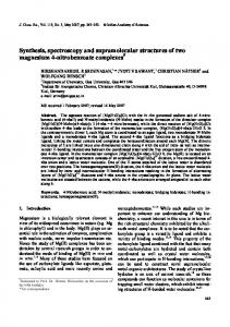
Figure 9. Solid-state fluorescent emission spectra of 3-NO2-bdcH2 (black), complex 1 (red) and complex 2 (blue) at room temperature.
Bull. Korean Chem. Soc. 2013, Vol. 34, No. 8
2379
Thermogravimetric (TG) Analyses. In order to study the framework stability of the title complexes, the thermogravimetric (TG) analysis was performed in N2 atmosphere on polycrystalline samples of complexes 1-2, and the TG curves are shown in Figure 8. The TG curve of 1 shows the first loss of 2.07% in the temperature range of 37-299 ºC, which indicates the exclusion of coordinated water molecules (calcd, 2.05%); The second stage occurs between 300 and 357 ºC, bthe anhydrous complex loses 36.15% of total weight, which is due to the decomposition of two 4,4'-bpy (calcd, 35.52%). The final weight loss of 45.56% (calcd, 47.57%) corresponds to the loss of two 3-NO2-bdc2− in the temperature range of 357-904 ºC. For 2, the weight loss attributed to the gradual release of watermolecules is observed in the range of 35-62 ºC (obsd, 3.91%; calcd, 4.07%). When the temperature holds on rising, the product lost 46.79% of the total weight in the temperature range of 62 to 317 ºC, which is related to the loss of 3-NO2-bdc2− (calcd, 47.28%). Beyond 317 ºC, one 4,4'-bpy processes with a total weight loss of 35.18% (calcd. 35.31%) was observed. The residual percentage weight at the end of the decomposition of the complex is observed 11.15%. Conclusion
Figure 10. The TG curves of two complexes.
action data, which confirms the phase purity of the as-prepared products. Fluorescence Properties. Recently, polymeric Zn(II) and Co(II) complexes with their metal cations adopting d10 configuration have been intensively investigated for attractive fluorescence properties and potential applications as new luminescent materials.20 For example, some zinc complexes have been used as organic light-emitting diodes (OLEDs).21 Hence, the solid state photoluminescence properties of 3NO2-bdcH2 ligand and polymers 1-2 were investigated at room temperature (Figure 7) under the same experimental conditions. In the solid state, the strongest emission peak for the free ligand 3-NO2-bdcH2 is at 438 nm with the excitation peak at 233 nm, which is attributed to the π*-n transitions 22. The strongest excitation peaks for 1-2 are at 275 and 285 nm, emission spectra mainly show strong peaks at 467 and 462 nm, respectively. The ligand chelation to the metal center may effectively increase the rigidity of the ligand and reduce the loss of energy by radiationless decay, thus causing the red shift in 1 and 2. Therefore, the luminescence behavior of complexes is caused by metal ligand charge transfer (MLCT).23
In summary, two metal-organic frameworks based on the connectivity co-effect between rigid aromatic dicarboxylic acids (3-NO2-bdcH2) and bridging ligand have been synthesized and characterized [Zn2(3-NO2-bdc)2(4,4'-bpy)2H2O]n (1), [Co(3-NO2-bdc)(4,4'-bpy)H2O]n (2). 1 and 2 are isostructural, which exhibit two-dimensional (2D) layer frameworks. In addition, the luminescence measurements reveal that two complexes exhibit good fluorescent emissions in the solid state at room temperature. Also, thermal decomposition process and powder X-ray diffraction of the complexes were investigated. Acknowledgments. The authors thank the National Natural Science Foundation of China (20761002), PR China, the Natural Science Foundation of Guangxi (053020), P.R. China and Guangxi University for Nationalities. And the publication cost of this paper was supported by the Korean Chemical Society. References 1. Henninger, S. K.; Habib, H. A.; Janiak, C. J. Am. Chem. Soc. 2009, 131, 2776-2777. 2. Habib, H. A.; Hoffmann, A.; Hoppe, H. A.; Janiak, C. Dalton Trans. 2009, 1742-1751. 3. Habib, H. A.; Sanchiz, J.; Janiak, C. Dalton Trans. 2008, 48774884. 4. Janiak, C. Dalton Trans. 2003, 2781-2804. 5. Higuchi, M.; Tanaka, D.; Horike, S.; Sakamoto, H.; Nakamura, K.; Takashima, Y.; Hijikata, Y.; Yanai, N.; Kim, J.; Kato, K.; Kubota, Y.; Takata, M.; Kitagawa, S. J. Am. Chem. Soc. 2009, 131, 1033610337. 6. Zhou, Y. L.; Zeng, M. H.; Wei, L. Q.; Li, B. W.; Kurmoo, M.
2380
Bull. Korean Chem. Soc. 2013, Vol. 34, No. 8
Chem. Mater. 2010, 22, 4295-4303. 7. Li, K. H.; Olson, D. H.; Seidel, J.; Emge, T. J.; Gong, H. W.; Zeng, H. P.; Li, J. J. Am. Chem. Soc. 2009, 131, 10368-10369. 8. Zeng, M. H.; Wang, Q. X.; Tan, Y. X.; Hu, S.; Zhao, H. X.; Long, L. S.; Kurmoo, M. J. Am. Chem. Soc. 2010, 132, 2561-2563. 9. Xu, G. C.; Ding, Y. J.; Okamura, T. A.; Huang, Y. Q.; Bai, Z. S.; Hua, Q.; Liu, G. X.; Sun, W. Y.; Ueyama, N. Cryst. Growth Des. 2009, 9, 395-403. 10. Wang, Y.; Huang, Y. Q.; Liu, G. X.; Okamura, T. A.; Doi, M.; Sheng, Y. W.; Sun, W. Y.; Ueyama, N. Chem. Eur. J. 2007, 13, 7523-7531. 11. Fan, J.; Hanson, B. E. Chem. Commun. 2005, 2327-2329. 12. Fan, J.; Yee, G. T.; Wang, G.; Hanson, B. E. Inorg. Chem. 2006, 45, 599-608. 13. Zheng, Y. Z.; Xue, W.; Tong, M. L.; Chen, X. M.; Zheng, S. L. Inorg. Chem. 2008, 47, 11202-11212. 14. Fan, L. L.; Li, C. J.; Meng, Z. S.; Tong, M. L. Eur. J. Inorg. Chem. 2008, 25, 3905-3909.
Wen-Jia Xu et al. 15. Liu, W. T.; Ou, Y. C.; Xie, Y. L.; Lin, Z. J.; Tong, M. L. Eur. J. Inorg. Chem. 2009, 28, 4213-4218. 16. Yaghi, O. M.; O’Keeffe, M.; Ockwig, N. W.; Chae, H. K.; Eddadoudi, M.; Kim, J. Nature 2003, 423, 705. 17. Higashi, T. Program for Absorption Correction, Rigaku Corporation: Tokyo, Japan, 1995. 18. Sheldrick, G. M. SHELXTL V5.1 software reference manual. Bruker AXS, Inc, Madison, Wisconsin, USA, 1997. 19. Guilera, G.; Steed, J. W. Chem. Commun. 1999, 1563-1564. 20. Fabbrizzi, L.; Licchelli, M.; Rabaioli, G.; Taglietti, A. Coord. Chem. Rev. 2000, 205, 85. 21. Evans, R. C.; Douglas, P.; Winscom, C. J. Coord. Chem. Rev. 2006, 250, 2093. 22. Chen, W.; Wang, J. Y.; Chen, C.; Yue, Q.; Yuan, H. M.; Chen, J. X.; Wang, S. N. Inorg. Chem. 2003, 42, 944-946. 23. Bauer, C. A.; Timofeeva, T. V.; Settersten, T. B.; Patterson, B. D.; Liu, V. H.; Simmons, B. A.; Allendorf, M. D. J. Am. Chem. Soc. 2007, 129, 7136-7144.