The Journal of Immunology
Genetic Manipulation of CD74 in Mouse Strains of Different Backgrounds Can Result in Opposite Responses to Central Nervous System Injury Hadas Schori,* Ravid Shechter,* Idit Shachar,† and Michal Schwartz1* The ability to recover from CNS injuries is strain dependent. Transgenic mice that weakly express the p41 CD74 isoform (an integral membrane protein functioning as a MHC class II chaperone) on an I-Ab genetic background have normal CD4ⴙ T cell populations and normal surface expression of MHC class II, but their B cell development is arrested while the cells are still immature. After a CNS injury, these mice recover better than their matched wild-type controls. We generated p41-transgenic mice on an I-Ad background (p41-I-Ad mice), and found that their recovery from CNS injuries was worse than that of controls. A correlative inverse effect was seen with respect to the kinetics of T cell and B cell recruitment to the injured CNS and the expression of insulin-like growth factor at the lesion site. These results, besides verifying previous findings that B cells function in the damaged CNS, demonstrate that the outcome of a particular genetic manipulation may be strain dependent. The Journal of Immunology, 2007, 178: 163–171.
T
he invariant chain protein CD74 (Ii)2 is a nonpolymorphic type II integral membrane protein with a short N-terminal cytoplasmic tail of 28 aa, followed by a single 24-aa transmembrane region and a luminal domain of ⬃150 aa. Differential splicing of the transcription products of CD74 in mice generates two different isoforms, p31 and p41. The CD74 chain was originally thought to function mainly as a MHC class II (MHC-II) chaperone, which promotes the exit of MHC-II proteins from the endoplasmic reticulum, directs them to endocytic compartments, prevents peptide binding within the endoplasmic reticulum, and contributes to peptide editing in the MHC-II compartment (1). However, in addition to its function as a chaperone, CD74 has been shown to play a role as an accessory-signaling molecule. CD74 is directly involved in the maturation of B cells (2) through a pathway that leads to the activation of transcription mediated by the NF-B p65/RelA homodimer and its coactivator, TAFII105 (3). NF-B activation is mediated by the cytosolic region of CD74 (CD74-ICD), which is liberated from the membrane (4). A transgenic mouse expressing low levels of the p41 isoform of murine CD74 on a genetic background of I-Ab haplotype (C57BL/6J mice) was generated in 1995 (5). In these p41 mice, in contrast to CD74 knockout (KO) mice, there is full reconstitution of the CD4⫹ T cell population and of MHC-II surface expression, whereas their B cell population is arrested at an immature stage.
*Departments of Neurobiology and †Immunology, The Weizmann Institute of Science, 76100 Rehovot, Israel Received for publication June 13, 2006. Accepted for publication September 19, 2006. The costs of publication of this article were defrayed in part by the payment of page charges. This article must therefore be hereby marked advertisement in accordance with 18 U.S.C. Section 1734 solely to indicate this fact. 1 Address correspondence and reprint requests to Dr. Michal Schwartz, Department of Neurobiology, The Weizmann Institute of Science, 76100 Rehovot, Israel. E-mail address:
[email protected] 2 Abbreviations used in this paper: Ii, invariant chain; MHC-II, MHC class II; KO, knockout; WT, wild type; IGF, insulin-like growth factor; RGC, retinal ganglion cell; I-B4, isolectin B4; BDNF, brain-derived neurotrophic factor.
Copyright © 2006 by The American Association of Immunologists, Inc. 0022-1767/06/$2.00 www.jimmunol.org
Recovery of rodents from acute and chronic CNS injuries has been shown to be dependent on a well-controlled T cell response to CNS Ags (6). Subsequent studies elucidated the underlying mechanism (7, 8) and suggested that additional immune cells, such as B cells, participate in fighting off injurious conditions in the CNS (9). Thus, for example, recovery from CNS injury in p41transgenic C57BL/6J mice is significantly better than in their matched wild-type (WT) C57BL/6J counterparts (9). Because these transgenic mice differ from the WT only in having no mature B cells, those findings implied that the effect of B cells on recovery from CNS injuries is harmful. This might not reflect the whole picture, however, since studies have shown that different strains vary in their T cell-dependent capacity for such recovery (10). Earlier studies by our group showed, moreover, that mice devoid of B cell function (MT KO) on a genetic background of I-Ab haplotype are better able than the corresponding WT to withstand CNS injury (9). That information led us to postulate that the observed negative role of B cells in C57BL/6J WT mice might be a characteristic of this particular mouse strain because of its I-Ab genetic background. Mice with an I-Ab haplotype show only limited spontaneous recovery from acute CNS injuries, whereas mice with an I-Ad haplotype (such as the BALB/c strain) can spontaneously fight off the aftermath of acute optic nerve injury (10 –13). The postulated strain-dependent effect of B cells on CNS recovery is further supported by our previous finding that the ability of SCID mice with I-Ab-haplotype to cope with CNS injuries is significantly better than that of their matched WT controls or of matched nude mice (i.e., mice congenitally devoid of T cells), whereas both SCID and nude mice with an I-Ad haplotype are significantly less able than their matched WT controls to cope with the same CNS injuries (9). To determine whether the observed negative effect of B cells on CNS recovery is common to all mouse strains or is unique to mice with the I-Ab haplotype (such as C57BL/6J), we established a new strain of p41-transgenic mice with a genetic background of I-Ad haplotype. These mice, which we named p41-I-Ad, closely resemble p41 mice with the I-Ab haplotype. Both strains have almost normal T cell populations whereas their B cell populations are
164
MANIPULATION OF CD74 IN CNS INJURIES
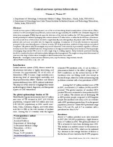
FIGURE 1. Production and characterization of p41-I-Ad-transgenic mice. Splenocytes from control (BALB/c and C57BL/6J WT) and p41-I-Ad mice were stained with anti-I-Ad Abs. A, Histograms show the results of FACS analysis of splenocyte expression of I-Ad; the p41-I-Ad mice express relatively large amounts of I-Ad. DNA was extracted from the tails of CD74⫺/⫺ and p41-I-Ad mice and was analyzed by PCR for p41 isoform expression. B, p41-I-Ad mice show positive expression of the p41 isoform. Splenocytes from control (BALB/c WT), p41, and p41-I-Ad mice were double-stained with anti-CD4 and anti-CD8 Abs. A dot plot shows the expression of CD4⫹ and CD8⫹ cells. C, The p41-I-Ad mice show normal expression of these cells compared with their matched WT controls. Splenocytes were tripled-stained with anti-B220, anti-IgM, and anti-IgD Abs. D, Dot plots present IgD and IgM expression on B220⫹ cells. Splenocytes were double-stained with anti-B220 and antiCD23 Abs. E, Histograms present CD23 expression on B220⫹ cells. Purified B cells from control (BALB/c WT), CD74⫺/⫺ with I-Ad haplotype, and p41-I-Ad mice were cultured with different concentrations of LPS or anti-IgM. Proliferation of cells was assessed by [3H]thymidine incorporation. F, B cells from p41-I-Ad mice proliferated normally in response to LPS, but did not respond to antiIgM stimulation.
arrested at an immature stage and thus cannot participate in the immune response. The results presented in this study suggested that manipulation of a single gene on a different genetic background has entirely different results in terms of the CNS response to an injury. Relative to their corresponding WT controls, transgenic (p41) mice of the C57BL/6J strain (i.e., with a genetic background of I-Ab haplotype) showed significantly better recovery from spinal cord injury or glutamate cytotoxicity in the eye, whereas recovery of transgenic (p41) mice of the BALB/c strain (with a genetic background of I-Ab haplotype) was significantly worse. Differences in recovery in the two strains correlated with their respective kinetics of T cell and B cell recruitment and their production of insulin-like growth factor (IGF)-I.
Materials and Methods Animals The mice used in this study were handled according to the Association for Research in Vision and Ophthalmology resolution on the use of animals in research. Male C57BL/6J p41-transgenic (5) and WT mice, BALB/c WT and nu/nu (nude) mice, and p41-I-Ad mice, all ages 8 –13 wk, were anesthetized by i.p. administration of ketamine (80 mg/kg) and xylazine (16 mg/kg). Before tissue excision, the mice were killed with a lethal dose of pentobarbitone (170 mg/kg).
Purification of murine CD⫹ T cells Lymph nodes (axillary, inguinal, superficial cervical, mandibular, and mesenteric) and spleens were harvested and mashed. T cells enriched by negative selection were purified on T cell columns (R&D Systems).
The Journal of Immunology
165
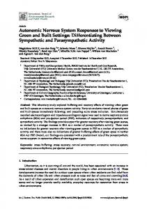
FIGURE 2. Transgenic p41 (I-Ab) mice are more resistant, and p41-I-Ad mice more susceptible, to the aftermath of CNS injuries than their respective matched WT controls. Control (WT C57BL/6J and BALB/c) and transgenic (p41 and p41-I-Ad) mice were each given a single i.v. injection of 200 nmol glutamate. Two groups of transgenic mice were injected i.v. with B cells from their corresponding WT counterparts. Surviving RGC were counted 7 days later. RGC survival (mean ⫾ SEM) is expressed as the number of RGC per square millimeter. ⴱ, p ⬍ 0.05 (Student’s t test); n ⫽ 8 –12 mice in each group. A, B cell transplantation restored the numbers of RGC to control values, indicating that the difference in recovery between the WT strains and their transgenic counterparts was B cell related. B, Recovery from spinal cord contusion in p41- and p41-I-Ad-transgenic mice compared with their matched WT controls. Spinal cord contusion at the level of T12 (200 kdynes, 1 s) was inflicted on mice in the four groups (n ⫽ 12 in each group), and for 30 days their movements were scored according to the Basso Motor Score. Comparison by twofactor repeated-measures ANOVA revealed: BALB/c vs p41-I-Ad, p ⬍ 0.05; C57BL/6J vs p41, p ⬍ 0.05. The results suggested that functional recovery in p41-I-Ad mice is significantly worse than in WT I-Ad mice (BALB/c) and that functional recovery in p41-I-Ab mice is significantly better than in WT I-Ab mice (C57BL/6J).
B cell isolation
Labeling of retinal ganglion cells
A cell suspension from the spleen was generated using standard methods. The B cell population was separated by incubation with CD19 microbeads (Miltenyi Biotec), then washed and subjected to magnetic separation in AutoMACS using the Positive Selection program. CD19-positive cells were collected. For negative selection of B cells, we used a B cell isolation kit (Miltenyi Biotec). The cell suspension was incubated with a mixture of biotin-conjugated Abs against CD43, CD4, and TER-119, as well as antibiotin microbeads. To collect the negative fraction, we used the Deplete AutoMACS program. These procedures yielded a population that was highly enriched in B cells.
Retinal ganglion cells (RGC) were labeled by a fluorescent dye injected stereotactically into the superior colliculus 72 h before tissue excision. For this purpose, mice were anesthetized and placed in a stereotactic device. The skull was exposed and kept dry and clean, and the bregma was identified and marked. The designated point of injection was 2.92 mm posterior to the bregma, 0.5 mm lateral to the midline, and at a depth of 2 mm from the brain surface. A window was drilled in the scalp above the designated coordinates in the right and left hemispheres. The neurotracer dye FluoroGold (5% solution in saline; Fluorochrome) was applied (1 l, at a rate of 0.5 l/min in each hemisphere) using a Hamilton syringe, and the skin over the wound was sutured.
Proliferation of B cells Purified B cells were cultured in 96-well plates at 2 ⫻ 105 cells/well in RPMI 1640 medium supplemented with 10% FCS, 2 mM glutamate, 100 U/ml penicillin, 100 g/ml streptomycin, and different concentrations of LPS (Sigma-Aldrich) or anti-IgM (Jackson ImmunoResearch Laboratories). Cell proliferation was assayed by pulsing the culture with 1 Ci of [3H]thymidine (Valeant Pharmaceuticals) for the last 18 h of a 2-day culture, after which the cells were harvested and counted. Assays were performed in triplicate.
Immunofluorescence staining for flow cytometry Freshly isolated splenocytes were washed with PBS containing 2% FBS and then incubated with one of the following Abs: anti-CD8, anti-CD4 (both from Sigma-Aldrich), anti I-Ab, anti-IgM (both from Southern Biotechnology Associates), and anti I-Ad, anti-B220, anti-CD23, or anti-IgD (all from BD Pharmingen). The cells were incubated with Ab for 30 min in staining buffer (PBS with 2% FBS) in a total volume of 100 l. Wells were then washed twice with staining buffer and the samples were subjected to FACS analysis.
DNA isolation and PCR Mouse-tail genomic DNA was isolated using the DNeasy Tissue Kit (Qiagen) and analyzed by PCR using primers designed for mouse p41 cDNA.
Glutamate injections Under a binocular microscope, the right eye of the anesthetized mouse was punctured with a 27-gauge needle in the upper part of the sclera and a 10-l Hamilton syringe with a 30-gauge needle was inserted as far as the vitreal body. Mice were injected with 1 l of L-glutamate (Sigma-Aldrich) dissolved in saline at a concentration of 200 nmol.
Assessment of RGC survival At the end of the experimental period, the mice were given a lethal dose of pentobarbitone (170 mg/kg). Their eyes were enucleated and the retinas were detached and prepared as flattened whole mounts in 4% paraformaldehyde in PBS. Labeled cells from four to six fields of identical size (0.076 mm2) were counted. The counted fields were located at approximately the same distance from the optic disk (0.3 mm) to allow for variations in RGC density as a function of distance from the optic disk. Fields were counted under the fluorescence microscope (original magnification, ⫻800) by observers blinded to the treatment received by the mice. The average number of RGC per field was calculated for each retina. The numbers of RGC in the contralateral (uninjured) eyes were also counted and served as an internal control.
166
MANIPULATION OF CD74 IN CNS INJURIES
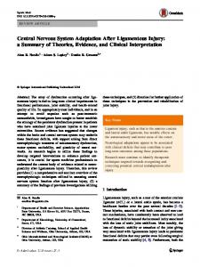
FIGURE 3. Kinetics of T cell infiltration into the site of the injury differs in different mouse strains. WT (C57BL/6J and BALB/c) mice and their respective transgenic (p41 and p41-I-Ad) counterparts were subjected to spinal cord injury. At different time points after the injury (days 1, 3, 7, or 14), the mice were killed and paraffin-embedded sections of their spinal cords were prepared. Immunohistochemical staining for T cells was performed using anti-CD3 Ab. Infiltration of T cells (indicated by arrows) into the site of the injury was observed in both mouse strains. However, infiltration in BALB/c WT mice (genetic background I-Ad) peaked on days 1–3 and in transgenic mice on the same background (p41-I-Ad) on day 7. The opposite picture is seen in mice with a genetic background of I-Ab. ⴱⴱⴱ, p ⬍ 0.0005 (two-tailed Student’s t test).
Spinal cord injury
Immunohistochemistry of paraffin sections
Mice were anesthetized as described above and their spinal cords were exposed by laminectomy at the level of T12. To stabilize the spinal cord, we applied adjustable forceps to the spinous processes of two vertebrae, the one proximal and the other distal to the laminectomy site. Using an Infinite Horizon spinal cord impactor (Research Precision Instruments), we placed the impactor tip on the exposed spinal cord for 1 s and at a force of 200 kdynes. From days 1–30 after the injury, we measured motor recovery of the mice at the indicated intervals using the Basso Motor Score, in which 0 represents complete paralysis and 9 denotes normal mobility. The results were analyzed using the Tukey-Kramer multiple comparisons test (ANOVA).
At the indicated time points after the injury, spinal cords were prepared for histology by perfusing the mouse via the left ventricle with 20 ml of PBS. The excised spinal cords were fixed in 10 ml of Bouin’s solution (75% saturated picric acid, 25% formaldehyde, and 5% glacial acetic acid) for 48 h and then placed in 70% EtOH. The tissue was hydrated in EtOH: xylene:paraffin over a gradient of 70:95:100% and then embedded in paraffin. Sections (6- to 7-m thick) were cut. For staining of B cells, we used rat anti-mouse CD45R/B220 mAb (BD Pharmingen). Slides were deparaffinated in xylene, EtOH (successively 100, 95, 70, and 50%), and PBS (15 min each). Endogenous peroxidase was inactivated (only for the diaminobenzidine peroxidase substrate) in 1% HCl/3% H2O2 in methanol for
The Journal of Immunology
167
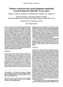
FIGURE 4. Kinetics of B cell infiltration into the site of the injury differs in different mouse strains. WT (C57BL/6J and BALB/c) mice and BALB/c nu/nu mice (devoid of T cells) were subjected to spinal cord injury. At different time points after the injury (days 1, 3, 7, or 14), the mice were killed and paraffin-embedded sections of their spinal cords were prepared. Immunohistochemical staining for T cells was performed using antiCD45R/B220 Ab. Infiltration of B cells into the site of the injury was observed in all mouse strains. However, infiltration in the C57BL/6J WT mice (genetic background I-Ab) peaked on day 7 and in the BALB/c WT mice (genetic background I-Ad) on days 1–3. In the BALB/c nu/nu mice, infiltration also peaked on days 1–3 (ⴱ, p ⬍ 0.05; ⴱⴱⴱ, p ⬍ 0.0005; two-tailed Student’s t test), but significantly fewer B cells infiltrated the site of injury than in the BALB/c WT mice.
15 min, and the slides were washed with PBS. They were then treated with 10 mM sodium citrate (pH 6.0) by heating in the microwave to boiling point, and then for a further 10 min at 20% microwave power. Blocking was performed with 20% normal rabbit serum for 60 min. Ab reaction was allowed to proceed for 24 h at room temperature before being stained by H&E and viewed by light microscopy. For T cell staining we used rat anti-human-CD3 Ab (Serotec). Sections were treated as described above. Ab reaction was allowed to proceed for 72 h at 4°C. Isolectin B4 (I-B4) was purchased from Sigma-Aldrich and chicken anti-brain-derived neurotrophic factor (anti-BDNF) from Promega. For anti-BDNF, sections were treated as described above. Blocking was performed with 20% normal horse serum for 60 min. Ab reaction was allowed to proceed for 24 h at room temperature and for a further 48 h at 4°C. For the secondary Ab reaction (50 min), we used Cy3D anti-chicken along with Cy2 I-B4 (Jackson ImmunoResearch Laboratories). To stain for IGF-I (R&D Systems), we used goat anti-IGF-I. Sections were treated as described above. We treated the sections with 0.1 M Tris (pH 9.0) in the microwave to boiling point and then for a further 10 min at 20% microwave power. Blocking was performed with 20% normal horse serum for 60 min. Ab reaction was allowed to proceeded for 48 h at 4°C. For the secondary Ab reaction (50 min), we used Cy3D anti-goat along with Cy2 I-B4 (Jackson ImmunoResearch Laboratories). Nuclei were labeled with Hoechst stain (Molecular Probes).
Quantification For microscopic analysis, we used a Zeiss LSM 510 confocal laser scanning microscope (original magnification, ⫻40) or a Nikon E800. BDNF immunoreactivity was quantified with Image-Pro Plus 4.5 software (Media Cybernetics) by measuring the mean intensity per unit surface area at the site of the injury. For this analysis, we used at least three spinal cord sections per group and at least two sections per mouse in each group. IGF-I-positive cells were counted at the site of the injury. We counted at least three spinal cord sections per group and at least two sections per mouse in each group. B cells and T cells were counted from the whole spinal cord. From each spinal cord, sections were taken at four different depths. For this analysis, we tested three mice from each group and at least three spinal cord sections per mouse.
The researcher who performed all of the measurements was blinded to the sources of the measured materials.
Results Generation of a mouse with I-Ad haplotype and no mature B cells To generate mice with an immature B cell population and a normal T cell repertoire on an I-Ad haplotype background, we crossed CD74 (invariant chain, Ii)⫺/⫺ BALB/c mice, whose B cells are arrested at an immature stage, with CD74⫺/⫺ (p41) mice that exclusively express the CD74 isoform p41 on an I-Ab background, and have almost normal CD4⫹ and CD8⫹ populations, but B cells that are also arrested at an immature stage (2, 5). We crossed the mice eight times, based on the reported purity of the transgene (14), and then screened for mice bearing I-Ad (Fig. 1A) and the CD74 p41 isoform (Fig. 1B). In this way, we obtained p41-transgenic mice on the I-Ad background (Fig. 1, A and B). Comparison to the previously characterized p41 strain showed that the phenotype of the newly generated strain of transgenic mice with its I-Ad background was identical to that of the p41-transgenic mice on an I-Ab background (5). To characterize the p41I-Ad strain, we first analyzed its T cell and B cell populations by FACS analysis. Its CD4 and CD8 T cell populations were almost normal (Fig. 1C), pointing to a normal positive selection of T cell populations in the thymus. However, surface markers indicated that its B cell development had been arrested at an immature stage (Fig. 1, D and E). To show directly that p41-I-Ad mice possess only B cells that are immature, we examined the functionality of B cells in the p41-I-Ad strain and the proliferation of purified B cells in response to the T cell-independent Ag LPS or to anti-IgM. LPS is a B cell mitogen capable of activating both immature and mature
168
MANIPULATION OF CD74 IN CNS INJURIES
FIGURE 5. Microglial expression of IGF-I, but not of BDNF, varies among mouse strains. WT (C57BL/6J and BALB/c), transgenic (p41 and p41-I-Ad), and BALB/c nu/nu mice (devoid of T cells) were subjected to spinal cord injury. At different time points after the injury (days 1, 3, 7, and 14), the mice were killed and paraffin-embedded sections of their spinal cords were prepared. A, BDNF staining in the injured spinal cord 7 days after injury is shown. This figure shows high magnification of positive staining for BDNF (red), microglia (green), and colocalized BDNF and microglia (yellow) (panels A1, A2, and A3). Quantification of BDNF immunoreactivity (in arbitrary units). B, At least three spinal cord sections per group were used for this analysis, with at least two sections per mouse in each group. C, IGF-I staining of the injured spinal cord 7 days after injury. Panels C1, C2, and C3 show high magnification of positive staining for IGF-I (blue), microglia (green), and colocalized IGF-I and microglia (light blue). Numbers of IGF-I⫹ cells are expressed as means ⫾ SEM. D, At least three spinal cord sections per group were counted, with at least two sections per mouse in each group (ⴱ, p ⬍ 0.05; ⴱⴱ, p ⬍ 0.005; ⴱⴱⴱ, p ⬍ 0.0005; two-tailed Student’s t test).
The Journal of Immunology B cells, whereas only mature B cells respond to anti-IgM stimulation. Control and p41-I-Ad B cells were found to proliferate similarly in response to LPS stimulation, whereas the transgenic B cells responded hardly at all to stimulation by anti-IgM (Fig. 1F). Taken together, the above results led us to conclude that B cells derived from the p41-I-Ad strain had been arrested while still immature. It is important to mention that CD74 (Ii)-deficient mice (such as the C57BL/6 strain) bearing MHC-II proteins of the H-2b haplotype preferentially exhibit a Th1-type of immune response and lack a Th2-type response in vivo. The Ii⫺/⫺ BALB/c mice display an inverted Th1:Th2 ratio with a Th1 preference similar to that in the case of the C57BL/6 mice (15), despite the known Th2-like immune tendency of BALB/c mice. In vivo, the Ii⫺/⫺ BALB/c mice exhibit a normal Th1 response but a significantly milder than normal Th2 inflammatory response in the asthma model. We found that the inverted Th1/Th2 phenotype is mediated by overexpression of the transcription factor T-, the main Th1 cell lineage activator (I. Topilski and I. Shachar, unpublished data). Opposite effects of B cells in I-Ad and I-Ab mice In a previous study, our group showed that the recovery from either glutamate toxicity or optic nerve crush injury in susceptible B cell-defective mice is better than that in their matched WT controls (9). After establishing our new strain of p41-I-Ad-transgenic mice, we compared the survival rate of neurons after a glutamate insult causing ocular cytotoxicity, or during recovery from a spinal cord injury, in susceptible (C57BL/6J) and resistant (BALB/c) mouse strains. Controls (C57BL/6J and BALB/c mice), p41-transgenic (C57BL/6J) mice, and transgenic BALB/c (p41-I-Ad) mice were subjected to spinal cord injury or intraocular glutamate intoxication. In the latter model, surviving RGC in the affected eyes were counted 7 days after the insult. To confirm the correlation observed between survival and lack of B cells, both groups of transgenic mice were replenished with B cells obtained from their matched WT controls (Fig. 2A). The spinal cord injury was a calibrated contusion at the level of T12 (16). Recovery was assessed by the Basso Motor Score, as described in Materials and Methods. In both injury models, the spontaneous recovery in C57BL/6J mice lacking mature B cells (I-Ab) was significantly better than the recovery in their matched WT controls, whereas the opposite was the case in BALB/c (I-Ad) mice (Fig. 2). In addition, the outcome of injury in B cell-replenished p41 mice of both genetic backgrounds was similar to that in their matched WT controls (Fig. 2A). These results suggested that genetic background determines the efficacy of the B cell contribution to spontaneous recovery from CNS injuries. Infiltration of T cells into the site of the injury varies between mouse strains Our group and others have shown that after passive or active vaccination, T cells infiltrate the site of CNS injury and interact there with resident microglia, thereby activating them to manifest a variety of effector functions (7). In an attempt to gain an insight into the cellular mechanism(s) responsible for the differences in injury outcome in the different strains of mice, we first examined whether T cells infiltrate the site of the injury. We subjected BALB/c and C57BL/6J WT mice, as well as their p41- and p41-I-Ad-transgenic counterparts, to spinal cord injury. At different time points after the injury, the mice were killed and their spinal cords were processed for immunohistochemical analysis using anti-CD3 Abs (which recognize T cells). We found that in all four mouse strains, T cells had infiltrated the site of the injury. However, whereas in strains with better spontaneous recovery from CNS injuries (WT on a BALB/c
169 background and p41 on a C57BL/6J background), the largest numbers of infiltrating T cells were seen on days 1 and 3; in strains with limited ability to recover spontaneously (WT on a C57BL/6J background and p41-I-Ad on a BALB/c background), the peaks occurred only on day 7. Notably, at all tested time points, the numbers of T cells in both transgenic strains were significantly lower than in their corresponding WT controls. These results might indicate that the kinetics of T cell infiltration into the site of the injury influences the ability to recover (Fig. 3). Infiltration of B cells into the site of the injury varies between mouse strains To determine whether B cell activity regulates the local response at the site of injury, we first examined whether, in naive mice, the injury site is in fact infiltrated by B cells. BALB/c and C57BL/6J WT mice were subjected to spinal cord injury, and at different time points thereafter the mice were killed and their spinal cords were processed for immunohistochemical analysis and B cell staining with anti-CD45R-B220. The results showed that B cells had indeed infiltrated the site of the injury; however, although the number of infiltrating B cells in BALB/c mice was largest on days 1 and 3, in C57BL/6J mice it peaked on day 7 (Fig. 4). B cells were detected at the site of injury and in the nerve parenchyma on both sides of the lesion site (17). To determine whether the infiltration of B cells is T cell dependent, we examined the numbers of B cells infiltrating the site of injury in BALB/c mice lacking T cells (BALB/c nu/nu mice). In the absence of T cells, the site of injury was infiltrated by significantly fewer B cells, but their patterns of peak infiltration were similar to those observed in their matched controls. These results pointed to the likelihood that T cells play a role in B cell recruitment and might suggest that unbalanced temporal control of B cell infiltration in susceptible mouse strains contributes to their negative effect (Fig. 4). Growth factor control by immune cells Microglia are the CNS-resident cells of the immune system. Activated microglia secrete neurotrophic factors such as BDNF, which acts as a crucial signaling molecule between microglia and neurons and encourages neurite growth (18 –21), and IGF-I, a growth factor whose presence at the site of the injury correlates with the presence of a protective phenotype of activated microglia there (8, 22, 23). To determine whether B cells play a role in the secretion of these factors, we subjected p41- and p41-I-Adtransgenic, BALB/c nu/nu, BALB/c WT, and C57BL/6J WT mice to spinal cord injury. At different time points after the injury, the mice were killed and their spinal cords were processed for immunohistochemical analysis by staining with anti-I-B4 (a marker of microglia), anti-BDNF, or anti-IGF-I Abs. The results showed that levels of BDNF expression by microglia were affected by both B and T cells, whereas the kinetics were affected by the T cells only (Fig. 5, A and B). In contrast, microglial IGF-I immunoreactivity, assessed on day 7 postinjury, was significantly higher in WT mice on an I-Ad genetic background (BALB/c) than in the matched transgenic mice, but significantly lower in WT mice on an I-Ab background (C57BL/6J) than in the corresponding transgenic strain (Fig. 5, C and D). Interestingly, in nude mice the levels of IGF-I were lower than in their WT controls (Fig. 5D).
Discussion In this study, we report the novel finding that manipulation of the same gene in mice of different genetic backgrounds results
170 in completely different phenotypic outcomes, both at the functional and at the cellular levels. We showed that in p41-transgenic mice (which lack mature B cells), on a genetic background of I-Ab, the spontaneous recovery after a CNS injury is significantly improved relative to their matched WT controls, whereas in mice transgenic for the same gene but having a genetic background of I-Ad the recovery relative to their WT controls is impaired. Strain-related variation was also observed in timing of the accumulation of both T cells and B cells that infiltrated the site of the injury. The spontaneous recovery in all cases was correlated with microglial expression of IGF-I, but not of BDNF. B cells are part of the adaptive immune system. Their various roles in the immune response include the following: 1) Ag uptake, processing, and presentation (in which B cells might act as critical APCs) and Ab production; 2) costimulation of T cells; 3) repair mediated by Abs in the presence or absence of complement; 4) recruitment and targeting of T cells and other cells to inflammatory sites; 5) influencing cytokine production toward a Th2 rather than a Th1 bias; and 6) inducing T cell anergy. The CNS is an immunologically specialized microenvironment, which for many years was perceived as an immune-privileged site. Recent evidence suggests, however, that not only T cells but also B cells can enter the CNS under physiological conditions. The brain parenchyma thus sustains the formation of ectopic lymphoid synapses (24 –26). Little information is available on the infiltration of B cells into injured neuronal tissues or on the contribution of B cells to spontaneous recovery from CNS injuries. The lack of data, as well as indications that the response to trauma differs in WT strains with different genetic backgrounds (10, 27–29), led us to investigate whether a particular genetic manipulation (such as gene knockout or introduction of transgenes) causing impairment of mature B cells might also affect the outcome of injury in a way that depends on the animal’s genetic background. To test this possibility, we established a new strain of transgenic mice expressing the p41 isoform of the CD74 Ag (p41I-Ad). By characterizing our transgenic mice at the molecular level (in terms of expression of the p41 isoform, expression of the I-Ad haplotype, distribution of CD4⫹ and CD8⫹ T cells, and expression of cellular markers of mature and immature B cell populations), we obtained similar results to those previously shown to characterize p41-transgenic mice on the background of I-Ab (5). Despite this similarity at the molecular level and at the level of immune cellular composition, we found in this study that in mice with an I-Ad background, manipulation of the CD74 gene leading to a deficiency in mature B cells (p41-I-Ad) was correlated with impaired recovery from CNS injuries, whereas the same genetic manipulation, when conducted in mice known to be susceptible to autoimmune disease development (p41), had the opposite effect, namely, improved recovery from CNS injuries. The connection between the present results and the role of B cells in the endogenous immune response evoked by CNS injury is further supported by our earlier findings that mice devoid of B cell function (MT KO) on a background of I-Ab are significantly better able than their matched WT controls to withstand CNS injuries, similarly to SCID mice on the same background, whereas SCID mice on a background of I-Ad cope significantly less well with such injuries than do their matched WT controls. Moreover, comparison of nude mice on both genetic backgrounds to their respective matched WT controls showed that in both cases the recovery from CNS injuries was worse in the nude mice. These results clearly demonstrate different effects of B cells on recovery from CNS injuries in mice of different genetic backgrounds (9).
MANIPULATION OF CD74 IN CNS INJURIES Trafficking of lymphocytes to the injured CNS is known to be regulated by local chemotactic signals as well as by the expression of adhesion and of relevant receptors on the lymphocytes (25). Our results showed that the kinetics of lymphocyte accumulation appear to be correlated with the ability to recover from the injury. Accumulation of both T cells and B cells at the site of the injury in mice whose recovery from CNS injury was impaired reached a peak 7 days after the injury, whereas in mice with improved recovery the peak of lymphocyte accumulation occurred as early as on days 1 and 3. In line with previous reports, we showed in this study that the arrival of T cells at the injured CNS is independent of B cells. T cell accumulation at the injured site, however, was B cell dependent, and B cell accumulation depended in turn on T cells. Elimination of one cell population by genetic manipulation thus led to a decrease in the number of lymphocytes that accumulate at the site of CNS injury. T cells are also known as a Th cells, and in helping they interact with microglia. The latter cells, depending on how they are activated, express different phenotypes, which can be either destructive or protective. Protective phenotypes are associated with the production of neurotrophic factors such as BDNF and of growth factors such as IGF-I and are capable of promoting neuronal tissue growth (22, 30, 31). BDNF, for example, mediates neuroprotection of adult rat RGC and promotes some regenerative activity after spinal cord injury. In vitro studies in chicks showed that both IGF-I and BDNF promote neurite outgrowth (18 –21, 23, 32). In the present study, we showed that expression of BDNF by microglia in the injured spinal cord, unlike IGF-I expression, is not affected by the I-A background. The postinjury mechanisms of B cell action in mice clearly require further investigation. Nevertheless, the present results show that although manipulation of the same gene leads to expression of the same phenotype, the outcome of a similar injury in mice of different genetic backgrounds is different. On the basis of these findings, we conclude that genetic background must be taken into account when defining the effect of a particular gene and that claims of universal application should be avoided.
Disclosures The authors have no financial conflict of interest.
References 1. Stumptner-Cuvelette, P., and P. Benaroch. 2002. Multiple roles of the invariant chain in MHC class II function. Biochim. Biophys. Acta 1542: 1–13. 2. Shachar, I., and R. A. Flavell. 1996. Requirement for invariant chain in B cell maturation and function. Science 274: 106 –108. 3. Matza, D., O. Wolstein, R. Dikstein, and I. Shachar. 2001. Invariant chain induces B cell maturation by activating a TAF(II)105-NF-B-dependent transcription program. J. Biol. Chem. 276: 27203–27206. 4. Matza, D., A. Kerem, H. Medvedovsky, F. Lantner, and I. Shachar. 2002. Invariant chain-induced B cell differentiation requires intramembrane proteolytic release of the cytosolic domain. Immunity 17: 549 –560. 5. Shachar, I., E. A. Elliott, B. Chasnoff, I. S. Grewal, and R. A. Flavell. 1995. Reconstitution of invariant chain function in transgenic mice in vivo by individual p31 and p41 isoforms. Immunity 3: 373–383. 6. Moalem, G., R. Leibowitz-Amit, E. Yoles, F. Mor, I. R. Cohen, and M. Schwartz. 1999. Autoimmune T cells protect neurons from secondary degeneration after central nervous system axotomy. Nat. Med. 5: 49 –55. 7. Shaked, I., Z. Porat, R. Gersner, J. Kipnis, and M. Schwartz. 2004. Early activation of microglia as antigen-presenting cells correlates with T cell-mediated protection and repair of the injured central nervous system. J. Neuroimmunol. 146: 84 –93. 8. Butovsky, O., A. E. Talpalar, K. Ben-Yaakov, and M. Schwartz. 2005. Activation of microglia by aggregated -amyloid or lipopolysaccharide impairs MHC-II expression and renders them cytotoxic whereas IFN-␥ and IL-4 render them protective. Mol. Cell Neurosci. 29: 381–393. 9. Schori, H., F. Lantner, I. Shachar, and M. Schwartz. 2002. Severe immunodeficiency has opposite effects on neuronal survival in glutamate-susceptible and resistant mice: adverse effect of B cells. J. Immunol. 169: 2861–2865.
The Journal of Immunology 10. Kipnis, J., E. Yoles, H. Schori, E. Hauben, I. Shaked, and M. Schwartz. 2001. Neuronal survival after CNS insult is determined by a genetically encoded autoimmune response. J. Neurosci. 21: 4564 – 4571. 11. Kerlero de Rosbo, N., I. Mendel, and A. Ben-Nun. 1995. Chronic relapsing experimental autoimmune encephalomyelitis with a delayed onset and an atypical clinical course, induced in PL/J mice by myelin oligodendrocyte glycoprotein (MOG)-derived peptide: preliminary analysis of MOG T cell epitopes. Eur. J. Immunol. 25: 985–993. 12. Gaupp, S., D. Pitt, W. A. Kuziel, B. Cannella, and C. S. Raine. 2003. Experimental autoimmune encephalomyelitis (EAE) in CCR2(⫺/⫺) mice: susceptibility in multiple strains. Am. J. Pathol. 162: 139 –150. 13. Bebo, B. F., Jr., E. Zelinka-Vincent, G. Adamus, D. Amundson, A. A. Vandenbark, and H. Offner. 1998. Gonadal hormones influence the immune response to PLP 139 –151 and the clinical course of relapsing experimental autoimmune encephalomyelitis. J. Neuroimmunol. 84: 122–130. 14. Wong, G. T. 2002. Speed congenics: applications for transgenic and knock-out mouse strains. Neuropeptides 36: 230 –236. 15. Topilski, I., A. Harmelin, R. A. Flavell, Y. Levo, and I. Shachar. 2002. Preferential Th1 immune response in invariant chain-deficient mice. J. Immunol. 168: 1610 –1617. 16. Hauben, E., T. Mizrahi, E. Agranov, and M. Schwartz. 2002. Sexual dimorphism in the spontaneous recovery from spinal cord injury: a gender gap in beneficial autoimmunity? Eur. J. Neurosci. 16: 1731–1740. 17. Ziemssen, T., and F. Ziemssen. 2005. The role of the humoral immune system in multiple sclerosis (MS) and its animal model experimental autoimmune encephalomyelitis (EAE). Autoimmun. Rev. 4: 460 – 467. 18. Namiki, J., A. Kojima, and C. H. Tator. 2000. Effect of brain-derived neurotrophic factor, nerve growth factor, and neurotrophin-3 on functional recovery and regeneration after spinal cord injury in adult rats. J. Neurotrauma 17: 1219 –1231. 19. Salie, R., and J. D. Steeves. 2005. IGF-1 and BDNF promote chick bulbospinal neurite outgrowth in vitro. Int. J. Dev. Neurosci. 23: 587–598. 20. Tauber, S. C., C. Stadelmann, A. Spreer, W. Bruck, R. Nau, and J. Gerber. 2005. Increased expression of BDNF and proliferation of dentate granule cells after bacterial meningitis. J. Neuropathol. Exp. Neurol. 64: 806 – 815. 21. Klocker, N., P. Kermer, J. H. Weishaupt, M. Labes, R. Ankerhold, and M. Bahr. 2000. Brain-derived neurotrophic factor-mediated neuroprotection of adult rat
171
22.
23.
24. 25. 26. 27.
28.
29. 30.
31.
32.
retinal ganglion cells in vivo does not exclusively depend on phosphatidyl-inositol-3⬘-kinase/protein kinase B signaling. J. Neurosci. 20: 6962– 6967. Butovsky, O., Y. Ziv, A. Schwartz, G. Landa, A. E. Talpalar, S. Pluchino, G. Martino, and M. Schwartz. 2006. Microglia activated by IL-4 or IFN-␥ differentially induce neurogenesis and oligodendrogenesis from adult stem/progenitor cells. Mol. Cell Neurosci. 31: 149 –160. Mason, J. L., S. Xuan, I. Dragatsis, A. Efstratiadis, and J. E. Goldman. 2003. Insulin-like growth factor (IGF) signaling through type 1 IGF receptor plays an important role in remyelination. J. Neurosci. 23: 7710 –7718. Wekerle, H. 2002. Tackling multiple sclerosis. Nature 420: 39 – 40. Uccelli, A., F. Aloisi, and V. Pistoia. 2005. Unveiling the enigma of the CNS as a B-cell fostering environment. Trends Immunol. 26: 254 –259. Hickey, W. F. 2001. Basic principles of immunological surveillance of the normal central nervous system. Glia 36: 118 –124. Helisch, A., S. Wagner, N. Khan, M. Drinane, S. Wolfram, M. Heil, T. Ziegelhoeffer, U. Brandt, J. D. Pearlman, H. M. Swartz, and W. Schaper. 2006. Impact of mouse strain differences in innate hindlimb collateral vasculature. Arterioscler. Thromb. Vasc. Biol. 26: 520 –526. Lee, K. J., S. K. Park, J. A. Im, S. K. Kim, G. H. Kim, G. Y. Kim, E. J. Yang, and Y. S. Ryang. 2004. Susceptibility of several strains of mice to Echinostoma hortense infection. Korean J. Parasitol. 42: 51–56. Shireman, P. K., and M. P. Quinones. 2005. Differential necrosis despite similar perfusion in mouse strains after ischemia. J. Surg. Res. 129: 242–250. abd-el-Basset, E., and S. Fedoroff. 1995. Effect of bacterial wall lipopolysaccharide (LPS) on morphology, motility, and cytoskeletal organization of microglia in cultures. J. Neurosci. Res. 41: 222–237. Hausler, K. G., M. Prinz, C. Nolte, J. R. Weber, R. R. Schumann, H. Kettenmann, and U. K. Hanisch. 2002. Interferon-␥ differentially modulates the release of cytokines and chemokines in lipopolysaccharide- and pneumococcal cell wall-stimulated mouse microglia and macrophages. Eur. J. Neurosci. 16: 2113–2122. Coull, J. A., S. Beggs, D. Boudreau, D. Boivin, M. Tsuda, K. Inoue, C. Gravel, M. W. Salter, and Y. De Koninck. 2005. BDNF from microglia causes the shift in neuronal anion gradient underlying neuropathic pain. Nature 438: 1017–1021.