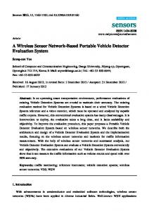
the layout to indicate chamber positions. (a) ... time, a rough estimate of the PSD ratio curves ... B. D. DeBusschere, D. A. Borkholder, and G. T. A. Kovacs, âDesign of an Integrated ... cells represented by the power spectral density (PSD) ratio.
SYSTEM REQUIREMENTS FOR A PORTABLE CELL BASED SENSOR B. Derek DeBusschere, David A. Borkholder, and Gregory T.A. Kovacs Electrical Engineering, Stanford University CIS 202X, Stanford, CA 94305-4075 Abstract The use of cell based biosensors outside of the laboratory has been limited due to many issues including preparation of the sample, maintenance of the biological environment, incorporation of the appropriate sensors, and integration of the electronics for data collection and analysis. This paper describes these system issues and briefly presents current efforts to address packaging, fluidics, and data interpretation. These include the development of an integrated silicon-PDMS cell cartridge system that provides reliable electrical and fluidic interconnect and a regulated cell environment, the development of low power and low overhead sensors and electronics, and the necessary fluidic sample preparation. Keywords: Cell based sensors, Portable biological sensors, Sample preparation 1. Introduction Cell based biosensors, which utilize living cells as the primary transducer (the interface between cell and electronics is the secondary transducer), have evolved from techniques originally used for biological and neurological research. There has been increased interest in expanding their sensitivity and broad based detection capability to general sensing applications [1]. The potential uses of cell based biosensors range from high-throughput drug discovery to chronic environmental monitoring and toxin detection. Although there has been significant progress toward producing practically useful biosensors [1-5], most of this work has been confined to a laboratory environment. 2. Cellular Requirements The living component of a cell based biosensor requires a controlled environment to sustain viability. The major environmental properties that must be controlled for cell culture are the incubation temperature, the substrate material (for anchorage dependent cells), the media, and the gas phase mixture. The optimal incubation temperature for a cell culture varies according to cell type and origin, but most mammalian cell lines will grow satisfactorily at 37 °C. Although temperatures a few degrees below optimal do not affect viability, cells cannot tolerate higher temperatures and will die rapidly at temperatures above 40 °C. Since both the metabolism and growth rate of cells are dependent on temperature, precision is more important than accuracy, and regulation should be controlled to within ± 0.5 °C [6]. The most significant constituents for the gas phase are oxygen (necessary for the oxidative metabolism of eukaryotic cells) and carbon dioxide (necessary only indirectly as it is involved in the bicarbonate buffer system). Most cell cultures are incubated in an atmosphere of 5 to 10% CO2 in air that equilibrates with the bicarbonate buffer in the media to provide the proper pH. However, for short durations, a non-bicarbonate buffer (such as HEPES) can be used and may be preferable since bicarbonate can form precipitates with heavy metals and other compounds. Unless the pH of the media is used as a sensor for cellular metabolism, the pH of the media should be regulated to within the range of 7.4 ± 0.2.
In addition to controlling pH, the culture medium provides the nutrients (inorganic salts, amino acids, glucose, and in many cell lines, serum) necessary for cell growth and survival and has the appropriate osmolarity. Although cells are tolerant of osmolalities in the range of 260 to 320 mM, consistency to within ± 10 mM should be maintained to avoid interfering with cellular function via osmotic stresses across the cell membrane [6]. 3. Sample Preparation and Injection Potential sample sources for a portable cell based biosensor include aerosol collection and water sampling. Aerosol collectors use vortex fans to collect airborne particles and concentrate the collected sample into a liquid form, while water collection could be as simple as manually filling a cup. In order to be utilized by the sensor, these samples must be prepared (conditioned) into a form that is characteristic of biological solutions. A liquid sample with unknown solutes must be converted to a sterile solution that can be perfused over the sensing cells without causing physiochemical damage due to pH or osmotic strength. Additionally, the solution must contain the biologically appropriate concentrations of ions and nutrients. The sample should be filter sterilized before entering the sample preparation system in order to prevent system contamination since fast growing microorganisms, such as bacteria, could overwhelm the sensing cells. Additionally, microorganisms growing in the fluidics system could release metabolites (toxins) that might affect the downstream cells. The osmotic strength of the solution must be adjusted to the biological level of approximately 300 mM by addition of specific concentrations of salts, glucose, and buffers. Since the cell is sensitive to the concentration of these compounds, care must be taken to ensure that the original sample does not contain high levels of any solutes. Variations of ± 10 mM are acceptable; thus, the collected sample needs to be diluted to a level of total dissolved solutes that is less than this. Finally, the pH of the sample solution needs to be adjusted to the physiological level of 7.4. A flowchart of an example sample preparation system is shown in Figure 1. Assumptions on the sample input are that it is in liquid form and has been filtered and sterilized. The first step in the sample preparation is to adjust the osmotic strength of the solution to less than 10 mM by dilution. If the incoming sample has a osmolarity that indicates a higher dissolved concentration, it must be diluted to a level below this threshold. The amount of dilution required will be dependent on the sample source; samples collected from soft lake water typically have an osmolarity of ~1 mM and thus require no dilution, while samples from hard river water may require a dilution of 3× [7]. The unfortunate and unavoidable effect of diluting the sample is that the target compound is also diluted. After the sample has been adjusted to or below the osmolarity threshold, it will be mixed with a concentrated flow of cell culture media. In Figure 1, concentrated 10× media is mixed at a volume ratio of 1:9 with the incoming sample stream which creates a slight (10%) but acceptable dilution of the sample. Sample Prep 10× Media Solution Sterilized Liquid Sample
Osmolarity Measure and Adjust
+
Cell Cartridge Dilute by 10×
Control Chamber
pH Adjust
Test Chamber
Electronics Environmental Monitor and control Signal Transduction and Analysis
Figure 1. Simplified block diagram of system showing the three major components: sample preparation, cell cartridge, and system electronics. The unknown sample is prepared into an appropriate state and then perfused through the test chamber. “Pure” media is perfused through a separate control chamber located within the same cell cartridge.
After the media is added (which contains a buffer), the pH needs to be adjusted (with HCl or NaOH) to 7.4, and then the sample is ready for application to the sensing cells. 4. Signal Transduction and Analysis Signal transduction for a cell based sensor involves two steps. The primary step is the transduction by the cell from chemical agent in the solution to a cellular signal, while the second transduction step is the conversion from cellular signal to an electrical signal. The primary transduction step requires a cell line that is sensitive to the desired agent and expresses a detectable biological activity that can be measured as an output signal. Some examples of cellular output signals are modifications in action potential activity [2,5], metabolic rate [4], or changes in cellular motility [3]. In most cases, the cellular response is broad-based (non-specific); however, this non-specific sensitivity is an advantage in the case of portable systems, where the presumed primary use would be the detection of biologically active agents such as toxins. The sensors and electronics that monitor the output of the primary signal must be compact and low power for a portable system, and the interface between sensor and cells must be mechanically robust to resist vibrations and shocks. The transduced electrical signal needs to be processed into an interpretable form, and in most cases real time analysis is desirable. The restrictions on power, size, and speed limit the amount of signal processing available and require the use of low overhead techniques for data analysis. An additional goal is minimal required user intervention. 5. Example Sensor and Electronics System While there are many sensing technologies under development, planar microelectrodes provide a simple, robust transduction method utilizing both action potential and impedance measurements. A cell cartridge that uses microelectrodes as primary sensors and that also Bondpads Alignment Holes
Silicon Die & Chambers
pH & Temperature Mux
Needle Port & Septa
Signal Muxes (a)
pH Electrode
Reference Temperature Electrodes Regulator (b)
Electrode Arrays
Digital Logic
Figure 2. (a) Top view of cell cartridge prototype. The silicon die is mounted directly on the PCB and enclosed on the top and sides (except for exposed bond pad areas) by the PDMS part. As the cartridge is lowered into a ZIF socket, the needles pierce septa to form the fluidic interconnect. The 6 x 9 mm silicon die can be seen as the dark rectangle beneath the two oval chambers. The 0.5 mm wide fluid channels on the right side have been highlighted in white. (b) Floorplan of the silicon die with analog amplifiers, 128 sense electrodes, temperature regulator, and digital logic. The PDMS part is outlined in black above the layout to indicate chamber positions.
addresses the requirements of sterile fluidic interconnect and environmental regulation is shown in Figure 2. The device consists of a PDMS (polydimethylsiloxane) part, a transparent cover that seals the chambers, and a silicon sensing die mounted on a printed circuit board. The PDMS part forms the fluidic channels, interconnect ports, septa, and two 10 µl chambers over the active sensing area. The silicon die will include microelectrodes and a temperature regulation system. Electrical and fluidic connections are made simultaneously as needles pierce septa on the cartridge when it is plugged into a zero-insertion force (ZIF) socket. An example data analysis technique for classifying changes in action potential (AP) shape has been described elsewhere [5]. This method of monitoring changes in the power spectral density (PSD) of an AP (as shown in Figure 3) can easily be implemented using analog band pass filters and peak detectors. By monitoring the peak value of the AP in narrow bands at low, mid and high frequencies over time, a rough estimate of the PSD ratio curves shown can be obtained. A microcontroller Figure 3. Drug modulation functions recorded from a sponsampling at low frequency (< 10 Hz) would be taneously beating culture of embryonic chick myocardial sufficient for this task. cells represented by the power spectral density (PSD) ratio 6. Conclusions
(PSDafter_drug/PSDbefore_drug) are shown. Epinephrine is a calcium channel agonist, verapamil is a calcium channel blocker, and tetrodotoxin (TTX) is a fast sodium channel blocker. The data shown is the average of eight different channels with error bars of one sigma.
The system requirements for building a portable cell based biosensor are sample preparation, environmental regulation, and low overhead data analysis. The most important issues in sample preparation are osmolarity and pH adjustment, and overall the sensing system must be compact, low power, and robust. A system that meets these requirements is under development. Acknowledgments DeBusschere is partially supported by a NDSEG Fellowship and project funding was provided by the DARPA MicroFlumes Program (Contract Number: N66001-96-C-8631). References 1. 2. 3. 4.
5.
6.
7.
L. Bousse, “Whole Cell Biosensors,” Sensors and Actuators B (Chemical), B34(1-3) (1996) 270-5. G. W. Gross, B. K. Rhoades, H. M. E. Azzazy, and M. Wu, “The Use of Neuronal Networks on Multielectrode Arrays as Biosensors,” Biosensors & Bioelectronics, 10(6-7) (1995) 553-67. C. R. Keese and I. Giaever, “A Biosensor that Monitors Cell Morphology with Electric Fields,” IEEE Engineering in Medicine and Biology, 13(3) (June/July 1994) 402-8. H. M. McConnell, J. C. Owicki, J. W. Parce, D. L. Miller, G. T. Baxter, H. G. Wada, and S. Pitchford, “The Cytosensor Microphysiometer: Biological Applications of Silicon Technology,” Science, 257 (25 September 1992) 1906-12. D. A. Borkholder, B. D. DeBusschere, and G. T. A. Kovacs, “An Approach to the Classification of Unknown Biological Agents With Cell Based Sensors,” Technical digest of the 1998 Solid-State Sensor and Actuator Workshop, Hilton Head Island, SC, 6/7-6/11, Transducer Research Foundation, Cleveland (1998) 178-82. B. D. DeBusschere, D. A. Borkholder, and G. T. A. Kovacs, “Design of an Integrated Silicon-PDMS Cell Cartridge,” Technical digest of the 1998 Solid-State Sensor and Actuator Workshop, Hilton Head Island, SC, 6/7-6/11, Transducer Research Foundation, Cleveland (1998) 358-62. R. Eckert and D. Randall, Animal Physiology, W.H. Freeman and Co., San Francisco, CA, 1978.