5 Apr 1989 - percentage for Proteus mirabilis and Klebsiella pneumoniae was not higher than ..... and Proteuis penneri can be easily distinguished from Pro-.
JOURNAL OF CLINICAL MICROBIOLOGY, JUIY 1989, P. 1646-1649 0095-1137/89/071646-04$02.00/0 Copyright © 1989, American Society for Microbiology
Vol. 27, No. 7
T-Mod Pathway, a Reduced Sequence for Identification of Gram-Negative Urinary Tract Pathogens FRANCESCA BERLUTTI,1 MARIA CRISTINA THALLER,l* BENEDETTO DAINELLI,2 AND RENATO PEZZI' Istituto di Microbiologia, Università "La Sapienza,"' and Dipartimento di Medicina Sperimentale e Scienze
Biochimiche,2 II Università, 00185 Rome, Italy Received 16 October 1987/Accepted
5
April 1989
In this paper, we describe a reduced sequence of identification that includes T-mod medium, a selective and differential isolation medium which allows accurate presumptive identification of the most common gramnegative bacteria encountered in urine samples. The present study, performed on bacteria isolated from 1,762 independent urine samples, has shown that a few selected tests (lysine and ornithine decarboxylase, urease and trehalose fermentation tests) improve the identification accuracy of T-mod, making it possible both to identify the less frequent species and to prevent some misidentifications of Klebsiella pneumoniae and Proteus mirabilis. The proposed work flow agreed with conventional identification protocols to a 99.3% extent and allowed identification of 87.4% of the isolates directly from the primary plate, 11.4% after 1 to 3 additional tests, and 1.2% after an identification gallery.
In a previous paper, we described a new selective and differential plating medium (T-mod) which yielded good results in the screening of the main gram-negative urinary tract pathogens (11). Although T-mod medium enabled us to differentiate the more frequent pathogens, the identification percentage for Proteus mirabilis and Klebsiella pneumoniae was not higher than about 80%. Some strains belonging to other species of the Proteeae tribe, indeed, were misidentified as P. mirabilis because of a falsely negative indole spot test on T-mod. Moreover, the reaction pattern of K. pneumoniae on this medium can be shared by some Enterobacter cloacae and Serratia marcescens strains. Both these species are infrequently recovered in outpatient populations, but their frequency increases when inpatient populations are considered. To improve the identification accuracy, therefore, we have selected some biochemical tests in order to devise a reduced sequence of identification. The aims of our study were (i) to increase, with a few additional tests, the agreement percentage of identification as far as the main species are concerned; (ii) to make possible the identification, at the species level, of other gram-negative urinary tract pathogens, especially those which are recovered from inpatient populations; and (iii) to lower the costs without a substantial loss of accuracy. A comparative evaluation with the practical work flow devised by Trepeta and Edberg (12) has also been performed. MATERIALS AND METHODS
Urine samples. A total of 1,762 independent urine specisubmitted to our laboratory from both inpatient and outpatient populations, was studied. Out of these, 1,266 were collected with the midstream clean-catch technique and 496 were collected by catheterization. The specimens arrived in the laboratory within 2 h of collection. After arrival and accessioning, the samples were refrigerated until being processed (no more than 3 h). Media. T-mod medium (Italian patent no. 47512A/87, Consiglio Nazionale delle Ricerche, January 1987), which mens,
*
Corresponding author.
combines ,-glucuronidase, adonitol, and phenylalanine deaminase tests and makes possible the performance of indole and cytochrome oxidase spot tests directly from the selective plate, was prepared as previously described (11). Methylumbelliferyl-p-D-glucuronide medium (FGM), which allows the detection of lactose fermentation and ,B-glucuronidase activity, was prepared by adding 150 mg of methylumbelliferyl-p-D-glucuronide (Sigma Chemical Co., St. Louis, Mo.) to MacConkey agar (Difco Laboratories, Detroit, Mich.), by the method of Trepeta and Edberg (12). Urea agar base was obtained from Difco; it was prepared according to the instructions of the manufacturer, and the results were read after 4 to 6 h. Lysine and ornithine decarboxylase tests were performed by using Decarboxylase base Moeller (Difco) with 5% agar plus 1% L-lysine or 1% L-ornithine, and the trehalose fermentation test was performed by using the standard method (10). The results of lysine, ornithine decarboxylase, and trehalose fermentation tests were read after 18 to 24 h. The ,-glucuronidase activity was detected, on T-mod and FGM plates, by the production of blue, fluorescent, isolated colonies under a 366-nm UV light lamp (12). The oxidase test was done with oxidase sticks (Oxoid Ltd., Basingstoke, Hampshire, United Kingdom) by following the instructions of the manufacturer. The indole test was done as a spot test (13); whenever a negative indole spot test occurred with a Proteus, Morganella, or Providencia isolate from T-mod (11), provided that it was recovered in pure culture, the T-mod plate was gently washed with sterile saline and a small amount (about 1 ml) of this suspension was tested with Kovacs reagent (0.5 ml) in a tube. 2-Aminoacetophenone production was detected by the method of Cox and Parker (2). API Rapid E was obtained from API Systems S.A. (La Balme Les Grottes, Montalieu Vercieau, France). Specimen processing. A 0.01-ml volume of the well-mixed, unspun urine samples was streaked onto blood agar, MacConkey, FGM, and T-mod plates. After an overnight incubation at 35°C, the plates were observed for growth, and if it was not present, they were reincubated and examined at 48 h. Blood agar plates were used for CFU counts and to perform indole and oxidase tests, if necessary. All the gram-negative isolates grown on MacConkey and blood agar 1646
IDENTIFICATION OF URINARY TRACT PATHOGENS
VOL. 27, 1989
1647
T-rod plate
ColCny color a
B1Ue
Yellcw
Y
E.coli
-.-.-
I C.diversusj1 +
IN Commercial rapid Identification kit
1
lm
+
S.marcescens
+
1-
P.-mirP.ilis
K.qcc
K4.leumoniae E.cloacae
+
U OD TR P .rettgeri + - P.alcalifaciens P.stuartii v - + *, _ M.mrorgarii
P.aeruginosa
a.
C. freundii E.cloacae S.rnarcescens K.pneurmiae
Brn
_
+
+ +
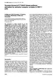
FIG. 1. T-mod work flow. Symbols: , test performed directly from the T-mod plate; O, additional tube test or commercial rapid identification gallery; Fluor, fluorescence detection under a 366-nm-light lamp; Ind, indole spot test; CO, oxidase test; LD, lysine decarboxylase test; OD, ornithine decarboxylase test; U, urease test; TR, trehalose fermentation test; Y, yes; N, no; +, positive; -, negative; v, variable.
were identified according to the criteria of Farmer and colleagues (3) and of Palleroni (9), by using standardized procedures (5, 10). The gram-negative organisms grown on FGM were processed according to the identification protocol devised by Trepeta and Edberg (12) (FGM pathway). Lactose fermentation and ,-glucuronidase activity were detected directly on the FGM plate; oxidase and indole tests were performed as spot tests from the companion blood agar plate. The lactosepositive,"-glucuronidase-positive, indole-positive, and oxidase-negative isolates were considered Escherichia coli. Oxidase-negative organisms that scored negative for at least one of the other three tests were identified by using a 4-h commercial identification system (API Rapid E). Oxidasepositive isolates were tested for 2-aminoacetophenone production (2), and if they were positive, they were identified as Pseudomonas aeruginosa; if they were negative, they were identified by conventional means (9). Gram-negative bacterial isolates grown on T-mod medium were processed and identified according to our pathway (T-mod pathway) as shown in Fig. 1. Cost analysis. The costs of T-mod and FGM media per plate were calculated by using the 1987 Difco and Sigma price lists at the maximum volume discount. The cost of a T-mod plate medium was based on that of the in-house-made medium; the cost of FGM was derived from the cost of commercially available MacConkey (Difco) plus that of 4-methylumbelliferyl-,3-D-glucuronide (Sigma). The costs of the API Rapid E gallery, the oxidase stick, lysine and ornithine decarboxylase media (Decarboxylase base Moeller with 5% agar plus 1% L-lysine or L-ornithine), the urease tube test (Urea Agar Base plus chemical urea), and the trehalose fermentation tube (peptonized water with Andrade's indicator plus 1% trehalose) were derived from 1987 API System, Oxoid, Difco, and Sigma price lists at the maximum volume discount. The costs of the identification to
species level of the tested isolates were calculated by adding to the cost of the primary plate the costs of the required additional tests, when necessary. For example, for the costs of identifying a Providencia rettgeri isolate by using the T-mod pathway, the costs of the urease, ornithine decarboxylase, and trehalose tests were added to the cost of the T-mod plate. RESULTS Unless specifically requested by clinicians, only urine samples with 105 CFU/ml or more were considered for processing. Out of the 1,762 examined urine samples, 642 had >105 CFU/ml and 7, for which counts had been requested, had between 103 and 104 CFU/ml. Therefore, there were 649 processed samples. Of these, 371 (57.2%) gave rise to the growth of a single gram-negative strain; 63 (9.7%) had a gram-negative and a gram-positive strain; 68 (10.4%) had two different gram-negative strains; 125 (19.3%) had only a gram-positive strain; and 22 (3.4%) had yeasts. The gramnegative isolates totaled 570 (Table 1), and all were recovered on MacConkey, FGM, and T-mod plates. Using the proposed T-mod pathway (Fig. 1), we scored 99.3% (566 out of the 570 examined strains) agreement with standard identification protocols (3, 9) (Table 1). The inconsistencies (0.7%) included three Pseudomonas putida strains that were identified as P. aeruginosa and one Enterobacter agglomerans isolate that was identified as Citrobacter freundii. Among the few Providencia isolates (Table 1), three strains (one Providencia rettgeri and two Providencia stuartii isolates) gave an apparent negative indole spot test on T-mod whereas they were positive when tested on blood agar plates.
1648
J. CLIN. MICROBIOL.
BERLUTTI ET AL.
TABLE 1. Results with 570 gram-negative strains, isolated from the infected urine samples, by using the T-mod pathway Organism (no. of isolates)
Escherichia coli (402) Proteus mirabilis
(42)
Pseudomonas aeruginosa (55) Pseudomonas putida (3) Providencia rettgeri (3) Providencia stuartii (3) Klebsiella pneumoniae (21) Enterobacter cloacae (15) Citrobacter freundii (10) Serratia marcescens (8) Klebsiella oxytoca (3) Morganella morganii (3) Citrobacter diversus (1) Enterobacter agglomerans (1)
No. of
Mis-
correctly identified
identification
isolates
(no.)b
Component Reason for misidentification
402
42 55
P. aeruginosa (3)
TABLE 2. Cost analysis of the single components employed in the T-mod and FGM pathways
P. putida is not included in our key
3c
T-mod plate FGM plateb Kovacs reagent' Oxidase stick Diethyl etherd Ornithine decarboxylase test tube Lysine decarboxylase test tube Trehalose test tube Urease test tube API Rapid E gallery
Technologist in Incubation Test used Cost labor time' time ($) which
0.43 0.86 0.001 0.49 0.06 0.06
T-mod FGM Both Both FGM T-mod
5 min 18-24 h
3 min 3 min 1 min 40 s 2 min 1 min
0.12
T-mod
18-24 h
1 min
0.02 0.02 4.13
T-mod T-mod Both
18-24 h 6h 4h
1 min 1 min 7 min
18-24 h 18-24 h
'Time required to perform the test and to read the results.
Y
b 20 ml of medium per plate. '1 drop to perform the indole spot test. d 5 ml to perform the 2-aminoacetophenone test.
21 15
moniae isolates, 3 K. oxytoca isolates, 15 Enterobacter cloacae isolates, 1 Enterobacter agglomerans isolate, 8 S. marcescens isolates, 10 C. freundii isolates, and 1 C. diversus isolate) needed a commercial identification gallery to be identified.
10 8
3 3 1
C. freundii Enterobacter agglomerans is not included (1) in our key
aPercent correctly identified, 99.3%. b Percent misidentified, 0.7%. C Two Providencia stuartii and one Providencia rettgeri isolate could have been misidentified as Proteus mirabilis, according to the indole spot test. They were correctly identified by following the technique described in the text (see Materials and Methods).
The calculated costs of the single components used in the two pathways are reported in Table 2. Table 3 summarizes the efficiency and the related costs for the identifications yielded by using both the studied pathways. Out of the 495 strains directly identified from the T-mod plate, 395 were E. coli, 55 were P. aeruginosa, 42 were Proteus mirabilis, and the remaining 3 strains were the above-mentioned P. putida. Out of the 68 isolates that were identified after one to three tests, 3 were Morganella morganii, 3 were Providencia rettgeri, 3 were Providencia stuartii, 21 were K. pneumoniae, 3 were Klebsiella oxytoca, 15 were Enterobacter cloacae, 10 were C. freundii, 1 was Citrobacter diversus, 8 were S. marcescens, and the remaining strain was the above-mentioned Enterobacter agglomerans. The seven strains that needed an identification gallery were ,-glucuronidase-negative E. coli. When the FGM pathway was used (Table 3), out of the 570 isolates, 361 (63.3%) were typical E. coli and 58 (10.2%) underwent the 2-aminoacetophenone test; of these, 55 were P. aeruginosa and 3 were P. putida. The remaining 151 (26.5%) isolates (i.e., 7 ,-glucuronidase-negative E. coli isolates, 34 indole-negative E. coli isolates, 42 Proteus mirabilis isolates, 3 M. morganii isolates, 3 Providencia rettgeri isolates, 3 Providencia stuartii isolates, 21 K. pneu-
DISCUSSION Urine specimens represent an important part of the workload in clinical microbiology laboratories, so that a protocol allowing a savings of time and materials without a loss of accuracy would be highly desirable. The work flow we devised improves the T-mod performances. With the use of T-mod plates, no additional tests are required to identify E. coli, Proteus mirabilis, and P. aeruginosa isolates (11). In other cases, no more than three tests are necessary to reach the identification to the species level. The use of the expensive commercial identification galleries is limited to the infrequent isolates which show blue, nonfluorescent, indolepositive colonies (e.g., P-glucuronidase-negative E. coli, Citrobacter amalonaticus, or phenylalanine deaminase-negative M. morganii). The proposed pathway increases the identification accuracy of the T-mod medium, which is now able to identify to species level the less frequently encountered species also without the use of commercial identification galleries. The proposed additional tests (ornithine and lysine decarboxylase, urease, and trehalose fermentation) are routine tests in microbiological laboratories with, perhaps, the exception of the trehalose fermentation that is needed to distinguish the emerging pathogen Providencia stuartii (1, 4, 14) from the other Providencia species. Also, as far as the Proteeae tribe is concerned, in performing the indole test, washing the plate (see Materials and Methods) improves the T-mod performances because the brown watersoluble pigments which can mask the indole spot reaction are not present in the alcoholic phase of Kovacs reagent, where the indole is extracted and revealed. In our experience, we did not find any adonitol-negative K. pneumoniae strain, but although 90% of K. pneumoniae strains are adonitol positive (3), negative strains can occur and have been considered in Fig. 1, where the atypical, adonitol-positive E. coli (3) and the infrequent Proteus penneri strains (3, 7) are not included. However, adonitolpositive E. coli isolates are easily recognized, in the T-mod plate, by screening the yellow colonies for fluorescence also,
IDENTIFICATION OF URINARY TRACT PATHOGENS
VOL. 27, 1989
1649
TABLE 3. Comparison of the results yielded by and costs of T-rnod and FGM pathways Directly from plates' Pathway
T-mod FGM
one totests threeAfter additional
After a complete gallery
Total
No. (%7c) of strains identified
Related cost ($)
No. (cX) of strains identified
Related cost (S)
No. (%/c) of strains identified
Related cost (S)
No. (%c) of strains identified
495 (86.8) 361 (63.3)
241.30 487.70
68 (11.9) 55 (9.6)
49.90 77.60
7 (1.2) 154 (27.1)'
31.90 844.20
566 (99.3)" 570 (100)
Cost
325.10
1,409.60
Indole and oxidase tests were considered only when actually performed. See Table 1. ' Including the three P. plaida strains fully identified after using conventional means (see text). `
b
and Proteuis penneri can be easily distinguished from Proteuis mirabilis by a simple ornithine decarboxylase test. P-Glucuronidase-positive Salmonella and Shigella strains have been described previously (6, 8); however, they have not been included in our pathway, because it is devised for urine samples. Almost all of the cytochrome oxidase-positive strains we have isolated belonged to the P. aeriuginosa species, and we believe, therefore, that further testing should be restricted to those strains lacking features, such as colonial morphology or grapefruit smell, peculiar to P. aeruginosa. The T-mod pathway is easy to perform but not as rapid as the FGM pathway because some of the additional tests need an overnight incubation. However, whenever saving time is essential, rapid identification can be obtained by using the API Rapid E galleries, which yield results within 4 h and contain the trehalose fermentation test. Moreover, the Tmod pathway is less expensive than the FGM work flow, even if commercial identification galleries have to be used instead of test tubes. That is because the biochemical reactions included in the T-mod medium allow a reduction in the number of galleries needed in the FGM pathway (for instance, in this study 154 galleries would have been needed for the FGM, in contrast with 75 for the T-mod work flow, with a total cost of $1,409.60 versus $552.60, respectively). The identifications obtained by using the T-mod reactions and the selected tests agreed very well with those obtained by using the standard methods. The incorrect identifications scored only 0.7% and concerned rather infrequent species. We believe, therefore, that the T-mod pathway could be advantageously used in clinical microbiology laboratories, whenever an urine sample has to be processed. ACKNOWLEDGMENTS
Thanks are due to M. Nicoletti for his help with the English language version of this paper and to D. Pasquetti and S. Gentili for their technical assistance. This work was supported by grant Progetto Finalizzato Controllo Malattie da Infezione no. 86.01616.52. from Consiglio Nazionale delle Ricerche, Italy. LITERATURE CITED 1. Cornaglia, G., B. Dainelli, F. Berlutti, and M. C. Thaller. 1988. Commercial identification systems often fail to identify Provi-
dencia .viuartii. J. Clin. Microbiol. 26:323-327. 2. Cox, C. D., and J. Parker. 1979. Use of 2-aminoacetophenone production in identification of Pseuedoinonas aceruginosa. J. Clin. Microbiol. 9:479-484. 3. Farmer, J. J., III, B. R. Davis, F. W. Hickman-Brenner, A. C. McWorter, G. P. Huntley-Carter, M. A. Asbury, C. Riddle, H. G. Wathen-Grady, C. Elias, G. R. Fanning, A. G. Steigerwalt, C. M. O'Hara, G. K. Morris, P. B. Smith, and D. J. Brenner. 1985. Biochemical identification of new species and biogroups of Enterobacteriaceae isolated from clinical specimens. J. Clin. Microbiol. 21:46-76. 4. Hawkey, P. M., S. J. Pedler, and A. Turner. 1983. Comparative in vitro activity of semisynthetic penicillins against Proteeae. Antimicrob. Agents Chemother. 23:619-621. 5. Hendrickson, D. A. 1985. Reagents and stains. p. 1093-1107. In E. H. Lennette. A. Balows, W. j. Hausler. Jr., and H. J. Shadomy (ed.). Manual of clinical microbiology, 4th ed. American Society for Microbiology, Washington, D.C. 6. Killian, M., and P. Bulow. 1976. Rapid diagnosis of Enterobacteriaceae. 1. Detection of bacteria glycosidases. Acta Pathol. Microbiol. Scand. Sect. B 84:245-251. 7. Krajden, S., M. Fuksa, C. Petrea, L. J. Crisp, and J. L. Penner. 1987. Expanded clinical spectrum of infections caused by Proteus penneri. J. Clin. Microbiol. 25:578-579. 8. Maddocks, J. L., and M. J. Greenan. 1975. A rapid method for identifying bacterial enzymes. J. Clin. Pathol. 28:686-687. 9. Palleroni, N. J. 1984. Genus I. Pseiidoitnonas Migula 1894. 237AL (Nom. cons. Opin. 5, Jud. Comm. 1952, 237), p. 141-199. In N. R. Krieg and J. G. Holt (ed.). Bergey's manual of systematic bacteriology. vol. 1. The Williams & Wilkins Co., Baltimore. 10. Phillips, E., and P. Nash. 1985. Culture media, p. 1051-1092. In E. H. Lennette. A. Balows. W. J. Hausler, Jr., and H. J. Shadomy (ed.), Manual of clinical microbiology. 4th ed. American Society for Microbiology, Washington, D.C. 11. Thaller, M. C., F. Berlutti, B. Dainelli, and R. Pezzi. 1988. New plate medium for screening and presumptive identifications of gram-negative urinary tract pathogens. J. Clin. Microbiol. 26: 791-793. 12. Trepeta, R. W., and S. C. Edberg. 1984. Methylumbelliferyl,-D-glucuronide-based medium for rapid isolation and identification of Escherichia coli. J. Clin. Microbiol. 19:172-174. 13. Vracko, R., and J. C. Sherris. 1963. Indole spot-test in bacteriology. Am. J. Clin. Pathol. 39:429-432. 14. Warren, J. W. 1986. Providencia stuartii: a common cause of antibiotic-resistant bacteriuria in patients with long-term indwelling catheters. Rev. Infect. Dis. 8:61-67.