The Journal of Neuroscience, December 1, 2001, 21(23):9142–9150
Targeted Mutations in the Syntaxin H3 Domain Specifically Disrupt SNARE Complex Function in Synaptic Transmission Tim Fergestad,1 Mark N. Wu,2 Karen L. Schulze,3 Thomas E. Lloyd,2 Hugo J. Bellen,2,3,4 and Kendal Broadie1 Department of Biology, University of Utah, Salt Lake City, Utah 84112-0840, and Departments of 2Molecular and Cellular Biology and 3Molecular and Human Genetics, 4Howard Hughes Medical Institute, Baylor College of Medicine, Houston, Texas 77030
1
The cytoplasmic H3 helical domain of syntaxin is implicated in numerous protein–protein interactions required for the assembly and stability of the SNARE complex mediating vesicular fusion at the synapse. Two specific hydrophobic residues (Ala240, Val-244) in H3 layers 4 and 5 of mammalian syntaxin1A have been suggested to be involved in SNARE complex stability and required for the inhibitory effects of syntaxin on N-type calcium channels. We have generated the equivalent double point mutations in Drosophila syntaxin1A (A243V, V247A; syx4 mutant) to examine their significance in synaptic transmission in vivo. The syx4 mutant animals are embryonic lethal and display severely impaired neuronal secretion, although nonneuronal secretion appears normal. Synaptic transmission is nearly abolished, with residual transmission delayed, highly
variable, and nonsynchronous, strongly reminiscent of transmission in null synaptotagmin I mutants. However, the syx4 mutants show no alterations in synaptic protein levels in vivo or syntaxin partner binding interactions in vitro. Rather, syx4 mutant animals have severely impaired hypertonic saline response in vivo, an assay indicating loss of fusion-competent synaptic vesicles, and in vitro SNARE complexes containing Syx 4 protein have significantly compromised stability. These data suggest that the same residues required for syntaxin-mediated calcium channel inhibition are required for the generation of fusion-competent vesicles in a neuronal-specific mechanism acting at synapses. Key words: Drosophila; SNARE complex; core complex; syntaxin; synaptotagmin; calcium channel
Syntaxin is a t-SNARE expressed ubiquitously in the plasma membrane, but it acts as the central member of the core complex mediating synaptic vesicle (SV) fusion only at presynaptic active zones (Jahn and Hanson, 1998; Weber et al., 1998; Chen et al., 1999). Targeted vesicle fusion is regulated by a large number of syntaxin-binding interactions that control its functional conformation (Bajjalieh and Scheller, 1995; Dulubova et al., 1999; Seagar et al., 1999; Brunger, 2000; Yang et al., 2000). Immunoprecipitation studies using anti-p35 (syntaxin) antibody suggested an interaction between syntaxin and N-type calcium channels (Bennett et al., 1992), an interaction widely postulated to tether synaptic vesicles at the sites of Ca 2⫹ influx (Rettig et al., 1997). Nevertheless, the significance of this proposed tethering interaction is unclear because synaptic vesicle docking occurs normally in Drosophila syntaxin null mutants (Broadie et al., 1995). More recently, coexpression of syntaxin and calcium channels in Xenopus oocytes has suggested a functional role for this interaction, because syntaxin appears to attenuate Ca 2⫹ influx and
slow the kinetics of channel inactivation (Bezprozvanny et al., 1995). Syntaxin cleavage by botulinum toxin C1 results in increased Ca 2⫹ influx in purified synaptosomes, further suggesting that syntaxin inhibits calcium channel function (Bergsman and Tsien, 2000). It has been proposed that ubiquitous syntaxin inhibits Ca 2⫹ influx in extrasynaptic membrane, permitting Ca 2⫹ influx only in the presence of a synaptic vesicle (SNARE complex formation) at the active zone. Thus, the formation and stability of the SNARE complex might be linked directly to the interaction of syntaxin and the calcium channel. Syntaxin, SNAP-25, and synaptotagmin all show biochemical and functional interactions with calcium channels (Leveque et al., 1992; Abe et al., 1993; Sheng et al., 1997; Wiser et al., 1999; Wu et al., 1999; Zhong et al., 1999). These proteins interact with each other to couple SNARE complex and calcium channel function (Wiser et al., 1996, 1997; Sheng et al., 1998; Tobi et al., 1998; Seagar et al., 1999). Formation of a quarternary complex of syntaxin, SNAP-25, synaptotagmin, and the calcium channel, termed the “excitosome” (Wiser et al., 1999), has been proposed to be required for efficient, synchronous neurotransmitter release at active zones (Kim and Catterall, 1997). Importantly, syntaxin inhibits N-type calcium channels through a site distinct from the synprint cytosolic loop of the calcium channel. This regulation is specifically disrupted by two point mutations (A240V and V244A) in the syntaxin H3 helical domain (Bezprozvanny et al., 2000). It has been proposed that this specific interaction provides a mechanism for coupling excitosome function with Ca 2⫹ influx at active zones. Our goal in this study was to introduce the comparable double point mutations (A243V and V247A; syx4 mutant) into Drosophila syntaxin1A to assay the impact on neurotransmitter release
Received April 20, 2001; revised Sept. 14, 2001; accepted Sept. 17, 2001. H.J.B. is supported by National Institutes of Health (NIH) Grant GM53571 and is an Investigator of the Howard Hughes Medical Institute. M.N.W. and T.E.L. are supported by predoctoral National Institute of Mental Health National Research Service Awards, and K.L.S. is supported by NIH Postdoctoral Training Grant 5T32HL07747 to the Baylor Section of Pulmonary and Critical Care Medicine. T.F. is supported by NIH Developmental Biology Training Grant 5T32HD07491, and K.B. is funded by NIH Grant GM54544. Special thanks to J. Rohrbough and J. Richmond for insightful discussions. We thank S. Harrison, G. Rubin, T. Su ¨dhof, and K. Zinsmaier for antibodies. We are grateful to D. Casso and T. Kornberg for sharing their Kr-GFP balancer flies. T.F. and M.N.W. contributed equally to this work. Correspondence should be addressed to Dr. Kendal Broadie, Department of Biology, University of Utah, 257 South 1400 East, Salt Lake City, UT 84112. E-mail:
[email protected]. Copyright © 2001 Society for Neuroscience 0270-6474/01/219142-09$15.00/0
Fergestad et al. • Core Complex Regulation in Neurotransmission
at the synapse. Binding assays with Syx 4 show normal biochemical interactions with syntaxin binding partners, Syx 4 displays in vitro SNARE complex formation, and these interactions are consistent with the normal non-neuronal secretion observed in mutants. However, syx4 displays severely compromised neurotransmission, including a high rate of failures. Residual responses display decreased amplitude and increased variability and are temporally uncoupled from the stimulus. The syx4 mutations also compromise the stability of the SNARE complex in vitro and severely reduce the response to hyperosmotic saline application in vivo. Our results indicate that the same H3 residues that mediate Ca 2⫹ channel inhibition also govern SNARE complexes through increased complex stability/assembly. We propose that these coupled processes ensure rapid SNARE complex formation and excitation–secretion coupling at the active zone.
MATERIALS AND METHODS Generation of the sy x 4 mutant. Site directed mutagenesis in vivo was performed as described (Wu et al., 1999). Briefly, mutations in the synta xin open reading frame (ORF) (A243V, V247A) were generated using the Quikchange kit (Stratagene, La Jolla, CA). After sequencing, the mutant ORF was subcloned (X baI–KpnI) into a 13.5 kb genomic rescue fragment in pC aSpeR3 (Pirrotta, 1988). Independent transgenic lines bearing this construct were generated as described (Rubin and Spradling, 1982) and crossed into the null syx229 background. Flies were balanced over TM6B, T b Hu (Lindsley and Z imm, 1992) or TM3, Kr-GFP (a gift of D. C asso and T. Kornberg, University of C alifornia San Francisco). Mutant embryos were identified by the absence of the Green Fluorescent Protein (GFP) balancer or by using outcrossed strains for which all non-hatchers were mutant embryos. Phenot ypic characterization of embr yos. Mutant embryonic Western blots were performed as described (Harrison et al., 1994), except that four embryos were used per lane rather than one. Proteins were detected by enhanced chemiluminescence (ECL; Amersham Pharmacia Biotech, Piscataway, NJ). I378 (anti-rat syntaxin) (Hata et al., 1993) was used at 1:5000, 4F8 [anti-ras-opposite promoter (ROP)] (Harrison et al., 1994) was used at 1:1000, Dsyt2 (anti-synaptotagmin) (Littleton et al., 1993a) was used at 1:2000, and 49/92 [anti-cysteine string protein (C SP)] (Z insmaier et al., 1990) was used at 1:1000. Cuticles were prepared as described (Ashburner, 1990) from control ( y w), y w; syx229, and y w; P{syx4}; syx229 embryos. y w; P{syxwt}; syx229 embryos resembled y w controls (data not shown). SDS-resistant comple x formation. Core complexes were formed during overnight incubation of glutathione S-transferase (GST)-syntaxin or GST-Sy x 4 (1.2 M, immobilized on glutathione-Sepharose beads) with SNAP-25 (4 M) and n-synaptobrevin (4 M) at 4°C in buffer A (50 mM H EPES, pH 7.4, 150 mM potassium acetate, 0.05% T ween 20). Samples were washed once with buffer A ⫹ 1 mg /ml gelatin and twice with buffer A ⫹ 5% glycerol. Samples were divided into eight tubes and resuspended in 1⫻ sample buffer (50 mM Tris, pH 6.8, 2.5% -mercaptoethanol, 2% SDS, 5% glycerol, and 0.025% bromophenol blue). Seven samples were incubated for 5 min at 25, 37, 42, 48, 52, 60, or 66°C using a Robocycler (Stratagene). The eighth aliquot was boiled for 5 min. The proteins were then resolved by SDS-PAGE, transferred to nitrocellulose membranes, and detected with a polyclonal n-syb antibody (R29) at 1:2000 using ECL. In vitro binding assays. For GST-syntaxin f usion protein constructs, the cytoplasmic domains (aa 1–272) of syntaxin and Sy x 4 were PCR amplified using P f u polymerase (Stratagene) and cloned into pGEX-4T-1 (Amersham Pharmacia Biotech). The open reading frame of each construct was sequenced entirely. Constructs expressing target proteins have been described previously (Wu et al., 1999). GST-f usion and His-tagged proteins were produced according to manufacturer’s protocols (Amersham Pharmacia Biotech, and Novagen, Milwaukee, W I, respectively). To exchange buffers for His-tagged proteins, proteins were concentrated using C entriprep columns (Amicon / Millipore, Bedford, M A) and washed twice with 10 ml of PBS (140 mM NaC l, 2.7 mM KC l, 10.1 mM Na2HPO4, 1.8 mM K H2PO4, pH 7.3). SDS-PAGE and Coomassie blue staining were used to estimate protein concentrations, using bovine serum albumin as a standard. T ypical binding incubations used 0.15– 0.30 M GST-syntaxin bound to glutathione-Sepharose beads and 2 M n-synaptobrevin, 1 M SNAP-25, 0.3– 0.6 M synaptotagmin I (Syt), 2 M synprint, or 1 M C SP in a total volume of 200 l with buffer A. Binding
J. Neurosci., December 1, 2001, 21(23):9142–9150 9143
was generally performed for 1–2 hr at 4°C, except for SNAP-25 and ternary core complex formation [overnight (O/ N)]. Beads were washed two times with buffer A ⫹ 1 mg /ml gelatin and three times with buffer A ⫹ 5% glycerol. Because no N-type synprint has been clearly identified in Drosophila, we used the mammalian N-type synprint as a surrogate, assuming conservation of structural homology between species. In our assay, because synaptotagmin and C SP showed nonspecific binding to beads, 20 –100 g of bacterial extract was added to those binding assays as a nonspecific competitor (Assubel, 1996). Because we were unable to produce soluble recombinant ROP, we detected the syntaxin –ROP interaction by performing pull-down experiments from head extracts. Briefly, fly heads were crushed in a mortar and pestle in buffer B (5 mM H EPES, pH 7.4, 100 mM NaC l; 2 ml /1 ml heads). After homogenization with a Dounce homogenizer, cuticular debris was pelleted at 5000 ⫻ g. Membranes were solubilized with 1% Triton X-100 at 4°C for 1 hr, and insoluble material was removed by spinning at 50,000 rpm for 20 min in a TL -100.2 rotor. GST-syntaxin protein (0.25 M) was incubated O/ N at 4°C with 500 g of head extract. Beads were washed as above. Proteins on beads were released by boiling in 20 l sample buffer, and bands were detected by Western blotting and ECL. Antibodies were used as described (Schulze et al., 1995). Synprint was detected using anti-Xpress antibody 1:5000 (Invitrogen, C arlsbad, CA). For dose –response binding curves, GST, GST-syntaxin, or GST-Sy x 4 bound to glutathioneSepharose beads was incubated with SNAP-25 (0.5 M) and n-synaptobrevin (0.01, 0.02, 0.05, 0.1, 0.2 M) or synaptotagmin (0.05, 0.02, 0.5, 1, 1.5 M) or C SP (0.02, 0.2, 0.5, 1, 1.5 M) in 200 l. Known amounts of n-synaptobrevin, synaptotagmin, or C SP were run on the same gel as standards. Bands were quantified using a Personal Densitometer SI (Molecular Dynamics, Sunny vale, CA). Binding curves with values that fell within the linear range were used. Binding of core complex proteins to GST-synaptotagmin I was performed as described (Gerona et al., 2000). Incubations of n-synaptobrevin, syntaxin, Sy x 4, and SNAP-25 (1 M) were performed overnight at 4°C in binding buffer (0.02 M H EPES, pH 7.6, 0.15 M potassium acetate, 0.5% Triton X-100, and 2.5% bovine serum albumin) to generate binary and ternary complexes. After the preincubation, complexes were diluted to 0.2 M with binding buffer and incubated with 2 g GST-Dsyt2 (aa 134 – 474 of synaptotagmin I, provided by J. Troy Littleton, Massachusetts Institute of Technology, C ambridge, M A) bound to glutathione-Sepharose beads. The reactions were supplemented with 2 mM EGTA or 1 mM C aC l2. After 2 hr incubation at room temperature, the beads were washed three times with 1 ml wash buffer (0.02 M H EPES, pH 7.6, 0.15 M potassium acetate, 0.5% Triton X-100). Beads were resuspended with 1⫻ sample buffer and boiled before electrophoresis, except those samples being tested for SDS-denaturation. The primary antibody used in Western detection was affinity purified anti-n-synaptobrevin (rat R29) at a 1:1000 dilution (Wu et al., 1999). Quantification of binding was performed by using an 125I-labeled secondary antibody (anti-rat Ig, whole antibody; Amersham Pharmacia Biotech). Electrophysiolog ical anal ysis. The syx4 mutants are late embryonic lethal, and therefore electrophysiological recordings were performed at the embryonic neuromuscular junction (NMJ) as reported previously (Broadie and Bate, 1993; Wu et al., 1999). All recordings were made at 18°C using standard whole-cell patch-clamp (– 60 mV) techniques from muscle 6 in anterior abdominal segments A2–A3 at 22–24 hr after fertilization (incubated at 25°C). E xcitatory junctional currents (EJC s) were evoked by brief stimulation of the motor nerve (1 msec) with positive current using a glass suction electrode. Mean EJC amplitudes were determined from 25 consecutive EJC s evoked at each frequency, including response failures. Data were acquired and analyzed using pCL AM P 6.0 software (Axon Instruments, Foster C ity, CA). All miniature EJC (mEJC) recordings were done in 0.1 M tetrodotoxin (TTX; Sigma, St. L ouis, MO) at 0.5 mM external C a 2⫹. mEJC amplitude and frequency were analyzed using Mini Analysis software 3.0 (Jaejin Software, Leonia, NJ). C alcium dependence of evoked transmission was characterized by the power relationship of basal EJC amplitudes at 0.1– 0.4 mM C a 2⫹ concentrations (Broadie et al., 1994). Hyperosmotic saline, consisting of bath saline with 850 mM sucrose added, was pressure ejected onto the neuromuscular junction for 3 sec using an unpolished patch pipette (Aravamudan et al., 1999). Statistical analyses were done with Instat (Graphpad software, San Diego, CA). All significance values were calculated using Mann –Whitney U tests.
9144 J. Neurosci., December 1, 2001, 21(23):9142–9150
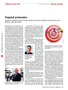
Figure 1. Syntaxin double point mutations lie in neighboring hydrophobic layers. a, Alignment of amino acids 229 –267 of the H3 domains of Drosophila, C. elegans, rat, squid, and yeast syntaxin-1A homologs. The central ionic layer is indicated by the zero, and the numbers indicate adjacent hydrophobic layers. These layer assignments are based on the crystal structure of the core complex (Sutton et al., 1998). The Syx 4 protein is altered for the two boxed amino acids. b, A schematic showing the position of the syx4 mutations in relation to the entire H3 domain of syntaxin. The solid bars indicate the syx4 mutations, and the hatched bars indicate the Ca 2⫹ effector domain (Wu et al., 1999).
RESULTS Targeted mutation of syntaxin H3 residues A243 and V247 The syntaxin H3 cytosolic domain, which mediates coiled-coil interactions with other members of the SNARE complex, is highly conserved across species and absolutely required for vesicular fusion (Schulze et al., 1995; Wu et al., 1999). Specific H3 residues support different protein-binding interactions, which both repress and enhance the efficiency of excitation–secretion coupling at Drosophila synapses (Wu et al., 1999). Two highly conserved H3 residues in mammalian syntaxin (Ala-240, Val-244) have been suggested to be required for SNARE complex stability and syntaxin-mediated inhibition of N-type calcium channels in vitro (Kee et al., 1995; Bezprozvanny et al., 2000). We generated double point mutations in the equivalent residues in Drosophila syntaxin1A using methods identical to our earlier mutational analyses of the H3 domain (Wu et al., 1999). The two point mutations (A243V, V247A) disrupt residues that lie at the end of the H3 coiled-coil domain in hydrophobic layers 4 and 5 within the core complex-forming bundle (Kee et al., 1995) and just outside the “Ca 2⫹ effector domain” characterized previously (Fig. 1a,b) (Wu et al., 1999). Both the mutant form of syntaxin (syx4) and wild-type syntaxin (syxwt) were introduced into the Drosophila genome using transgenic constructs (see Materials and Methods). Transgenic animals bearing either the genomic rescue syxwt construct or the genomic syx4 construct were crossed into a syntaxin null deletion mutant (syx229) background (Schulze et al., 1995). Multiple insertion lines of each construct were compared with Western blots for protein expression levels. Figure 2 shows that two different lines of both syxwt and syx4 constructs in the syx229 null background express similar levels of syntaxin protein. These data show that the syx4 mutations do not significantly alter levels of syntaxin protein in vivo, compared with syxwt controls. Likewise, different transgenic lines for both constructs display similar levels of syntaxin expression (Fig. 2), showing that there are no significant position effects on transgene expression. To
Fergestad et al. • Core Complex Regulation in Neurotransmission
Figure 2. syx4 mutant animals exhibit normal levels of synaptic proteins. The levels of syntaxin, ROP, synaptotagmin I, and CSP are unchanged in syx4 mutants, compared with controls (syxwt). Westerns were performed on extract obtained from four embryos of the appropriate genotype, i.e., syxwt-1, syxwt-2, syx4 –1, and syx4 –2. Bands shown for a given protein were taken from the same exposure of a single gel. The smaller synaptotagmin I band represents a degradation product (Littleton et al., 1993b).
determine whether the syx4 mutations alter the expression of other proteins implicated in synaptic transmission, Western blots were probed for ROP (Munc-18 homolog), synaptotagmin I, and CSP. As shown in Figure 2, the levels of these proteins are similar between syxwt and syx4 embryos and also between different transgenic lines of each construct. To examine the spatial and temporal localization of syntaxin and synaptotagmin I, immunocytochemical staining of embryos was performed. Immunocytochemistry revealed an indistinguishable level and distribution of both proteins in multiple syxwt and syx4 lines (data not shown). Hence, protein levels and distribution of all proteins tested in syx4 mutants were indistinguishable from wild-type controls. We used these transgenic animals to assay the function of the disrupted residues in vesicle fusion in vivo.
syx 4 mutants display defects in neuronal but not non-neuronal secretion We first assessed the gross phenotypes of the syx4 mutants. All syx4 phenotypic analyses were performed in the syx null (syx229) background. We have shown previously that syx229 embryos are late embryonic lethal (Schulze et al., 1995). The wild-type genomic construct (syxwt) can rescue null (syx229) mutants to adulthood (Fig. 3a), demonstrating the normal function of the transgenic protein. In contrast, the genomic construct containing the syx4 mutation is fully embryonic lethal in the null background (Fig. 3a). Hence, the syx4 mutations must cause a severe loss of syntaxin function. We and others have shown previously that syntaxin is absolutely required for both neuronal and non-neuronal secretory events in Drosophila (Schulze et al., 1995; Schulze and Bellen, 1996; Burgess et al., 1997). For example, epidermal cells secrete cuticular proteins from their apical surface; hence, this process represents a polarized form of vesicle transport similar to neurotransmission. Mature wild-type embryos display numerous cuticular structures, most obviously including segmental denticle belts and anterior mouth hooks (Fig. 3b, control ). In contrast, syx null mutant embryos (syx229/syx229) fail to secrete detectable cuticle and show a complete absence of denticle belts and mouth hooks (Fig. 3b, syx229). Surprisingly, the cuticular features of syx4 mutants (syx4/syx4;syx229/syx229) are indistinguishable from wild-type
Fergestad et al. • Core Complex Regulation in Neurotransmission
Figure 3. Gross phenotypes of syx 4 mutants suggest defects in neuronal but not non-neuronal secretion. a, Lethal phase and movement in the syx null (syx229) background. Spontaneous muscle contractions of embryos 22–24 hr after egg laying were observed for 5 min and quantified. Spontaneous peristaltic contractions of syx4 embryos are significantly reduced, compared with syxwt embryos. Evoked contractile responses, determined after a brisk tactile stimulation, were present in syx4 although absent in the null allele. b, Cuticle secretion is not impaired in syx4 mutants. Cuticles from control, syx229, and syx4 (in syx229 background) embryos were imaged using dark-field microscopy. Anterior is to the left. Cuticular structures, including denticle belts and mouth hooks, are absent in the null mutant but present in the control and syx4 mutant.
controls and contain normal segmental denticles and mouth hooks developed from cuticle secretion (Fig. 3b, syx4). The syx4 mutant embryos appear to have normally structured tissues in general, in sharp contrast to syx229 nulls, which display grossly abnormal gut and nerve cord development (data not shown) (Schulze et al., 1995). These data show that the syx4 mutations do not detectably impair non-neuronal secretion, suggesting that H3 residues A243/V247 are not required for vesicular fusion in constitutive secretory processes. Mature wild-type embryos display robust, neurally driven peristaltic muscle contractions before hatching. Spontaneous contractions strongly resemble postembryonic locomotory movement, and tactile stimulation increases the strength and frequency of this movement. Null syx229 mutants display a complete absence of both evoked and spontaneous coordinated movement attributable to a complete block of neurotransmission (Fig. 3a) (Schulze et al., 1995). As expected, the syxwt genomic construct rescues both spontaneous and touch-evoked movement phenotypes (Fig. 3a). Likewise, the syx4 mutant embryos, unlike syx229 mutants, show spontaneous movement and touch-evoked muscle contraction. However, both behaviors are impaired, suggesting that neuromuscular transmission is reduced but not abolished (Fig. 3a). The syx4 mutants display an approximately fourfold reduction in the
J. Neurosci., December 1, 2001, 21(23):9142–9150 9145
Figure 4. syx 4 mutants display a profound reduction in evoked neurotransmission. a, Representative excitatory junctional current (EJC) traces are shown for syxwt; syx229 and syx4; syx229 embryos. Recordings were performed at the embryonic muscle 6 NMJ (22–24 hr after egg laying). In addition to a severe reduction in the EJC amplitude in syx4, neurotransmitter release is clearly asynchronous. Four traces are superimposed; arrow indicates nerve stimulation artifact. b, Mean EJC amplitudes for syxwt-1 (n ⫽ 5), syxwt-2 (n ⫽ 9), syxwt-3 (n ⫽ 5), syx4 –1 (n ⫽ 3), syx4 –2 (n ⫽ 2), and syx4 –3 (n ⫽ 3) embryos. c, Mean latency (time to peak) data are pooled for syxwt (n ⫽ 21) and syx4 (n ⫽ 8) embryos. d, Evoked neurotransmission is variable in syx4 mutants, compared with syxwt. Coefficient of variation (EJC amplitude SD/EJC amplitude) is plotted for syxwt-1, syxwt-2, syxwt-3, syx4 –1, syx4 –2, and syx4 –3 embryos. e, Percentage of failures of evoked response after nerve stimulation in 1.8 mM Ca 2⫹ for syxwt and syx4 embryos. syxwt transmission almost never fails, whereas syx4 fails 50% of all stimuli. Error bars signify SEM. **p ⬍ 0.01.
frequency of muscular contraction waves compared with wildtype controls (syxwt/syxwt;syx229/syx229 ⫽ 1.7 ⫾ 0.2 contractions per minute, n ⫽ 20; syx4/syx4;syx229/syx229 ⫽ 0.4 ⫾ 0.1 contractions per minute, n ⫽ 11; p ⬍ 0.01). These results suggest that the H3 residues A243 and V247 play an important role in a process specific for neuronal secretion at the synapse.
syx 4 mutants display severely impaired excitation–secretion coupling Targeted mutations in Drosophila syntaxin cause striking alterations in synaptic transmission, ranging from a complete loss of transmission in null mutants and a H3 deletion through marked elevated transmission in some H3 point mutations, revealing different regulatory functions of specific protein interactions (Broadie et al., 1995; Schulze et al., 1995; Wu et al., 1999). To address the in vivo role of H3 residues A243 and V247 in neurotransmission, whole-cell patch-clamp recordings were performed at the NMJ of syxwt and syx4 transgenic embryos. As shown in Figure 4, a and b, evoked EJC amplitude is severely reduced in syx4 mutants, to ⬃10% of the levels of syxwt transgenic controls
9146 J. Neurosci., December 1, 2001, 21(23):9142–9150
(1.1 ⫾ 0.1 nA for syxwt, n ⫽ 19; 0.12 ⫾ 0.02 nA for syx4, n ⫽ 8; p ⬍ 0.0001). An identical phenotype was observed in three independent transgenic lines (Fig. 4b). Transmission in syx4 NMJs was similarly severely reduced in all external [Ca 2⫹] from 0.2 to 1.8 mM, but the Ca 2⫹ cooperativity of transmission was similar for syx4 and syxwt controls (1.59 for syxwt; 1.47 for syx4). These data show that H3 residues A243 and V247 play a central, but nonessential, function in synaptic transmission and explain the embryonic lethality and severe loss of movement observed in the syx4 mutants. Neurotransmission in syx4 mutants is characterized by several other obvious defects, which have been observed previously only in synaptotagmin null mutants in Drosophila (Broadie et al., 1994) and after disruption of the excitosome by synprint peptide injection (Mochida et al., 1996; Wiser et al., 1999). Specifically, as shown in Figure 4, a and c–e, neurotransmitter release in syx4 mutants is strikingly asynchronous, demonstrates low fidelity to identical stimuli, and exhibits a high failure rate. In control syxwt NMJ synapses, stimulation-evoked transmission occurs consistently within ⬃5 msec after nerve stimulation, whereas in syx4 mutants, evoked release occurs at delayed (twofold) latencies (Fig. 4c) (5.8 ⫾ 0.2 msec for syxwt, n ⫽ 21; 10.4 ⫾ 0.5 msec for syx4, n ⫽ 8; p ⬍ 0.0001). This increased latency suggests reduced kinetics of excitation–secretion coupling. As shown in Figure 4d, syx4 mutants also show dramatically increased (fourfold) variability in the amount of neurotransmitter released per stimulus, compared with controls (coefficient of variation 0.31 ⫾ 0.04 for syxwt, 1.2 ⫾ 0.08 for syx4; p ⬍ 0.0001). Finally, although syxwt controls always release neurotransmitter in response to nerve stimulation in 1.8 mM extracellular Ca 2⫹ (no failures), syx4 mutants fail to respond ⬎50% of the time (Fig. 4e). Together, the strongly reduced, asynchronous, delayed, and variable release, combined with a high failure of evoked neurotransmission, indicate that excitation–secretion coupling of neurotransmitter release is severely impaired in syx4 mutants. We next assayed spontaneous vesicle fusion in the absence of action potentials by recording mEJCs in the presence of TTX. As shown in Figure 5, mEJC amplitude is slightly increased in syx4 mutants (0.19 ⫾ 0.02 nA for syxwt, n ⫽ 10; 0.26 ⫾ 0.01 nA for syx4, n ⫽ 12; p ⬍ 0.01), but no changes in the kinetics of transmitter release were observed. The underlying reason for the increase in quantal size is unclear, but the increase is specific for the syx4 mutation, because other syntaxin point mutations that we have analyzed do not show an increase (Wu et al., 1999). Thus, because mEJC amplitude is increased in syx4 mutants, this result demonstrates that the postsynaptic receptor field is present and the severe decrease in syx4 neurotransmission is attributable to a presynaptic defect. In line with evoked defects, syx4 mutants reveal a significant decrease in mEJC frequency (0.042 ⫾ 0.010 Hz for syxwt, n ⫽ 11; 0.023 ⫾ 0.006 Hz for syx4, n ⫽ 13; p ⬍ 0.05). This result suggests that core complexes containing Syx 4 protein show a decreased ability to mediate vesicular fusion. In conclusion, syx4 mutants reveal a striking impairment, but not abolishment, of both evoked (Fig. 4) and spontaneous (Fig. 5) fusion events at the synapse.
Syntaxin interactions are maintained with the syx 4 mutations, but core complex stability is impaired What role do the syntaxin H3 residues A243 and V247 play that is so crucial to excitation–secretion coupling in the presynaptic terminal? Can we explain why these residues are central to secretion at presynaptic terminals but appear to play no role in
Fergestad et al. • Core Complex Regulation in Neurotransmission
Figure 5. The frequency of spontaneous mEJCs is reduced in syx4 mutants. a, Representative mEJC traces are shown for syxwt; syx229 and syx4; syx229 animals. Recordings were performed in 0.5 mM Ca 2⫹ ⫹ TTX as described in Materials and Methods. b, Mean mEJC frequency in syxwt (n ⫽ 11) and syx4 (n ⫽ 13) animals. Data from individual transgenic lines were not statistically different and thus were pooled. syx4 mutants show a 50% reduction in mEJC frequency relative to controls. c, Mean mEJC amplitude in syxwt and syx4 animals. syx4 mutants display a slight, but significant, increase in quantal amplitude. Error bars represent SEM. *p ⬍ 0.05; **p ⬍ 0.01.
non-neuronal secretion? We have shown previously that point mutations in the H3 domain of syntaxin can alter binding of specific syntaxin partners (Wu et al., 1999). Many of these proteins act as specific mediators of neuronal, but not non-neuronal, syntaxin function. One possibility is that the Syx 4 mutations disrupt one or more of these known syntaxin interactions. Specifically, the physiological phenotype was very suggestive of an impairment of Syx 4 interaction with synaptotagmin I, a putative Ca 2⫹ sensor (Broadie et al., 1994). To test whether the mutant Syx 4 protein has altered interactions with known syntaxin binding partners, GST-pull-down assays were performed with GST alone (Fig. 6a, GST ), GSTsyntaxin (Fig. 6a, Syxwt), and GST-Syx 4 (Fig. 6a, Syx4). GST alone did not bind any of the assayed proteins, including SNAP-25, n-synaptobrevin (within the ternary complex), ROP, synaptotagmin I, synprint, or CSP. However, GST-syntaxin and GST-Syx 4 were both capable of interacting similarly with each of these binding partners (Fig. 6a). As shown previously by Kee et al. (1995), GST-Syx 4 does show a reduction in this binary binding assay with n-synaptobrevin, compared with GST-syntaxin (data not shown). However, as described previously, this binary interaction is weak, easily disrupted, and unlikely to be physiologically significant (Kee et al., 1995; Wu et al., 1999). More detailed binding assays were performed to specifically examine core complex formation as well as the possible interactions with synaptotagmin I and CSP (Fig. 6b–d). Similar dose– response binding curves for Syx wt and Syx 4 were obtained for each of these proteins: n-synaptobrevin, synaptotagmin I, and CSP. Together, these data suggest that the syx4 mutations do not detectably alter binding of syntaxin to synaptotagmin I, CSP, SNAP-25, ROP, and synprint and do not alter core complex formation, as measured by a steady-state assay. The C2A and C2B domains of synaptotagmin I bind the four-
Fergestad et al. • Core Complex Regulation in Neurotransmission
Figure 6. Mutant Syx 4 protein interacts normally with syntaxin binding partners. a, GST pull-down assays were performed by incubating GST, GST-Syx wt, or GST-Syx 4 with recombinant target proteins or Drosophila head extract, and bands were detected by immunoblotting as described in Materials and Methods. Bands shown for a given target protein were taken from a single exposure of a single gel. b, The syx4 mutation does not significantly alter ternary complex formation. Ternary complex formation was assessed by incubating immobilized GST-Syx wt or GST-Syx 4 with SNAP-25 and increasing amounts of n-synaptobrevin. Bound n-synaptobrevin was determined by immunoblotting and ECL, followed by densitometry with known standards. c, Dose–response binding for GST-Syx wt and GST-Syx 4 to synaptotagmin I. Increasing amounts of synaptotagmin I were incubated with either GST-Syx wt or GST-Syx 4, and bound synaptotagmin I was determined by immunoblotting and ECL. d, Syx 4 and Syx wt show similar dose–response binding to CSP.
helical bundle of the SNARE complex (Davis et al., 1999; Gerona et al., 2000) and may mediate or trigger Ca 2⫹-dependent exocytosis. Furthermore, the role of synaptotagmin in exocytosis may begin very early in SNARE complex formation (vesicle docking) (Reist et al., 1998) because it has been shown recently to also accelerate core complex formation in vitro (Littleton et al., 2001). Therefore, to further test whether the syx4 mutation affects the ability of the core complex to bind synaptotagmin, GST fused with the cytoplasmic domain of synaptotagmin was immobilized on glutathione-Sepharose beads and exposed to n-synaptobrevin alone, preformed binary complexes (n-synaptobrevin-SNAP-25; n-synaptobrevin-syntaxin, or n-synaptobrevin-Syx 4), or preformed ternary complexes (n-synaptobrevin-SNAP-25-syntaxin or Syx 4). GST-synaptotagmin did not bind monomeric n-synaptobrevin or binary complexes of n-synaptobrevin-SNAP25, n-synaptobrevin-syntaxin, or n-synaptobrevin-Syx 4 in the presence or absence of Ca 2⫹ (Fig. 7a, lanes 1– 6 ). Ternary complexes composed of syntaxin and Syx 4 were both bound by GST-synaptotagmin (Fig. 7a, lanes 7–14 ). Quantification of this binding using 125I-labeled secondary antibody indicates that complexes composed of syntaxin and Syx 4 bound GSTsynaptotagmin with equal intensity in the absence of Ca 2⫹. Both forms of syntaxin demonstrated slightly increased binding in the presence of Ca 2⫹ (Fig. 7b). We point out that synaptotagmin binding was enormously more variable in complexes containing Syx 4 than in those containing Syx wt (Fig. 7b); however, the mean binding was indistinguishable, and the significance of the increased variability in Syx 4 is presently unclear. We conclude,
J. Neurosci., December 1, 2001, 21(23):9142–9150 9147
Figure 7. Ternary complexes containing Syx 4 mutant protein interact normally with synaptotagmin I. Binary and ternary complexes of n-synaptobrevin, syntaxin, Syx 4, and SNAP-25 were compared for their ability to bind GST-synaptotagmin in the presence and absence of Ca 2⫹. a, Complexes were detected by Western blotting with anti-synaptobrevin antibody. Lanes 1– 6 are control proteins and binary combinations that do not bind GST-synaptotagmin. Lanes 7–14 show that core complexes formed with mutant Syx 4 protein bind GST-synaptotagmin as readily as wild type (lanes 7–10 are boiled and lanes 11–14 are unboiled). b, The graph shows the percentage binding to GST-Syt as quantified using 125 I-labeled secondary antibody. Percentage binding was normalized, with the highest pixel value for each individual experiment being assigned 100%, from four independent experiments: syntaxin with EGTA, 59%; syntaxin with Ca 2⫹, 99%; Syx 4 with EGTA, 58%; Syx 4 with Ca 2⫹, 98%. Each bar represents the average of four independent experiments ⫾ SEM. *p ⬍ 0.05.
therefore, that the syx4 mutation does not appear to consistently alter the binding between the core complex and synaptotagmin I. Can the Syx 4 mutations affect SNARE complex function directly? The core complex normally forms a highly stable fourhelical bundle. The center of this SNARE bundle contains an ionic “layer” flanked by hydrophobic layers that mediate stabilizing interactions within the bundle of the core complex (Fasshauer et al., 1998; Sutton et al., 1998). The syx4 mutations lie in these stabilizing layers (⫹4 and ⫹5) of the H3 coiled-coil domain (Fig. 1). Hayashi et al. (1994) have shown that the ternary core complex is resistant to SDS-denaturation up to 60°C. We assayed Drosophila SNARE complex stability containing either Syx wt or Syx 4. We bound soluble His-synaptobrevin and His-SNAP-25 to immobilized GST-syntaxin and GST-Syx 4 to examine the SDS resistance and heat lability of the complexes containing these variant proteins. As shown in Figure 8a, SDS-resistant core complexes migrate at ⬃110 kDa, and higher molecular weight bands are also present that likely represent a dimeric form (Hayashi et al., 1994; Hao et al., 1997), recently found to be increased by synaptotagmin I in the presence of Ca 2⫹ (Littleton et al.,
9148 J. Neurosci., December 1, 2001, 21(23):9142–9150
Fergestad et al. • Core Complex Regulation in Neurotransmission
Figure 8. Core complex stability in vitro is impaired by the syx 4 mutation. The heat lability of SDS-resistant core complexes containing Syx 4 is increased, compared with core complexes containing Syx wt. Complexes were formed by incubating His-tagged SNAP-25 and n-synaptobrevin overnight with GST-Syx wt or GST-Syx 4 immobilized on glutathioneSepharose beads. a, Syx 4-containing complexes are resistant to 2% SDS at 25°C, similar to control complexes (Syxwt). b, Complexes were challenged in sample buffer for 5 min at the temperature shown. The lower molecular weight complexes (asterisk) correspond to the trimeric SNARE complex, whereas the higher molecular weight bands likely represent a dimeric form. Note the increased instability of complexes containing Syx 4 relative to Syx wt protein.
2001). The wild-type core complex is stable through 54°C and partially denatured at 60°C in a sample buffer containing 2% SDS (Fig. 8b). In contrast, core complexes made with Syx 4 denature at much lower temperatures. The Syx 4 complexes remain stable up to 25°C (Fig. 8a) but are degraded at 37°C and undetectable above ⬃48°C (Fig. 8b). These observations show that the syx4 mutation impairs the stability of the core complex and may provide a mechanistic explanation for the impaired excitation–secretion coupling in syx4 synapses.
syx 4 mutant synapses have significantly fewer SNARE complexes To test the SV fusion competence in syx4 mutants, the synaptic response to hyperosmotic saline application, which requires functional core complexes, was assayed (Rosenmund and Stevens, 1996; Aravamudan et al., 1999). A 3 sec focal burst of hypertonic saline was applied to syx4 and control (syxwt) embryonic synapses (Aravamudan et al., 1999). Synaptic responses from control animals consisted of many repetitive, high-frequency secretion events (Fig. 9a). In contrast, the hyperosmotic response of syx4 mutant animals is extremely reduced (Fig. 9a). The total charge elicted for syx wt, syx4, and syx229 (null) is shown in Figure 9b (222 nA 䡠 msec for syxwt; 47 nA 䡠 msec for syx4; 20.1 nA 䡠 msec for syx229). syx4 dramatically reduces hyperosmotic saline response, a phenotype comparable to, but slightly less severe than, complete deletion of syntaxin. Note also that there is an increased latency in the hyperosmotic response in syx4 (Fig. 9a), similar to the delay in stimulation-evoked neurotransmission (Fig. 4c). Because syx4 mutant transmission is phenotypically very similar to synaptotagmin I null mutants (sytA D4) (Broadie et al., 1994), the hyperosmotic response in sytA D4 was assayed in parallel. Hyperosmotic saline application to sytA D4 yielded reduced secretion
Figure 9. Core complex function is impaired in syx 4 mutant synapses. a, Representative current traces are shown for syxwt; syx229, syx4; syx229, syx229, and sytA D4 animals in response to 3 sec application of hyperosmotic saline (1175 mOsm; diagonally striped bar). The syxwt synapse responds with robust, high-frequency secretory events, whereas the syx null (syx229) displays no detectable response. Note the obvious response reduction with increased latencies in both syx4 and sytA D4. b, Total charge induced by transmitter release was measured from the area of the current trace responses (nanoamperes times millisecond). Average responses are shown for syxwt (n ⫽ 6), syx4 (n ⫽ 7), syx229 (n ⫽ 5), and sytA D4 (n ⫽ 6) animals. Data from individual transgenic lines were not statistically different and thus were pooled. Error bars represent SEM. **p ⬍ 0.01; ***p ⬍ 0.001.
events that were qualitatively (Fig. 9a) and quantitatively (Fig. 9b) very similar to syx4. The defects in both syx4 and sytA D4 animals shown here are reminiscent of unc13 null mutants in Drosophila, Caenorhabditis elegans, and mouse (Aravamudan et al., 1999; Augustin et al., 1999; Richmond et al., 1999), where formation of the SNARE complex is severely reduced. Thus, these data suggest that the syntaxin H3 domain residues A243 and V247 are required for normal levels of fusion-competent SVs, and this explains both the severely compromised neurotransmission and the embryonic lethality in syx4 mutants.
DISCUSSION Specific amino acids in the hydrophobic “layers” of the SNARE complex interact with a number of regulatory proteins to control the efficacy of neurotransmission (Littleton et al., 1998; Saifee et al., 1998; Wu et al., 1999). The two specific amino acids in the syntaxin H3 domain investigated here (A243, V247) have been proposed to mediate SNARE complex stability (Kee et al., 1995) and, more recently, to mediate calcium channel inhibition (Bez-
Fergestad et al. • Core Complex Regulation in Neurotransmission
prozvanny et al., 2000). Our aim was to mutate these residues (syx4 mutant) to assay their significance during in vivo secretory events. In Drosophila, syntaxin1A is absolutely required for all vesicular fusion events throughout the animal (Schulze et al., 1995); null syntaxin mutants abolish both non-neuronal and neuronal secretion. In contrast, syx4 mutants display no detectable defects in non-neuronal secretion but rather specifically impaired synaptic transmission. These data show that constitutive vesicle fusion does not require residues A243 and V247 in the syntaxin H3 domain, implicating this site in mediating a process specifically involved in calcium-dependent synaptic vesicle fusion. Interaction with N-type Ca 2⫹ channels is an obvious and attractive explanation for this synapse-specific function (Bezprozvanny et al., 2000). However, this interaction has been proposed to inhibit Ca 2⫹ influx, which is not necessarily consistent with observed phenotypes. The syx4 mutants display a striking impairment of synaptic excitation–secretion coupling: action potential-evoked release reduced by ⬃90% and residual transmission, which is highly asynchronous, variable, and prone to failure. Thus, syx4 mutants are not capable of properly triggering robust, synchronized synaptic vesicle fusion in response to a Ca 2⫹ influx. These defects are more consistent with an inability to rapidly generate functional SNARE complexes, as predicted (Kee et al., 1995). The syx4 synaptic phenotypes are clearly distinct from those associated with other engineered point mutations in the H3 domain of syntaxin (Wu et al., 1999). However, the phenotypes are strikingly similar to those described previously for both the synaptotagmin I null mutant (Broadie et al., 1994) and the syxH3-C mutant, which deletes the Ca 2⫹ effector domain to severely reduce binding to synaptotagmin I (Wu et al., 1999). The syx4 phenotypes also resemble the unreliable transmission observed in wild-type synapses at low (⬍0.4 mM) extracellular Ca 2⫹ concentrations (Broadie et al., 1994; Wu et al., 1999). On the basis of these phenotypic similarities, it appears possible that core complex function in vivo is modulated at least in part by synaptotagmin I and that the syx4 mutations impair this regulation. We tested this hypothesis by assaying the protein binding properties of syx4 but were unable to identify impaired binding to synaptotagmin I, CSP, ROP/MUNC-18, the Ca 2⫹ channel synprint site, or other members of the core complex. In particular, in numerous assays synaptotagmin I binding of the Syx 4 core complex was not significantly different from controls, other than a dramatic increase in the variability of binding in the presence of Ca 2⫹ (Fig. 7b). The increased variability of synaptotagmin I binding to the Syx 4 core complex may possibly indicate that rapid core complex formation in syx4 mutants is impaired, because synaptotagmin I has recently been shown to accelerate core complex formation (Littleton et al., 2001). This is consistent with the evidence provided here showing a strong reduction of hyperosmotic saline-induced transmitter release in both synaptotagmin null (sytA D4) and syx4 mutant synapses (Fig. 9). Although Syx 4 containing core complexes can be formed in vitro, on the basis of a steady-state assay, the resulting complexes display impaired stability manifested by increased heat lability. These observations suggest that the formation of the SNARE complex in vivo, which underlies neurotransmission, may be more rapid and substantially different from complex formation in vitro. These observations might reasonably explain why syx4 does not detectably perturb the slow, constitutive vesicle fusion in non-neuronal tissues, whereas it dramatically impairs the fast, Ca 2⫹-dependent fusion at synapses.
J. Neurosci., December 1, 2001, 21(23):9142–9150 9149
Syntaxin, synaptotagmin, and SNAP-25 all dynamically interact with calcium channels and modify channel current properties (Wiser et al., 1996; Wiser et al., 1997; Catterall, 1998). Through these interactions, calcium channels have also been implicated in SNARE complex formation (Sheng et al., 1998; Seagar et al., 1999), possibly through an intermediate termed the excitosome where syntaxin, SNAP-25, and synaptotagmin all bind the channel in a complex awaiting the vesicle and its v-SNARE, synaptobrevin (Wiser et al., 1999). Simplistically, the inhibition of Ca 2⫹ influx by syntaxin (Bezprozvanny et al., 1995) predicts a negative role for the syntaxin–calcium channel interaction on neurotransmission. Therefore, removal of syntaxin-mediated inhibition of Ca 2⫹ influx should result in increased presynaptic Ca 2⫹ levels and increased vesicle fusion and transmission. However, we show that the double point mutations that remove syntaxin-mediated inhibition of calcium channels in vitro (Bezprozvanny et al., 2000) result in severely reduced transmission. We show here that these same residues of syntaxin are critical for normal response to hyperosmotic saline application. Therefore, these residues may play a coupled role in the regulation of Ca 2⫹ channels and SNARE complexes, perhaps through the formation of an excitosome intermediate (Catterall, 1998; Wiser et al., 1999). In Drosophila, we do not know which Ca 2⫹ channels are present at presynaptic active zones and interact with the presynaptic SNARE complex. Therefore, we can provide no direct evidence for Drosophila syntaxin inhibiting calcium channels. However, the syntaxin interaction is maintained through different calcium channel types in vertebrates (Bezprozvanny et al., 1995; Wiser et al., 1999), and the specific residues mediating the interaction are highly conserved in Drosophila (Fig. 1). Thus, one focus of this study was to examine the significance of these calcium channel-inhibiting residues in vivo. Aberrant calcium channel openings, in the absence of syntaxin-mediated inhibition, might result in impaired excitation–secretion; however, because voltage activation of the channel is unaffected (Bezprozvanny et al., 2000) and mEJCs are less frequent in syx4 mutants (Fig. 4b), this is unlikely. Presently, the only functional link for the syntaxin– calcium channel interaction is through syntaxin residues 240 and 244 (243 and 247 in Drosophila). Therefore, alteration of these residues may impair the function of the SNARE complex by disruption of a calcium channel/excitosome intermediate. If we have disrupted the only conserved syntaxin–Ca 2⫹ channel interaction, as we believe, these data provide strong evidence for a positive role for this interaction. This model does not exclude an inhibitory role for syntaxin in calcium channel gating (Bezprozvanny et al., 2000) but suggests that these syntaxin residues, and the syntaxin–calcium channel interaction, are important for more than just inhibiting inappropriate Ca 2⫹ influx. Examination of the interaction between syntaxin and Ca 2⫹ channels may best be done by altering the Ca 2⫹ channel instead of the multifunctional syntaxin, once the non-synprint site of interaction is identified.
REFERENCES Abe T, Saisu H, Horikawa HP (1993) Synaptocanalins (N-type Ca channel-associated proteins) form a complex with SNAP-25 and synaptotagmin. Ann NY Acad Sci 707:373–375. Aravamudan B, Fergestad T, Davis WS, Rodesch CK, Broadie K (1999) Drosophila Unc-13 is essential for synaptic transmission. Nat Neurosci 2:965–971. Ashburner M (1990) Drosophila: A laboratory Handbook. Cold Spring Harbor, NY: Cold Spring Harbor Laboratory. Assubel F (1996) Analysis of protein interactions. New York: Wiley. Augustin I, Rosenmund C, Sudhof TC, Brose N (1999) Munc13–1 is
9150 J. Neurosci., December 1, 2001, 21(23):9142–9150
essential for fusion competence of glutamatergic synaptic vesicles. Nature 400:457– 461. Bajjalieh SM, Scheller RH (1995) The biochemistry of neurotransmitter secretion. J Biol Chem 270:1971–1974. Bennett MK, Calakos N, Scheller RH (1992) Syntaxin: a synaptic protein implicated in docking of synaptic vesicles at presynaptic active zones. Science 257:255–259. Bergsman JB, Tsien RW (2000) Syntaxin modulation of calcium channels in cortical synaptosomes as revealed by botulinum toxin C1. J Neurosci 20:4368 – 4378. Bezprozvanny I, Scheller RH, Tsien RW (1995) Functional impact of syntaxin on gating of N-type and Q-type calcium channels. Nature 378:623– 626. Bezprozvanny I, Zhong P, Scheller RH, Tsien RW (2000) Molecular determinants of the functional interaction between syntaxin and N-type Ca2⫹ channel gating. Proc Natl Acad Sci USA 97:13943–13948. Broadie KS, Bate M (1993) Development of the embryonic neuromuscular synapse of Drosophila melanogaster. J Neurosci 13:144 –166. Broadie K, Bellen HJ, DiAntonio A, Littleton JT, Schwarz TL (1994) Absence of synaptotagmin disrupts excitation-secretion coupling during synaptic transmission. Proc Natl Acad Sci USA 91:10727–10731. Broadie K, Prokop A, Bellen HJ, O’Kane CJ, Schulze KL, Sweeney ST (1995) Syntaxin and synaptobrevin function downstream of vesicle docking in Drosophila. Neuron 15:663– 673. Brunger AT (2000) Structural insights into the molecular mechanism of Ca(2⫹)-dependent exocytosis. Curr Opin Neurobiol 10:293–302. Burgess RW, Deitcher DL, Schwarz TL (1997) The synaptic protein syntaxin1 is required for cellularization of Drosophila embryos. J Cell Biol 138:861– 875. Catterall WA (1998) Structure and function of neuronal Ca2⫹ channels and their role in neurotransmitter release. Cell Calcium 24:307–323. Chen YA, Scales SJ, Patel SM, Doung YC, Scheller RH (1999) SNARE complex formation is triggered by Ca2⫹ and drives membrane fusion. Cell 97:165–174. Davis AF, Bai J, Fasshauer D, Wolowick MJ, Lewis JL, Chapman ER (1999) Kinetics of synaptotagmin responses to Ca2⫹ and assembly with the core SNARE complex onto membranes. Neuron 24:363–376. Dulubova I, Sugita S, Hill S, Hosaka M, Fernandez I, Sudhof TC, Rizo J (1999) A conformational switch in syntaxin during exocytosis: role of munc18. EMBO J 18:4372– 4382. Fasshauer D, Eliason WK, Brunger AT, Jahn R (1998) Identification of a minimal core of the synaptic SNARE complex sufficient for reversible assembly and disassembly. Biochemistry 37:10354 –10362. Gerona RR, Larsen EC, Kowalchyk JA, Martin TF (2000) The C terminus of SNAP25 is essential for Ca(2⫹)-dependent binding of synaptotagmin to SNARE complexes. J Biol Chem 275:6328 – 6336. Hao JC, Salem N, Peng XR, Kelly RB, Bennett MK (1997) Effect of mutations in vesicle-associated membrane protein (VAMP) on the assembly of multimeric protein complexes. J Neurosci 17:1596 –1603. Harrison SD, Broadie K, van de Goor J, Rubin GM (1994) Mutations in the Drosophila Rop gene suggest a function in general secretion and synaptic transmission. Neuron 13:555–566. Hata Y, Slaughter CA, Sudhof TC (1993) Synaptic vesicle fusion complex contains unc-18 homologue bound to syntaxin. Nature 366:347–351. Hayashi T, McMahon H, Yamasaki S, Binz T, Hata Y, Sudhof TC, Niemann H (1994) Synaptic vesicle membrane fusion complex: action of clostridial neurotoxins on assembly. EMBO J 13:5051–5061. Jahn R, Hanson PI (1998) Membrane fusion. SNAREs line up in new environment. Nature 393:14 –15. Kee Y, Lin RC, Hsu SC, Scheller RH (1995) Distinct domains of syntaxin are required for synaptic vesicle fusion complex formation and dissociation. Neuron 14:991–998. Kim DK, Catterall WA (1997) Ca2⫹-dependent and -independent interactions of the isoforms of the alpha1A subunit of brain Ca2⫹ channels with presynaptic SNARE proteins. Proc Natl Acad Sci USA 94:14782–14786. Leveque C, Hoshino T, David P, Shoji-Kasai Y, Leys K, Omori A, Lang B, el Far O, Sato K, Martin-Moutot N (1992) The synaptic vesicle protein synaptotagmin associates with calcium channels and is a putative Lambert-Eaton myasthenic syndrome antigen. Proc Natl Acad Sci USA 89:3625–3629. Lindsley D, Zimm G (1992) The genome of Drosophila melanogaster. San Diego: Academic. Littleton JT, Stern M, Schulze K, Perin M, Bellen HJ (1993a) Mutational analysis of Drosophila synaptotagmin demonstrates its essential role in Ca(2⫹)-activated neurotransmitter release. Cell 74:1125–1134. Littleton JT, Bellen HJ, Perin M (1993b) Expression of synaptotagmin
Fergestad et al. • Core Complex Regulation in Neurotransmission
in Drosophila reveals transport and localization of synaptic vesicles to the synapse. Development 118:1077–1088. Littleton JT, Chapman ER, Kreber R, Garment MB, Carlson SD, Ganetzky B (1998) Temperature-sensitive paralytic mutations demonstrate that synaptic exocytosis requires SNARE complex assembly and disassembly. Neuron 21:401– 413. Littleton JT, Bai J, Vyas B, Desai R, Baltus AE, Garment MB, Carlson SD, Ganetzky B, Chapman ER (2001) Synaptotagmin mutants reveal essential functions for the C2B domain in Ca 2⫹-triggered fusion and recycling of synaptic vesicles in vivo. J Neurosci 21:1421–1433. Mochida S, Sheng ZH, Baker C, Kobayashi H, Catterall WA (1996) Inhibition of neurotransmission by peptides containing the synaptic protein interaction site of N-type Ca2⫹ channels. Neuron 17:781–788. Pirrotta V (1988) Vectors for P-mediated transformation in Drosophila. Biotechnology 10:437– 456. Reist NE, Buchanan J, Li J, DiAntonio A, Buxton EM, Schwarz TL (1998) Morphologically docked synaptic vesicles are reduced in synaptotagmin mutants of Drosophila. J Neurosci 18:7662–7673. Rettig J, Heinemann C, Ashery U, Sheng ZH, Yokoyama CT, Catterall WA, Neher E (1997) Alteration of Ca 2⫹ dependence of neurotransmitter release by disruption of Ca 2⫹ channel/syntaxin interaction. J Neurosci 17:6647– 6656. Richmond J, Davis WS, Jorgensen EM (1999) Unc-13 is required for synaptic vesicle fusion. Nat Neurosci 2:959 –964. Rosenmund C, Stevens CF (1996) Definition of the readily releasable pool of vesicles at hippocampal synapses. Neuron 16:1197–1207. Rubin GM, Spradling AC (1982) Genetic transformation of Drosophila with transposable element vectors. Science 218:348 –353. Saifee O, Wei L, Nonet ML (1998) The Caenorhabditis elegans unc-64 locus encodes a syntaxin that interacts genetically with synaptobrevin. Mol Biol Cell 9:1235–1252. Schulze KL, Bellen HJ (1996) Drosophila syntaxin is required for cell viability and may function in membrane formation and stabilization. Genetics 144:1713–1724. Schulze KL, Broadie K, Perin MS, Bellen HJ (1995) Genetic and electrophysiological studies of Drosophila syntaxin-1A demonstrate its role in nonneuronal secretion and neurotransmission. Cell 80:311–320. Seagar M, Leveque C, Charvin N, Marqueze B, Martin-Moutot N, Boudier JA, Boudier JL, Shoji-Kasai Y, Sato K, Takahashi M (1999) Interactions between proteins implicated in exocytosis and voltagegated calcium channels. Philos Trans R Soc Lond B Biol Sci 354:289 –297. Sheng ZH, Yokoyama CT, Catterall WA (1997) Interaction of the synprint site of N-type Ca2⫹ channels with the C2B domain of synaptotagmin I. Proc Natl Acad Sci USA 94:5405–5410. Sheng ZH, Westenbroek RE, Catterall WA (1998) Physical link and functional coupling of presynaptic calcium channels and the synaptic vesicle docking/fusion machinery. J Bioenerg Biomembr 30:335–345. Sutton RB, Fasshauer D, Jahn R, Brunger AT (1998) Crystal structure of a SNARE complex involved in synaptic exocytosis at 2.4 A resolution. Nature 395:347–353. Tobi D, Wiser O, Trus M, Atlas D (1998) N-type voltage-sensitive calcium channel interacts with syntaxin, synaptotagmin and SNAP-25 in a multiprotein complex. Receptors Channels 6:89 –98. Weber T, Zemelman BV, McNew JA, Westermann B, Gmachl M, Parlati F, Sollner TH, Rothman JE (1998) SNAREpins: minimal machinery for membrane fusion. Cell 92:759 –772. Wiser O, Bennett MK, Atlas D (1996) Functional interaction of syntaxin and SNAP-25 with voltage-sensitive L- and N-type Ca2⫹ channels. EMBO J 15:4100 – 4110. Wiser O, Tobi D, Trus M, Atlas D (1997) Synaptotagmin restores kinetic properties of a syntaxin-associated N-type voltage sensitive calcium channel. FEBS Lett 404:203–207. Wiser O, Trus M, Hernandez A, Renstrom E, Barg S, Rorsman P, Atlas D (1999) The voltage sensitive Lc-type Ca2⫹ channel is functionally coupled to the exocytotic machinery. Proc Natl Acad Sci USA 96:248 –253. Wu MN, Fergestad T, Lloyd TE, He Y, Broadie K, Bellen HJ (1999) Syntaxin 1A interacts with multiple exocytotic proteins to regulate neurotransmitter release in vivo. Neuron 23:593– 605. Yang B, Steegmaier M, Gonzalez LC, Scheller RH (2000) nSec1 binds a closed conformation of syntaxin1A. J Cell Biol 148:247–252. Zhong H, Yokoyama CT, Scheuer T, Catterall WA (1999) Reciprocal regulation of P/Q-type Ca2⫹ channels by SNAP-25, syntaxin and synaptotagmin. Nat Neurosci 2:939 –941. Zinsmaier KE, Hofbauer A, Heimbeck G, Pflugfelder GO, Buchner S, Buchner E (1990) A cysteine-string protein is expressed in retina and brain of Drosophila. J Neurogenet 7:15–29.