Oncotarget, Vol. 6, No.4
www.impactjournals.com/oncotarget/
Targeting a ribonucleoprotein complex containing the caprin-1 protein and the c-Myc mRNA suppresses tumor growth in mice: an identification of a novel oncotarget Ya-Qi Qiu1,2, Cheng-Wei Yang1, Yue-Zhi Lee1, Ruey-Bing Yang3, Chih-Hao Lee4, Hsing-Yu Hsu1, Chien-Chung Chang2 and Shiow-Ju Lee1 1
Institute of Biotechnology and Pharmaceutical Research, National Health Research Institutes, Miaoli, Taiwan
2
Graduate Program of Biotechnology in Medicine, Institute of Molecular & Cellular Biology, National Tsing Hua University, Hsinchu, Taiwan 3
Institute of Biomedical Sciences, Academia Sinica, Taipei, Taiwan
4
Department of Genetics and Complex Diseases, Division of Biological Sciences, Harvard School of Public Health, Boston, Massachusetts, USA Correspondence to: Shiow-Ju Lee, email:
[email protected] Keywords: Caprin-1, c-Myc, Cyclin D, G3BP1, Processing Body Received: October 02, 2014
Accepted: December 09, 2014
Published: December 10, 2014
This is an open-access article distributed under the terms of the Creative Commons Attribution License, which permits unrestricted use, distribution, and reproduction in any medium, provided the original author and source are credited.
ABSTRACT Tylophorine compounds have been the focus of drug development for decades. Tylophorine derivatives exhibit anti-cancer activities but their cellular targets remain unknown. We used a biotinylated tylophorine derivative to probe for the interacting cellular target(s) of tylophorine. Tylophorine directly binds to caprin-1 and consequently enhances the recruitment of G3BP1, c-Myc mRNA, and cyclin D2 mRNA to form a ribonucleoprotein complex. Subsequently, this tylophorine targeted ribonucleoprotein complex is sequestered to the polysomal fractions and the protein expressions of the associated mRNA-transcripts are repressed. Caprin-1 depleted carcinoma cells become more resistant to tylophorine, associated with decreased formation of the ribonucleoprotein complex targeted by tylophorine. Consequently, tylophorine downregulates c-Myc and cyclins D1/D2, causing hypophosphorylation of Rb and suppression of both processing-body formation and the Warburg effect. Gene expression profiling and gain-of-c-Myc-function experiments also revealed that the downregulated c-Myc contributes to the anti-oncogenic effects of tylophorine compounds. Furthermore, the potent tylophorine derivative dibenzoquinoline-33b elicited a similar effect, as c-Myc protein levels were also decreased in xenograft tumors treated with dibenzoquinoline-33b. Thus, tylophorine compounds exert anticancer activity predominantly by targeting and sequestering the caprin-1 protein and c-Myc mRNA associated ribonucleoprotein complex.
INTRODUCTION
recruiting miRNA let-7-loaded RISC (RNA miRNAinduced silencing complex) to the c-Myc 3’UTR [6]. This raises the possibility of developing c-Myc inhibitors by targeting its associated RNP. Cyclin D (cyclin D1, cyclin D2, and cyclin D3 encoded in human cells) is a member of the cyclin protein family. These proteins are involved in the regulation of cell cycle progression for the transition of G1 to S phase [7]. c-Myc has been shown to regulate transcription of genes involved in cell cycle including cyclin D[1]. The
c-Myc is a transcriptional factor that regulates a variety of cellular processes including the activity of cyclin D-Cdk (cyclin-dependent kinase) [1], the Warburg effect in cancer cell metabolism[2, 3], and cellular mRNP processing[4, 5]. Myc expression is deregulated in a wide range of human cancers and is therefore a therapeutic target. Recently, it was found that the RNA-binding protein human antigen R inhibits c-Myc expression by www.impactjournals.com/oncotarget
2148
Oncotarget
retinoblastoma tumor suppressor protein (pRb) is one of the best known substrates of active cyclin D-Cdk4/6 complexes. Thus, the phosphorylated pRb is a common downstream effector of cyclins D1[8, 9], D2[10, 11], and c-Myc[1, 7]. The tumor suppressor protein pRb is dysregulated in several major cancers. pRb is a stable protein and is phosphorylated by several Cdks, which downregulates its activity. Active pRb acts as a tumor suppressor by inhibiting cell cycle progression[7]. In the hypophosphorylated active form, pRb sequesters the transcriptional factor E2F. When pRb is inactivated by hyperphosphorylation, it releases E2F, permitting E2F to execute its cell cycle regulatory functions. Therefore, the hyperphosphorylation of pRb during late G1 phase enables the activation of E2F-dependent transcription, e.g cyclin A2[12]. Caprin-1(Cytoplasmic activation- and proliferationassociated protein 1) is ubiquitously expressed, and its phosphorylation is required for normal cell cycle progression from the G1 to S phase. Caprin-1 co-localizes with G3BP1 (Ras GTPase-activating protein-binding protein 1) within the cytoplasmic RNA granules that are associated with microtubules. This caprin-1/G3BP1 complex is thought to regulate the transport and translation of mRNAs whose protein products are involved in proliferation and migration in multiple cell types because the carboxyl-terminal region of caprin-1 selectively binds to c-Myc or cyclin D2 mRNAs[13]. Collectively, the caprin-1, G3BP1, c-Myc mRNA, and Cyclin D2 mRNA complex represents a function-specific RNP complex and is required for important cellular events. Natural products constitute a successful and promising resource for drug discovery. The elucidation of the cellular targets of therapeutic natural products not only contributes to the identification of valuable novel drug targets for future development, but also guides subsequent molecular pharmacology and mode-of-action studies[14, 16]. Tylophorine-related natural compounds, which are found in the Asclepiadaceae and Moraceae plant families, exhibit multiple biological activities (e.g., anticancer, anti-coronavirus, and anti-inflammatory activities) [17-24]. Tylophorine compounds have been the focus of drug development as therapeutic agents since the 1960s, whereas tylocrebrine failed phase I clinical trials due to neurotoxicity [17]. Although several modes of action for anti-cancer tylophorine compounds have been reported[20, 25-28], their direct cellular target(s) and related pathways remain to be elucidated[24]. In particular, the fundamental mechanisms underlying the broad spectrum, drug-resistant cancer cell inhibitory activities of tylophorine compounds remain to be investigated. We designed and synthesized a biotinylated tylophorine compound which retained the molecular targeting moiety, enabling the probing of direct cellular target(s). In combination with proteomic approaches and www.impactjournals.com/oncotarget
further molecular mechanism studies, the anti-cancer activities of the tylophorine compounds were elucidated. Our results provide a mechanistic basis for developing the tylophorine derived compounds into anti-cancer drugs. The identified cellular target(s) of tylophorine represent novel drug targets for anti-cancer therapeutic applications.
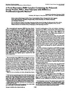
RESULTS Synthesis of a biotinylated tylophorine analogue Previously, we reported that the non-planar structure between the two major phenanthrene and indolizidine/ quinolizidine moieties, as well as the planarity and rigidity of the indolizidine/quinolizidine moiety of tylophorine compounds, significantly affect the activities of tylophorine analogues[19]. Recently, we successfully synthesized potent tylophorine-derived dibenzoquinolines that elicit the same biological functions as traditional pentacyclic tylophorine compounds[23]. Therefore, we concluded that the angular dibenzoquinoline moiety in tylophorine compounds is responsible for the interactions with their direct target molecule(s). Thus, we sought to design and synthesize a biotinylated tylophorine-derived dibenzoquinoline (Fig. 1A), referred to herein as the “biotinylated tylophorine”, to probe direct interacting molecules in cells. The biotinylated tylophorine retained anti-cancer cell activity with IC50 values of ~13-30 µM against HONE-1, MCF7, or NUGC-3 carcinoma cells (see Supplemental Table 1).
Identification of direct targets of tylophorine via pull-down experiments with biotinylated tylophorine HONE-1 cell lysates were incubated with the vehicle control (dimethyl sulfoxide, DMSO), biotin-XSSE, or biotinylated tylophorine prior to the addition of streptavidin in a pull-down experiment. The pulleddown complex was subjected to sodium dodecyl sulfatepolyacrylamide gel electrophoresis (SDS-PAGE) and silver staining to analyze the proteins that specifically interacted with the biotinylated tylophorine. A total of 6 protein bands were specifically associated with biotinylated tylophorine compared to biotin-X-SSE (Fig. 1B); these bands were excised for further matrix-assisted laser desorption ionization-time of flight (MALDI-TOF) mass spectrometry (MS) proteomics analyses. The potential tylophorine-interacting proteins were identified by Mascot data analysis (Fig. 1C and Supplemental Table 2), and the specific interaction of these proteins with biotinylated tylophorine was validated by western blot analysis (Fig. 1D). Caprin-1, α-actinin-4, nucleolin, the splicing factor (PSF), polyadenylate-binding protein-1 2149
Oncotarget
Tylophorine associates with an RNP complex containing caprin-1, G3BP-1, c-Myc mRNA, and Cyclin D2 mRNA
(PABP-1), heterogeneous nuclear ribonucleoprotein M3M4 (hnRNPM3-M4), hnRNPQ, TATA-binding proteinassociated factor 2N isoform 1 (TAF-15), G3BP1, 40S ribosomal protein S6 (S6), phosphorylated S6 (p-S6), and ribosomal protein S3 were specifically pulled down by biotinylated tylophorine compared to biotin-X-SSE or DMSO, but the expression levels of these proteins in tylophorine-treated HONE-1 or MCF7 carcinoma cells were not significantly altered compared with DMSOtreated cells, with the exception of p-S6, which was significantly increased (Fig. 1E). The pull-down of ribosome proteins by biotinylated tylophorine was not unexpected because tylocrebrine was previously reported to inhibit the translocation of peptidyltRNA in ribosomes by 54%, although a tylocrebrine concentration as high as 100 µM was used[29]. Because tylophorine compounds exert significant anti-cancer activities, generally with IC50 values ranging from hundreds to a few nM[22-24], we further examined other proteins identified to seek specific targets for anti-cancer.
Caprin-1 was identified among the molecules that were pulled down by biotinylated tylophorine, and we focused on this protein because it is ubiquitously expressed and required for normal progression through the G1-S phase[13]. We performed co-immunoprecipitation using a caprin-1 antibody to assess the associated complex members. We identified α-actinin-4, G3BP1, and p-S6 within the caprin-1 complex in the presence of biotinylated tylophorine (Fig. 2A). Because caprin-1 and G3BP1 were previously reported to associate and co-localize within RNA-rich cytoplasmic granules[13], we further dissected the association of c-Myc mRNA and cyclin D2 mRNA in HONE-1 cells by RT-PCR analysis of the products from the pull-down experiment with biotinylated tylophorine (Fig. 2B). We also demonstrated the direct, but weak, interaction of biotinylated tylophorine with caprin-1 in
Figure 1: The application of biotinylated tylophorine to probe its direct interacting cellular targets in carcinoma cells.
A. Scheme for the synthesis of biotinylated tylophorine (BT), a dibenzoquinoline derivative. B & C. Pull-down experiment in HONE-1 cell lysates to identify potential molecular targets of tylophorine. BT and biotin-X-SSE were used to pull down the interacting molecules by using streptavidin beads. The lane labelled “3x sample” was loaded with 3-fold-amount of the BT pull-down and the lane labelled “3x lysate” was loaded with the BT pull-down from a 3-fold-amount of the lysate. The indicated bands in the silver-stained SDS-PAGE gel (B) were subjected to proteomic studies for Mascot analysis as potential targets (C). D. Immunoblot analyses with the indicated antibodies to confirm the molecules identified by Mascot analysis as potential tylophorine-interacting targets in the pulled-down complexes. E. Immunoblot analysis of the identified intracellular targets of biotinylated tylophorine in the total carcinoma cell lysates of HONE-1 or MCF7 cells treated with tylophorine for 24 h. The downregulation of cyclin A2[20] and upregulation of c-Jun[25] protein expression induced by tylophorine treatment were considered as internal controls in addition to the loading control GAPDH. The results shown are representative of 3 independent experiments. www.impactjournals.com/oncotarget
2150
Oncotarget
another pull-down experiment using purified recombinant caprin-1 protein (Fig. 2C). Furthermore, sedimentation fractionation revealed that caprin-1, G3BP1, p-S6, c-Myc mRNA, and cyclin D2 mRNA co-localized within polysomal fractions upon tylophorine treatment (Fig. 2D-a); this colocalization did not occur in vehicle-treated HONE-1 cells (Fig. 2Db). Moreover, a low abundance of cyclin D2 has been observed in certain carcinoma cell lines[30-32], such as MCF7 (Fig. 3A); under certain conditions, the loss of a specific cyclin can be compensated by the presence of others [33, 34]. Further investigation revealed that cyclin
D1 mRNA co-localizes with the caprin-1, G3BP1, and c-Myc mRNA-associated RNP complex (Fig. 2B), as well as within polysomal fractions (Fig. 2D). These results indicate that either cyclin D1 or D2 mRNAs might be sequestered by biotinylated tylophorine (Fig. 2B & 2D) or associated with a tylophorine-targeted RNP complex in tylophorine-treated HONE-1 cells (Fig. 2D).
Figure 2: The association of caprin-1 and G3BP1 with c-Myc and cyclins D1/D2 mRNAs through tylophorine. A.
The co-immunoprecipitation of caprin-1 with α-actinin-4, G3BP1, and p-S6 from the HONE-1 cell lysate in the presence of biotinylated tylophorine was detected by immunoblot analyses with the indicated antibodies. B. Biotinylated tylophorine pulled down the mRNAs for c-Myc and cyclins D1 and D2 from the HONE-1 cell lysate as detected by RT-PCR with the indicated primer pairs. C. Purified recombinant caprin-1 protein physically associates with biotinylated tylophorine. Human caprin-1 was transiently expressed in HEK-293 cells, affinity-purified as described in the Methods section, and applied to a pull-down experiment with biotinylated tylophorine. The protein was subsequently detected by immunoblot analysis using an antibody against human caprin-1. D. Polysome profile analysis (by sucrose gradient sedimentation) and protein (by western, upper panel) and RNA (by qRT-PCR, lower panel) analyses of sucrose gradient fractions for the co-localization of caprin-1, G3BP1, c-Myc mRNA, cyclin D1 mRNA, and cyclin D2 mRNA in the tylophorine-induced RNP complex from the lysates of HONE-1 cells treated with either with tylophorine (2 µM) (Fig. 2D-a) or vehicle (0.1% DMSO) (Fig. 2D-b) for 24 h. BT, biotinylated tylophorine. The results shown are representative of 3 independent experiments. www.impactjournals.com/oncotarget
2151
Oncotarget
Tylophorine represses the protein expression of c-Myc and cyclins D1/D2 but does not decrease their mRNA levels through association with caprin-1
(Fig. 3A). However, c-Myc and cyclin D1 protein levels were diminished in HONE-1, MCF7, or NUGC-3 cells upon tylophorine treatment (Fig. 3B). In addition, and as expected, cyclin D2 protein levels were decreased in tylophorine treated HONE-1 and NUGC-3 but no cyclin D2 were detectable in MCF7 cells (Fig. 3B). Furthermore, we used TAMRA to label the newly synthesized protein after tylophorine treatment and followed by immunoprecipitation and western blot to detect the proteins of interest. We found that the de novo protein syntheses of c-Myc and cyclinD1/D2 were diminished upon tylophorine treatment but not that of G3BP1 protein (Fig. 3C). When HONE-1 cells were depleted with caprin-1 by siRNA, the resultant cells became more
We examined the effect of tylophorine on the expression of c-Myc and cyclins D1/D2 based on the above pull-down results. Tylophorine treatment increased the mRNA levels of c-Myc and cyclin D1 in both HONE-1 and MCF7 cells. Cyclin D2 mRNA levels were increased only in HONE-1 cells upon tylophorine treatment since cyclin D2 mRNA level was undetectable in MCF7 cells as reported [30-32], regardless of tylophorine treatment
Figure 3: The effect of tylophorine through caprin-1 on c-Myc, cyclin D1, and cyclin D2 expression in carcinoma cells. A. Semi-quantitative RT-PCR analyses of the effect of tylophorine on the mRNA levels of c-Myc, cyclin D1, and cyclin D2. The relative expression levels of each mRNA were normalized with their respective internal loading control GAPDH. B. Immunoblot analyses of the effects of tylophorine on protein expressions related to the caprin-1-associated RNP complex and their common downstream target pRb. The carcinoma cells were treated with tylophorine (2 µM) for 24 h prior to semi-quantitative RT-PCR or western blotting analyses with the indicated gene primer pairs or antibodies. C. Tylophorine repressed the de novo protein syntheses of c-Myc and cyclins D1/D2. TAMRAlabeled newly synthesized proteins from tylophorine treated HONE-1 cells were immunoprecipitated with specific antibody as indicated and then detected by western blotting with indicated antibody. D. Depletion of caprin-1 increased carcinoma cells’ resistance to tylophorine or DBQ 33b treatments. HONE-1 cells were transfected with control shRNA or CAPRIN1 shRNA plasmids respectively, and then selected with puromycin. The resultant cells were analyzed by western blotting for validation of caprin-1 depletion before subjected to measurement of cell growth IC50 values by tylophorine or DBQ 33b. A 2-tailed unpaired Student’s t test was used to evaluate the p-value between two groups. E & F. The effect of caprin-1 depletion on the association of tylophorine targeted RNP complex. Caprin-1 depleted HONE-1 lysates described in D were subjected to pull-down assays with biotinylated tylophorine or biotin-X-SSE. Immunoblot (E) and Semi-RT-qPCR analyses (F) were used to detect the protein and mRNA components in the tylophorine-associated RNP complex. The sequences of the gene primer pairs used are listed in Supplemental Table 3. BT, biotinylated tylophorine. The results shown are representative of 3 independent experiments. *, p