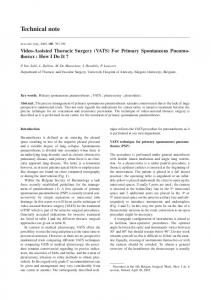
boy (patient 1) with ureteral stents. A, Distal end of internalized stent is within posterior urethra (arrowheads). Also seen is second externalized stent with tip near ...
Technical A Simplified Method for Migrated Ureteral Stents
Repositioning
Note
Distally
Brian 0. CoIey1 and Mark J. Hogan
T
he use ofureteral
et al. in 1967
stenting
is typically
[1]. In pediatrics, performed
teral stunt placement involving the distal
which
are rare,
we
disease
report
have devised these stents
oroscopy
suite,
ing room
and cystoscopy.
Patient
ureteral
for stone
two
boys with ureteral stunts in ends were malpositioned in the
posterior urethra. We method for repositioning
Subjects
practice,
by Zims-
and for difficult ureteropelvic Although complications of ure-
before lithotripsy junction repairs. cases
has become
the first description
since
kind
stunts
part of urologic
a standard
obviating
a return
a simple in the flu-
to the operat-
and Methods I
This 5-year-old boy was born prematurely and had a history of diuretic use for bronchopulmonary dysplasia
complicated
sis. After
conservative
by symptomatic management
nephrolithiafailed,
an open
nephrolithotomy was performed at a hospital that did not have pediatric stents. A 7-French ureteral stent, the smallest adult ureteral stent available, was placed. The patient was transferred to our institution for postoperative
care,
where
he complained
of moderately
severe pelvic and perineal pain. An abdominal film revealed the distal end of the ureteral stent to be within
the posterior
urethra
(Fig.
1A). Because
ofthe
patient’s pulmonary status, the urology service sought to avoid another general anesthetic exposure. Received 1 Both
January
Fig. 1-5-year-old boy (patient 1) with ureteral stents. A, Distal end of internalized stent is within posterior urethra (arrowheads). Also seen is second externalized stent with tip near right ureterovesical junction (arrow). B, Guidewire has been placed into urethral catheter (arrow), balloon inflated with contrast material, and stent pushed back into bladder (arrowheads).
The patient was brought
to the interventional
radiol-
sedation was achieved with IV sodium pentobarbital and morphine. A Foley catheter and the glans penis were sterilized and a loogy suite
and conscious
1% lidocaine gel. was injected into the urethra. With gentle traction on the Foley catheter, a 0.035-inch straight guidewire (SF-35-80: Cook,
cal anesthetic,
Bloomington,
IN)
was advanced
under
fluoroscopic
13, 1997; accepted without revision February 14, 1997.
authors: Children’s
Radiological
Institute,
Columbus Children’s
Hospital,
700
Children’s Dr., Columbus,
OH 43205. Address
correspondence
to B. D. Coley.
AJR1997:169:567-568 0361-803X/97/1692-567 ©American Roentgen Ray Society
AJR:169, August 1997
567
Coley
and
Hogan
stents,
Aubert
distal rected
[21 reported
et al.
the stent
external
to the
patient.
edge, no other reports correct distal migration ureteral
to push
the stent.
through
the
amount
tal to the stent,
of contrast
material
was
used
to opacify
and
distend the Foley balloon. The stiffened Foley catheter was advanced, and the stent engaged and pushed back
into
the
bladder
(Fig.
IB)
where
it reformed
and remained in satisfactory position ftr the remainder of the patient’s care. Symptoms of pelvic and perineal pain resolved after stent repositioning.
Patient
2
from
the 4.5-French
stent
after
placement.
A
radiograph obtained during recovery showed the distal end of the stent to be within the prostatic urethra (Fig. 2A). The patient was brought to the interventional
radiology
suite
and
local
preparations
and anesthesia were performed as in patient 1 . No additional sedation was used because the patient was
platz
recovering
568
from
guidewire
(Medi-Tech,
support.
catheter was brought With
back
As into
and the balloon
advancement
before,
the
Foley
the urethra
just
was gently
inflated.
of the Foley
catheter
and
dis-
with
Watertown.
general
a 6-cm MA)
anesthesia.
soft
An
straight
was placed
Am-
end
through
the
PD,
teral splints I 20:840-844
Discussion
in children, they are still useful in certain cases of hydronephrosis [2] and stone disease. While generally well-tolerated by patients,
ureteral
and stent with
uncommon, reported,
many including
erosion, stone proximal migration manipulation either
approaches
transurethral
focus
complications stent fracture,
formation, [4]. Most on
reports
[6, 7] after proximal
of
retrieval
stent migraureter.
and was technique
cystoscopy
in
well-tolerhas
ad-
because
it of
Fetter
In
ureteral
TR,
Wilkerson
indwelling
inserted
JL. Clinical
silicone
cystoscopically.
rubber
ure-
J Uro! 1967;
ureteral
stents. J Urn! 1988:139:37-38 M, Pattnaik PK. Knotted
Kundargi P. Bansal per
end:
a new
complication
in
dwelling ureteral stent. J Uro! 5. Low RK, Kogan BA, Stoller retrieval
double-J
[5, 6] or percutaneous
tion from the bladder into the distal a review of 16 children with double-J
4.
wire
knotting,
stent
cases.
repositioning
2. Aubert D, Rigaud P. Zoupanos G. Double pigtail ureteral stent in pediatric urology. Eur J Pediatr Surg 1993;3:281-283 3. Pollard 5G. Macfarlane R. Symptoms arising from double-J
some may experience urinary tract symptoms and pain with indwelling ureteral stents [3]. been
support
in both
References use of long-term
Although
and ure-
avoids general anesthesia and the expense an operating room and staff.
I. Zimskind
have
stents
This
repeat
stiff
fluoroscopic
puncture
simple
department
over
not
a guidewire
the stiffening
by the patients.
vantages
catheter
was
wire,
the stent was engaged and pushed into the bladder where it reformed normally (Fig. 2B).
Ureteral stents are common in adult urologic practice. Although less frequently used
This 5-year-old boy had a duplication ofthe left renal collecting system and obstructive hydronephrosis of the lower pole moiety. He underwent pyeloplasty and ureteral stenting via cystoscopy, with the urologist reporting difficulty removing the wire
stiffening
to of
the stent in
(with
catheter
allowed
the radiology to
of
knowl-
Foley
Placing
catheter
to reposition
method
the catheter
provided
injury)
ated provide
Foley
to avoid
required
control
with
enough
thral
under fluoroscopic
portion
To our
to reposition
the soft
guidance
the Foley catheter
of cor-
describe techniques or malpositioning
However,
alone.
control to the tip of the catheter. The balloon was deflated and the catheter withdrawn just distal to the ureteral stent within the posterior urethra. A small
attempt
I was made
patient
Our
girl,
stents.
Our first
Fig. 2-5-year-old boy (patient 2) with ureteral stent A, Postoperative radiograph shows ureteral stent within posterior urethra (arrow). B, After manipulation with urethral catheter, stent has reformed and is normally positioned (arrow).
one case
migration in a 1-month-old by excision of the exposed
of a proximally
ureteral
stent.
the
use
upan
in-
1994;l5l:995-996
ML. migrated
J Uro!
of
Intraluminal pediatric
1995; 154:223-224
6. Slaton JW, Kropp KA. Proximal ureteral stent migration: an unavoidable complication? J Urn! 1996; 155:58-61 7. LeRoy AJ, Williams HJ Jr. Segura JW, Patterson DE. Benson RC Jr. Indwelling ureteral stents: percutaneous management of complications. Radio!ogv
1986;
158:219-222
AJR:169, August 1997


![technical note - USDA [PDF]](https://m.moam.info/img/260x300/technical-note-usda-pdf_647a4d52098a9eae448b4600.jpg)


