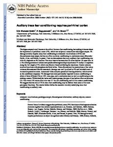
Behavioral Neuroscience 2004, Vol. 118, No. 2, 395– 402
Copyright 2004 by the American Psychological Association 0735-7044/04/$12.00 DOI: 10.1037/0735-7044.118.2.395
Temporal Encoding in Fear Conditioning Revealed Through Associative Reflex Facilitation Derick H. Lindquist and Thomas H. Brown Yale University Temporal encoding in Pavlovian fear conditioning was examined through conditional facilitation of the short-latency (R1) component of the rat eyeblink reflex. Rats were fear-conditioned to a tone conditional stimulus (CS) with either a 3- or 9-s interstimulus interval (ISI) between CS onset and the onset of the grid-shock unconditional stimulus (US). R1 facilitation was tested over 2 days, in counterbalanced order, at a latency of 3 s and 9 s from CS onset. CS-produced R1 facilitation, the conditional response (CR), was 3– 4 times larger when the test latency equaled the conditioning ISI. These results, coupled with the known neurophysiology of R1 facilitation, suggest that this CR could disclose differences in the time course of CS-generated output from the amygdala when driven by cortical versus subcortical CS–CR pathways.
develop in the cerebellum— one in the cerebellar cortex and the second in the deep nuclei (Garcia & Mauk, 1998; Medina et al., 2002; Perrett & Mauk, 1995; Steinmetz, 2000; Thompson, 1986, 1990, 1991). The two engrams are theorized to encode time differently (Mauk & Donegan, 1997; Ohyama et al., 2003). Relatively little is known about ISI encoding in the amygdaladependent memory system, where much of the research has focused on aversive conditioning in rats. Common defensive CRs, such as freezing (Blanchard & Blanchard, 1969; Fanselow, 1997), show no evidence of being sharply timed to correspond to the expected arrival of the US (Choi & Brown, 2003). The motor circuits that control freezing may be triggered by output from the amygdala (Choi & Brown, 2003; LeDoux, Iwata, Cicchetti, & Reis, 1988), but they clearly remain active long after the expected time of the US. There would appear to be no adaptive utility for freezing to occur just before an expected US. To understand the precision with which the amygdala-dependent memory system can encode the ISI requires a punctate CR that is neurophysiologically and biomechanically simple and well understood. For this purpose, we used a modern version of the original conditional reflex facilitation paradigm, which was developed half a century ago by J. S. Brown, Kalish, and Farber (1951). Whereas Brown and coworkers examined CS-produced facilitation of the whole-body, acoustic startle response, we switched to the shortlatency (R1) electromyographic (EMG) component of the rat eyeblink reflex. The R1 response is recorded in the orbicularis oculi (OO) muscle, which is responsible for the active downward force during a reflex blink (Evinger, Manning, & Sibony, 1991; Manning & Evinger, 1986), and is elicited by direct electrical stimulation of the supraorbital (SO) branch of the fifth cranial nerve. The trisynaptic R1 reflex is much simpler to interpret and is far better understood across species, including humans, than the whole-body startle reaction to an aversively loud noise (Canli & Brown, 1996; Choi, Lindquist, & Brown, 2001b; Evinger, Shaw, Peck, Manning, & Baker, 1984; Lam, Wong, Canli, & Brown, 1996; Powers, Schicatano, Basso, & Evinger, 1997; Trigo, Gruart, & Delgado-Garcia, 1999).
Recognition that there are multiple memory systems (T. H. Brown et al., 2003; Kim & Baxter, 2001; Klein, Cosmides, Tooby, & Chance, 2002; Poldrack & Packard, 2003; Stanton, 2000; White & McDonald, 2002) has caused increasing interest in understanding similarities and differences among them (Medina, Christopher Repa, Mauk, & LeDoux, 2002; Thompson & Kim, 1996). The two best understood memory systems are those associated with the cerebellum (Lavond, Kim, & Thompson, 1993; Thompson & Krupa, 1994) and the amygdala (Fanselow & LeDoux, 1999; Gallagher & Holland, 1994; LeDoux, 2000; Maren, 2001; Rogan & LeDoux, 1996). Of these, cerebellum-dependent conditioning is much better understood in the time domain, which is essential for creating an adequate neurophysiological theory of learning and memory (Ivry, Spencer, Zelaznik, & Diedrichsen, 2002; Mauk & Donegan, 1997; Ohyama, Nores, Murphy, & Mauk, 2003; Yarom & Cohen, 2002). In terms of timing, the most extensively analyzed, cerebellumdependent conditional responses (CRs) are the rabbit eyeblink and nictitating membrane responses (Kehoe, Graham-Clarke, & Schreurs, 1989; Lavond et al., 1993; Ohyama et al., 2003). The timing of these cerebellum-dependent CRs demonstrates that this memory system encodes the interstimulus interval (ISI) between the conditional stimulus (CS) and the unconditional stimulus (US). In particular, CR production is timed to anticipate the US (Atwell, Ivarsson, Millar, & Yeo, 2002; T. H. Brown et al., 2003; Mauk & Ruiz, 1992; Medina, Garcia, Nores, Taylor, & Mauk, 2000; Millenson, Kehoe, & Gormezano, 1977; Moore & Choi, 1997). One hypothesis proposes that dual CS–CR connections, or “engrams,”
Derick H. Lindquist and Thomas H. Brown, Department of Psychology, Yale University. This research was supported by National Institutes of Health Grant MH58405. Predoctoral support to Derick H. Lindquist was furnished by National Research Service Award Grant MH64331. We thank Timothy Allen and Sharon Furtak for useful comments on the manuscript. Correspondence concerning this article should be addressed to Thomas H. Brown, Department of Psychology, Yale University, 2 Hillhouse Avenue, New Haven, CT 06520. E-mail:
[email protected] 395
LINDQUIST AND BROWN
396
The R1 component of the eyeblink reflex is cerebellum independent, unlike the longer latency R2 component, which is also sometimes seen in EMGs elicited in the OO muscle by SO stimulation (Evinger et al., 1991; Tamai, Iwamoto, & Tsujimoto, 1982). Conditional R1 facilitation is completely blocked by lesions of the central nucleus of the amygdala (Choi et al., 2001b) and is highly correlated with three other fear-related CRs—freezing, 22kHz ultrasonic vocalization, and defecation (Lindquist & Brown, 2004). All four of these CRs are similarly affected by manipulations of amygdalar function (Lindquist & Brown, 2004). The SO-elicited R1 reflex does not rapidly habituate or easily fatigue (Basso, Strecker, & Evinger, 1993; Choi et al., 2001b; Lindquist & Brown, 2004), unlike the polysynaptic R2 response (Basso et al., 1993) and acoustic startle reaction (Borszcz, Cranney, & Leaton, 1989; Davis, 1972; Davis & Wagner, 1969; Leaton, Cassella, & Borszcz, 1985; Pilz & Leaton, 1999). Having a stable baseline is critical for studies of CR facilitation because the experimental designs invariably entail repetitive CR testing, if only to achieve reasonable statistical power (see Choi et al., 2001b; Lindquist and Brown, 2004). We discuss the experimental significance of response stability for understanding the neurophysiology of amygdala-dependent CR timing in subcortical versus cortical CS–CR pathways.
Method Subjects Seventy-one experimentally naive male Sprague–Dawley rats (Charles River, Kingston, NY) underwent surgery. Each rat was housed individually with ad-lib access to food and water and kept in a vivarium with a 12-hr light– dark cycle. Rats were handled at least 2 days (3–5 min per day) before surgery. At the time of surgery, subjects weighed between 200 and 260 g. All of the following procedures were in strict compliance with the Yale Animal Resource Center guidelines.
inserted beneath the skin were connected to a five-pin male amphenol connector that was cemented to the skull.
Apparatus The head set of each subject was attached to a commutator (CAY-675– 12; Airflyte, Bayonne, NJ) at the top of the conditioning and testing chambers, of which there were two (modified from Model ENV-001; Med Associates, Fairfield, VT). Both structures had a standard grid floor consisting of parallel steel rods and two Plexiglas walls. The chambers were equipped with an infrared light source and video camera (CB-21; Circuit Specialists, Mesa, AZ) to unobtrusively observe the rat’s behavior. The conditioning chambers were housed in sound-attenuating cubicles (ENV018S and ENV-018XX; MED Associates). A ventilation fan within each cubicle gave a constant background noise of 65 dB. The insides of the cubicles were not illuminated in the visible spectrum. The tone CS (10 s, 4 kHz, 75 dB) was delivered by a speaker mounted on the side of each chamber. The grid-shock US (500 ms, 0.8 mA) was produced by a regulated small-animal shock generator (Coulbourn Instruments, Allentown, PA). The grid current was measured conventionally (Choi et al., 2001b) from the voltage drop across the smaller of two resistors (1 k⍀ and 100 k⍀) connected in series between adjacent grid bars.
Electrical Stimulation and Recording Nerve-cuff stimulation (NCS) and EMG recording methods were as described elsewhere (Canli & Brown, 1996; Choi et al., 2001b; Lam et al., 1996). The NCS was controlled by a programmable pulse generator (Master-8; A.M.P.I., Jerusalem, Israel) and delivered through a constant-current stimulus-isolation unit (Model BSI-2; Bak Electronics, Germantown, MD). The NCS current was adjusted for each rat to a level that consistently produced suitable R1 responses (defined below). Output from both EMG electrodes was connected to a differential AC amplifier (Model 1700; A-M Systems, Carlsborg, WA). The EMG response was amplified (1000⫻) and band-pass filtered (0.3 kHz and 5 kHz; 4-pole Bessel filter, 40 dB/decade) before being digitized at 10 kHz. Presentation of stimuli and sampling and storage of EMG responses were controlled by a multifunction data acquisition system (Datawave Technologies Corp., Longmont, CO).
Surgery Subjects were anesthetized with an intraperitoneal injection of Ketamine (90 mg/kg) and Xylazine (10 mg/kg). The surgical procedures were originally developed by Evinger and coworkers (Manning & Evinger, 1986; Powers et al., 1997). Each rat was implanted with a unilateral nerve cuff and ipsilateral EMG recording electrodes in the OO muscle (Canli & Brown, 1996; Choi et al., 2001b; Lam et al., 1996). The nerve cuff was made of a section of longitudinally split polyethylene tubing (PE 50; ⬃ 2 mm) and two Teflon-coated stainless-steel wires. Teflon was removed from the portion of the wires inside the nerve cuff. During surgery, the SO branch of the fifth nerve was isolated and the nerve cuff was placed around it. Three stainless steel screws were fastened to the skull to anchor a head set. The nerve cuff, EMG electrodes, and a bare ground wire that was
Experimental Design All rats experienced 1 day of habituation, 4 days of fear conditioning, and 2 days of testing (see Table 1). Each subject was habituated and trained in one of two conditioning chambers, randomly assigned, and then switched to the alternate chamber for the 2 days of testing. The chamber shift for testing was intended to reduce the effects of context conditioning, which might otherwise have introduced a ceiling effect (cf. Lee, Choi, Brown, & Kim, 2001). Rats were randomly assigned to one of four groups (see Table 1). Two groups were conditioned with a 3-s ISI, and two were conditioned with a 9-s ISI. On the 2 test days, CS-produced R1 facilitation was measured, in counterbalanced order, at latencies of 3 s and 9 s from the CS onset (see Table 1). The two variables of primary interest were
Table 1 Counterbalanced Testing at Two Interstimulus Intervals (ISIs) in the Two Conditioning Groups Day 1
Days 2–5
Day 6
Day 7
Group
Habituation (Groups I & II)
3-s conditioning ISI
3-s testing ISI 9-s testing ISI
9-s testing ISI 3-s testing ISI
I (n ⫽ 11) II (n ⫽ 11)
Habituation (Groups III & IV)
9-s conditioning ISI
3-s testing ISI 9-s testing ISI
9-s testing ISI 3-s testing ISI
III (n ⫽ 12) IV (n ⫽ 12)
TEMPORAL ENCODING AND REFLEX FACILITATION conditioning ISI (3 s or 9 s) and test latency (3 s or 9 s). Counterbalancing across days introduced two additional variables (test day and group), which were also evaluated where appropriate. The between-subjects variable (group) determined the conditioning ISI (3 s or 9 s; see Table 1). The within-subjects variable (test day) determined the latency (3 s or 9 s) from CS onset at which the R1 reflex was elicited (see Table 1). Regardless of the group or day, the CS used for testing was always the same 10-s tone that was used for conditioning.
Habituation, Conditioning, and Testing Seven to 10 days after surgery, subjects were habituated for 40 min in one of the two conditioning chambers (Day 1 in Table 1). Each chamber was cleaned with a vinegar/water (1:3) solution or Windex prior to the rat being placed in the chamber. The R1 response quality was evaluated at the end of the habituation session. The R1 response was deemed satisfactory for further use if the stimulus artifact (SA in Figure 1) produced by NCS did not contaminate (temporally overlap) the R1 response and the R1 onset latency was 4 –10 ms from the SA onset. Any subject that did not meet both requirements was dropped from the study. The first of 4 days of conditioning began the day after habituation (Days 2–5 in Table 1). The quality of the R1 response was reevaluated immediately before each day of conditioning and the NCS intensity was adjusted as needed. Each of the 4 days of conditioning consisted of 10 pairings of the 10.0-s tone CS and the 0.5-s US. The intertrial interval (ITI) was chosen
397
from a preselected uniform distribution (30-s bins) ranging from 180 to 300 s (mean of 240 s). After 4 days of conditioning, each rat received 2 days of testing in the shifted context. In an attempt to reduce the effects of context conditioning on CRs to the cue, the conditioning and testing chambers were made to differ in three respects (cf. Choi et al., 2001b). First, during testing, the grid bars in each chamber were covered with a sheet of Plexiglas angled 5° from horizontal. Second, the two chambers were cleaned with different solutions (a vinegar/ water solution or Windex) immediately before the rat was placed inside. Third, the Plexiglas walls of the two chambers were visually different. Testing consisted of 10 NCS ⫹ tone trials and 10 NCS-alone trials (see Figure 1). On the NCS ⫹ tone trials, the NCS occurred either 3 s or 9 s after tone onset, depending on the conditioning group and the particular test day (Days 6 and 7 of Table 1). The NCS-alone trials occurred between CS presentations. These two trial types were presented in a random order, with the restriction that neither trial type occurred consecutively more than twice in a row. The ITI was the same for conditioning and testing trials. At the end of each session, the rats were removed from the testing chamber and returned to their home cage. Note that NCS was never paired with the grid-shock US during any stage of the experiment.
R1 Response Analysis The EMG data were analyzed offline with BLINK, a custom program written in C (Choi, 2000). BLINK displayed the raw EMG waveform of individual trials; computed the baseline noise level; rectified the EMG waveforms; detected the onset and offset of the R1 component of the EMG within a user-defined time window; and calculated the area under the rectified R1 waveform, an example of which is shown in Figure 1. The integration window (between the arrows in Figure 1) was selected to contain the entire R1 response uncontaminated by the stimulus artifact (SA in Figure 1) or the R2 response (asterisk in Figure 1). The baseline EMG noise level was defined as the root mean square (RMS) of the 10-ms interval prior to the stimulus artifact. As in previous studies (Choi et al., 2001b; Lam et al., 1996), traces with an RMS noise level (calculated on the nonrectified EMG) that exceeded 50 V were discarded (to filter movement artifacts), resulting in a rejection rate of 0.7% (7 trials out of 920). CS-produced changes in the R1 response were first assessed conventionally, on the basis of a percentage of R1 facilitation (F) for each rat, as in previous studies (Choi et al., 2001b; Lam et al., 1996): F ⫽ 兵共⌺ 共NCS ⫹ Tone兲 i/⌺ 共NCS ⫺ Alone兲 i兲 ⫺ 1其 䡠 100%, n
(1)
n
where n is the number of trials (usually 10 in these experiments) over which the R1 areas are summed. Analysis of variance (ANOVA) and contrasts were performed on data based on the F values for each rat. We also measured the difference (D) in R1 areas between NCS-alone trials and NCS ⫹ tone trials for each rat at both test latencies: D ⫽ ⌺ 共NCS ⫹ Tone trials兲 i ⫺ ⌺ 共NCS ⫺ Alone trials兲 i. n
Figure 1. Rectified and then averaged electromyograph (EMG) responses recorded in the orbicularis oculi muscle of the eye and elicited by direct electrical stimulation of the supraorbital (SO) branch of the fifth nerve. Time is measured from the onset of the stimulus artifact (SA) produced by nerve-cuff stimulation (NCS) of SO. The R1 area is computed from the integrals of the rectified EMG waveforms summed across individual trials. The arrows are positioned to include the limits of the R1 responses in these averaged waveforms. The R1 area was compared in the presence and absence of the tone conditional stimulus (CS), respectively, on NSC ⫹ tone trials and NSC-alone trials. Conditional R1 facilitation is the CS-produced increase in R1 area. Facilitation was quantified both as an area ratio converted to a percentage (Equation 1; F) and as an area increase (Equation 2; D). The asterisk marks an R2 response, which is also sometimes observable in the EMGs. R1 ⫽ short-latency, trisynaptic component of eyeblink reflex; R2 ⫽ long-latency, polysynaptic component of the eyeblink reflex.
(2)
n
A repeated-measures t test evaluated the null hypothesis that the theoretical mean difference is zero (D ⫽ 0). Note that the theoretical distribution of D is unbounded, unlike the distribution of F, which has a lower limit of ⫺100% (from which we assume that F ⬎ 0). In other contexts, D might also be more suitable for revealing inhibitory conditioning. There are no previous reports of R1 facilitation based on Equation 2, which has the advantage of being dimensionally connected to the electrophysiology.
Results Subjects A total of 71 rats underwent surgery. Twenty-five rats were dropped from the study as a result of noisy or inconsistent R1 responses, leaving 46 subjects for data analysis.
LINDQUIST AND BROWN
398 NCS Current and NCS-Alone Trials
Temporal Specificity of R1 Facilitation
The mean (⫾ SE) level of NCS current required to elicit a usable R1 response was compared in rats conditioned with a 3-s or 9-s ISI. The NCS current for rats conditioned with a 3-s ISI, averaged across both test days, was 0.80 ⫾ 0.16 mA. The mean NCS current in rats conditioned with a 9-s ISI, again averaged across both test days, was 0.89 ⫾ 0.15 mA. Because the current used to elicit an R1 response in each rat was held constant over both test days, the average current was identical across test days and test latencies. A one-way ANOVA revealed no significant differences in NCS current as a function of conditioning ISI, F(1, 90) ⫽ 0.60, p ⫽ .44. On NCS-alone trials (see Figure 1, bottom trace), the average R1 area was 1,250 ⫾ 190 V䡠ms among rats conditioned with a 3-s ISI and 998 ⫾ 173 V䡠ms among rats conditioned with a 9-s ISI. An ANOVA revealed no significant differences in R1 area as a function of conditioning ISI, F(1, 88) ⫽ 3.80, p ⫽ .054, or test latency, F(1, 88) ⫽ 0.21, p ⫽ .65, and the ISI ⫻ Latency interaction was also insignificant, F(1, 88) ⫽ 0.03, p ⫽ .87.
The overall pattern of R1 facilitation (see Figure 2) immediately suggests some degree of ISI encoding. The asterisks in Figure 2 indicate significant differences in CS-produced R1 facilitation ( p ⬍ .05, two-tailed t tests for independent samples) between the comparison groups. R1 facilitation as an area ratio. Among rats conditioned with a 3-s ISI (Groups I and II; n ⫽ 11 for both groups) R1 facilitation calculated from Equation 1 (a ratio converted to a percentage) was 4-fold greater when tested at a 3-s latency than when tested at a 9-s latency (see Figure 2A). When tested at a 3-s latency, mean R1 facilitation was 87.3 ⫾ 17.9% on Test Day 1 and 86.6 ⫾ 26.4% on Test Day 2 (see Figure 2A). When these same rats were tested at a 9-s latency, R1 facilitation was 16.5 ⫾ 12.2% on Test Day 1 and 24.5 ⫾ 9.2% on Test Day 2. An ANOVA of percent R1 facilitation revealed no significant main effect of test day, F(1, 40) ⫽ 0.04, p ⫽ .84, or group, F(1, 40) ⫽ 0.06, p ⫽ .81, but there was a significant Day ⫻ Group interaction, F(1, 40) ⫽ 14.09, p ⬍ .01.
Figure 2. Associative R1 facilitation as a function of conditioning interstimulus interval (ISI) and test latency from CS onset. R1 facilitation was measured as an area ratio converted to a percentage (Equation 1; Panels A–C) and as an area difference (Equation 2; Panel D). Asterisks denote significant (␣ ⫽ .05 per comparison) group differences (two-tailed t tests for independent samples). A: Two groups of rats were conditioned with a 3-s ISI. On the 1st day of testing, Group I (n ⫽ 11) was tested at a latency of 3 s from the conditional stimulus onset and Group II (n ⫽ 11) was tested at a 9-s latency. The latencies were reversed on the 2nd day of testing. B: Two groups of rats were conditioned with a 9-s ISI. On the 1st day of testing, Group III (n ⫽ 12) was tested at a 3-s latency and Group IV (n ⫽ 12) was tested at a 9-s latency. The latencies were reversed on the 2nd day of testing. C: R1 facilitation, measured as an area ratio converted to a percentage increase, in data pooled across both days of testing (N ⫽ 46). D: R1 facilitation, measured as an area difference, in data pooled across both days of testing (N ⫽ 46). R1 ⫽ short-latency, trisynaptic component of eyeblink reflex.
TEMPORAL ENCODING AND REFLEX FACILITATION
Among rats conditioned with a 9-s ISI (Groups III and IV; n ⫽ 12 for both groups), R1 facilitation was 3 times larger when tested at a 9-s latency than when tested at a 3-s latency (see Figure 2B). When tested at a 9-s latency, the mean R1 facilitation was 108.0 ⫾ 18.0% on Test Day 1 and 84.7 ⫾ 26.2% on Test Day 2. When tested at a 3-s latency, the mean R1 facilitation on Test Days 1 and 2 was, respectively, 24.4 ⫾ 11.4% and 39.9 ⫾ 21.4%. An ANOVA of R1 facilitation revealed no significant effect of test day, F(1, 44) ⫽ 0.04, p ⫽ .85, or group, F(1, 44) ⫽ 0.95, p ⫽ .34, but the Day ⫻ Group interaction was significant, F(1, 44) ⫽ 10.32, p ⬍ .01. The interaction between conditioning ISI and test latency is clearly evident in the pooled data from all four groups (see Figure 2C; N ⫽ 46). In the combined data set, rats conditioned with a 3-s ISI (left two bars in Figure 2C) exhibited 86.9 ⫾ 15.6% R1 facilitation when tested at a 3-s latency and 20.5 ⫾ 7.5% facilitation when tested at a 9-s latency, a 4-fold difference. Rats conditioned with a 9-s ISI (right two bars in Figure 2C) exhibited 96.4 ⫾ 15.7% facilitation when tested at a 9-s latency and 32.2 ⫾ 12.0% when tested at a 3-s latency, a 3-fold difference. Two-way ANOVA revealed no significant main effects for conditioning ISI, F(1, 88) ⫽ 0.64, p ⫽ .43, or test latency, F(1, 88) ⫽ 0.01, p ⫽ .93, but there was a significant ISI ⫻ Latency interaction, F(1, 88) ⫽ 24.36, p ⬍ .01. R1 facilitation as an area difference. The overall pattern of reflex facilitation was the same when this CR was measured as an R1 area difference (Equation 2; see Figure 2D) rather than as an area ratio (Equation 1; see Figure 2C). The shape of the frequency distributions of F (Eq. 1) and D (Eq. 2) turned out to be very similar (data not shown) in spite of the fact that F has a lower bound and D does not. In what follows, we examine the differences in R1 areas between NCS-alone trials and NCS ⫹ tone trials (see the Method section). Among rats conditioned at a 3-s ISI and tested at that same latency, the mean (⫾ SE) increase in R1 area on Test Day 1 was 1,024 ⫾ 282 V䡠ms, corresponding to t(10) ⫽ 3.64, p ⬍ .05. Statistical significance implies that the null hypothesis, of no CS effect (D ⫽ 0), can be rejected (see Method). Among rats conditioned at a 3-s ISI and tested on Day 2 at this same latency, the mean increase in R1 area was 797 ⫾ 159 V䡠ms, corresponding to t(10) ⫽ 5.03, p ⬍ .01. When rats conditioned with a 3-s ISI were tested with a 9-s latency, the mean area increase was 83 ⫾ 80 V䡠ms on Day 1, t(10) ⫽ 1.04, p ⫽ .32, and 229 ⫾ 75 V䡠ms on Day 2, t(10) ⫽ 3.04, p ⬍ .05. When rats conditioned with a 9-s ISI were tested at a 9-s latency, the mean (⫾ SE) increase in R1 area was 858 ⫾ 198 V䡠ms on Day 1, t(11) ⫽ 4.33, p ⬍ .01, and 675 ⫾ 175 V䡠ms on Day 2, t(11) ⫽ 3.85, p ⬍ .01. When these same rats were tested with a 3-s latency on Day 1, the mean area increase was 241 ⫾ 149 V䡠ms, t(11) ⫽ 1.62, p ⫽ .13, and 376 ⫾ 236 V䡠ms on Day 2, t(11) ⫽ 1.59, p ⫽ .14. Figure 2D shows these area changes averaged across test days. The mean R1 area increase was always significantly greater when the test latency equaled the conditioning ISI. Regardless of the test latency, however, the null hypothesis of no CS effect (D ⫽ 0) could be rejected in all four groups (see Method). The temporal generalization gradient appears to span several seconds. Among rats conditioned at a 3-s ISI and tested at this same latency, the overall mean (⫾ SE) increase in R1 area was 911 ⫾ 160 V䡠ms, t(21) ⫽ 5.70, p ⬍ .01. When these same rats were tested with a 9-s
399
latency, the area increase was only 156 ⫾ 56 V䡠ms, but the CS effect remained statistically significant, t(21) ⫽ 2.79, p ⬍ .05. Among rats conditioned with a 9-s ISI and tested at that same latency, the mean area increase was 766 ⫾ 131 V䡠ms, representing a significant CS effect, t(23) ⫽ 5.86, p ⬍ .01. When these same subjects were tested with a 3-s latency, the area increase was reduced to 308 ⫾ 137 V䡠ms, but the CS effect remained statistically significant, t(23) ⫽ 2.25, p ⬍ .05. An ANOVA applied to the pooled area changes revealed no significant effect of conditioning ISI, F(1, 88) ⫽ .001, p ⫽ .98, or test latency, F(1, 88) ⫽ 1.35, p ⫽ .25, but there was a significant ISI ⫻ Latency interaction, F(1, 88) ⫽ 22.47, p ⬍ .01.
Discussion The present investigation was motivated partly by an interest in similarities and differences between the amygdala-dependent memory system and the cerebellum-dependent memory system (cf. T. H. Brown et al., 2003; Medina et al., 2002). Overall, the results show that CS-produced R1 facilitation, an amygdala-dependent CR, like cerebellum-dependent CRs, clearly does show a degree of temporal specificity—meaning, in this case, that conditional responding is greater when the CR is tested at the training ISI. One interesting difference between the two memory systems is the range of ISIs that can support conditioning. Effective ISIs for cerebellum-dependent CRs are limited to approximately 0.5 to 2.5 s, whereas ISIs more than 10 times longer can be effective in supporting amygdala-dependent CRs. Differences in the range of ISIs that can be encoded by these two memory systems are hypothesized to reflect differences in the firing latencies in the critical learning circuits, as explained below.
Demonstration of ISI Encoding Through R1 Facilitation Conditional R1 facilitation—measured either as an R1 area ratio converted to a percentage (Equation 1; see Figure 2C) or as an R1 area change (Equation 2; see Figure 2D)—is greater when the test latency equals the conditioning ISI. The asterisks in Figure 2 indicate significant differences (␣ ⫽ .05 per comparison, twotailed t tests for independent samples) between the indicated comparisons. Overall, CS-produced R1 facilitation was approximately 3 to 4 times larger when the test latency equaled the conditioning ISI. When they were equal, the mean percent R1 facilitation (F, from Equation 1) was 91.5% and the mean increase in R1 area (D, from Equation 2) was 839 V䡠ms. When they were unequal, the mean percent R1 facilitation was reduced to 26.5% and the average increase in R1 area declined to 232 V䡠ms. Although the CS effect was smaller when the conditioning ISI was different from the test latency, the null hypothesis of no CS effect (D ⫽ 0) could be rejected, implying that the temporal generalization gradient is not extremely steep across intervals of several seconds. In absolute terms, the precision of cerebellumdependent CR timing appears to be much greater. However, additional studies of the kind suggested below will be needed to elucidate the actual time course of conditional R1 facilitation when elicited through cortical versus subcortical pathways. Furthermore, a scalar metric (see Gallistel & Gibbon, 2000; Tieu, Keidel, McGann, Faulkner, & Brown, 1999) might offer more insight,
400
LINDQUIST AND BROWN
because the range of effective ISIs is so different for these two memory systems.
Comparison of New and Old CR Facilitation Procedures We decided not to use the original measure of CR facilitation, developed by J. S. Brown et al. (1951), because it suffers from severe interpretational limitations, elaborated in detail elsewhere (Choi et al., 2001b; Leaton & Cranney, 1990; Lindquist & Brown, 2004). Most of the problems associated with this measure are direct or indirect consequences of three facts about the wholebody, acoustic startle response: It rapidly habituates (Davis, 1972; Davis & Wagner, 1969; Leaton et al., 1985); the adequate startle stimulus is sufficiently aversive to support cue and possibly context conditioning during testing (Leaton & Cranney, 1990); and the behavior is neurophysiologically and biomechanically complex and poorly understood (Choi et al., 2001a, 2001b). These caveats notwithstanding, it is worth noting that the only two studies that used the acoustic startle response to evaluate the timing of a fear CR (Davis, Schlesinger, & Sorenson, 1989; Siegel, 1967) both concluded that there was some degree of temporal specificity.
Temporal Specificity of CRs Elicited by Cortical and Subcortical CS Pathways The R1 reflex should be useful for tracking the time course of conditional reflex facilitation with reasonable precision. Unlike the acoustic startle response, the trisynaptic R1 reflex can be repetitively elicited without habituation or fatigue (Basso et al., 1993). The time course of reflex facilitation might be measurable by eliciting a train of R1 responses in the presence and absence of the CS. Such information is pertinent to current hypotheses regarding the content of the acquired representations associated with the two CS pathways to the amygdala. In particular, CS-related neural activity can reach the amygdala via subcortical and cortical routes (Romanski & LeDoux, 1992), both of which are hypothesized to create functional CS–CR connections, or engrams (LeDoux, 2000; Medina et al., 2002). R1 facilitation procedures might afford sufficient temporal resolution to distinguish—when combined with reversible inactivation of the appropriate brain regions— between a single engram with a shallow temporal gradient and the combined effects of one that does encode temporal information and a second that does not. Both possibilities are consistent with the present data. In the case of cerebellum-dependent CR timing, performance has been suggested to reflect the cortical engram (Medina et al., 2002; Perrett & Mauk, 1995).
Neurophysiological Hypothesis for Amygdala-Dependent CR Timing On the basis of computational considerations (Faulkner, Tieu, & Brown, 1997), and for reasons considered below, we have tentatively assumed that amygdala-dependent temporal encoding depends on cortical processing. More specifically, our working hypothesis (Faulkner & Brown, 1999; Faulkner et al., 1997) has been that a critical aspect of the neurophysiological mechanism includes perirhinal–amygdala circuits that contain “late-spiking” neurons. In response to sustained synaptic excitation, these extraordinary neurons can delay firing for several seconds and then maintain
firing to continuing excitation (Beggs, Moyer, McGann, & Brown, 2000; Moyer & Brown, 1998). Neurons with firing latencies this long appear to be uniquely and remarkably abundant in parts of the perirhinal cortex and amygdala (Beggs et al., 2000; Faulkner & Brown, 1999; McGann, Moyer, & Brown, 2001; Moyer & Brown, 1998; Moyer, McNay, & Brown, 2002). Simulations of perirhinal–amygdala circuits (Faulkner et al., 1997; McGann & Brown, 2000; Tieu et al., 1999) show that relatively small networks containing Hebb-type synapses (T. H. Brown, Furtak, & Lindquist, in press; T. H. Brown, Ganong, Kairiss, & Keenan, 1990) and late-spiking neurons can easily encode the ISIs explored in the present experiments. The latter, it should be noted, are outside the range that can support the acquisition of cerebellum-dependent CRs. The enormous difference in neural firing latencies in these two memory circuits furnishes the most obvious neurophysiological and computational explanation for why they function over such different time domains. Both the remarkable abundance of late-spiking neurons in perirhinal cortex and the conspicuously low levels of myelin in this region (Burwell, 2001) are consistent with the supposition that this cortical CS pathway to the amygdala selectively processes information associated with much longer intervals than those that support cerebellum-dependent CRs.
References Atwell, P. J., Ivarsson, M., Millar, L., & Yeo, C. H. (2002). Cerebellar mechanisms in eyeblink conditioning. In S. M. Highstein & W. T. Thach (Eds.), Annals of the New York Academy of Sciences: Vol. 978. The cerebellum: Recent developments in cerebellar research (pp. 79 –92). New York: New York Academy of Sciences. Basso, M. A., Strecker, R. E., & Evinger, C. (1993). Midbrain 6-hydroxydopamine lesions modulate blink reflex excitability. Experimental Brain Research, 94, 88 –96. Beggs, J. M., Moyer, J. R., Jr., McGann, J. P., & Brown, T. H. (2000). Prolonged synaptic integration in perirhinal cortical neurons. Journal of Neurophysiology, 83, 3294 –3298. Blanchard, R. J., & Blanchard, D. C. (1969). Crouching as an index of fear. Journal of Comparative and Physiological Psychology, 67, 370 –375. Borszcz, G. S., Cranney, J., & Leaton, R. N. (1989). Influence of long-term sensitization on long-term habituation of the acoustic startle response in rats: Central gray lesions, preexposure, and extinction. Journal of Experimental Psychology: Animal Behavioral Processes, 15, 54 – 64. Brown, J. S., Kalish, H. I., & Farber, I. E. (1951). Conditioned fear as revealed by magnitude of startle response to an auditory stimulus. Journal of Experimental Psychology, 41, 317–328. Brown, T. H., Byrne, J. H., LaBar, K., LeDoux, J., Lindquist, D. H., Thompson, R. F., & Teyler, T. J. (2003). Learning and memory: Basic mechanisms. In J. H. Byrne & J. L. Roberts (Eds.), From molecules to networks: An introduction to cellular and molecular neuroscience (pp. 499 –574). San Diego, CA: Academic Press. Brown, T. H., Furtak, S., & Lindquist, D. H. (in press). Hebbian synapses. In G. Adelman & B. H. Smith (Eds.), Encyclopedia of neuroscience (3rd ed.). New York: Elsevier Science. Brown, T. H., Ganong, A. H., Kairiss, E. W., & Keenan, C. L. (1990). Hebbian synapses: Biophysical mechanisms and algorithms. Annual Review of Neuroscience, 13, 475–512. Burwell, R. D. (2001). Borders and cytoarchitecture of the perirhinal and postrhinal cortices in the rat. Journal of Comparative Neurology, 437, 17– 41. Canli, T., & Brown, T. H. (1996). Amygdala stimulation enhances the rat eyeblink reflex through a short-latency mechanism. Behavioral Neuroscience, 110, 51–59.
TEMPORAL ENCODING AND REFLEX FACILITATION Choi, J.-S. (2000). BLINK. Unpublished computer software, Department of Psychology, Yale University. Choi, J.-S., & Brown, T. H. (2003). Central amygdala lesions block ultrasonic vocalization and freezing as conditional but not unconditional responses. Journal of Neuroscience, 23, 8713– 8721. Choi, J.-S., Lindquist, D. H., & Brown, T. H. (2001a). Amygdaladependent conditioned enhancement of a simple reflex. Society for Neuroscience Abstracts, 27, 743.4. Choi, J.-S., Lindquist, D. H., & Brown, T. H. (2001b). Amygdala lesions prevent conditioned enhancement of the rat eyeblink reflex. Behavioral Neuroscience, 115, 764 –775. Davis, M. (1972). Differential retention of sensitization and habituation of the startle response in the rat. Journal of Comparative and Physiological Psychology, 78, 260 –267. Davis, M., Schlesinger, L. S., & Sorenson, C. A. (1989). Temporal specificity of fear conditioning: Effects of different conditioned stimulus– unconditioned stimulus intervals on the fear-potentiated startle effect. Journal of Experimental Psychology: Animal Behavior Processes, 15, 295–310. Davis, M., & Wagner, A. R. (1969). Habituation of startle response under incremental sequence of stimulus intensities. Journal of Comparative and Physiological Psychology, 67, 486 – 492. Evinger, C., Manning, K. A., & Sibony, P. A. (1991). Eyelid movements: Mechanisms and normal data. Investigative Ophthalmology & Visual Science, 32, 387– 400. Evinger, C., Shaw, M. D., Peck, C. K., Manning, K. A., & Baker, R. (1984). Blinking and associated eye movements in humans, guinea pigs, and rabbits. Journal of Neurophysiology, 52, 323–339. Fanselow, M. S. (1997). Species-specific defense reactions: Retrospect and prospect. In M. E. Bouton (Ed.), Learning, motivation, and cognition (pp. 321–341). Washington, DC: American Psychological Association. Fanselow, M. S., & LeDoux, J. E. (1999). Why we think plasticity underlying Pavlovian fear conditioning occurs in the basolateral amygdala. Neuron, 23, 229 –232. Faulkner, B., & Brown, T. H. (1999). Morphology and physiology of neurons in the rat perirhinal–lateral amygdala area. Journal of Comparative Neurology, 411, 613– 642. Faulkner, B., Tieu, K. H., & Brown, T. H. (1997). Mechanisms for temporal encoding in fear conditioning. In J. Bower (Ed.), Computational neuroscience (pp. 641– 645). New York: Plenum Press. Gallagher, M., & Holland, P. C. (1994). The amygdala complex: Multiple roles in associative learning and attention. Proceedings of the National Academy of Sciences, USA, 91, 11771–11776. Gallistel, C. R., & Gibbon, J. (2000). Time, rate, and conditioning. Psychological Review, 107, 289 –344. Garcia, K. S., & Mauk, M. D. (1998). Pharmacological analysis of cerebellar contributions to the timing and expression of conditioned eyelid responses. Neuropharmacology, 37, 471– 480. Ivry, R. B., Spencer, R. M., Zelaznik, H. N., & Diedrichsen, J. (2002). The cerebellum and event timing. In S. M. Highstein & W. T. Thach (Eds.), Annals of the New York Academy of Sciences: Vol. 978. The cerebellum: Recent developments in cerebellar research (pp. 302–317). New York: New York Academy of Sciences. Kehoe, E. J., Graham-Clarke, P., & Schreurs, B. G. (1989). Temporal patterns of the rabbit’s nictitating membrane response to compound and component stimuli under mixed CS–US intervals. Behavioral Neuroscience, 103, 283–295. Kim, J. J., & Baxter, M. G. (2001). Multiple brain-memory systems: The whole does not equal the sum of its parts. Trends in Neuroscience, 24, 324 –330. Klein, S. B., Cosmides, L., Tooby, J., & Chance, S. (2002). Decisions and the evolution of memory: Multiple systems, multiple functions. Psychological Review, 109, 306 –329. Lam, Y., Wong, A., Canli, T., & Brown, T. H. (1996). Conditioned
401
enhancement of the early component of the rat eyeblink reflex. Neurobiology of Learning and Memory, 66, 212–220. Lavond, D. G., Kim, J. J., & Thompson, R. F. (1993). Mammalian brain substrates of aversive classical conditioning. Annual Review of Psychology, 44, 317–342. Leaton, R. N., Cassella, J. V., & Borszcz, G. S. (1985). Short-term and long-term habituation of the acoustic startle response in chronic decerebrate rats. Behavioral Neuroscience, 99, 901–912. Leaton, R. N., & Cranney, J. (1990). Potentiation of the acoustic startle response by a conditioned stimulus paired with acoustic startle stimulus in rats. Journal of Experimental Psychology: Animal Behavior Processes, 16, 279 –287. LeDoux, J. E. (2000). Emotion circuits in the brain. Annual Review of Neuroscience, 23, 155–184. LeDoux, J. E., Iwata, J., Cicchetti, P., & Reis, D. J. (1988). Different projections of the central amygdaloid nucleus mediate autonomic and behavioral correlates of conditioned fear. Journal of Neuroscience, 8, 2517–2529. Lee, H. J., Choi, J.-S., Brown, T. H., & Kim, J. J. (2001). Amygdalar N-methyl-D-aspartate (NMDA) receptors are critical for the expression of multiple conditioned fear responses. Journal of Neuroscience, 21, 4116 – 4124. Lindquist, D. H., & Brown, T. H. (2004). Amygdalar NMDA receptors control the expression of reflex facilitation and three other conditional responses. Behavioral Neuroscience, 18, 36 –52. Manning, K. A., & Evinger, C. (1986). Different forms of blinks and their two-stage control. Experimental Brain Research, 64, 579 –588. Maren, S. (2001). Neurobiology of Pavlovian fear conditioning. Annual Review of Neuroscience, 24, 897–931. Mauk, M. D., & Donegan, N. H. (1997). A model of Pavlovian eyelid conditioning based on the synaptic organization of the cerebellum. Learning & Memory, 4, 130 –158. Mauk, M. D., & Ruiz, B. P. (1992). Learning-dependent timing of Pavlovian eyelid responses: Differential conditioning using multiple interstimulus intervals. Behavioral Neuroscience, 106, 666 – 681. McGann, J. P., & Brown, T. H. (2000). Fear conditioning model predicts key temporal aspects of conditioned response production. Psychobiology, 28(3), 303–313. McGann, J. P., Moyer, J. R., Jr., & Brown, T. H. (2001). Predominance of late-spiking neurons in layer VI of rat perirhinal cortex. Journal of Neuroscience, 21, 4969 – 4976. Medina, J. F., Christopher Repa, J., Mauk, M. D., & LeDoux, J. E. (2002). Parallels between cerebellum- and amygdala-dependent conditioning. Nature Reviews Neuroscience, 3, 122–131. Medina, J. F., Garcia, K. S., Nores, W. L., Taylor, N. M., & Mauk, M. D. (2000). Timing mechanisms in the cerebellum: Testing predictions of a large-scale computer simulation. Journal of Neuroscience, 20(14), 5516 –5525. Millenson, J. R., Kehoe, E. J. G., & Gormezano, I. (1977). Classical conditioning of the rabbit’s nictitating membrane response under fixed and mixed CS-US intervals. Learning and Motivation, 8, 351–366. Moore, J. W., & Choi, J.-S. (1997). Conditioned response timing and integration in the cerebellum. Learning & Memory, 4, 116 –129. Moyer, J. R., Jr., & Brown, T. H. (1998). Methods for whole-cell recording from visually preselected neurons of perirhinal cortex in brain slices from young and aging rats. Journal of Neuroscience Methods, 86, 35–54. Moyer, J. R., Jr., McNay, E. C., & Brown, T. H. (2002). Three classes of pyramidal neurons in layer V of rat perirhinal cortex. Hippocampus, 12, 218 –234. Ohyama, T., Nores, W. L., Murphy, M., & Mauk, M. D. (2003). What the cerebellum computes. Trends in Neuroscience, 26, 222–227. Perrett, S. P., & Mauk, M. D. (1995). Extinction of conditioned eyelid
402
LINDQUIST AND BROWN
responses requires the anterior lobe of cerebellar cortex. Journal of Neuroscience, 3(Pt. 1), 2074 –2080. Pilz, P. K., & Leaton, R. N. (1999). Short-term and long-term habituation of the acoustic startle response as a function of stimulus rise time in rats. Psychobiology, 27, 402– 414. Poldrack, R. A., & Packard, M. G. (2003). Competition among multiple memory systems: Converging evidence from animal and human brain studies. Neuropsychologia, 41(3), 245–251. Powers, A. S., Schicatano, E. J., Basso, M. A., & Evinger, C. (1997). To blink or not to blink: Inhibition and facilitation of reflex blinks. Experimental Brain Research, 113, 283–290. Rogan, M. T., & LeDoux, J. E. (1996). Emotion: Systems, cells, and synaptic plasticity. Cell, 35, 469 – 475. Romanski, L. M., & LeDoux, J. E. (1992). Equipotentiality of thalamoamygdala and thalamo-cortico-amygdala circuits in auditory fear conditioning. Journal of Neuroscience, 12, 4501– 4509. Siegel, A. (1967). Stimulus generalization of a classically conditioned response along a temporal dimension. Journal of Comparative and Physiological Psychology, 64, 461– 466. Stanton, M. E. (2000). Multiple memory systems, development and conditioning. Behavioural Brain Research, 110, 25–37. Steinmetz, J. E. (2000). Brain substrates of classical eyeblink conditioning: A highly localized but also distributed system. Behavioural Brain Research, 110, 13–24. Tamai, Y., Iwamoto, M., & Tsujimoto, T. (1982). Reactivated response of blink reflex in the cat. Japan Journal of Physiology, 32, 761–769. Thompson, R. F. (1986, August 29). The neurobiology of learning and memory. Science, 233, 941–947. Thompson, R. F. (1990). Neural mechanisms of classical conditioning in
mammals. Philosophical Transaction of the Royal Society of London, B, Biological Sciences, 329, 161–170. Thompson, R. F. (1991). Are memory traces localized or distributed? Neuropsychologia, 29(6), 571–582. Thompson, R. F., & Kim, J. J. (1996). Memory systems in the brain and localization of a memory. Proceedings of the National Academy of Sciences, USA, 93, 13438 –13444. Thompson, R. F., & Krupa, D. J. (1994). Organization of memory traces in the mammalian brain. Annual Review of Neuroscience, 17, 519 –549. Tieu, K. H., Keidel, A. L., McGann, J. P., Faulkner, B., & Brown, T. H. (1999). Perirhinal-amygdala circuit-level computational model of temporal encoding in fear conditioning. Psychobiology, 27(1), 1–25. Trigo, J. A., Gruart, A., & Delgado-Garcia, J. M. (1999). Discharge profiles of abducens, accessory abducens, and orbicularis oculi motoneurons during reflex and conditioned blinks in alert cats. Journal of Neurophysiology, 81, 1666 –1684. White, N. M., & McDonald, R. J. (2002). Multiple parallel memory systems in the brain of the rat. Neurobiology of Learning and Memory, 77, 125–184. Yarom, Y., & Cohen, D. (2002). The olivocerebellar system as a generator of temporal patterns. In S. M. Highstein & W. T. Thach (Eds.), Annals of the New York Academy of Sciences: Vol. 978. The cerebellum: Recent developments in cerebellar research (pp. 122–34). New York: New York Academy of Sciences.
Received April 25, 2003 Revision received August 11, 2003 Accepted August 13, 2003 䡲