Uncorrected Version. Published on June 29, 2009 as DOI:10.1189/jlb.0908548
Meeting Article
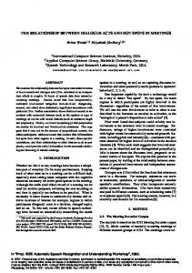
The alarmin HMGB1 acts in synergy with endogenous and exogenous danger signals to promote inflammation ¨ stberg,†,1 Heidi Wa Hulda Sigridur Hreggvidsdottir,*,1 Therese O ¨ha ¨maa,* Hanna Schierbeck,† † † Ann-Charlotte Aveberger, Lena Klevenvall, Karin Palmblad,† Lars Ottosson,† Ulf Andersson,† and Helena Erlandsson Harris*,2 *Department of Medicine, Rheumatology Research Unit, Karolinska Hospital, and †Woman and Child Health, Pediatric Unit, Astrid Lindgren Children’s Hospital, Karolinska Institutet, Stockholm, Sweden RECEIVED SEPTEMBER 12, 2008; REVISED MARCH 30, 2009; ACCEPTED APRIL 12, 2009. DOI: 10.1189/JLB.0908548
ABSTRACT The nuclear protein HMGB1 has previously been demonstrated to act as an alarmin and to promote inflammation upon extracellular release, yet its mode of action is still not well defined. Access to highly purified HMGB1 preparations from prokaryotic and eukaryotic sources enabled studies of activation of human PBMC or synovial fibroblast cultures in response to HMGB1 alone or after binding to cofactors. HMGB1 on its own could not induce detectable IL-6 production. However, strong enhancing effects on induction of proinflammatory cytokine production occurred when the protein associated with each of the separate proinflammatory molecules, rhIL-1, the TLR4 ligand LPS, the TLR9 ligand CpG-ODN, or the TLR1-TLR2 ligand Pam3CSK4. The bioactivities were recorded in cocultures with preformed HMGB1 complexes but not after sequential or simultaneous addition of HMGB1 and the individual ligands. Individual A-box and B-box domains of HMGB1 had the ability to bind LPS and enhance IL-6 production. Heat denaturation of HMGB1 eliminated this enhancement. Cocultures with HMGB1 and other proinflammatory molecules such as TNF, RANKL, or IL-18 did not induce enhancement. HMGB1 thus acts broadly with many but not all immunostimulatory molecules to amplify their activity in a synergistic manner. J. Leukoc. Biol. 86: 000 – 000; 2009.
Introduction The initial trigger for an inflammatory response is conveyed by DAMPs. These can be of endogenous origin released from stressed or injured cells or of exogenous origin and
Abbreviations: CBA⫽cytometric bead array, DAMP⫽danger-associated molecular pattern, DC⫽dendritic cell, h⫽human, HMGB1⫽high mobility group box protein 1, OA⫽osteoarthritis, ODN⫽oligodeoxynucleotide, Pam3CSK4⫽palmitoyl-3-cysteine-serine-lysine-4, Poly I:C⫽polyinosinic: polycytidylic acid, RA⫽rheumatoid arthritis, RAGE⫽receptor for advanced glycated end products, RANKL⫽receptor activator of NF-B ligand, tHMGB1⫽thymus HMGB1
0741-5400/09/0086-0001 © Society for Leukocyte Biology
generally derived from microbial organisms. Exogenous DAMPs have been studied extensively during the last two decades, and DAMP-recognizing receptors include TLRs. Less is known about endogenous DAMPs, also denoted alarmins, regarding molecular patterns, mechanisms of release, or receptor interactions [1]. HMGB1 was discovered initially as a nuclear protein and studied with respect to its interaction with DNA [2]. During necrotic cell death, HMGB1 leaks out from cells, while in contrast, the main part of HMGB1 remains tightly bound to DNA during apoptotic processes and is hence released in lesser amounts from apoptotic cells [3]. In addition to passive release, HMGB1 can be actively translocated and secreted upon stimulation of certain cell types (reviewed in ref. [4]). Extracellular HMGB1 has been ascribed multiple inflammatory functions using in vivo systems. In vitro, HMGB1 has been reported to induce the production of proinflammatory cytokines to up-regulate adhesion molecules on endothelial cells and to activate neutrophils [2, 5–7]. Corroborating these proinflammatory properties are reports demonstrating the beneficial effects of HMGB1-specific therapy in experimental models of inflammation [8 –10]. Based on its proinflammatory properties, its release from necrotic and apoptotic cells, and its active secretion from macrophages, monocytes, and DCs, HMGB1 is regarded as an alarmin. Thus, on one hand, extracellular HMGB1 has been demonstrated to possess proinflammatory activity, conversely, extracellular HMGB1 is also known to induce cell migration and cellular differentiation in noninflammatory settings. HMGB1 (initially named amphoterin), secreted from neurons, promotes neurite outgrowth, and mouse erythroleukemia cells secrete HMGB1 and differentiate in response to HMGB1 [11, 12]. In these examples, as well as in other de1. These authors contibuted equally to this work. 2. Correspondence: Department of Medicine, Rheumatology Research Unit, CMM L8:04, Karolinska Hospital, Karolinska Institutet, S-171 76 Stockholm, Sweden. E-mail:
[email protected]
Volume 86, September 2009
Journal of Leukocyte Biology
Copyright 2009 by The Society for Leukocyte Biology.
1
scribed settings, no inflammatory reactions are evident. Extracellular HMGB1 has also been demonstrated to promote healing by inducing cell migration and differentiation without inducing inflammation-enhancing activities. Intramural injection of HMGB1 in a model of heart muscle ischemia induced tissue regeneration through activation of stem cells [13]. How these separate functions can be performed by one protein has been a focus of intense interest lately. Inconsistent results, depending on the origin of the HMGB1 used, have also raised the question of whether HMGB1 is truly a proinflammatory molecule or whether other molecules potentially present in the HMGB1 preparations have affected the results. In addition, different research groups have used different experimental set-ups and doses of HMGB1 in their studies, rendering data comparisons difficult. HMGB1 consists of a long stretch of C-terminally located, negatively charged amino acids, whereas in the N-terminal region, the molecule contains multiple interspersed, positively charged lysine residues. HMGB1 also contains two DNA binding regions (the A-box and the B-box) and an uneven number of cysteine residues. In addition to its wellstudied DNA-binding properties, HMGB1 can bind lipid molecules such as phosphatidylserine [14] and sulfatide lipids [15] and should, theoretically, be able to react with other proteins via its odd cysteine. Taken together, the biochemical properties of HMGB1 should allow for interactions with multiple molecules, which could potentially affect its bioactivity. In support of this theory, HMGB1 has been reported to bind to synthetic CpG-ODN and thereby enhance TLR9-dependent cytokine production [16]. Extracellular HMGB1 was demonstrated to accelerate the delivery of CpG-ODN to endosomal TLR9, thereby regulating the activity of CpG oligonucleotides [17]. The group of Youn and co-workers [18] demonstrated that HMGB1 bound to LPS facilitates the transfer of LPS to CD14 and enhances LPSmediated TNF-␣ production in monocytes. HMGB1 has also been reported to interact with IL-1, which resulted in acquisition of proinflammatory activity in an inert preparation of HMGB1 [19]. Finally, it was reported recently that HMGB1-nucleosome complexes can mediate proinflammatory activity that neither HMGB1 nor nucleosomes can achieve alone [20]. The aim of our study was to expand the knowledge reported previously and to test our hypothesis that HMGB1 is an evolutionary conserved molecule that is used as an enhancer of exogenous and endogenous DAMPs to promote proinflammatory activities. To function as an enhancer, HMGB1 needs to associate with minute amounts of DAMP molecules, whereas on its own, HMGB1 does not possess proinflammatory activity. We thus set out to simultaneously compare the proinflammatory activities of highly purified HMGB1 obtained from different sources, alone or complexed in parallel experiments with LPS (TLR4 ligand), CpG-ODN (TLR9 ligand), Pam3CSK4 (TLR1/2 ligand), or IL-1. 2 Journal of Leukocyte Biology
Volume 86, September 2009
MATERIALS AND METHODS
HMGB1 and cofactors used Rat rHMGB1(a) with a calmodulin-binding protein tag was expressed in Escherichia coli strain BL21 (for sequence, see ref. [21]). The proteins were purified first with ion exchange chromatography (MonoS 5/50 GL column, GE Healthcare, Chalfont St. Giles, UK) and then run through a calmodulin affinity column (Calmodulin Sepharose 4B, GE Healthcare). Protein was stored at – 80°C in a buffer containing 20 mM 3-(Nmorpholino)propanesulfonic acid, 400 mM NaCl, 20 mM EGTA, and 10 mM DTT at pH 8.0. Endotoxin was removed by filtering through Acodisc units with Mustang E membrane (0.25 m, Pall Life Sciences, East Hills, NY, USA), yielding endotoxin levels below 0.03 EU/g protein, as measured using the Limulus assay. Endotoxin- and tag-free rat rHMGB1(b) as well as endotoxin-free Abox (aa 2–79) and B-box (aa 89 –163) were obtained from HMGBiotech (Milan, Italy). Native HMGB1 purified from bovine tHMGB1, as described previously [22], was a kind gift from Michael Bustin (NCI, Bethesda, MD, USA). rHMGB1, produced in the baculovirus system [rHMGB1(c)], as described previously [23], was a kind gift from Heikki Rauvala (University of Helsinki, Helsinki, Finland). All HMGB1 proteins were stored at – 80°C until day of use. LPS extracted from E. coli 055:B5 was purchased from Sigma-Aldrich (St. Louis, MO, USA) and rhIL-1 was from R&D Systems (Minneapolis, MN, USA). CpG-ODN of type B (ODN2006), Pam3CSK4, and biotinylated LPS from E. coli 0111:B4 were all purchased from InvivoGen (San Diego, CA, USA). DMEM, OPTIMEM, PBS, penicillin, streptomycin, Trypsin-EDTA, and Hepes were obtained from Invitrogen (Carlsbad, CA, USA).
Preparation of HMGB1 complexes HMGB1 in PBS was mixed together with LPS/Pam3CSK4/CpG-ODN/ IL-1 in different ratios to give indicated final concentrations in cell cultures. The mixtures were incubated at 4°C for 16 h before addition to cell cultures, apart from complexes used in the experiment (see Fig. 2B), where HMGB1 was incubated together with LPS for various time periods. To denature formed complexes, the mixture was boiled for 5 min in a water bath and then cooled before addition to cell cultures.
In vitro cell cultures PBMCs were purified using Ficoll centrifugation (Ficoll-Paque Plus, GE Healthcare) from the blood of healthy donors drawn into sodium heparin tubes. Cells were plated at 1–3 ⫻ 105 cells/well in a 96-well plate in OPTIMEM, supplemented with 100 U/ml penicillin and 100 g/ml streptomycin and cultured at 37°C in 5% CO2. Cell cultures were stimulated with HMGB1, cofactors, or HMGB1-cofactor complexes (prepared as described above) at the final concentrations indicated in each figure. Supernatants were harvested after 20 or 40 h (indicated in each figure) and stored at –20°C until analysis. Synovial fibroblasts from four RA and five OA patients (Asterand, Detroit, MI, USA) were cultured in DMEM, supplemented with 10% heatinactivated FCS (PAA Laboratories, Pasching, Austria), 100 U/ml penicillin, 100 g/ml streptomycin, and 10 mM Hepes at 37°C in 5% CO2. Cells were grown to confluence, trypsinized with Trypsin-EDTA, and washed once with medium. Cell viability was assessed in every experimental setup and was determined to be 95–100% using Trypan blue exclusion (Merck, Rahway, NJ, USA). Cells were suspended in complete DMEM and plated at 4 ⫻ 104 cells/well in a 12-well plate and allowed to rest for 15–17 h. Medium was discarded, and cells were washed twice with OPTIMEM, supplemented with 100 U/ml penicillin and 100 g/ml streptomycin and stimulated as indicated with HMGB1, IL-1, or HMGB1-IL-1 complexes. Supernatants were collected after 24 h of stimulation and stored at –20°C until analysis.
www.jleukbio.org
Hreggvidsdottir et al. HMGB1 promotes inflammation through complex formation
Cytokine analysis of supernatants IL-6 levels were measured in supernatants from PBMC and fibroblast cultures by ELISA using hIL-6 ELISA DuoSet威 kits (R&D Systems), according to the manufacturer’s instructions. Flow CBA from BD Biosciences (PharMingen, San Diego, CA, USA) was used to analyze IL-12, IL-10, TNF-␣, IL-1, and IL-6 levels in supernatants from PBMC cultures.
Pull-down assay for HMGB1-LPS complexes Biotinylated LPS (10 g) was incubated with 30 l streptavidin sepharose high-performance beads (GE Healthcare) for 3 h at 4°C. rHMGB1(a) (3 g) were added after a washing step and incubated overnight at 4°C. After three washes, the beads were boiled with sample buffer containing SDS and -ME. Solutions were centrifuged, and the supernatants were separated in a 12.5% SDS-polyacrylamide gel (BioRad, Hercules, CA, USA). The gel was stained with Coomassie brilliant blue for protein visualization.
Statistical analysis Kruskal-Wallis nonparametric ANOVA was used to test statistical significance. All pair-wise comparisons were adjusted by using Dunn’s multiple comparisons test. Data were normalized by denoting the cytokine value from complexed HMGB1 stimulations as 100%. Data were analyzed for statistical significance using the Mann-Whitney U test for independent groups when comparing cytokine production with CBA flow. A P value below 0.05 was considered statistically significant. The computer software program GraphPad Prism, Version 5 for Windows (GraphPad Software, San Diego, CA, USA), was used for all tests.
whether extracellular HMGB1 promotes cytokine induction. Carefully purified tissue-derived or rHMGB1 did not induce any detectable IL-6 release on its own when added to cultures of human PBMCs obtained from healthy blood donors (Fig. 1A). Consistent results were observed in all experiments using natural calf tHMGB1 or rHMGB1 from eukaryotic and prokaryotic sources. A lack of biological activity by HMGB1 could theoretically be caused by protein damage during extensive purification procedures. This explanation was not supported by analysis of PBMC cultures supplemented with the same HMGB1 batches complexed with the TLR4 ligand LPS. All tested batches of purified HMGB1 acted in strong synergy with LPS to induce IL-6 production in PMBC cultures (Fig. 1B). As little as 0.75 ng/ml LPS, associated with 0.5 or 2.0 g/ml HMGB1, induced comparable levels of released IL-6, as did 100 ng/ml LPS alone. A titration of HMGB1 levels required for the enhanced LPS response demonstrates a synergy at and above 62.5 ng/ml HMGB1 (Fig. 1C). To define whether the stimulatory capacity of HMGB1 complexes is an IL-6-specific effect or whether it is a more general, proinflammatory effect, we investigated the induction of TNF-␣, IL-1, IL-6, IL-10, and IL-12 by flow CBA further. The production of TNF-␣, IL-1, IL-6, and IL-10 was enhanced in similar manners by HMGB1-LPS complex stimulation, although much lower levels of IL-10 were recorded than of the other cytokines (P⬍0.05). No IL-12 production could be detected (Fig. 1D).
Synergy depends on formation of HMGB1-LPS complexes
RESULTS
Activity of HMGB1 alone or in complex with LPS Access to highly purified HMGB1 protein from multiple sources enabled us to readdress the important question of
To investigate further whether the proinflammatory activity mediated by HMGB1 was dependent on a direct association of HMGB1 with LPS, we first incubated biotin-labeled LPS
Figure 1. Endotoxin-free HMGB1 does not induce IL-6 production on its own but enhances the cytokine20 20 inducing ability of LPS. Highly puri15 15 fied HMGB1, produced in E. coli [rHMGB1(a), rHMGB1(b)] or eu10 10 karyotic cells [rHMGB1(c)] or puri5 5 fied from thymic tissue (tHMGB1), was used to stimulate freshly isolated 0 0 LPS (ng/ml) – 100 – – – – – – – – LPS (ng/ml) – 100 + + + + + + + + + PBMCs, alone (A) or in a complex HMGB1 (µg/ml) – – 2 0,5 2 0,5 2 0,5 2 0,5 HMGB1 (µg/ml) – – 2 0,5 2 0,5 2 0,5 2 0,5 – with 0.75 ng/ml LPS (B and C). LPS rHMGB1(a)) rHMGB1(b) rHMGB1(c) tHMGB1 rHMGB1(a) HMGB1( ) rHMGB1(b) HMGB1(b) rHMGB1(c) HMGB1( ) tHMGB1 (100 ng/ml) was used as a positive control. IL-6 concentrations were Unstimulated C D # measured in supernatants after 20 h 2 µg/ml rHMGB1(a) 4 50 stimulation. Representative results 2 µg/ml rHMGB1(a), low LPS 30 0,5 µg/ml rHMGB1(a), low LPS from three to five separate experi25 40 Low LPS ments. (A and B) Significant differ3 High LPS 20 ences were evident between the 30 HMGB1-LPS complex stimulation and 2 15 LPS (P⬍0.05) or HMGB1 (P⬍0.005) 20 10 stimulation. (D) Supernatants from 1 culture of PBMCs stimulated for 20 h 10 5 with rHMGB1(a), alone or in com0 0 0 plex with LPS, were analyzed with LPS (ng/ml) – – + + + + + + + + + + + + + 100 IL-12 IL-10 TNF IL-1β IL-6 β flow CBA to measure IL-12, IL-10, rHMGB1(a) – – – TNF-␣, IL-1, and IL-6 levels. Low (ng/ml) LPS (0.75 ng/ml) and high LPS (100 ng/ml); #, above 50 ng/ml. Significant difference between HMGB1-LPS complex stimulation and HMGB1 stimulation could be seen for IL-10, TNF, IL-1, and IL-6 (P⬍0.05) but not IL-12.
B
25
IL-6 (ng/ml)
25
1
2
4
8
16
32
125
62,5
250
500
1000
2000
2000
www.jleukbio.org
(ng g/ml)
(ng/m ml)
IL-6 (ng/ml)
IL-6 (ng/ml)
A
Volume 86, September 2009
Journal of Leukocyte Biology
3
Figure 2. Formation of HMGB1-LPS complexes is important for the synergistic effects. (A) HMGB1 can bind to LPS, as depicted by pull-down assay. Molecular weight marker (I); rHMGB1(a) (II); streptavidin beads, biotinylated LPS, and rHMGB1(a) (III); streptavidin beads and biotinylated LPS (IV); streptavidin beads and rHMGB1(a) (V). (B) PBMC cultures were stimulated for 20 h with HMGB1, LPS, or HMGB1-LPS together, and IL-6 levels were measured in supernatants. HMGB1 and LPS were added directly to PBMC cultures or incubated for 1, 3, 6, 12, or 24 h before addition. (C) PBMC cultures were stimulated for 20 h with HMGB1, A-box and B-box alone or in complexes with low doses of LPS, and IL-6 levels measured in the supernatants. HMGB1 (5000, 1250, and 500 ng/ml) gives equal molar concentrations as 2000, 500, and 200 ng/ml to A-box or B-box, respectively. Representative results from two separate experiments.
and rHMGB1 and performed a pull-down assay (Fig. 2A). rHMGB1 was readily demonstrated to form a complex with LPS, as expected. To address the issue of whether the formation of HMGB1-LPS complexes was critical for the observed biological activity, we added preformed rHMGB1-LPS complexes or rHMGB1 and LPS separately to the PBMC cultures. The results presented in Figure 2B demonstrate that HMGB1 and LPS needed to be physically associated to mediate synergy. Simultaneous addition of uncomplexed HMGB1 and LPS gave low levels of IL-6, and incubation of LPS and HMGB1 for 12 or 24 h, prior to addition to the cell cultures, generated more active complexes than after a shorter incubation period. Heat inactivation of HMGB1-LPS complexes eliminated their capacity to enhance LPS-induced IL-6 formation completely, and heat-treated LPS alone retained activity (data not shown). To further delineate the interaction of HMGB1 with LPS, we assessed the interaction of LPS and the B-box part of the molecule, demonstrated previously as being cytokine-inducing [24], and of LPS and the A-box part of the molecule. Neither A-box nor B-box alone could elicit IL-6 production in PBMCs, whereas A-box and B-box preincubated with lowdose LPS-elicited comparable IL-6 production (Fig. 2C). Thus, the data indicate that HMGB1 has more than one interaction site for LPS.
Heterogeneous in vitro response to HMGB1-LPS complexes among blood donors IL-6 synthesis in PBMC cultures from 12 different healthy blood donors following stimulation with rHMGB1, LPS, or rHMGB1-LPS complexes was investigated to study the pattern of individual responses. A synergistic effect of HMGB1 on LPS-induced IL-6 production was recorded in cultures 4 Journal of Leukocyte Biology
Volume 86, September 2009
from all donors, although the amount of IL-6 released from different donors differed widely, with levels ranging from 100 to 24,000 pg/ml (P⬍0.01; Fig. 3).
HMGB1 acts in synergy with additional TLR ligands The described results with HMGB1-LPS complexes stimulating PBMCs concord with a previous publication using measurement of TNF as a read-out system [18]. That report concluded that the mechanism for this stimulation was based on an ability of HMGB1 to replace LPS-binding pro-
Figure 3. HMGB1-LPS complexes induce synergistic IL-6 production in 12 different donors. PBMCs were isolated from 12 different healthy donors to compare individual responses with HMGB1-LPS complexes. Cell cultures from each donor were stimulated with 0.4 g/ml rHMGB1(a), 0.15 ng/ml LPS, or HMGB1-LPS complex at the same concentrations or were left untreated. Supernatants were collected after 20 h stimulation, and IL-6 levels were measured. Significant differences were evident between HMGB1-LPS complex stimulation and LPS or HMGB1 stimulation (P⬍0.01).
www.jleukbio.org
Hreggvidsdottir et al. HMGB1 promotes inflammation through complex formation
tures (Fig. 4B). CpG-ODN was a less-potent stimulator of IL-6 synthesis than LPS or Pam3CSK4 but was clearly more potent when acting in preformed complexes with rHMGB1. Higher HMGB1 doses were required to generate synergism than needed in complex formations with LPS (P⬍0.05). Importantly, the synergistic effect of HMGB1 with the described TLR ligands was ligand-specific, as preincubation of HMGB1 with the cytokines TNF-␣, RANKL, or IL-18 or the TLR3 ligand Poly I:C did not result in enhanced IL-6 production in human PBMCs upon addition to the cell cultures (data not included).
HMGB1 acts in synergy with IL-1
tein. In the experiments described below, we used highly purified rHMGB1 to study whether the collaboration between HMGB1 and LPS represents a unique feature or if the effect could also occur with other TLR ligands complexed to HMGB1. We thus first used Pam3CSK4, which is a synthetic derivate of triacylated lipoproteins that specifically stimulates the TLR2-TLR1 heterodimer structure. The performance of the synthetic ligand alone to induce IL-6 formation in human PBMCs was compared with the ligand complexed with rHMGB1 (Fig. 4A). IL-6 release was distinctly enhanced after stimulation with HMGB1- Pam3CSK4 complexes in comparison with Pam3CSK4 alone (P⬍0.05). However, the HMGB1 concentration needed for this enhancement was considerably higher than that required for association with LPS. Microbial CpG-DNA or its synthetic analog CpG-ODN are ligands for intracellular TLR9. This interaction may activate DCs, macrophages, or monocytes to produce cytokines. We used class B CpG-ODN, alone or complexed with rHMGB1, to study subsequent IL-6 production in human PBMC cul-
www.jleukbio.org
100 000
RA OA
10 000
IL-6 (log scale)
Figure 4. HMGB1 acts in synergy with Pam3CSK4 and CpG-ODN. PBMC cultures were stimulated with HMGB1 alone, TLR ligand alone [Pam3CSK4 (A) and CpG-B ODN (B)], or HMGB1 complexed with TLR ligand. Supernatants were collected after 20 h (A) or 20 – 40 h (B) of stimulation, and IL-6 levels were measured. Representative results from three to five separate experiments. Significant differences were evident between HMGB1-Pam3CSK4 complex stimulation and Pam3CSK4 or HMGB1 stimulation (P⬍0.05), and HMGB1-CpG-B ODN complex stimulation and HMGB1 stimulation (P⬍0.05).
It has been demonstrated recently that HMGB1 may also develop proinflammatory activity through forming complexes with IL-1, stimulating enhanced cytokine synthesis in cocultures with a murine macrophage cell line [19]. We thus addressed whether these results could be verified using primary cell cultures. However, attempts to activate human PBMCs with IL-1 or preformed HMGB1- IL-1 complexes did not result in detectable IL-6 synthesis in the culture supernatants (data not included). We speculate that the lack of response might be a result of the low IL-1 type I receptor expression on resting human monocytes. Instead, early passages of cultured synovial fibroblasts obtained from intraarticular biopsies from patients with RA or OA were demonstrated to produce IL-6 in response to rIL-1 stimulation at doses equal to or above 0.5 ng/ml (data not included). HMGB1 (100 ng/ml) alone or a lower dose of rIL-1 (0.05 ng/ml) alone, did not stimulate IL-6 release in the cultured fibroblasts, while preformed complexes of low-dose rIL-1 and rHMGB1 induced potent activation (P⬍0.01; Fig. 5). The HMGB1 concentration of 100 ng/ml, sufficient for exhibiting synergy with IL-1, was in the same range as that required for LPS enhancement in PBMC cultures. No difference in the IL-6 response between fibroblasts derived from OA or RA patients could be detected (Fig. 5). A similar re-
1 000 100 10 1
IL-1β (ng/ml)
0,05
–
0,05
–
rHMGB1(a) (ng/ml)
100
100
–
–
Figure 5. HMGB1 enhances the cytokine-inducing ability of IL-1 on synovial fibroblasts, which from four RA and five OA patients, were stimulated with HMGB1 alone, IL-1 alone, or HMGB1 complexed to IL-1. Supernatants were collected after 24 h stimulation, and IL-6 levels were measured. Significant differences were evident between the HMGB1-IL-1 complex stimulation and IL-1 or HMGB1 stimulation (P⬍0.01).
Volume 86, September 2009
Journal of Leukocyte Biology
5
sponse pattern, although much weaker, was detected in cultures of dermal fibroblasts derived from adult individuals (data not shown).
DISCUSSION There are several reports documenting extracellular HMGB1 as a prototypic alarmin, promoting inflammation in sterile as well as microorganism-induced tissue damage (reviewed in refs. [4, 25]). However, the mode of action of HMGB1 is still an unresolved issue. The present study adds essential pieces of information to this important knowledge. HMGB1 may form complexes with different proinflammatory ligands to induce cytokine production. This molecular collaboration results in strong synergy compared with the activity of the ligand alone. The use of highly purified HMGB1 from multiple sources demonstrated that HMGB1 alone did not induce proinflammatory cytokine production. This observation is in agreement with recent publications [16 –19, 26] but differs from our original report that HMGB1 on its own may induce cytokine synthesis [27]. The reason for this discrepancy will be discussed later. Uniform negative cell stimulations with uncomplexed HMGB1 were recorded in our present experiments using a battery of carefully purified HMGB1 preparations. These batches were derived from extraction from primary cells (obtained from M. Bustin) or were produced as recombinant proteins by eukaryotic (obtained from H. Rauvala) or prokaryotic cells (HMGBiotech; our laboratory). When extracellular HMGB1 operated in an environment with selected proinflammatory mediators, it acted as a highly potent enhancer of inflammation. Establishment of cell cultures and preformed complexes with HMGB1 and proinflammatory ligands, including LPS, IL-1, Pam3CSK4, or CpG-ODN, led to a marked, synergistic inflammatory response, in particular, in association with LPS or IL-1. This biological activity required an association of HMGB1 with each of the studied cofactors, as the simultaneous addition of the individual components from the initiation of the cultures did not generate any enhancement. Five recent independent publications emphasize the importance of complex formation with HMGB1 by LPS, CpG-ODNs, IL-1, or nucleosomes to enable enhanced inflammatory responses [16 – 20]. HMGB1 acted selectively to induce synergy through complex formation in our experimental settings, as HMGB1 together with TNF-␣, RANKL, IL-18, or the TLR3 ligand Poly I:C did not augment IL-6 synthesis (data not included). Analogous results were reported recently by Sha and coworkers [19] describing that HMGB1 avidly bound IL-1 but hardly at all associated to TNF-␣ or IFN-␥. Our original report, later confirmed by many other research groups, identifying rHMGB1 as a potent inducer of proinflammatory cytokines, was based on the use of recombinant protein produced in E. coli. It is highly likely that these earlier results were caused by the complex formation described herein between HMGB1 and small amounts of LPS and potentially with other molecules of microbial origin. This property of HMGB1 is an important complication 6 Journal of Leukocyte Biology
Volume 86, September 2009
of the interpretation of experimental results based on the use of rHMGB1 or HMGB1 purified from natural sources. However, it is not a mere issue of whether HMGB1 is “contaminated” in in vitro experiments but rather represents what we believe is a key regulatory function of the protein in vivo. The extracellular presence of the nuclear protein HMGB1 functions to enhance inflammatory responses by binding other coexisting danger molecules. In the in vitro experiments presented, we have used levels of HMGB1 ranging from 1 ng/ml to 30 ug/ml and detected enhancing effects of HMGB1 from 62.5 ng/ml for HMGB1-LPS complexes, whereas enhancing effects by complexes formed with HMGB1 and Pam3CSK4 or CpG-ODN required higher amounts of HMGB1. The levels of HMGB1 used in our in vitro system are all relevant for in vivo systems, as we have recorded microgram levels of HMGB1 in synovial fluid of RA patients [28]. Another important aspect about HMGB1 formation of complexes with other molecules concerns the reduced ability of HMGB1-specific antibodies to function in a consistent manner in therapeutic interventions or in analytical assays. Although highly purified HMGB1 preparations were used in the present in vitro experiments, we must be cautious not to rule out the possibility of the existence of an in vivo version of HMGB1 promoting cytokine production directly without a necessity for complex formation with other molecules. There is a risk that the multiple ex vivo purification steps might modify and impair the function of the protein. PBMCs from all of the 12 tested healthy blood donors produced IL-6 in response to HMGB1-LPS complexes, and only half of the tested donors responded to the low dose of LPS alone when used at the same concentration as in the complexes (Fig. 3). The fact that all tested donors responded to HMGB1 complexes may indicate that the principle of HMGB1 enhancement is of general importance in inflammation biology. The range of IL-6 levels produced following HMGB1-LPS stimulation varied extensively between blood donors. In our experience, the degree of response to HMGB1 challenge can be dependent on interindividual differences in the composition of the PBMC fraction used as well as on intraindividual differences; i.e., PBMC purified from the same donor at different occasions can respond differently, possibly as a result of the immune status of the individual at the time of sampling. The critical question of how the HMGB1-dependent synergy is functionally mediated at a receptor level was not addressed directly in the present study. However, based on the fact that HMGB1 and its cofactors need to be physically associated to be most biologically active, we suggest that the complex may augment inflammation by promoting dual receptor activation, as also indicated in previous studies [16, 29] using CpG-ODNs or CpG-DNA as cofactors. Our present report extends the validity of these observations by demonstrating the functional importance of HMGB1 complex formation with chemically distinctly different molecules including several TLR ligands and a cytokine. The involved receptor collaboration could include RAGE (reviewed by ref. [30]) combined with either of the appropriate TLRs, IL-1R
www.jleukbio.org
Hreggvidsdottir et al. HMGB1 promotes inflammation through complex formation
type I, or as-yet unidentified receptors. There are presently at least two reports supporting the notion that an intimate collaboration between RAGE and other molecules in or on the same cell will enhance inflammation selectively [16, 29]. A novel HMGB1-dependent pathway for inflammatory cell recruitment and activation was identified recently in neutrophils [29]. This activity required a functional, lateral cellsurface interplay between the 2-integrin membrane-activated complex 1 and RAGE. RAGE was the first cellular receptor identified for extracellular HMGB1 bioactivity. Subsequently, TLR2 and TLR4 have been reported to bind HMGB1 and to convey activating signals upon HMGB1 ligation [31–33]. To molecularly define whether it is HMGB1 per se that interacts with these three suggested receptors or if complex formation of HMGB1 with various cofactors is necessary for receptor engagement remains to be elucidated in detail. A potential mechanism of action for the inflammatory properties of HMGB1 could be a requirement for HMGB1 to bind TLR ligands or other inflammation-promoting molecules, such as IL-1, to enhance cytokine production. This scenario would occur in the early phase of an inflammatory response and be mediated by signaling via RAGE and an additional receptor such as TLR4. During the subsequent resolution phase of an inflammatory response that is characterized by regeneration and repair, the tissue levels of microbial products as well as IL-1 will decrease, and HMGB1 levels will remain abundant. HMGB1, which is no longer complexed with cofactors, will then signal primarily via RAGE, resulting in cell migration, proliferation, and differentiation but not cytokine production. Two of the HMGB1 preparations used in our study—HMGB1 obtained from H. Rauvala and from HMGBiotech— have been demonstrated to promote RAGE-dependent cell migration. Neither of these two HMGB1 preparations could induce cytokine production in our assays. This hypothesis about the mode of action of HMGB1 requires further investigations that also take into account that HMGB1 may undergo substantial post-translational modifications such as acetylation, phosphorylation, methylation, and oxidation. The main message of the present study is that HMGB1, associated with certain proinflammatory molecules, mediates biological activities distinctly different from those induced by uncomplexed HMGB1.
ACKNOWLEDGMENTS The study was supported by the regional agreement on medical training and clinical research (ALF) between Stockholm County Council and Karolinska Institutet, Åke Wiberg’s Foundation, Stiftelsen Allma¨nna Barnhuset, the Freemason Lodge Barnhuset in Stockholm, the Swedish Association against Rheumatism, the Swedish Medical Research Council, and King Gustaf V:s Foundation. We are grateful to Michael Bustin for the kind gift of tHMGB1 and to Heikki Rauvala for the kind gift of rHMGB1 produced in baculoviruses. We thank Dr. R. A. Harris for critical reading of the manuscript.
www.jleukbio.org
REFERENCES
1. Matzinger, P. (2001) Introduction to the series. Danger model of immunity. Scand. J. Immunol. 54, 2–3. 2. Bustin, M. (1999) Regulation of DNA-dependent activities by the functional motifs of the high-mobility-group chromosomal proteins. Mol. Cell. Biol. 19, 5237–5246. 3. Scaffidi, P., Misteli, T., Bianchi, M. E. (2002) Release of chromatin protein HMGB1 by necrotic cells triggers inflammation. Nature 418, 191–195. 4. Bianchi, M. E., Manfredi, A. A. (2007) High-mobility group box 1 (HMGB1) protein at the crossroads between innate and adaptive immunity. Immunol. Rev. 220, 35– 46. 5. Fiuza, C., Bustin, M., Talwar, S., Tropea, M., Gerstenberger, E., Shelhamer, J. H., Suffredini, A. F. (2003) Inflammation-promoting activity of HMGB1 on human microvascular endothelial cells. Blood 101, 2652–2660. 6. Park, J. S., Arcaroli, J., Yum, H. K., Yang, H., Wang, H., Yang, K. Y., Choe, K. H., Strassheim, D., Pitts, T. M., Tracey, K. J., Abraham, E. (2003) Activation of gene expression in human neutrophils by high mobility group box 1 protein. Am. J. Physiol. Cell Physiol. 284, C870 – C879. 7. Treutiger, C. J., Mullins, G. E., Johansson, A. S., Rouhiainen, A., Rauvala, H. M., Erlandsson-Harris, H., Andersson, U., Yang, H., Tracey, K. J., Andersson, J., Palmblad, J. E. (2003) High mobility group 1 B-box mediates activation of human endothelium. J. Intern. Med. 254, 375–385. 8. Kokkola, R., Li, J., Sundberg, E., Aveberger, A. C., Palmblad, K., Yang, H., Tracey, K. J., Andersson, U., Harris, H. E. (2003) Successful treatment of collagen-induced arthritis in mice and rats by targeting extracellular high mobility group box chromosomal protein 1 activity. Arthritis Rheum. 48, 2052–2058. 9. Yang, H., Ochani, M., Li, J., Qiang, X., Tanovic, M., Harris, H. E., Susarla, S. M., Ulloa, L., Wang, H., DiRaimo, R., Czura, C. J., Roth, J., Warren, H. S., Fink, M. P., Fenton, M. J., Andersson, U., Tracey, K. J. (2004) Reversing established sepsis with antagonists of endogenous highmobility group box 1. Proc. Natl. Acad. Sci. USA 101, 296 –301. 10. Yang, R., Harada, T., Mollen, K. P., Prince, J. M., Levy, R. M., Englert, J. A., Gallowitsch-Puerta, M., Yang, L., Yang, H., Tracey, K. J., Harbrecht, B. G., Billiar, T. R., Fink, M. P. (2006) Anti-HMGB1 neutralizing antibody ameliorates gut barrier dysfunction and improves survival after hemorrhagic shock. Mol. Med. 12, 105–114. 11. Merenmies, J., Pihlaskari, R., Laitinen, J., Wartiovaara, J., Rauvala, H. (1991) 30-kDa Heparin-binding protein of brain (amphoterin) involved in neurite outgrowth. Amino acid sequence and localization in the filopodia of the advancing plasma membrane. J. Biol. Chem. 266, 16722–16729. 12. Sparatore, B., Passalacqua, M., Patrone, M., Melloni, E., Pontremoli, S. (1996) Extracellular high-mobility group 1 protein is essential for murine erythroleukaemia cell differentiation. Biochem. J. 320, 253–256. 13. Palumbo, R., Sampaolesi, M., De Marchis, F., Tonlorenzi, R., Colombetti, S., Mondino, A., Cossu, G., Bianchi, M. E. (2004) Extracellular HMGB1, a signal of tissue damage, induces mesoangioblast migration and proliferation. J. Cell Biol. 164, 441– 449. 14. Rouhiainen, A., Imai, S., Rauvala, H., Parkkinen, J. (2000) Occurrence of amphoterin (HMG1) as an endogenous protein of human platelets that is exported to the cell surface upon platelet activation. Thromb. Haemost. 84, 1087–1094. 15. Mohan, P. S., Laitinen, J., Merenmies, J., Rauvala, H., Jungalwala, F. B. (1992) Sulfoglycolipids bind to adhesive protein amphoterin (P30) in the nervous system. Biochem. Biophys. Res. Commun. 182, 689 – 696. 16. Tian, J., Avalos, A. M., Mao, S. Y., Chen, B., Senthil, K., Wu, H., Parroche, P., Drabic, S., Golenbock, D., Sirois, C., Hua, J., An, L. L., Audoly, L., La Rosa, G., Bierhaus, A., Naworth, P., Marshak-Rothstein, A., Crow, M. K., Fitzgerald, K. A., Latz, E., Kiener, P. A., Coyle, A. J. (2007) Toll-like receptor 9-dependent activation by DNA-containing immune complexes is mediated by HMGB1 and RAGE. Nat. Immunol. 8, 487– 496. 17. Ivanov, S., Dragoi, A. M., Wang, X., Dallacosta, C., Louten, J., Musco, G., Sitia, G., Yap, G. S., Wan, Y., Biron, C. A., Bianchi, M. E., Wang, H., Chu, W. M. (2007) A novel role for HMGB1 in TLR9-mediated inflammatory responses to CpG-DNA. Blood 110, 1970 –1981. 18. Youn, J. H., Oh, Y. J., Kim, E. S., Choi, J. E., Shin, J. S. (2008) High mobility group box 1 protein binding to lipopolysaccharide facilitates transfer of lipopolysaccharide to CD14 and enhances lipopolysaccharide-mediated TNF-␣ production in human monocytes. J. Immunol. 180, 5067–5074. 19. Sha, Y., Zmijewski, J., Xu, Z., Abraham, E. (2008) HMGB1 develops enhanced proinflammatory activity by binding to cytokines. J. Immunol. 180, 2531–2537. 20. Urbonaviciute, V., Furnrohr, B. G., Meister, S., Munoz, L., Heyder, P., De Marchis, F., Bianchi, M. E., Kirschning, C., Wagner, H., Manfredi, A. A., Kalden, J. R., Schett, G., Rovere-Querini, P., Herrmann, M., Voll, R. E. (2008) Induction of inflammatory and immune responses by HMGB1nucleosome complexes: implications for the pathogenesis of SLE. J. Exp. Med. 205, 3007–3018. 21. Wang, H., Bloom, O., Zhang, M., Vishnubhakat, J. M., Ombrellino, M., Che, J., Frazier, A., Yang, H., Ivanova, S., Borovikova, L., Manogue, K. R., Faist, E., Abraham, E., Andersson, J., Andersson, U., Molina, P. E., Abumrad, N. N., Sama, A., Tracey, K. J. (1999) HMG-1 as a late mediator of endotoxin lethality in mice. Science 285, 248 –251.
Volume 86, September 2009
Journal of Leukocyte Biology
7
22. Bustin, M., Hopkins, R. B., Isenberg, I. (1978) Immunological relatedness of high mobility group chromosomal proteins from calf thymus. J. Biol. Chem. 253, 1694 –1699. 23. Parkkinen, J., Raulo, E., Merenmies, J., Nolo, R., Kajander, E. O., Baumann, M., Rauvala, H. (1993) Amphoterin, the 30-kDa protein in a family of HMG1-type polypeptides. Enhanced expression in transformed cells, leading edge localization, and interactions with plasminogen activation. J. Biol. Chem. 268, 19726 –19738. 24. Li, J., Kokkola, R., Tabibzadeh, S., Yang, R., Ochani, M., Qiang, X., Harris, H. E., Czura, C. J., Wang, H., Ulloa, L., Warren, H. S., Moldawer, L. L., Fink, M. P., Andersson, U., Tracey, K. J., Yang, H. (2003) Structural basis for the proinflammatory cytokine activity of high mobility group box 1. Mol. Med. 9, 37– 45. 25. Klune, J. R., Dhupar, R., Cardinal, J., Billiar, T. R., Tsung, A. (2008) HMGB1: endogenous danger signaling. Mol. Med. 14, 476 – 484. 26. Rouhiainen, A., Tumova, S., Valmu, L., Kalkkinen, N., Rauvala, H. (2007) Pivotal advance: analysis of proinflammatory activity of highly purified eukaryotic recombinant HMGB1 (amphoterin). J. Leukoc. Biol. 81, 49 –58. 27. Andersson, U., Wang, H., Palmblad, K., Aveberger, A. C., Bloom, O., Erlandsson-Harris, H., Janson, A., Kokkola, R., Zhang, M., Yang, H., Tracey, K. J. (2000) High mobility group 1 protein (HMG-1) stimulates proinflammatory cytokine synthesis in human monocytes. J. Exp. Med. 192, 565–570. 28. Kokkola, R., Sundberg, E., Ulfgren, A. K., Palmblad, K., Li, J., Wang, H., Ulloa, L., Yang, H., Yan, X. J., Furie, R., Chiorazzi, N., Tracey, K. J., Andersson, U., Harris, H. E. (2002) High mobility group box chromosomal protein 1: a novel proinflammatory mediator in synovitis. Arthritis Rheum. 46, 2598 –2603. 29. Orlova, V. V., Choi, E. Y., Xie, C., Chavakis, E., Bierhaus, A., Ihanus, E., Ballantyne, C. M., Gahmberg, C. G., Bianchi, M. E., Nawroth, P. P.,
8 Journal of Leukocyte Biology
Volume 86, September 2009
30. 31.
32.
33.
Chavakis, T. (2007) A novel pathway of HMGB1-mediated inflammatory cell recruitment that requires Mac-1-integrin. EMBO J. 26, 1129 – 1139. Rauvala, H., Rouhiainen, A. (2007) RAGE as a receptor of HMGB1 (Amphoterin): roles in health and disease. Curr. Mol. Med. 7, 725– 734. Apetoh, L., Ghiringhelli, F., Tesniere, A., Obeid, M., Ortiz, C., Criollo, A., Mignot, G., Maiuri, M. C., Ullrich, E., Saulnier, P., Yang, H., Amigorena, S., Ryffel, B., Barrat, F. J., Saftig, P., Levi, F., Lidereau, R., Nogues, C., Mira, J. P., Chompret, A., Joulin, V., Clavel-Chapelon, F., Bourhis, J., Andre, F., Delaloge, S., Tursz, T., Kroemer, G., Zitvogel, L. (2007) Toll-like receptor 4-dependent contribution of the immune system to anticancer chemotherapy and radiotherapy. Nat. Med. 13, 1050 –1059. Park, J. S., Gamboni-Robertson, F., He, Q., Svetkauskaite, D., Kim, J. Y., Strassheim, D., Sohn, J. W., Yamada, S., Maruyama, I., Banerjee, A., Ishizaka, A., Abraham, E. (2006) High mobility group box 1 protein interacts with multiple Toll-like receptors. Am. J. Physiol. Cell Physiol. 290, C917–C924. Park, J. S., Svetkauskaite, D., He, Q., Kim, J. Y., Strassheim, D., Ishizaka, A., Abraham, E. (2004) Involvement of Toll-like receptors 2 and 4 in cellular activation by high mobility group box 1 protein. J. Biol. Chem. 279, 7370 –7377.
KEY WORDS: cell activation 䡠 innate cell-mediated immunity
www.jleukbio.org