49-7071-29-73350; Fax: 49-7071-29-3332; E-mail: robert.feil@ uni-tuebingen.de. ..... Ghofrani, H. A., Osterloh, I. H., and Grimminger, F. (2006) Nat. Rev. Drug.
THE JOURNAL OF BIOLOGICAL CHEMISTRY VOL. 284, NO. 1, pp. 556 –562, January 2, 2009 © 2009 by The American Society for Biochemistry and Molecular Biology, Inc. Printed in the U.S.A.
The Commonly Used cGMP-dependent Protein Kinase Type I (cGKI) Inhibitor Rp-8-Br-PET-cGMPS Can Activate cGKI in Vitro and in Intact Cells* Received for publication, August 8, 2008, and in revised form, November 3, 2008 Published, JBC Papers in Press, November 13, 2008, DOI 10.1074/jbc.M806161200
Nadejda Valtcheva‡, Peter Nestorov‡, Alexander Beck§, Michael Russwurm¶, Matthias Hillenbrand‡, Pascal Weinmeister储, and Robert Feil‡1 From the ‡Interfakulta¨res Institut fu¨r Biochemie, Universita¨t Tu¨bingen, 72076 Tu¨bingen, the §Zentrum fu¨r Klinische Massenspektrometrie GmbH, 74076 Heilbronn, the ¶Institut fu¨r Pharmakologie und Toxikologie, Universita¨t Bochum, 44780 Bochum, and the 储Institut fu¨r Pharmakologie und Toxikologie, Technische Universita¨t Mu¨nchen, 80802 Mu¨nchen Germany
cGMP is a cyclic-nucleotide second messenger with multiple targets and functions. Small-molecule modulators of cGMP
* This work was supported by grants from the Deutsche Forschungsgemeinschaft. The costs of publication of this article were defrayed in part by the payment of page charges. This article must therefore be hereby marked “advertisement” in accordance with 18 U.S.C. Section 1734 solely to indicate this fact. 1 To whom correspondence should be addressed: Interfakulta¨res Institut fu¨r Biochemie, Universita¨t Tu¨bingen, Hoppe-Seyler-Str. 4, 72076 Tu¨bingen, Germany. Tel.: 49-7071-29-73350; Fax: 49-7071-29-3332; E-mail: robert.feil@ uni-tuebingen.de.
556 JOURNAL OF BIOLOGICAL CHEMISTRY
generators and effectors are important biochemical tools as well as established and prospective drugs for the treatment of human diseases, such as erectile dysfunction, pulmonary hypertension, and various cardiovascular disorders (1–3). cGMP is generated by nitric oxide- or natriuretic peptide-stimulated guanylyl cyclases and can bind to and modulate the activity of at least three classes of cGMP effector proteins: cyclic nucleotide-hydrolyzing phosphodiesterases (PDEs),2 cyclic nucleotide-gated cation channels, and cGMP-dependent protein kinases (cGKs, also known as protein kinase G or PKG) (4). Based on pharmacological and genetic studies, the cGK type I (cGKI) is considered the major mediator of cGMP signaling in many tissues including the cardiovascular system (5– 8). The mammalian cGKI is a cytosolic Ser/Thr protein kinase comprising an N-terminal regulatory domain with two cGMPbinding sites and a C-terminal catalytic domain. It exists in two isoforms termed cGKI␣ and cGKI. The isozymes have identical cGMP-binding sites and catalytic domains but differ in their N-terminal ⬃100 amino acids, which contribute to homodimerization, sensitivity to cGMP activation, and interaction with anchoring and substrate proteins. Recent in vivo studies with transgenic mice demonstrated that both isoforms can induce smooth muscle relaxation and vasodilation (9), but the respective molecular mechanisms behind these effects are controversial (6, 10). Similarly, opposing effects of cGMP/cGKI signaling have been reported on the growth and phenotype of vascular smooth muscle cells (VSMCs) (11, 12). The inconsistency of the results concerning the function of cGKI might in part be related to unexpected effects of the pharmacological cGKI activators and inhibitors that are commonly used to distinguish between cGKI-dependent and cGKI-independent signaling. For instance, cGMP analogues can bind to multiple 2
The abbreviations used are: PDE, phosphodiesterase; cGKI, cGMP-dependent protein kinase type I; PET, 8-Br-PET-cGMP; Rp-PET, Rp-8-Br-PETcGMPS; 8-Br-PET-cGMP, -phenyl-1,N2-etheno-8-bromoguanosine-3⬘,5⬘cyclic monophosphate; Rp-8-pCPT-cGMPS, 8-(4-chlorophenylthio)guanosine-3⬘, 5⬘-cyclic monophosphorothioate, Rp-isomer; Rp-8-BrPET-cGMPS, -phenyl-1,N2-etheno-8-bromoguanosine-3⬘,5⬘-cyclic monophosphorothioate, Rp-isomer; VASP, vasodilator-stimulated phosphoprotein; VSMC, vascular smooth muscle cell; MTS, 3-(4,5-dimethylthiazol-2-yl)-5-(3-carboxymethoxyphenyl)-2-(4-sulfophenyl)-2H-tetrazolium, inner salt; CFP, cyan fluorescent protein; YFP, yellow fluorescent protein; ESI-MS, electrospray ionization-mass spectrometry; FRET, fluorescence resonance energy transfer; Mes, 2-morpholineethanesulfonic acid.
VOLUME 284 • NUMBER 1 • JANUARY 2, 2009
Downloaded from http://www.jbc.org/ by guest on November 7, 2015
Small-molecule modulators of cGMP signaling are of interest to basic and clinical research. The cGMP-dependent protein kinase type I (cGKI) is presumably a major mediator of cGMP effects, and the cGMP analogue Rp-8-Br-PET-cGMPS (RpPET) (chemical name: -phenyl-1,N2-etheno-8-bromoguanosine-3ⴕ,5ⴕ-cyclic monophosphorothioate, Rp-isomer) is currently considered one of the most permeable, selective, and potent cGKI inhibitors available for intact cell studies. Here, we have evaluated the properties of Rp-PET using cGKI-expressing and cGKI-deficient primary vascular smooth muscle cells (VSMCs), purified cGKI isozymes, and an engineered cGMP sensor protein. cGKI activity in intact VSMCs was monitored by cGMP/cGKI-stimulated cell growth and phosphorylation of vasodilator-stimulated phosphoprotein. Unexpectedly, Rp-PET (100 M) did not efficiently antagonize activation of cGKI by the agonist 8-Br-cGMP (100 M) in intact VSMCs. Moreover, in the absence of 8-Br-cGMP, Rp-PET (100 M) stimulated cell growth in a cGKI␣-dependent manner. Kinase assays with purified cGKI isozymes confirmed the previously reported inhibition of the cGMP-stimulated enzyme by Rp-PET in vitro. However, in the absence of the agonist cGMP, Rp-PET partially activated the cGKI␣ isoform. Experiments with a fluorescence resonance energy transfer-based construct harboring the cGMP binding sites of cGKI suggested that binding of Rp-PET induces a conformational change similar to the agonist cGMP. Together, these findings indicate that Rp-PET is a partial cGKI␣ agonist that under certain conditions stimulates rather than inhibits cGKI activity in vitro and in intact cells. Data obtained with Rp-PET as cGKI inhibitor should be interpreted with caution and not be used as sole evidence to dissect the role of cGKI in signaling processes.
Rp-8-Br-PET-cGMPS Can Activate cGKI
EXPERIMENTAL PROCEDURES Materials— cGMP (catalogue number G 001), 8-Br-cGMP (catalogue number B 004), 8-Br-PET-cGMP (PET; catalogue number P 003), Rp-8-Br-PET-cGMPS (Rp-PET; catalogue number P 007), and Rp-8-pCPT-cGMPS (catalogue number C 013) were purchased from Biolog Life Science Institute. Compounds were dissolved in water, and stock solutions (100 mM for cGMP and 8-Br-cGMP and 10 mM for the other compounds) were stored at ⫺20 °C. Intact Cell Studies (Cell Culture, Growth Assays, Western Blotting, and Antibodies)—Primary VSMCs were obtained from aortae of 3– 6-week-old cGKI-deficient (cGKIL⫺/L⫺) (25) and litter-matched control (cGKI⫹/⫹ or cGKI⫹/L⫺) mice or from aortae of 8-week-old SM-I␣ or SM-I smooth muscle rescue mice (9). Cells were isolated by enzymatic digestion and cultured in Dulbecco’s modified Eagle’s medium supplemented with 10% (v/v) fetal calf serum and penicillin/streptomycin as described previously (26). To determine cell growth, primary VSMCs were plated on 96-well culture plates (20,000 cells/well) in the absence or presence of 8-Br-cGMP and/or Rp-PET. After 72 h, the cell number was determined by the MTS assay (CellTiter 96威 AQueous, Promega) or by staining with toluidine blue O. The MTS assay was performed according to the manufacturer’s protocol. Briefly, cells were washed once with serum-free medium to remove non-adherent cells. Subsequently, 20 l of the MTS reagent were added to 100 l of serum-free medium in each well. The JANUARY 2, 2009 • VOLUME 284 • NUMBER 1
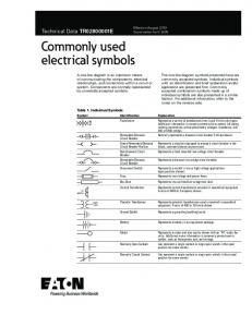
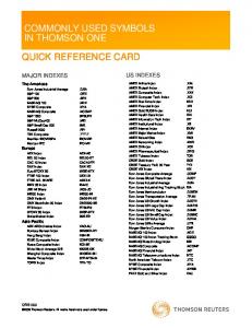
FIGURE 1. Chemical structure of (A) cGMP and (B) the cGMP analogues PET and Rp-PET. PET is a potent agonist of cGKI. Replacement of one of the free oxygen atoms of the phosphate moiety by sulfur in the equatorial position with respect to the sugar ring results in Rp-PET, which is reported to be a competitive inhibitor of cGKI.
A492 was measured after a 30- and 60-min incubation at 37 °C in a humidified, 6% CO2 atmosphere. For toluidine blue O staining, cells were washed once with phosphate-buffered saline and then fixed and stained for 10 min in 100 l of ice-cold toluidine blue O solution (0.5% (w/v) toluidine blue O in phosphate-buffered saline containing 2% (v/v) formaldehyde and 0.2% (v/v) glutaraldehyde). Excess dye was removed by five washes with phosphate-buffered saline. Stained cells were incubated in 100 l of 1% (w/v) SDS to release the dye and the A620 was determined. Phosphorylation of the vasodilator-stimulated phosphoprotein (VASP) was detected by Western blotting via the band shift to a higher apparent molecular weight when VASP is phosphorylated at Ser-157 (8). Primary VSMCs were plated on 6-well culture plates (100,000 cells/well) and grown for 7 days to a confluence of 80 –90%. Subsequently, cells were maintained in serum-free medium for a further 48 h. Then cells were preincubated for 30 min with Rp-PET or vehicle followed by a 30-min incubation in the presence or absence of 8-Br-cGMP. Cells were washed once with phosphate-buffered saline and lysed in lysis buffer (20 mM Tris-HCl, pH 8.0, 0.7% (w/v) SDS, 1.7% (v/v) -mercaptoethanol, 0.2 mM phenylmethylsulfonyl fluoride). Cell lysates were incubated for 5 min at 95 °C and used for SDS-PAGE and Western blot analysis with polyclonal rabbit antibodies against VASP (Alexis Biochemicals, catalogue number Alx-210-725, 1:2000), Akt (Cell Signaling Technology, catalogue number 9272, 1:1000), and cGKI (1:5000). The rabbit polyclonal cGKI antiserum, termed “cGKI common (DH),” detects both cGKI␣ and cGKI. It was raised against recombinant bovine cGKI␣ and affinity-purified using cGKI␣ coupled JOURNAL OF BIOLOGICAL CHEMISTRY
557
Downloaded from http://www.jbc.org/ by guest on November 7, 2015
cGMP receptors and can elevate the cAMP level by inhibiting the cAMP-degrading PDE3 (8, 13, 14). Moreover, there is evidence that one of the most frequently used cGKI inhibitors, KT5823, does not at all inhibit cGKI activity both in vitro and in intact cells and may in fact inhibit other protein kinases (15– 17). Consequently, other classes of cGKI inhibitors are increasingly used for intact cell studies. These inhibitors are based on cell-permeable modified peptide substrates (18) or on the allosteric activator cGMP (see Fig. 1A) (19). Agonistic cGMP analogues have been converted to antagonists by exchanging one oxygen atom of the cyclic phosphate moiety with sulfur in the equatorial position with respect to the sugar ring (20). The resulting antagonistic Rp-phosphorothioate cGMP analogues are supposed to bind to the cGMP-binding sites of cGKI without inducing the conformational change crucial for allosteric activation of the enzyme (21, 22). For instance, the cGMP analogue 8-Br-PET-cGMP (PET) is a cGKI agonist (23), whereas Rp-8-Br-PET-cGMPS (Rp-PET) (see Fig. 1B) competitively inhibits activation of purified cGKI by cGMP (24). Rp-PET is currently considered one of the most permeable, selective, and potent cGKI inhibitors available for intact cell studies (14, 19). In the present study, we intended to validate the selectivity and efficacy of Rp-PET as a cGKI inhibitor in intact cells by comparing its effects on cGKI-expressing and cGKI-deficient VSMCs. Surprisingly, Rp-PET did not efficiently inhibit but rather stimulated cGKI-mediated processes in VSMCs. In vitro experiments with purified cGKI isozymes and an engineered cGKI-based cGMP sensor protein supported these findings, suggesting that Rp-PET is a partial agonist rather than an antagonist of cGKI␣.
Rp-8-Br-PET-cGMPS Can Activate cGKI
558 JOURNAL OF BIOLOGICAL CHEMISTRY
VOLUME 284 • NUMBER 1 • JANUARY 2, 2009
Downloaded from http://www.jbc.org/ by guest on November 7, 2015
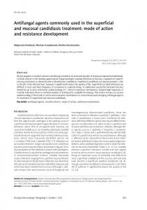
to a final concentration of 0.5 mM. For spiking, PET was added to the Rp-PET aliquot in a 30:70 (v/v) ratio. Solutions were infused (4 l/min) into the ESI source using a Parmer Infusion 74900 series syringe pump (Cole-Parmer Instrument Co.). Mass spectra were acquired in the negative ion mode using a HCT Plus ion trap mass spectrometer equipped with a standard ESI source (Bruker-Daltonics). Spectra (50 –1000 m/z) were acquired in the standard enhanced mode (scan rate 8100 m/z per second). Dry gas (5 liters/min) temperature was set to 300 °C, the nebulizer was set to 10.0 p.s.i., and the electrospray voltage was set to 4000 V. Maximal accumulation FIGURE 2. Effects of Rp-PET on cGKI-mediated processes in intact primary VSMCs. Cell growth (upper time was set to 200 ms. Loading of panels) and VASP phosphorylation (lower panels) were monitored in control (ctr, left panels) and cGKI-deficient (ko, right panels) VSMCs. Cells were incubated with or without 8-Br-cGMP (100 M) and Rp-PET (100 M) as the trap was controlled by the indicated. The cell number was determined after 72 h by the toluidine blue assay and normalized to control instrument (ICC 70000). growth in the absence of drugs. Data of two to five separate cell preparations were pooled and presented as Fluorescence Resonance Energy means ⫾ S.E. The number of wells measured under each condition (n) is indicated in the respective columns. Transfer (FRET) Measurements—A *** indicates p ⬍ 0.001 versus control in the absence of drugs (Student’s t test). The cell number in the presence of 8-Br-cGMP and Rp-PET was not significantly different from 8-Br-cGMP alone (Student’s t test). Similar results fusion protein (cGi-500, Ref. 30) were obtained by the MTS assay (data not shown). Phosphorylation of VASP was measured after a 30-min preincubation with or without Rp-PET followed by 30 min with or without 8-Br-cGMP. Phospho-VASP (p-VASP) consisting of the tandem cGMPwas monitored by immunodetection of the band shift to a higher apparent molecular weight when VASP is binding domains of bovine cGKI phosphorylated at Ser-157 (8). Staining of cGKI confirmed the presence and absence of the kinase in control sandwiched between the cyan and and cGKI-deficient cells, respectively. Akt was used as a loading control. rel. cell number, relative cell number. yellow fluorescent proteins CFP and YFP, respectively, was transiently to BrCN-Sepharose. The cGKI␣ protein was expressed in Sf9 expressed in HEK-293 cells using the FuGENE 6 transfection reainsect cells and purified as described (27). gent according to the instructions of the manufacturer (Roche In Vitro Kinase Assay—A radioactive assay was used to deter- Applied Science). 1 ⫻ 107 cells were lysed in homogenization mine the kinase activity of purified recombinant bovine cGKI␣ buffer (25 mM triethanolamine/HCl, pH 7.4, containing 2 mM diand cGKI. Both isozymes were expressed in Sf9 insect cells thiothreitol and a 100-fold dilution of protease inhibitor mixture, and purified by affinity chromatography (27, 28). The phospho- Sigma-Aldrich) by sonication (1 pulse, 5 s), and a cytosolic fraction rylation reaction was carried out at 30 °C in a total volume of was obtained (100,000 ⫻ g, 40 min, 4 °C). Fluorescence measure100 l. The reaction mix contained 50 mM Mes, 0.4 mM EGTA, ments were performed on a Cary eclipse spectrofluorometer 1 mM magnesium acetate, 10 mM NaCl, 10 mM dithiothreitol, equipped with a microplate accessory (Varian Inc., excitation at 0.1% (w/v) bovine serum albumin, 0.1 mM ATP (⬃100 cpm/ 436 nm) in white half-area microplates (Greiner, catalogue numpmol [␥-32P]ATP), 40 M substrate peptide GRTGRRNSI-am- ber 675075) using 5 l of the cytosol containing the indicator in a ide, and various concentrations of cGMP and/or Rp-PET. Reac- total volume of 100 l of buffer A (25 mM triethanolamine/HCl, tions were started by adding 10 ng of purified enzyme. After 5 pH 7.4, 2 mM dithiothreitol, 10 mM MgCl2). Concentration-remin, 80 l of the reaction mix were spotted onto Whatman P81 sponse curves for the compounds were assessed by recording of phosphocellulose paper (2.5 ⫻ 3.0 cm). Then the filter papers CFP and YFP emissions at 475 and 525 nm, respectively, for 5 min, were washed three times for 10 min in 85 mM phosphoric acid, subtracting the background emission of a water-filled well and dried, and put into scintillation vials to measure 32P incorpora- calculating the emission ratio of 475 to 525 nm. tion. Activity was calculated as mol of phosphate transferred per minute and mg of kinase. Ka values for reaching half-max- RESULTS AND DISCUSSION imal activity were determined from the inflection points of the The effects of Rp-PET (Fig. 1B) on intact cells were studied in activation curves. Ki values for inhibition of the enzyme to half- murine primary aortic VSMCs, which express both cGKI␣ and maximal activity were determined by Dixon plots (29). cGKI (31). VSMCs obtained from control or cGKI-deficient Electrospray Ionization Mass Spectrometry (ESI-MS)—Aque- mice (25) were compared. This cell culture system has been ous Rp-PET and PET stock solutions (10 mM, sodium salt) were proven useful to identify cGKI-dependent functions. Previous stored at ⫺20 °C. Before MS analysis, they were allowed to ther- studies have shown that the stimulation of cell growth and mally equilibrate at room temperature for 30 min. For ESI-MS, VASP phosphorylation by the membrane-permeable cGMP stock solutions were diluted with water/acetonitrile (50:50, v/v) analogue 8-Br-cGMP is indeed mediated via activation of cGKI
Rp-8-Br-PET-cGMPS Can Activate cGKI
JANUARY 2, 2009 • VOLUME 284 • NUMBER 1
JOURNAL OF BIOLOGICAL CHEMISTRY
559
Downloaded from http://www.jbc.org/ by guest on November 7, 2015
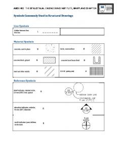
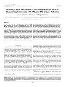
M) had no significant effect on 8-Br-cGMP (100 M)-induced growth and phospho-VASP. When applied under basal conditions, i.e. in the absence of 8-Br-cGMP, RpPET even stimulated cell growth and VASP phosphorylation in control VSMCs (Fig. 2, left panels). Growth stimulation by Rp-PET alone was reproducible and highly significant, but clearly weaker than with 8-Br-cGMP. Importantly, RpPET had no effects on cGKI-deficient cells (Fig. 2, right panels), demonstrating that its apparent agonistic activity in control VSMCs was indeed mediated by cGKI. To further investigate the potential partial agonistic activity of RpPET, its effects on the activity of purified cGKI␣ and cGKI were examined. Both enzymes displayed cGMP-dependent kinase activity with characteristic Ka values for FIGURE 3. Effect of Rp-PET on purified cGMP-activated cGKI␣ (A) and cGKI (B). Upper panels show plots of kinase activity against increasing cGMP concentrations. cGMP activation curves were obtained in the absence stimulation with cGMP (Ka ⬃0.1 (filled squares) and presence of 0.01 M (open circles), 0.1 M (filled triangles), 1 M (open squares), and 50 M and ⬃1 M for cGKI␣ and cGKI, (filled inverse triangles) Rp-PET. Kinase activity was normalized to maximal cGMP stimulation in the absence of respectively; Fig. 3, no inhibitor, and Rp-PET. The broken arrows and the black arrows indicate the effect of 50 M Rp-PET on kinase activity in the absence of cGMP and on cGMP-stimulated kinase activity in the presence of 10⫺4 M cGMP, respectively. Out of Fig. 4). Increasing concentrations of three independent kinase assays, one representative experiment is shown. Data are given as means ⫾ S.D. (n ⫽ Rp-PET caused a right shift of the 3 per data point). Lower panels show corresponding Dixon plots to estimate the half-maximal inhibitory concGMP activation curves for both the centration (Ki) for Rp-PET inhibition of cGMP-activated cGKI␣ (A) and cGKI (B). The Ki values were determined from the median of the interception points of the lines obtained by plotting the reciprocal values of the kinase cGKI␣ isoform (Fig. 3A, upper activity against the Rp-PET concentration for three cGMP concentrations as indicated. Note that each line was panel) and the cGKI isoform (Fig. based on three data points. However, for a clearer presentation of the interception points, a zoomed section of 3B, upper panel), as expected for an the diagram is shown. inhibitor. By using Dixon plots (29), Ki values of 0.03 and 0.05 M were determined for cGKI␣ (Fig. 3A, lower panel) and cGKI (Fig. 3B, lower panel), respectively. These Ki values were similar to Ki values reported previously for the inhibition of the cGKI isozymes by Rp-PET in vitro (24). Interestingly, inhibition of cGMP-activated cGKI␣ by Rp-PET was less complete than inhibition of cGKI (Fig. 3, upper panels, black arrows). Moreover, when added in the absence FIGURE 4. Effect of Rp-PET on the activity of purified cGKI␣ (A) and cGKI (B) in the absence of added of cGMP, Rp-PET appeared to cGMP. Kinase activity was plotted against increasing concentrations of Rp-PET (open circles) and compared with cGMP activation curves (filled squares). Kinase activity was normalized to maximal cGMP stimulation. Out increase the activity of cGKI␣ but not of three independent kinase assays, one representative experiment is shown. Data are given as means ⫾ S.D. cGKI above basal levels (Fig. 3, (n ⫽ 3 per data point). upper panels, broken arrows). These findings were consistent with the par(31–33). Therefore, it was tested whether Rp-PET could inhibit tial agonistic effect of Rp-PET observed in intact cells and sugcGMP/cGKI-stimulated VSMC growth and phosphorylation gested that Rp-PET might preferentially activate the cGKI␣ of VASP. As expected, the cGKI agonist 8-Br-cGMP (100 M) isozyme. Indeed, Rp-PET alone increased the kinase activity of induced cell growth and VASP phosphorylation in cGKI-ex- cGKI␣ in a concentration-dependent manner with a Ka value of 1 pressing cells (Fig. 2, left panels) but was ineffective in cGKI- M (Fig. 4A). In line with a partial agonistic activity, the maximal knock-out cells (Fig. 2, right panels). Surprisingly, Rp-PET (100 kinase activity that could be induced with Rp-PET was lower than
Rp-8-Br-PET-cGMPS Can Activate cGKI
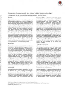
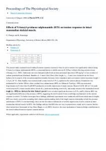
with cGMP, reaching ⬃38% of the enzyme activity in the presence of saturating cGMP concentrations. In contrast, RpPET did not significantly alter the kinase activity of the cGKI isoform (Fig. 4B). It seemed possible that the apparent activation of purified cGKI␣ by Rp-PET was caused by spontaneous exchange of the sulfur atom of the cyclic phosphorothioate moiety of Rp-PET with oxygen, resulting in conversion of the Rp-analogue to the corresponding agonistic compound, PET (Fig. 1B; see Technical Information about Rp-8-Br-PET-cGMPS, update October 15, 2007, Biolog Life Science Institute). To exclude this possibility and to analyze the homogeneity of the Rp-PET compound, the aliquot used for the kinase assays was examined by ESI-MS. Only peaks at m/z ratios corresponding to the molecular mass of Rp-PET could be detected (Fig. 5, left panel), indicating that the compound was pure and that exchange of sulfur with oxygen or other chemical alterations had not occurred. Spiking of Rp-PET with the potential conversion product PET, which is 16 Da lighter, confirmed that these compounds could clearly be resolved by the experimental setup (Fig. 5, right panel). Thus, the mass spectrum confirmed that Rp-PET itself exerted the partial agonistic effect on cGKI␣ that was observed in the kinase assays. To further verify its partial agonistic activity, the effects of Rp-PET on the isolated cGMP-binding domains of cGKI were studied by FRET measurements. For these measurements, the two cGMP-binding sites of bovine cGKI were sandwiched between CFP and YFP, respectively (30). This construct contains only the cGMP-binding sites, which are identical in cGKI␣ and cGKI but lacks the N-terminal region that differs between the isozymes. Binding of the agonist cGMP to the FRET construct induces a conformational change that can be monitored as altered FRET signal (30), whereas binding of an antagonist does not produce a FRET change. In line with a partial agonistic activity of Rp-PET, increasing concentrations
560 JOURNAL OF BIOLOGICAL CHEMISTRY
FIGURE 7. Effects of 8-Br-cGMP (100 M) and Rp-PET (100 M) on the growth of primary SM-I␣ or SM-I smooth muscle rescue VSMCs, which express only the cGKI␣ or cGKI isoform, respectively. The experiments were performed as described in the legend for Fig. 2. Cells were incubated with drugs as indicated, and their growth was determined by the MTS assay. Relative cell numbers (rel. cell number) are given as means ⫾ S.E. The number of wells measured under each condition (n) is indicated in the respective columns. *** indicates p ⬍ 0.001 versus control in the absence of drugs (Student’s t test).
of Rp-PET caused a similar, albeit weaker, FRET change as the known agonists cGMP and PET (Fig. 6). The combined results of the in vitro kinase assays with purified cGKI isoforms (Figs. 3 and 4) and of the FRET measurements (Fig. 6) suggest the following model. Rp-PET binds to the cGMP-binding sites of both cGKI isozymes. Although it acts as an antagonist of cGKI, its binding to cGKI␣ induces a conformational change that partially activates the enzyme. To confirm that Rp-PET activates cGKI␣ but not cGKI in intact cells, growth assays were performed with so-called SM-I␣ or SM-I smooth muscle rescue VSMCs, which express only the cGKI␣ or the cGKI isoform, respectively (9). Indeed, Rp-PET (100 M) stimulated the growth of SM-I␣ but not SM-I cells (Fig. 7). 8-Br-cGMP (100 M) was effective in both cell preparations, VOLUME 284 • NUMBER 1 • JANUARY 2, 2009
Downloaded from http://www.jbc.org/ by guest on November 7, 2015
FIGURE 5. Analysis of the Rp-PET aliquot by mass spectrometry. Negative ion ESI mass spectra were obtained for the Rp-PET aliquot used for the kinase assays (left panel). As a control, the Rp-PET solution was spiked with 30% (v/v) PET (right panel). The peaks corresponding to the sulfur-containing Rp-PET (m/z of 538 and 540) are clearly distinguishable from those of the oxygencontaining PET (m/z of 522 and 524), which is 16 Da lighter. Peak doublets with a 2-Da mass difference were due to the 1:1 occurrence of the 79Br and 81 Br isotopes.
FIGURE 6. Conformational changes of the cGMP-binding domains of cGKI induced by cGMP, PET and Rp-PET as observed by FRET measurements. The ratio between CFP emission at 475 nm and YFP emission at 525 nm upon excitation at 436 nm was plotted against increasing concentrations of cGMP (filled squares), PET (open circles), or Rp-PET (filled triangles). Data are presented as means ⫾ S.D. (n ⱖ3 per data point).
Rp-8-Br-PET-cGMPS Can Activate cGKI
demonstrating that, in principle, both cGKI isozymes can promote VSMC growth. The differential sensitivity of cGKI␣ versus cGKI to the partial agonistic effect of Rp-PET must be related to their different N termini, which apparently differ in their ability to couple ligand binding to activation of the catalytic region. Indeed, the N termini determine the differential sensitivity of the isozymes to cGMP activation, the cGKI␣ isoform being ⬃10 times more sensitive to cGMP than cGKI (34). Target-specific effects have also been reported for the Rp-phosphorothioate cGMP analogue Rp-8-pCPT-cGMPS, which is an antagonist of the olfactory cyclic nucleotide-gated channel but an agonist of the photoreceptor cyclic nucleotide-gated channel (35). Because Rp-8-pCPT-cGMPS is also used as cGK inhibitor (36), we tested its effect on cGKI activity in the absence of cGMP. Similar to Rp-PET, Rp-8-pCPT-cGMPS did not alter the basal kinase activity of cGKI but partially activated the cGKI␣ isozyme to ⬃35% of the maximal cGMP-stimulated activity with a Ka value of 1 M (data not shown). In the original reports on the use of Rp-8-pCPT-cGMPS (36) and Rp-PET (24) as cGK inhibitors, the effects of these compounds on cGKI activity in the absence of cGMP were not determined. However, in line with the present results, other investigators have noticed partial agonistic effects of both Rp-PET and Rp-8-pCPT-cGMPS on cGKI (37, 38). Partial agonistic activity has also been reported for Rp-phosphorothioate cAMP analogues that are used as inhibitors of the cAMP-dependent protein kinase (39). JANUARY 2, 2009 • VOLUME 284 • NUMBER 1
Acknowledgments—We thank S. Feil and A. Gerling for providing mice, M. Zeeb, Y. Heubach, and D. Hildebrand for help with the generation of the cGKI antibody, P. Ruth for the cGKI baculovirus, H. Echner for synthesis of the cGKI substrate peptide, T. Haug for support in the radioisotope laboratory, and H. Bisswanger and the members of the Feil laboratory for helpful discussions.
REFERENCES 1. Evgenov, O. V., Pacher, P., Schmidt, P. M., Hasko, G., Schmidt, H. H., and Stasch, J. P. (2006) Nat. Rev. Drug Discov. 5, 755–768 2. Ghofrani, H. A., Osterloh, I. H., and Grimminger, F. (2006) Nat. Rev. Drug Discov. 5, 689 –702 3. Kemp-Harper, B., and Feil, R. (2008) Sci. Signal. 1, pe12 4. Beavo, J. A., and Brunton, L. L. (2002) Nat. Rev. Mol. Cell. Biol. 3, 710 –718 5. Foller, M., Feil, S., Ghoreschi, K., Koka, S., Gerling, A., Thunemann, M., Hofmann, F., Schuler, B., Vogel, J., Pichler, B., Kasinathan, R. S., Nicolay, J. P., Huber, S. M., Lang, F., and Feil, R. (2008) Proc. Natl. Acad. Sci. U. S. A. 105, 6771– 6776 6. Hofmann, F., Feil, R., Kleppisch, T., and Schlossmann, J. (2006) Physiol. Rev. 86, 1–23 7. Lincoln, T. M., Dey, N., and Sellak, H. (2001) J. Appl. Physiol. 91, 1421–1430 8. Lohmann, S. M., and Walter, U. (2005) Front. Biosci. 10, 1313–1328 9. Weber, S., Bernhard, D., Lukowski, R., Weinmeister, P., Worner, R., Wegener, J. W., Valtcheva, N., Feil, S., Schlossmann, J., Hofmann, F., and Feil, R. (2007) Circ. Res. 101, 1096 –1103 10. Mendelsohn, M. E. (2005) J. Clin. Investig. 115, 840 – 844 11. Feil, R., Feil, S., and Hofmann, F. (2005) Trends Mol. Med. 11, 71–75
JOURNAL OF BIOLOGICAL CHEMISTRY
561
Downloaded from http://www.jbc.org/ by guest on November 7, 2015
FIGURE 8. Inhibition of 8-Br-cGMP-stimulated VSMC growth (upper panel) and VASP phosphorylation (lower panel) by Rp-PET. The experiments were performed with cGKI-expressing primary VSMCs as described in the legend for Fig. 2, with the exception that the concentration of 8-Br-cGMP was reduced to 50 M and that of Rp-PET was increased to 200 M. Cell growth was determined by the MTS assay, and p-VASP (Ser-157) was determined by the upshift of the VASP band. Relative cell numbers (rel. cell number) are given as means ⫾ S.E. The number of wells measured under each condition (n) is indicated in the respective columns. *** indicates p ⬍ 0.001 versus 8-Br-cGMP (Student’s t test).
As a partial agonist, Rp-PET should, in principle, be able to inhibit cGMP-activated cGKI in intact cells. The failure of our initial attempts to inhibit cGKI-mediated VSMC growth and VASP phosphorylation in the presence of 100 M 8-Br-cGMP by an equimolar concentration of Rp-PET (Fig. 2) might be related to an inappropriate intracellular ratio of 8-Br-cGMP to Rp-PET. Indeed, when the nominal 1:1 ratio was decreased to 1:4, i.e. 50 M 8-Br-cGMP and 200 M Rp-PET, cGMP/cGKImediated cell growth and VASP phosphorylation were significantly inhibited by Rp-PET (Fig. 8). These results are consistent with a previous study showing that Rp-PET can inhibit cGMPactivated cGKI in intact cells under certain conditions (24). Considering the 46-fold higher lipophilicity (14) and the 4-fold higher concentration of Rp-PET versus 8-Br-cGMP used in our experiment, it appears that the inhibitory potential of Rp-PET in intact cells is quite moderate and that it must be present in large excess to inhibit the effect of a strong agonist such as 8-Br-cGMP. Taken together, the present study indicates that the cGMP analogue Rp-PET, which is frequently used as cGKI inhibitor, is a cGKI antagonist and partial cGKI␣ agonist and, therefore, affects cGKI activity in a complex isoform-dependent manner. In intact cells, Rp-PET may either inhibit or activate cGKImediated pathways depending on the intracellular cGMP level and the prevalence of the cGKI␣ or cGKI isoform. Other studies have shown that Rp-PET can also exert cGKI-independent effects (40, 41), perhaps via modulation of PDEs (3, 14). The unpredictable behavior of Rp-PET complicates the interpretation of intact cell studies. Rp-PET should be used with caution as cGKI inhibitor and not as sole proof for the involvement of cGKI in signaling pathways.
Rp-8-Br-PET-cGMPS Can Activate cGKI
562 JOURNAL OF BIOLOGICAL CHEMISTRY
and Feil, R. (2000) Genesis 28, 15–22 27. Feil, R., Kellermann, J., and Hofmann, F. (1995) Biochemistry 34, 13152–13158 28. Ruth, P., Pfeifer, A., Kamm, S., Klatt, P., Dostmann, W. R., and Hofmann, F. (1997) J. Biol. Chem. 272, 10522–10528 29. Dixon, M. (1953) Biochem. J. 55, 170 –171 30. Russwurm, M., Mullershausen, F., Friebe, A., Jager, R., Russwurm, C., and Koesling, D. (2007) Biochem. J. 407, 69 –77 31. Feil, R., Gappa, N., Rutz, M., Schlossmann, J., Rose, C. R., Konnerth, A., Brummer, S., Kuhbandner, S., and Hofmann, F. (2002) Circ. Res. 90, 1080 –1086 32. Weinmeister, P., Lukowski, R., Linder, S., Traidl-Hoffmann, C., Hengst, L., Hofmann, F., and Feil, R. (2008) Mol. Biol. Cell 19, 4434 – 4441 33. Wolfsgruber, W., Feil, S., Brummer, S., Kuppinger, O., Hofmann, F., and Feil, R. (2003) Proc. Natl. Acad. Sci. U. S. A. 100, 13519 –13524 34. Ruth, P., Landgraf, W., Keilbach, A., May, B., Egleme, C., and Hofmann, F. (1991) Eur. J. Biochem. 202, 1339 –1344 35. Kramer, R. H., and Tibbs, G. R. (1996) J. Neurosci. 16, 1285–1293 36. Butt, E., Eigenthaler, M., and Genieser, H. G. (1994) Eur. J. Pharmacol. 269, 265–268 37. Taylor, M. S., Okwuchukwuasanya, C., Nickl, C. K., Tegge, W., Brayden, J. E., and Dostmann, W. R. (2004) Mol. Pharmacol. 65, 1111–1119 38. Vaandrager, A. B., Edixhoven, M., Bot, A. G., Kroos, M. A., Jarchau, T., Lohmann, S., Genieser, H. G., and de Jonge, H. R. (1997) J. Biol. Chem. 272, 11816 –11823 39. Gjertsen, B. T., Mellgren, G., Otten, A., Maronde, E., Genieser, H. G., Jastorff, B., Vintermyr, O. K., McKnight, G. S., and Doskeland, S. O. (1995) J. Biol. Chem. 270, 20599 –20607 40. Gambaryan, S., Geiger, J., Schwarz, U. R., Butt, E., Begonja, A., Obergfell, A., and Walter, U. (2004) Blood 103, 2593–2600 41. Marshall, S. J., Senis, Y. A., Auger, J. M., Feil, R., Hofmann, F., Salmon, G., Peterson, J. T., Burslem, F., and Watson, S. P. (2004) Blood 103, 2601–2609
VOLUME 284 • NUMBER 1 • JANUARY 2, 2009
Downloaded from http://www.jbc.org/ by guest on November 7, 2015
12. Lincoln, T. M., Wu, X., Sellak, H., Dey, N., and Choi, C. S. (2006) Front. Biosci. 11, 356 –367 13. Aizawa, T., Wei, H., Miano, J. M., Abe, J., Berk, B. C., and Yan, C. (2003) Circ. Res. 93, 406 – 413 14. Poppe, H., Rybalkin, S. D., Rehmann, H., Hinds, T. R., Tang, X. B., Christensen, A. E., Schwede, F., Genieser, H. G., Bos, J. L., Doskeland, S. O., Beavo, J. A., and Butt, E. (2008) Nat. Meth. 5, 277–278 15. Bain, J., McLauchlan, H., Elliott, M., and Cohen, P. (2003) Biochem. J. 371, 199 –204 16. Burkhardt, M., Glazova, M., Gambaryan, S., Vollkommer, T., Butt, E., Bader, B., Heermeier, K., Lincoln, T. M., Walter, U., and Palmetshofer, A. (2000) J. Biol. Chem. 275, 33536 –33541 17. Wyatt, T. A., Pryzwansky, K. B., and Lincoln, T. M. (1991) Res. Commun. Chem. Pathol. Pharmacol. 74, 3–14 18. Dostmann, W. R., Taylor, M. S., Nickl, C. K., Brayden, J. E., Frank, R., and Tegge, W. J. (2000) Proc. Natl. Acad. Sci. U. S. A. 97, 14772–14777 19. Schwede, F., Maronde, E., Genieser, H., and Jastorff, B. (2000) Pharmacol. Ther. 87, 199 –226 20. Butt, E., van Bemmelen, M., Fischer, L., Walter, U., and Jastorff, B. (1990) FEBS Lett. 263, 47–50 21. Chu, D. M., Corbin, J. D., Grimes, K. A., and Francis, S. H. (1997) J. Biol. Chem. 272, 31922–31928 22. Wall, M. E., Francis, S. H., Corbin, J. D., Grimes, K., Richie-Jannetta, R., Kotera, J., Macdonald, B. A., Gibson, R. R., and Trewhella, J. (2003) Proc. Natl. Acad. Sci. U. S. A. 100, 2380 –2385 23. Sekhar, K. R., Hatchett, R. J., Shabb, J. B., Wolfe, L., Francis, S. H., Wells, J. N., Jastorff, B., Butt, E., Chakinala, M. M., and Corbin, J. D. (1992) Mol. Pharmacol. 42, 103–108 24. Butt, E., Pohler, D., Genieser, H. G., Huggins, J. P., and Bucher, B. (1995) Br. J. Pharmacol. 116, 3110 –3116 25. Wegener, J. W., Nawrath, H., Wolfsgruber, W., Kuhbandner, S., Werner, C., Hofmann, F., and Feil, R. (2002) Circ. Res. 90, 18 –20 26. Kuhbandner, S., Brummer, S., Metzger, D., Chambon, P., Hofmann, F.,
Mechanisms of Signal Transduction: The Commonly Used cGMP-dependent Protein Kinase Type I (cGKI) Inhibitor Rp-8-Br-PET-cGMPS Can Activate cGKI in Vitro and in Intact Cells Nadejda Valtcheva, Peter Nestorov, Alexander Beck, Michael Russwurm, Matthias Hillenbrand, Pascal Weinmeister and Robert Feil
Access the most updated version of this article at doi: 10.1074/jbc.M806161200 Find articles, minireviews, Reflections and Classics on similar topics on the JBC Affinity Sites. Alerts: • When this article is cited • When a correction for this article is posted Click here to choose from all of JBC's e-mail alerts This article cites 41 references, 24 of which can be accessed free at http://www.jbc.org/content/284/1/556.full.html#ref-list-1
Downloaded from http://www.jbc.org/ by guest on November 7, 2015
J. Biol. Chem. 2009, 284:556-562. doi: 10.1074/jbc.M806161200 originally published online November 13, 2008