D28 and CTLA-4, with particular emphasis on the role played by the PI3K-dependent signalling cascade. K ey words: T cell; CD28; CTLA-4; PI3K; signalling. N.
�
Archivum Immunologiae et Therapiae Experimentalis, 1999, 47, 69–75 PL ISSN 0004-069X �
�
Review
The Complexities of CD28 and CTLA-4 Signalling: PI3K and Beyond S. G. Ward: CD28 and CTLA-4 Signalling
STEPHEN G. WARD Department of Pharmacy and Pharmacology, Bath University, Claverton Down, Bath BA2 7AY, United Kingdom
Abstract. A successful immune response requires a set of non-cognate cell-cell interactions which provide the second “costimulatory” signal to the T cells. The best characterized costimulatory receptor expressed on resting T cells is CD28 which provides poorly-defined cyclosporin-resistant biochemical signal(s) that promote expression of several cytokines/chemokines. Another major effect of CD28 ligation is the promotion of cell survival which is thought to occur via the up-regulation of Bcl-xL expression. CD28 shares its ligands B7.1 and B7.2 with the related CTLA-4, which plays an inhibitory role in T cell activation. Manipulation of CD28/CTLA-4 interactions with their natural ligands has provided exciting results in transplantation and tumor therapy settings and also has potential in the treatment of several diseases such as arthritis and multiple sclerosis, asthma and protection against HIV infection. The biochemical basis for the different functional outcomes of CD28 and CTLA-4 ligation has been the subject of intense investigation over the past few years. This review will focus on our current understanding of the biochemical signals that may be involved in regulating the different functional outcomes of CD28 and CTLA-4, with particular emphasis on the role played by the PI3K-dependent signalling cascade. �
�
�
�
�
�
�
�
�
�
Key words: T cell; CD28; CTLA-4; PI3K; signalling.
Nearly 30 years ago, BRETSCHER and COHN4 first proposed that the generation of an antigen-specific lymphocyte response required at least two distinct signals from the antigen presenting cell4. We now know that the first signal is provided by engagement of the antigen-specific TCR by a peptide antigen bound to a MHC protein. This interaction confers antigen specificity to a T cell response, although occupancy of the TCR is usually insufficient to initiate a productive T cell response. A successful immune response usually requires a set of additional non-cognate cell-cell interactions which provide the second “costimulatory” signal to the T cells. The best characterized costimulatory receptor expressed on resting T cells is CD28 which binds the natural ligands B7.1 (CD80) or B7.2 (CD86). CD28 provides poorly-defined cyclosporin-resistant �
�
�
�
�
�
�
�
�
�
�
�
�
�
biochemical signal(s) that promote IL-2 production and cellular proliferation in the presence of an additional signal(s) provided by either the TCR or phorbol esters20. Another major effect of CD28 ligation is the promotion of cell survival which is thought to occur via the up-regulation of Bcl-xL expression2. CD28 also up-regulates expression of several other cytokines/chemokines (e. g. IL-4, IL-13, IFN-γ, TNF, GM-CSF, IL-8 and RANTES)45, the IL-2 receptor8 and modulates expression of the chemokine receptors CCR5, CCR1, CCR4, CXCR1 and CXCR26, 46. CD28 shares its ligands B7.1 and B7.2 with the related receptor CTLA-4. There is now strong evidence from studies using cross-linked or immobilised CTLA-4 Abs in murine T cells and CTLA-deficient mice which has demonstrated that CTLA-4 plays an inhibitory role in T cell activation9. �
�
�
�
�
�
�
�
�
�
�
�
�
70 -
S. G. Ward: CD28 and CTLA-4 Signalling 173
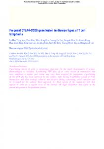
YMNM) that forms a potential binding site for interaction with the SH2 domain containing p85 regulatory subunit of phosphoinositide 3-kinase (PI3K)51 (Fig. 1). Moreover, CD28 also contains two proline rich motifs (178PRRP and 190PYAP) which conform to the PXXP SH3 binding consensus sequence and these regions of the CD28 tail may mediate interactions with signalling proteins (Fig. 1). The primary amino acid sequence of the CTLA-4 cytoplasmic domain contains tyrosine residues (164Y and 181Y) which lie in potential SH2 domains binding motifs. In addition, CTLA-4 contains a potential binding motif for an SH3 domain (Fig. 1). There has been no direct evidence to indicate whether either of the tyrosine residues are phosphorylated in vivo.
Manipulation of CD28/CTLA-4 interactions with their natural ligands has provided exciting results in transplantation and tumor therapy12 and also has potential in the treatment of several diseases such as arthritis48, multiple sclerosis49, asthma43 and protection against HIV infection22. An improved understanding of the molecular interactions and signals regulated by both CD28 and CTLA-4 is therefore of considerable therapeutic importance. This review will focus on our current understanding of the biochemical signals that may be involved in regulating the different functional outcomes of CD28 and CTLA-4. �
�
�
�
�
�
�
�
�
�
�
�
�
"
�
#
�
�
�
�
�
"
�
�
�
Potential Signalling Domains of CD28 and CTLA-4 Cytoplasmic Tails �
�
$
"
Coupling of CD28 to Protein Tyrosine Kinases Whilst the extracellular domains have limited sequence homology, the ligand binding sites of CD28 and CTLA-4 are localised to a conserved sequence MYPPPY in the CDR3-like region of both CD28 and CTLA-415. There is also only approximately 30% amino acid conservation between CD28 and CTLA-4 in the cytoplasmic domain, perhaps suggesting an ability to generate different signals. The cytoplasmic domain of CD28 lacks any direct enzymatic activity and is therefore presumed to signal via the recruitment of cellular protein tyrosine kinases (PTK) such as Lck, Fyn or Itk/EMT. In this respect, CD28 contains 4 tyrosine residues (173Y, 188Y, 191Y and 200Y) which can potentially be phosphorylated and mediate interactions with other proteins such as Src-homology 2 (SH2) domain-containing proteins. Of specific interest is 173Y which lies within a consensus sequence motif ((p) �
�
�
�
�
�
�
�
!
%
One of the earliest biochemical events stimulated by CD28 is the activation of the Src family PTKs Lck and Fyn as well as the Tec family kinase Itk/EMT which have been shown to be physically associated with the receptor1, 17, 35. The consequences of PTK activation are probably several fold, but one important effect is the phosphorylation of tyrosine residues within the CD28 cytoplasmic tail which is likely to result in the recruitment and activation of other SH2 containing proteins. Under in vitro conditions, Lck has been demonstrated to phosphorylate only Tyr173, whilst Itk/EMT phosphorylates all 4 tyrosine residues within the CD28 cytoplasmic tail17. The precise sequence of events leading to PTK recruitment and activation of these PTKs remains unclear, although it appears that Itk binding to the PRRP motif of CD28 via its SH3 domain, is sufficient to result in increased intrinsic kinase activity of Itk27. Recent studies have indicated that not all the tyrosine phosphorylation events observed following CD28 ligation might necessarily lead to promotion of T cell activation. For instance, studies with Itk-deficient mice have revealed that Itk actually negatively regulates induction of T cell proliferation by CD28 costimulation23. Hence, the functional outcome of phosphorylation of 188Y, 191Y and 200Y remains unclear.
�
�
&
�
�
�
�
�
�
�
�
�
�
�
�
�
�
�
�
�
�
PI3K Recruitment by CD28 and CTLA-4
�
,
+
*
�
Fig. 1. Potential signalling domains of the CD28 and CTLA-4 cytoplasmic tails. The sequence of the CD28 and CTLA-4 cytoplasmic domains is shown. Established and putative SH2, SH3 and AP-2 binding sites are indicated �
PI3K controls an alternative inositol phospholipid metabolic pathway which phosphorylates the membrane phosphatidylinositol lipids at the 3 position on the inositol ring and can result in the generation of D-3 phosphoinositide lipids (e. g. PtdIns(3)P, PtdIns(3,4) P2 and PtdIns(3,4,5)P3)44. Considerable importance has been attached to this pathway, particularly concerning )
'
'
(
�
-
7
S. G. Ward: CD28 and CTLA-4 Signalling 4
the putative role of these D-3 phosphoinositides as regulatory molecules. Hence, PtdIns(3) P is thought to be involved primarily in the regulation of membrane trafficking, whilst PtdIns(3,4)P2 and PtdIns(3,4,5)P3 are thought to be important components of a receptor-activated signalling cascade. The first indication that CD28 could indeed couple to PI3K was the demonstration that ligation of CD28 by B7.1 induced the accumulation of PtdIns(3,4,5)P3 in the leukemic T cell line Jurkat47. The heterodimeric PI3K (consists of p85 regulatory and p110 catalytic subunits) interacts directly with CD28 by virtue of the p85 subunit binding to the (p)173YMNM motif as predicted32. This interaction depends on phosphorylaton of the 173Y and the PTK responsible for phosphorylating 173Y is thought to be Lck. However, quite how this fits in with the observation that CD28 can still recruit and activate PI3K in Lck-deficient Jurkat cells26 is not clear. Receptor activation of PI3K in T lymphocytes is not unique to CD28 and can also be stimulated by the TCR47. However, it is interesting to note that the accumulation of PtdIns(3,4,5)P3 following CD28 ligation occurs with delayed kinetics, greater magnitude and prolonged duration when compared with the accumulation in response to ligation of the TCR alone47. Ligation of CTLA-4 with anti-CTLA-4 mAbs also triggers binding of the p85 subunit of PI3K to the 164YVKM motif in the CTLA-4 cytoplasmic domain, although tyrosine phosphorylation of CTLA-4 has not yet been demonstrated38. �
�
'
2, thus making these cells much more relevent models in which to study the costimulatory signalling cascades regulating IL-2 production. In addition, mutation of the human CD28 cytoplasmic tail 173Y to Phe and subsequent expression in a murine T cell hybridoma, prevents CD28-PI3K association, activation of PI3K and production of IL-2 in response to CD28 ligation32. However, similar mutations of murine CD28 expressed in the human Jurkat model, prevented ligation-induced association of CD28 with PI3K, but CD28 ligation still resulted in IL-2 production in these models41. This data further emphasizes the differences that have been observed in the role of PI3K in CD28-mediated stimulation in Jurkat cells and indicates that PI3K may mediate multiple CD28 functions. "
'
(
�
.
�
.
(
�
(
.
�
�
�
(
�
/
�
"
�
�
�
�
/
CD28 Activates the PI3K Effector Protein Kinase B: Relevence to Cell Survival Recent evidence has indicated that ligation of CD28 results in activation of protein kinase B (PKB)33. PKB is recognised as being both a downstream effector target of PI3K and a key mediator of growth factor-induced cell survival29. PKB can phosphorylate a critical serine residue on the death promoting protein BAD, causing it to dissociate from and thus, activating the cell survival factor Bcl-xL10. This is an interesting observation given that CD28 not only activates PKB33 but also up-regulates Bcl-xL expression which correlates with enhanced resistance to apoptosis induced by γ-irradiation, or Abs to Fas or CD32. (
�
'
0
�
(
�
�
�
�
�
�
�
"
�
71
(
�
(
5
�
$
�
�
Is PI3K Required for CD28-dependent Costimulation? 1
�
3
�
�
/
�
�
�
2
�
�
�
�
PKB and Transcriptional Regulation Both pharmacological and genetic approaches have been used to assess the functional relevance of PI3K activation with respect to CD28-mediated costimulation of IL-2 production, although these have yielded controversial results. Hence, in the leukemic T cell line Jurkat or murine CD4+ T cells, PI3K inhibitors such as wortmannin can either potentiate or have no effect on CD28-mediated costimulation of IL-2 production from these cells41, 47. The use of Jurkat cells to study CD28mediated costimulation of IL-2 production could be misleading however, since transformed cell lines may be poor models of Signal 2 because they have been selected to be independent of co-stimulatory signals. Of major importance therefore, is the observation that wortmannin inhibits CD28-mediated costimulation of IL-2 production from purified resting T cells and human T lymphoblasts47. In resting T cells and human T lymphoblasts, optimal IL-2 production is usually much more dependent on the provision of Signal �
�
Apart from promoting cell survival, PKB activation is known to have at least one other major outcome which may be relevent to the functional outcome of CD28 ligation in T cells, namely p70S6 kinase activation5. Activation of p70S6 kinase has been implicated in the regulation of the transcription factor NFκB by CD2845. Since CD28 can activate both PKB and p70S6 kinase in a wortmannin-sensitive manner33, 34, a PI3K/ /PKB/p70S6 kinase cascade may mediate the effects of CD28 on transcription factor regulation. There is evidence that this may not be the case in normal T lymphoblasts however, since experiments using human T lymphoblasts indicate that CD28-induced activation of NFκB is unaffected by pre-treatment with PI3K inhibitors11. It is also interesting to note that the inhibitory effects of constitutively active PI3K on TCR-mediated NF-AT responses are not mimicked by PKB mutants, emphasising that PKB is not the only downstream ef#
�
�
�
$
$
�
�
�
6
(
72 -
S. G. Ward: CD28 and CTLA-4 Signalling
fector of PI3K36. Nevertheless, this does not exclude the possibility that PKB is involved in the biochemical wiring that mediates the CD28-dependent modulation of different transcription factors that may be involved in transcriptional regulation of other cytokine/chemokine genes. In this respect, both PI3K and PKB have been shown to be necessary and sufficient for induction of activity of the transcription factor E2F3. (
�
�
�
�
with AP50 resulting in accumulation of CTLA-4 at the cell surface. It is likely that phosphorylation of 164Y will also facilitate recruitment of the p85 subunit of PI3K38 which may therefore be involved in the mechansisms by which cell surface expression of CTLA-4 is up-regulated. In addition, PI3K binding to CTLA-4 may result in T cell inhibition if CTLA-4 sequesters sufficient PI3K to prevent it from participating in TCR and/or CD28 signal transduction. Alternatively, there may be secondary conditions that lead CTLA-4 to bind and activate PI3K and which may account for the ability of CTLA-4 to act as a costimulatory receptor under some in vitro conditions9. �
�
(
�
(
�
�
�
�
�
Regulation of the Surface Expression of CD28 and CTLA-4: a Role for PI3K? �
�
�
�
Given the opposing effects of CD28 and CTLA-4 on T cell activation, it is interesting to note that CTLA4 is not expressed constitutively and is only expresed maximally 2-3 days after T cell activation by TCR and CD28 ligation. Moreover, CD28 engagement by B7.1 induces a transient down-regulation of CD28 and prolonged unresponsiveness to CD28 signalling24, 25. This co-ordinated set of events probably facilitates the inhibitory effects of CTLA-4 and contributes to the tight control of T cell activation. Ligation-stimulated CD28 receptor endocytosis, like that observed for endocytosis of receptors for transferrin, PDGF or EGF, occurs via clathrin-coated pits which are shuttled to lysosomes for proteolytic degradation and/or recycled to the cell surface7. PI3K has been reported to associate with clathrin-coated vescicles and the microtubule cytoskeleton and to influence the endocytic destiny of internalised PDGF-R14. It is fitting therefore that PI3K also been implicated in the control of ligation-stimulated endocytosis of CD28 since CD28-PI3K complexes are prefentially endocytosed and 173Y mutations that alter PI3K binding correlates with a decrease in the efficacy of endocytosis7. Cell surface expression of CTLA-4 is limited by its rapid clearance from the surface of activated T cells through clathrin-mediated endocytosis39. Although, the phoshorylated 164YVKM motif within CTLA-4 is similar to that found in CD28 around 173Y, there is evidence that these sites may not be functionally equivalent in CD28 and CTLA-4, since the CTLA-4 motif is located within an intracellular localisation motif (160TTGVYVKMPPT) that restricts CTLA-4 expression to intracellular membranes such as the perinuclear Golgi or post-Golgi compartment21. An unphosphorylated 6 amino acid sequence (GVYVKM) within this domain can bind specifically to the medium chain subunit (AP50) of the clathrin-associated protein complex AP-239. This AP-2 complex plays an important role in targeting cell surface glycoproteins for endocytosis and mutation or tyrosine phosphorylation of 164Y abrogates the interaction �
/
9
�
8
�
�
�
�
�
:
�
CD28 Regulation of Rac and Rho Proteins: Relevence of PI3K
�
�
�
�
;
$
�
?
'
-
7
S. G. Ward: CD28 and CTLA-4 Signalling
*
B
C
D
�
�
�
�
�
�
�
E
potential to exert a major influence on T lymphocyte biology. For instance, substrates for JNK and p38 MAPK include transcription factors such as ATF-2, c/Jun and Ets family proteins. JNK is an anchor kinase of the Rac/Cdc42 small G protein/p21-activated kinase (PAK)/MEKK-1/SEK-1 pathway. Indeed, PAK and MEKK-1 are stimulated by CD28 ligation, whilst JNK is activated by the combined triggering of TCR and CD2816, 45. Furthermore, induced deficiency of SEK-1 (which lies downstream of PAK and MEKK-1) in mice, inhibits CD28-mediated T cell costimulation30. Although, active mutants of PI3K can induce a selective subset of Rac/Rho-mediated cellular responses that control the cortical actin cytoskeleton in other systems, these mutants are not sufficient to stimulate the full range of Rac- or Rho-coordinated pathways that couple to nuclear responses of transcription factor activation37. It is interesting to note therefore that recent evidence indicates that ceramide may be involved in coupling CD28 to PAK and MEKK-116. With regard PtdIns(4) P 5-kinase, this kinase controls the cellular levels of D-5 phosphoinositides such as PtdIns(4,5)P2 which is crucial for TCR signalling pathways in T cells, since its hydrolysis by phospholipase C generates Ins(1,4,5) P3 and diacylglycerols that regulate intracellular calcium and PKC, respectively. In addition, PtdIns(4,5)P2 is a substrate for PI3K-mediated formation of PtdIns(3,4,5)P3 which along with its degradation product PtdIns(3,4)P2, is an important modifier of intracellular signal transduction pathways at multiple stages of lymphocyte activation and growth. Thus, the putative regulation of PtdIns(4)P 5-kinase in T lymphocytes by Rac/Rho may have a major impact on T cell biology and it will be important to define whether there is direct coupling between Rac/Rho and lipid kinases in T lymphocytes. CD28 stimulates tyrosine phosphorylation of a 62 kDA adaptor protein31 now identified as p62dok. This protein forms a protein complex with p120 RasGAP �
�
Fig. 2. Schematic diagram indicating the interactions of the PI3K-dependent signalling cascade with downstream effector pathways. Known and putative interactions are depicted. See text for further details. *Tyrosine phosphorylation
and p190 RhoGAP. It is postulated that tyrosine phosphorylation of p62dok will modulate the cellular distribution of associated RasGAPs and RhoGAPs and sequester these negative regulators away from active Ras and Rac molecules, thereby prolonging the activation of Rac or Rho effector pathways. Hence, CD28 by virtue of its coupling to Vav and p62dok, is uniquely coupled to the regulatory networks that control the activity of Rho family GTPases. Given that p62dok contains a PH domain there is also considerable potential for regulation of this protein by the D-3 phosphoinositide lipids that accumulate after CD28 ligation (Fig. 2). �
@
�
�
�
�
�
@
�
�
@
�
�
(
�
73
Activation of Protein Tyrosine Phosphatases by CTLA-4 A
(
'
�
�
�
There have been some important findings relating to the association of CTLA-4 with SH2-containing tyrosine phosphatase-2 (SHP-2). This interaction is mediated by the SH2-domain of SHP-2 which probably binds to the SH2-binding domain of CTLA-4 around 164 Y. This correlates well with the observation in T cells of CTLA-4 deficient mice that the TCR-associated kinases Fyn, Lck and ZAP-70 were found to be constitutively activated and there was a profound increase in the tyrosine phosphorylation of intracellular protein. In addition, CTLA-4-associated SHP-2 had phosphatase activity against the Ras regulatory protein p52SHC28. Hence, CTLA-4 might initiate a signal transduction cascade that result in either directly or indirectly, in the dephosphaorylation of the TCR-associated kinases and their substrates.
'
�
�
�
�
�
�
�
�
�
(
�
�
'
�
�
(
�
�
�
�
�
?
�
�
�
�
�
(
�
>
�
@
�
�
In summary, our knowledge and understanding of the exact make-up and biological function of the effector pathways that mediate cellular repsonse to CD28activated PI3K remains fragmentary. However, it is clear that the PI3K-dependent signalling cascade has
74
�
�
�
�
�
�
�
-
S. G. Ward: CD28 and CTLA-4 Signalling
the potential to influence signal transduction at multiple points in the signalling machinary and so, this pathway can make a major contribution to the signals required for T cell activation. In contrast to CD28, very little is known about the biochemical signals elicited by CTLA-4 ligation and which facilitate the inhibitory actions of CTLA-4 on T cell activation. A future challange will be to establish whether activation of PI3K by different immune receptors can achieve different functional outcomes by diversity of different effectors.
S
T
�
13. U
O
D
7
15. W
17. W
C
�
18. Z
H
[
19. ^
20.
O
O
U
_
O
H
�
22. [
H
O
7
c
24.
J
`
+
H
C
L
25. e
L
O
N
L
�
26. U
F
R
H
5
L
d
O
L
N
L
J
`
H
F
J
J
D
�
O
H
J
F
F
F
�
23.
H
F
O
b
Q
H
O
H
a
U
P
F
O
F
O
L
5
V
`
F
N
O
�
21.
H
I
N
]
N
W
�
N
O
J
F
\
D
M
F
N
5
E
H
F
O
I
Y
X
I
F
L
H
F
�
K
O
J
G
J
H
O
F
F
H
J
F
E
H
K
V
�
References
H
O
K
H
*
F
F
�
14.
16.
1. AUGUST A., GIBSON S., KAWAKAMI Y., KAWAKAMI T., MILLS G. B. and DUPONT B. (1994): CD28 is associated with and induces the immediate tyrosine phosphorylation and activation of the Tec family kinase ITK/EMT in the human Jurkat leukaemic T cell line. Proc. Natl. Acad. Sci. USA, 91, 9347–9351. 2. BOISE L. H., MINN A. J., NOEL P. J., JUNE C. H., ACCAVITTI M. A., LINDSTEN T. and THOMPSON C. B. (1995): CD28 costimulation can promote T cell survival by enhancing the expression of Bcl-xL. Immunity, 3, 87–98. 3. BRENNAN P., BABBAGE J., BURGERING B. M. T., GRONER B., REIF K. and CANTRELL D. A. (1997): PI3K couples IL-2 receptor to the cell cycle regulator E2F. Immunity, 7, 679–689. 4. BRETSCHER J. F. and COHN M. (1970): A theory of self-nonself discrimination. Science, 169, 1042–1046. 5. BURGERING B. M. T. and COFFER P. J. (1995): Protein kinase B (c-Akt) in PI3K signal transduction. Nature, 376, 599–602. 6. CARROLL R. G., RILEY J. L., LEVINE B. L., FENG Y., KAUSHAL S., RITCHEY D. W., BERNSTEIN W., WEISLOW O. S., BROWN C. R., BERGER E. A., JUNE C. H. and ST. LOUIS D. C. (1997): Differential regulation of HIV-1 fusion cofactor expression by CD28 costimulation of CD4+ T cells. Science, 276, 273–276. 7. CEFAI D., SCHNEIDER H., MATANGKASOMBUT O., KANG H., BRODY J. and RUDD C. E. (1998): CD28 receptor endocytosis is targeted by mutations that disrupt PI3K binding and costimulation. J. Immunol., 160, 2223–2230. 8. CERDAN C., MARTIN Y., COURCOUL M., BRAILLY H., MAWAS C., BIRG F. and OLIVE D. (1992): Prolonged IL-2Rα/CD25 expression after T cell activation via the adhesion molecules CD2 and CD28. J. Immunol., 149, 2255–2261. 9. CHAMBERS C. A., KRUMMEL M. F., BOITEL B., HURWITZ A., SULLIVAN T. J., FOURNIER S., CASSELL D., BRUNNER M. and ALLISON J. P. (1996): Role of CTLA-4 in the regulation and initiation of T cell responses. Immunol. Rev., 153, 27–46. 10. DATTA S. R., DUDEK H., TAO X., MASTERS S., FU H., GOTOH Y. and GREENBERG M. E. (1997): Akt phosphorylation of BAD coupled survival signals to the cell-intrinsic death machinary. Cell, 91, 231–241. 11. EDMEAD C., PATEL Y. I., WILSON A., BOULOUGOURIS G., HALL N. D., WARD S. G. and SANSOM D. M. (1996): Induction of AP-1 and NFκB by CD28 stimulation involves both PI3K and acidic sphingomyelinase signals. J. Immunol., 157, 3290–3297. 12. GUINAN E. C., GRIBBEN J. G., BOUSSIOTIS V. A., FREEMAN G. J. and NADLER L. M. (1994): Pivotal role of the B7: CD28 path-
way in transplantation, tolerance and tumor immunity. Blood, 84, 3261–3282. HAN J., LUBY-PHELPS K., DAS B., SHU X., XIA Y., MOSTELLER R. D., KRISHNA M., FALCK J. R., WHITE M. and BROEK D. (1998): Role of substrates and products of PI3K in regulating activation of Rac-related guanosine trisphosphatases by Vav. Science, 279, 558–561. JOLY M., KAZLAUSKAS A., FAY F. and CORVERA S. (1994): Disruption of PDGF receptor trafficking by mutation of its PI3K binding sites. Science, 263, 684–687. JUNE C. H., BLUESTONE J. A., NADLER L. M. and THOMPSON C. B. (1994): The B7 and CD28 receptor families. Immunol. Today, 15, 321–332. KAGA S., RAGG S., ROGERS K. A. and OCHI A. (1998): Activation of p21-Cdc42/Rac-activated kinases by CD28 signalling: p21-activated kinase (PAK) and MEK kinase 1 (MEKK1) may mediate the interplay between CD3 and CD28 signals. J. Immunol., 160, 4182–4189. KING P. D., SADRA A., TENG J. M., LIU X. R., HAN A., SELVAKUMAR A., AUGUST A. and DUPONT B. (1997): Analysis of CD28 cytoplasmic tail tyrosine residues as regulators and substrates for the protein tyrosine kinases EMT and Lck. J. Immunol., 158, 580–590. KLASEN S., PAGES F., PEYRON J. F., CANTRELL D. A. and OLIVE D. (1998): Two distinct regions of the CD28 intracytoplasmic domain are involved in the tyrosine phosphorylation of Vav and GTPase activating protein-associated p62 protein. Int. Immunol., 10, 481–489. KYRIAKIS J. M. and AVRUCH J. (1996): Protein kinase cascades activated by stress and inflammatory cytokines. Bioessays, 18, 567–577. LENSCHOW D. J., WALUNAS T. L. and BLUESTONE J. A. (1996): CD28/B7 system of T cell costimulation. Ann. Rev. Immunol., 14, 233–250. LEUNG H. T., BRADSHAW J., CLEVELAND J. S. and LINSLEY P. S. (1995): CTLA-4, a high avidity receptor for CD80 and CD86 contains a intracellular localisation motif in its cytoplasmic tail. J. Biol. Chem., 270, 25107–25114. LEVINE B. L., MOSCA J. D., RILEY J. L., CARROLL R. G., VAHEY M. T., JAGODZINSKI L. L., WAGNER K., MAYERS D. L., BURKE D. S., WEISLOW O. S., ST. LOIUS D. C. and JUNE C. H. (1996): Antiviral effect and ex vivo CD4+ T cell proliferation in HIV-positive patients as a result of CD28 costimulation. Science, 272, 1939–1943. LIAO X., FOURNIER S., KILLEEN N., WEISS A., ALLISON J. P. and LITTMAN D. R. (1997): Itk negatively regulates induction of T cell proliferation by CD28 costimulation. J. Exp. Med., 186, 221–228. LINSLEY P. S., BRADSHAW J., GROSMAIRE L. and LEDBETTER J. A. (1993): CD28 engagement by B7/BB-1 induces transient down-regulation of CD28 synthesis and prolonged unresponsiveness to CD28 signalling. J. Immunol., 150, 3161–3169. LINSLEY P. S., GREENE J. L., TAN P., BRADSHAW J., LEDBETTER J. A., ANASETTI C. and DAMLE N. K. (1992): Coexpression and functional cooperation of CTLA-4 and CD28 on activated T lymphocytes. J. Exp. Med., 176, 1595–1604. LU Y., PHILLIPS C. A., BJORNDAHL J. M. and TREVILLYAN J. (1994): CD28 signal-transduction: tyrosine phosphorylation and receptor association of PI3K correlate with Ca2+-independent costimulatory activity. Eur. J. Immunol., 24, 2732–2739. D
L
_
f
N
-
7
g
S. G. Ward: CD28 and CTLA-4 Signalling 27. MARENGERE L. E. M., OKKENHAUG K., CLAVREUL A., COUEZ D., GIBSON S., MILLS G. B., MAK T. W. and ROTTAPEL R. (1997): The SH3 domain of ITK/EMT binds to proline-rich sequences in the cytoplasmic domain of the T cell costimulatory receptor CD28. J. Immunol., 159, 3220–3229. 28. MARENGERE L. E. M., WATERHOUSE P., DUNCAN G. S., MITTRUCKER H. W., FENG G. S. and MAK T. W. (1996): Regulation of T cell receptor signalling by tyrosine phosphatase SYP association with CTLA-4. Science, 272, 1170–1173. 29. MARTE B. and DOWNWARD J. (1997): PKB/Akt: connecting PI3K to cell survival and beyond. Trends Biochem. Sci., 22, 355–358. 30. NISHINA H., BACHMANN M., OLIVEIRA-DOS-SANTOS A., KOZIERADZKI I., FISCHER K. D., ODERMATT B., WAKEHAM A., SHAHINIAN A., TAKIMOTO H., BERNSTEIN A., MAK T. W., WOODGETT J. R., OHASHI P. S. and PENNINGER J. M. (1997): Impaired CD28-mediated IL-2 production and proliferation in SEK1/MKK4-deficient T lymphocytes. J. Exp. Med., 186, 941–953. 31. NUNES J., TRUNEH A., OLIVE D. and CANTRELL D. A. (1996): Signal transduction by CD28 costimulatory receptor on T cells: B7.1 and B7.2 regulation of tyrosine kinase adaptor molecules. J. Biol. Chem., 271, 1591–1598. 32. PAGES F., RAGUENEAU M., ROTTAPEL R., TRUNEH A., NUNES J., IMBERT J. and OLIVE D. (1994): Binding of PI3K to CD28 is required for T cell signalling. Nature, 369, 327. 33. PARRY R. V., REIF K., SMITH G., SANSOM D., HEMMINGS B. A. and WARD S. G. (1997): Ligation of the T cell co-stimulatory receptor CD28 activates the serine-threonine protein kinase protein kinase B. Eur. J. Immunol., 27, 2495–2501. 34. PARRY R. V. and WARD S. G. (1996): Involvement of PI3K in the activation of p70S6 kinase by the T cell costimulatory molecule CD28. Biochem. Soc. Trans., 24, 88S. 35. RAAB M., CAI Y. C., BUNNELL S. C., HEYECK S., BERG L. J. and RUDD C. E. (1995): p56Lck and p59Fyn regulate CD28 binding to PI3K, Grb-2 and Itk: implications for T cell costimulation. Proc. Natl. Acad. Sci. USA, 92, 8891. 36. REIF K., LUCAS S. and CANTRELL D. A. (1997): A negative role for PI3K in T cell antigen receptor signalling. Curr. Biol., 7, 285–293. 37. REIF K., NOBES C. D., THOMAS G., HALL A. and CANTRELL D. A. (1996): PI3K signals activate a selective subset of Rac/Rho-dependent effector pathways. Curr. Biol., 6, 1445– 1455. 38. SCHNEIDER H., PRASAD K. V. S., SHOELSON S. and RUDD C. E. (1995): CTLA-4 binding to the lipid kinase PI3K in T cells. J. Exp. Med., 181, 351–355. 39. SHIRATORI T., MIYATAKE S., OHNO H., NAKASEKO C., ISONO K., BONIFACINO J. and SPITO T. (1997): Tyrosine phosphorylah
O
i
*
H
5
g
H
g
O
O
O
F
D
p
W
J
H
�
41.
F
7
42.
n
�
j
H
k
J
H
K
C
43. D
44. r
C
45.
V
H
m
F
`
s
5
j
j
O
N
O
m
N
47.
�
j
H
*
c
D
49.
j
I
L
K
E
s
50. u
51.
K
n
o
t
F
F
[
O
N
H
H
H
K
K
J
v
H
N
W
N
J
I
x
F
n
H
N
J
r
�
j
5
O
N
O
F
F
�
48.
F
N
I
O
E
F
j
H
�
46.
M
j
H
J
O
F
j
n
F
N
�
j
q
H
O
l
tion controls internalisation of CTLA-4 by regulating its interaction with clathrin-associated adaptor complex AP-2. Immunity, 6, 583–589. TAPON N. and HALL A. (1997): Rho, Rac, and Cdc42 GTPases regulate the organisation of the actin cytoskeleton. Curr. Opin. Cell Biol., 9, 86–92. TRUITT K. E., NAGEL T., SUEN L. F. and IMBODEN J. B. (1996): Structural requirements for CD28-mediated costimulation of IL-2 production in Jurkat T cells. J. Immunol., 156, 4539–4541. TSUYUKI S., TSUYUKI J., EINSLE K., KOPF M. and COYLE A. J. (1997): Costimulation through B7.2 is required for the induction of a lung mucosal T helper cell 2 (Th2) immune response and altered airway responsiveness. J. Exp. Med., 185, 1671–1679. TUOSTO L., MICHEL F. and ACUTO O. (1996): p95Vav associates with tyrosine-phosphorylated SLP-76 in antigen-stimulated T cells. J. Exp. Med., 184, 1161–1166. VANHAESEBROECK B., LEEVERS S. A., PANAYOTOU G. and WATERFIELD M. D. (1997): Phosphoinositide 3-kinases: a conserved family of signal transducers. Trends Biochem. Sci., 22, 267–272. WARD S. G. (1996): CD28: a signalling perspective. Biochem. J., 318, 361–377. WARD S. G., BACON K. B. and WESTWICK J. (1998): Chemokines and T lymphocytes: more than an attraction. Immunity, 9, 1–11. WARD S. G., JUNE C. H. and OLIVE D. (1996): PI3K: a pivotal pathway in T cell activation. Immunol. Today, 17, 187–197. WEBB L., WALMSLEY M. J. and FELDMANN M. (1996): Prevention and amelioration of collagen-induced arthritis by blockade of the CD28 costimulatory pathway: requirement for both B7.1 and B7.2. Eur. J. Immunol., 26, 2320–2328. WINDHAGEN A., NEWCOMBE J., DANGOND C., STRAND C., WOODROOFE N., CUZNER M. L. and HAFLER D. A. (1995): Expression of costimulatory molecules B7.1 and B7.2 and IL-12 cytokine in multiple sclerosis lesions. J. Exp. Med., 182, 1985–1996. WU J., MOTTO D. G., KORETZKY G. A. and WEISS A. (1996): Vav and SLP-76 interact and functionally cooperate in IL-2 gene activation. Immunity, 4, 593–602. ZHOU S. Y., SHOELSON S. E., CHAUDHURI M., GISH G., PAWSON T., HASER W. G., KING F., ROBERTS T., RATNOFSKY S., LECHLEIDER R. J., NEEL B. G., BIRGE R. B., FAJARDO J. E., CHOU M. M., HANAFUSA H., SCHAFFHAUSEN B. and CANTLEY L. (1993): SH2 domains recognise specific phosphopeptide sequences. Cell, 72, 767–778. �
40.
w
75
�
Received in July 1998 Accepted in August 1998
O
O