malian motoneurons (Jahr and Yoshioka, 1986; Flatman et al.,. 1987) and to monosynaptic EPSPs produced in rat dorsal horn neurons by DRG cells in vitro ...
The Journal
The Development of Sensorimotor Synaptic Lumbosacral Cord of the Chick Embryo Matt
T. Lee,
Mark
J. Koebbe,
and
Michael
of Neuroscience,
Connections
July
1988,
8(7):
2530-2543
in the
J. O’Donovan
Department of Physiology and Biophysics, The University of Iowa, Iowa City, Iowa 52242
We have examined the development of synaptic connections between afferents and motoneurons in the lumbosacral spinal cord of the chick embryo betweeen stages 28 and 39. The central projection of afferents was visualized following injection of dorsal root ganglia with HRP. Afferent fibers first entered the dorsal gray matter between stages 29 and 31. They grew in a ventrolateral direction, reaching motoneuron dendrites by stage 32. Quantitative analysis of axon numbers suggested that individual axons did not begin to branch extensively until they approached the lateral motor column at stage 38. Connectivity between afferents and motoneurons was assessed by stimulating dorsal roots or nerves supplying the femorotibialis muscle and recording the resulting motoneuron synaptic potentials intracellularly or from the cut ventral roots. At stages 37-39, low-intensity stimulation produced a short-latency positive potential that was followed at higher stimulus currents by slower positive potentials. All of these potentials were abolished in solutions that block chemical synaptic transmission (zero Ca2+/2 mM Mn2+). The early potential, which includes the monosynaptic EPSP produced by muscle afferents, persisted in the presence of the Kmethylo-aspartate antagonist, 2-amino-5-phosphonovaleric acid (APV), but was largely eliminated by the more general excitatory amino acid antagonist, kynurenic acid. Therefore, in the chick, as in other species, a glutamate-like transmitter appears to be released at the synapses between muscle afferents and motoneurons. The APV-resistant potential was reduced in amplitude during bath application of the glycine and GABA antagonists, strychnine and picrotoxin, suggesting that it was composed of depolarizing inhibitory as well as excitatory components at these stages. The monosynaptic EPSP could be recorded in ventral roots as early as stages 32-33, when muscle afferents first grew into the vicinity of motoneuron dendrites. The EPSP in these young embryos was unaffected by picrotoxin and strychnine, but responded to APV and kynurenate in a manner similar to that at later stages. Between stages 28 and 32, only longlatency, slowly rising potentials could be evoked in the ventral roots by afferent activation. These potentials were abol-
Received July 27, 1987; revised Nov. 12, 1987; accepted Nov. 13, 1987. We thank P. Getting for the use of his computer-assisted reconstruction program and equipment, H. Duer for assistance with the reconstructions, D. Lawrence for writing software for the quantification of morphological data, and A. McClellan for comments on the manuscript. This work was supported by NIH Grant NS22559. Correspondence should be addressed to Dr. Matt T. Lee, Department of Physiology and Biophysics, Bowen Science Building, The University of Iowa, Iowa City, IA 52242. Copyright 0 1988 Society for Neuroscience 0270-6474/88/072530-14$02.00/O
ished by superfusion with zero Ca2+/2 mM MnZ+, APV, or kynurenic acid, and could be revealed before stage 31 only by removing Mg*+ from the bath. The early appearance of synapses between muscle afferents and motoneurons in the chick at a time when both cell populations are making peripheral connections and decreasing in number through cell death raises the possibility that these processes may be important in regulating the formation of sensorimotor connections in this species.
The monosynaptic reflex involving Ia muscle afferents and a-motoneurons representsone of the most extensively studied pathways in the vertebrate spinal cord (for reviews, seeBurke and Rudomin, 1977; Redman, 1979). Despite our knowledge about the structure and function of this pathway in adults, relatively little is known about the mechanismscontrolling its formation during development. Early studies documented the growth of afferent collateralsinto the spinal gray matter and the onsetof reflexive behavior in a number of species(Windle, 1934; Windle and Orr, 1934; Windle and Baxter, 1936; Visintini and Levi-Montalcini, 1939;Levi-Montalcini and Levi, 1943).More recently, the use of precise anatomical labeling methods and electrophysiologicalrecording hasmadeit possibleto follow the early development of specific reflex pathways at the cellular level. Of the investigations that have focused on the ontogeny of the Ia pathway, only those in the bullfrog (Frank and Westerfield, 1983) and the rat (Saito, 1979; Kudo and Yamada, 1987) have examined in detail the properties of direct sensorimotor connectionsat the time thoseconnectionsfirst develop. Someevidence suggeststhat the mechanismscontrolling the formation of sensorimotorsynapsesmight differ amongspecies. In the bullfrog, muscleafferentsappear to establishappropriate synaptic contacts with motoneuronsde nova without substantial error (Frank and Westerfield, 1983). In contrast, Saito (1979) demonstrated that intersegmentalreflexes evoked by stimulating dorsal root afferents in the rat were more widespreadin the fetus than in the adult, although he did not examine the pattern of connectivity between afferents from particular musclesand different motoneuron pools. Further support for the possibility of interspeciesdifferencesin the regulation of sensorimotorconnectivity is provided by the results of experimental manipulations in the frog and chick. While bullfrog motoneurons deprived of their normal afferent inputs by prior deletion of a dorsal root ganglion receive foreign synapsesfrom an adjacent ganglion (Frank and Westerfield, 1982b), chick motoneurons arepermanently deprived of their afferent inputs by partial neural crest removal (Eide et al., 1982). In order to learn more about the mechanismsgoverning the formation of sensorimotorconnections, we have examined the
The Journal
development, physiology, and pharmacology of afferent projections to motoneurons in the lumbosacral spinal cord of the chick embryo. The chick embryo is particularly suitable for developmental studies of sensory connectivity because of its brief incubation, its amenability to chronic surgical and pharmacological manipulations, and the wealth of accumulated information about the development of its spinal afferents and efferents (Windle and Orr, 1934; Oppenheim et al., 1975; Hamburger, 1976; Hamburger et al., 198 1; Honig, 1982). Previous descriptions of the ontogeny of the muscle afferent-motoneuron pathway in the chick have been behavioral (Visintini and LeviMontalcini, 1939) or anatomical (Johnson et al., 1986) or have concentrated on the relatively mature connections that are established just before hatching occurs (Eide et al., 1982). Our results extend those observations and indicate that the first direct, functional contacts between afferents from the femorotibialis muscle and motoneurons form early in embryonic development, before cell death in these neuronal populations is complete. Some of our findings have been published in abstract form (Koebbe and O’Donovan, 1985, 1987; Lee and O’Donovan, 1987). Materials and Methods All experiments were performed using White Leghorn chicken embryos obtained from a local supplier (Welp) and incubated at 37°C in a forceddraft incubator. Embryos were staged according to the criteria of Hamburger and Hamilton (195 1). Anatomical demonstration of aferent projections to lumbosacral motoneurons. Embryos were removed from their shells, decapitated, and transferred to a dissection dish continuously superfused at a rate of lO-
30ml/minwith oxygenated (95%0,/5% CO,)Tyrode’ssolutionat room temperature (2 l-24°C). The viscera were removed, a ventral laminectomy was performed over the thoracic and lumbosacral (LS) segments of the spinal cord, and the ventral roots (VRs) and dorsal root ganglia (DRGs) of the anterior 3 LS segments were exposed and separated. Afferents or motoneurons were labeled with a 5-40% (wt/vol) aqueous solution of HRP containing 3% lysophosphatidyl choline (Frank et al., 1980). To label afferents, this solution was pressure-injected into l-3 DRGs with a glass micropipette. In some experiments, afferents were labeled on one side and contralateral motoneurons ofthe same segments were retrogradely labeled by injecting HRP into the VRs. Generally, each DRG and VR was injected several times. To reduce the possibility that adjacent, noninjected roots might take up HRP, we cut them close to the cord and washed the area thoroughly with Tyrode’s solution. Following the injections, the bath temperature was raised to 29-3 1°C and the superfusion was continued for 5-7 hr. Injected embryos were fixed for 24-48 hr in phosphate buffer (100 mM, pH 7.2) containing 2% glutaraldehyde or a mixture of 2% glutaraldehyde and 1% paraformaldehyde. Either 3,3’-diaminobenzidine tetrahydrochloride (DAB; Polysciences) or tetramethylbenzidine (TMB; Sigma) was used as a substrate for HRP. When DAB served as the substrate, the tissue was rinsed several times in 50 mM Tris (pH 7.2) and then incubated in DAB in 100 mM Tris (1 mgml) for 2.5 hr at 4°C. Hydrogen peroxide was added to make a final concentration of 0.03%, and the reaction was allowed to proceed for 35 hr at room temperature. The tissue was then dehydrated in ethanol and xylene, embedded in paraffin, and sectioned transversely at 10 or 20 pm on a microtome. In experiments where TMB was to be used as the chromogen, the fixed tissue was placed in 25% sucrose in phosphate buffer for 3-24 hr, frozen in O.C.T. compound (Miles Scientific), and sectioned at 40 pm on a cryostat or frozen-stage sliding microtome. Sections were reacted with TMB at room temperature by one of 2 procedures. In the first (Mesulam, 1978), the sections were incubated for 15-20 min in 10 mM acetate buffer (pH 3.3) containing 0.005% TMB and 0.1% sodium nitroferricyanide. Hydrogen peroxide was added (final concentration, approximately O.Ol%), and the sections were reacted for 20-30 min. In the second procedure (Olucha et al., 1985) sections were incubated for 15-20 min in 100 mM phosphate buffer (pH 6.0) containing 0.005%
of Neuroscience,
July
1988,
8(7)
2531
TMB and 0.25% ammonium heptamolybdate. Hydrogen peroxide was added (final concentration, approximately 0.0 lo/o), and the reaction was monitored under a microscope and stopped when staining was judged to be optimal (usually at 15-30 min). This method resulted in reduced spicule formation, less tissue shrinkage, and lower background staining than the first method. Reacted sections were mounted on slides, cleared in ethanol and xylene, and coverslipped. Labeled afferents and motoneurons were reconstructed in sections from the first and second lumbosacral segments (LSl and LS2), using a computer-controlled tracing device mounted on a microscope. Reconstructed images were digitized at a resolution of 3 pm and drawn on a plotter (Hewlett-Packard 7475A). To quantify the dorsoventral growth of afferent axons and motoneuronal dendrites at different stages, a single transverse section showing the most extensive afferent or motoneuronal projection was selected from each embryo. That section was divided into lo-20 regions of equal width by a set of parallel, mediolateral lines, and the relative length of axons below or dendrites above each region was expressed as a percentile. Thus, 100% of the total length of afferent axons occurs below the dorsal roof of the cord and successively smaller fractions occur more ventrally, while the converse is true for motoneuronal dendrites. In addition, the number of afferent axons crossing each mediolateral line was counted in order to estimate the extent of axonal branching in each region. Physiological and pharmacological techniques. The ventral surface of the spinal cord was exposed by the procedure described above, and the meninges and dura mater were removed. The cord was then transected longitudinally along the midline, and remnants of the vertebrae were extracted from between the spinal roots on one side (usually the left). The half of spinal cord on that side was lifted out of the vertebral canal. along with the LS dorsal roots (DRs), VRs, and selected hindlimb muscle nerves. This preparation was then pinned to a layer of Sylgard on the bottom ofa 25 x 75 mm plexiglass chamber, through which oxygenated Tyrode’s was superfused at 11 ml/min. All experiments were performed at room temperature. In most of our experiments, we stimulated afferents by shocking one or more of the nerve branches innervating the femorotibialis muscle. We chose this muscle because its large nerves make a substantial afferent contribution to the spinal cord and are easy to identify and dissect, even in young embryos. In a few embryos, afferents were activated by shocking the DRs of LS2 or LS3. Stimuli were delivered with suction electrodes and consisted of 0.1-0.5 msec pulses of either polarity. The efficacy of these stimuli was monitored by recording the afferent volley in LS3 DR or LS3 DRG, using either suction electrodes (bandwidths, 0.1 Hz to 1 or 5 kHz) or tungsten microelectrodes whose tips were coated with platinum black (tip diameter, 25 wrn, bandwidth 10 Hz to 5 kHz; Medical Systems). Motoneuron responses to DR or femorotibialis nerve stimulation were recorded from the VR of LS2 or LS3 and intracellularly from LS3 motoneurons. We chose LS2 and LS3 because the majority of femorotibialis motoneurons are located in those segments (Landmesser, 1978; Hollyday, 1980), and because muscle afferents in other species usually project most strongly to motoneurons that innervate their own muscles (Eccles et al., 1957; Frank and Westerfield, 1982a). Recordings from the VRs were accomplished with tight-fitting suction electrodes and were filtered at 0.1 Hz to 1 or 5 kHz. At this bandwidth, motoneuron synaptic potentials, as well as action potentials, were preserved without significant distortion. Intracellular recordings from motoneurons, identified by antidromic activation from the VRs or muscle nerves, were made with glass microelectrodes filled with 2 M potassium citrate and having resistances of 90-130 MR. A motorized drive (Newport) was used to advance the microelectrodes into cells. Recordings were accepted only from cells in which the resting membrane potential stabilized at or below -40 mV within a few minutes after penetration, although the resting potential was not monitored throughout the duration of impalement in all motoneurons. Amplified signals were digitized at 10 kHz using a commercially available data-acquisition package (Computerscope, RC Electronics) run on an IBM XT and stored on a hard disk. Selected signals were reproduced on a graphics printer or a plotter with one of the data-conditioning programs included in that package or with another commercially available graphics program (PrintGraph, Lotus). Normal Tyrode’s had the following composition (mM): NaCl, 139; KCl, 2.9; NaHCO,, 17; CaCl,, 3; MgCl,, 1; glucose, 12.2 (pH 7.4). Mg*+free saline was prepared by removing MgCl, from normal Tyrode’s without substitution. The pharmacology of sensorimotor transmission
2532
Lee et al. - Development
of Sensorimotor
Connections
in the Chick
32 :
Figure 1. Photomicrographs of transverse sections of the lumbosacral spinal cord (LSl or LS2) at several stages of development, illustrating the relative growth of afferents and motoneurons. HRP was injected into l-3 DRGs on the right side and the corresponding VRs on the left side. All sections were cut at 40 pm and processed with TMB. Spinal cord boundaries are indicated by dotted lines.
30-
20-
10-
s
$
s
20
.-. . . . .a*-..* :. . . ...** .:
28
CO:
LENGT$%)
30
80
100 VENTRAL
32
STAGE
E 2 z
b
s-l
0 DORSAL
0
4
8
12
i7i g 20 5 z 16
28
t? z 24
C
34
20 COii
LENCTi’(%)
37
80
100 VENTRAL
100 urn
lmm
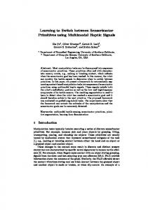
Figure 2. Quantification of the growth of afferents in the spinal cord. A, Computer reconstructions of afferent fibers in thi right half of the cord at LS2, following injection of LSl-LS3 DRGs with HRP. Each reconstruction was drawn from a single 40 pm section that exhibited the most ventral afferent projection at that stage. Medial is at the left and dorsal is at the top in each section. The boundaries of the lateral motor column are indicated by dotted lines. B, The dorsoventral projection of afferent axons at different developmental stages. At each stage, the fraction of the cumulated length of afferent axons below successive 10% increments in the dorsoventral length of the cord is plotted against the cord length. C, Number of axon segments crossing successive 10% increments of the dorsoventral cord length. Data in C and D were obtained from 2-3 embryos at each stage and are plotted as mean k SD (for n > 3).
0 I 0 DORSAL
50-
60-
2
40-
70-
p3
2
80-
E
g
go-
100
5
B
4-J
A
? L
2534
Lee et al. - Development
of Sensorimotor
Connections
in the Chick
32
Figure 3. Quantification of the growth of motoneurons during stages 29-32. A, Computer reconstructions of motoneurons in LS 1 and LS2 following injection of VRs with HRP. The 2 sections on the left were cut at 10 pm and processed using DAB as the chromogen; those on the right were cut at 40 pm and processed with TMB. Each reconstruction was drawn from a single section that showed the most dorsal dendritic extension with each chromogen. B, The ventrodorsal growth of motoneuronal dendrites at stages 29-30 and 32. At each group of stages, the fraction of the cumulated length of motoneuronal neurites (axons and dendrites) above successive 5% increments in the dorsoventral length of the cord is plotted against the cord length. Data were obtained from 3 embryos at each stage and are plotted as mean + SD. Portions of the curves for stages 30 and 32 from Figure 2B have been reproduced to illustrate the development of overlap between afferents and motoneuronal dendrites during these stages.
32
B g
80
z
70-
i? !I
60-
E I4 3 $
i
50 40-
SENSORY AXONS : : ::
3020-
0 DORSAL
20
40 CORD
60 LENGTH (X)
80
100 VENTRAL
The Journal
was studied by superfusing the preparation with solutions of the following agents added to normal or Mg2+-free Tyrode’s: APV (200 PM; Sigma and Cambridge Research Biochemicals); kynurenic acid (1.25 mM; Sigma); picrotoxin ( 100 PM; Sigma); and strychnine (10 PM; Sigma). To block chemical synaptic transmission, CaCl, was replaced with 2 mM MnCl, in normal or Mg2+-free Tyrode’s. High-calcium/high-magnesium solution contained 12 mM Ca2+ and 5 mM Mg2+; in 1 of 2 experiments in which this solution was used, [NaCl] was reduced to 119.5 mM to maintain normal osmolarity.
Results Anatomical analysis of afferent and motoneuron development The growth of afferent fibers into the spinal cord wasexamined by injecting HRP into the DRGs of the first 3 lumbosacral segmentsin 24 embryos between stages28 and 37. In 15 of theseembryos, the dendritic development of contralateral motoneurons in the samesegmentswas also monitored, following HRP injections into the VRs. Three embryos were processed with DAB, 6 with TMB/nitroferricyanide, and 15 with TMB/ heptamolybdate (seeMaterials and Methods). Although qualitatively similar results were obtained with all 3 methods, the TMB proceduresproved more useful for quantitative measures becauseof their lower background staining and higher sensitivity. Representative photomicrographs of labeled afferents and motoneurons are illustrated in Figure 1, and reconstructionsof sensoryand motor projections are shownin Figures2A and 3A, respectively. In 2 embryos injected with HRP at stage28, labeled sensory fibers had penetratedthe spinalcord but appearedto be confined to a dorsolateralband of white matter. Approximately 1 d later, at stages29-30 (4 embryos), sensoryaxons could be detected in the gray matter. At thesestages,90% of the cumulative length of labeled afferent axons occurred in the dorsal 30% of the cord (ca. 50 Km), and no axons extended below the dorsal 40% (Fig. 2B). The total number of labeledafferentswithin the gray matter increasedsubstantially by stage 32, although more than 80% were still found in the dorsalthird of the cord. As development proceededthe sensoryprojection continued to grow ventrolaterally so that, by stages36-37, sensoryaxons were distributed widely in the spinal cord and had reached the lateral motor column in all 7 embryos examined. Inspection of the afferent morphologiesillustrated in Figures 1 and 2A suggeststhat sensoryaxons do not branch significantly until after stage 34. To quantify the degree of branching at different stages,we counted the number of axon segmentsthat crossedmediolateral lines at 10% intervals along the dorsoventral axis of the cord (Fig. 2C). The number of axon crossings peaked in the dorsal 20% of the cord at all stages.At stages30 and 32, this number declined ventrally, as would be expected if little branching occurred. Betweenstages34 and 36, however, a second peak in the number of axon crossingsappearedjust below the dorsoventral midline of the cord, suggestingthat individual axons beganto branch there during that period of development. This area of branching coincides with the dorsal portion of the lateral motor column (Fig. 1). The elaboration of motoneuronal dendrites at stages29-30 and 32 wasmeasuredin a manner similar to that usedto assess the growth of afferents (Fig. 3B). This measurementwas not performed past stage32 becausewe were unable to obtain consistent labeling of dendrites in older embryos. At stages29-30, more than 80% of the cumulative dendritic length occurred in the ventral third of the cord, and there was no overlap between
of Neuroscience,
July
1988,
8(7)
2535
the territories occupied by afferents and motoneuronal dendrites. In contrast, 3 of 6 embryos at stage32 contained dendrites of labeledmotoneurons within the dorsal third of the gray matter, in the vicinity of the most ventrally projecting sensoryaxons. Therefore, the morphological basisfor a monosynaptic connection between afferents and motoneurons is present by at least stage32. While it is conceivable that undetected contacts between very small afferent terminals and dendrites exist prior to stage32, sensorimotor synapsesalso become resolvable electrophysiologically at this stage(seebelow). Physiological analysis of sensorimotor connectivity Propertiesof sensorimotorconnectivity in stage 37-39 embryos In 15 embryos examined at stages37-39, brief shocksof moderate intensity, delivered to a femorotibialis nerve branch or a DR in normal Tyrode’s, elicited composite, positive-going potentials in LS2 or LS3 VR (Fig. 4A). With femorotibialis nerve stimulation, the earliest component of these potentials began approximately 5 msecafter the arrival of the afferent volley in the correspondingDRG (range,3.9-6.0 msec;10embryos).This component usually fused with potentials appearing at longer latencies,including large, cyclic potentials produced by the central pattern generator for rhythmic limb movements (O’Donovan and Landmesser,1987).The early component wasthe first to appearasthe stimulusintensity wasgradually increasedfrom levels that were subthresholdfor afferent activation (Fig. 4B), and often its appearancecould be correlated with that of the fastest afferent spikes in the DRG (not shown). A small, predominantly negative deflection coincident with the afferent volley in the DRG record typically preceded the positive-going potential in the VR trace at these stages(seeFig. 11). In most cases,this deflection probably representedthe extracellular field potential of afferent spikeswithin the spinal cord, sincesimilar potentials have been associatedwith afferent invasion of the spinal cord in cats (Munson and Sypert, 1979). However, in peparationswhere the recording electrode on the VR was very closeto the DRG, the VR electrode may have picked up the afferent volley directly from the DRG. This latter possibility wasespecially likely in the smaller cords dissectedfrom young embryos. In order to determine whether the composite potentials recorded from the VRs accurately reflected the synaptic and spiking activity occurring in individual motoneurons, we recorded from LS3 VR and intracellularly from 31 LS3 motoneurons during stimulation of musclenerves in 10 embryos.Antidromic spike amplitudes ranged from 22 to 70 mV (mean, 5 1 f 12 mV; n = 28 cells) following VR or muscle nerve stimulation. Extracellular field potentials resulting from muscleafferent activation were not detectable. While most cells could be held for no more than approximately 10 min, in a few casesthe impalementswere stable for 3-4 hr, long enough to examine the effectsof severalchangesin superfusatecomposition (seebelow). In the vast majority of the cellswe recorded from (26 of 3 l), femorotibialis nerve stimulation elicited a depolarization in the motoneuron that was very similar to the VR potential, with both early and later componentswhen thesewere presentin the VR (Fig. 5, A-C, also seeFigs. 6-8B). The latency of the early PSP in these motoneurons was equal to that of the early component of the VR potential. The remaining 5 motoneuronswere either unresponsive to stimulation of the femorotibialis nerve or were hyperpolarized by it (Fig. 5D). Two of thesemotoneurons, both in stage39 embryos, were depolarized by stimulation
2536
Lee et al. * Development
of Sensorimotor
Connections
in the Chick
100 ms
5ms
Figure 4. Gradual recruitment of the various components of muscle nerve-evoked VR potentials with increasing stimulus intensity. A, Recordings from LS3 VR in a stage 39 embryo during stimulation of a femorotibialis nerve branch in normal Tyrode’s. At low stimulus intensity (bottom), only the early component was elicited. At an intermediate intensity (middle), this component increased in amplitude and began to merge with longer-latency potentials. High-intensity stimuli (top) evoked motoneuron spikes on the early component and later, during the first of several large potentials produced by the locomotor pattern generator. B, Recordings of the afferent volley produced in LS3 DRG by the stimuli delivered in A.
of the antagonistic sartorius nerve (Fig. 5E), indicating that at least somemotoneurons respond differentially to activation of afferents from antagonistic musclesby this stageof development. Several lines of evidence suggestedthat the shortest-latency component of the VR potentials included the monosynaptic EPSPgeneratedin motoneuronsby Ia muscleafferents.As shown above, the early component could be elicited in associationwith the firing of low-threshold, rapidly conducting sensory axons, which may belong to Ia afferents (Brinley, 1974). This component persistedin high-calcium/high-magnesiumsolutionsthat blocked later events (2 embryos; data not shown). In addition, its latency matched that of the earliest potentials recorded intracellularly in motoneurons and remained constant in each preparation, even during repetitive stimulation at lo-20 Hz. Moreover, the reaction of the early VR potential to pharmacologicalmanipulations wascharacteristic of the Ia EPSPin motoneurons of the newborn rat (Jahr and Yoshioka, 1986). In the presenceof the N-methyl-D-aspartate (NMDA) antagonist, APV (200 KM; 8 embryos),the early component remained,while most or all of the later components were abolished (Fig. 6). Becauseof the fusion of the various components of the VR potential, it was impossible to determine precisely the time courseand amplitude of the monosynaptic component in normal Tyrode’s. Therefore, potentials obtained during drug administration were compared with the maximum amplitude attained during the initial 10msecof the VR potential in normal Tyrode’s. The APV-resistant potential had a time-to-peak of approximately 6-l 0 msecand a maximum amplitudethat ranged from 68 to 96%of the amplitude of the early potential in normal saline.In contrast, the nonselective inhibitor of excitatory amino acid receptors,kynurenic acid (1.25 mM; 5 embryos),reduced the amplitude of the early component to 15-22% of its value in normal Tyrode’s and eliminated later potentials (Fig. 6). In one embryo from which recordingswere made simultaneously from LS3 VR and an LS3 motoneuron, the effects of APV and kynurenic acid on VR potentials were paralleled by similar changesin the motoneuron (Fig. 6). No recovery of the VR potential wasseenwhen superfusionwith theseagentswasmaintained (up to 1 hr for APV and 40 min for kynurenic acid), but their actions were completely reversed upon return to saline lacking the drugs.
The presenceof depolarizing IPSPs hasbeenreported in motoneuronsof the newborn rat (Takahashi, 1984;Jahr and Yoshioka, 1986) and the chick embryo (Velumian, 1984). To determine whether
any part of the APV-resistant
component
of muscle
nerve-evoked potentials in our preparationsmight be inhibitory, we examined the actions of picrotoxin and strychnine, blockers of GABA- and glycine-basedIPSPs, respectively, on the potential isolated by APV (3 embryos, all at stage39). In all cases, the addition of theseinhibitory blockers reducedthe amplitude of the later portion of the VR response,leaving a potential that appearedat the samelatency asthe early component. We believe this potential representsthe monosynaptic EPSP.In one of these experiments, the effects recorded in the VR were corroborated by simultaneous, intracellular recordings from an LS3 motoneuron (Fig. 7). Subtraction of the traces obtained in APV, picrotoxin, and strychnine from those in APV alone yielded positive-going potentials in the motoneuron and the VR that beganapproximately 5 msecafter the onsetof the monosynaptic EPSP. Theseresultsimply that musclenerve-evoked potentials isolated by APV were not purely monosynaptic in our preparations; they must have contained di- or perhapspolysynaptic potentials mediated by inhibitory transmitters. Extracellular Mg*+ at concentrations above 10 PM has been shown to be an effective depressantof motoneuron depolarizations induced by NMDA and by afferent activation (Ault et al., 1980). This observation raised the possibility that some portion
of the afferent input to motoneurons
in our preparations
might be inhibited in normal Tyrode’s, which contained 1 mM MgZ+.To addressthis question, we recorded VR potentials elicited by femorotibialis nerve stimulation in salinelacking MgZ+ (4 embryos). Changing the superfusatefrom normal to Mg2+free Tyrode’s markedly enhancedthe VR response,causinga larger underlying synaptic potential and a higher incidence of spiking (Fig. 8A). Furthermore, when the preparation was returned to normal Tyrode’s after the exposureto Mg2+-freesaline, the VR potential wasdepressedbelow its original magnitude in normal Tyrode’s (Fig. 8A). After approximately 1.5 hr in normal saline,the VR potential recovered partially, but wasstill smaller than the responseevoked initially in normal Tyrode’s. These solution changeshad very little effect on the size of the afferent volley (Fig. 8A). To determine whether alterations in [Mg*+] affectedthe mono-
The Journal
of Neuroscience,
July
1988.
8(7)
2537
D
I
20mV
MN
I
500
pv
I
VR ,,
20mV
lJ 100
ms
200
ms
Figure 5. Correlation between nerve-evoked potentials recorded in VRs and motoneurons. Intracellular recordings from LS3 motoneurons (MN; upper)are paired with simultaneous recordings from LS3 VR (lower)in 3 embryos at stage 39. A-C, Three motoneurons that showed a synaptic response very similar to that elicited in the VR. All 3 were recorded in normal Tvrode’s durina stimulation of the same femorotibialis nerve branch. Records in A were from the same VR and motoneuron as are shown in Figure-6; those in iand C were obtained from another embryo. D, E, A motoneuron that was hyperpolarized by stimulation of the femorotibialis nerve (0) and depolarized by sartorius nerve stimulation (E). Both nerves produced positive-going potentials in the VR. The recordings were made in Mg2+-free Tyrode’s.
synaptic EPSP produced by muscle afferents, we isolated the EPSP in normal Tyrode’s containing APV, picrotoxin, and strychnine, while recording from LS3 VR and an LS3 motoneuron (one embryo; Fig. 8B). Switching to Mg2+-freeTyrode’s containing theseagentsresulted in a slight increasein the size of the EPSP in the motoneuron but almost no change in the VR potential. Theseresultssuggestthat most of the potentiation of sensorimotor transmissionby Mg*+ removal occurs in polysynaptic pathways. The mechanismsthrough which Mg*+ might act in this systemare consideredin the Discussion. Development of sensorimotorconnectivity The development of functional connections between afferents and motoneurons was examined by recording afferent-evoked potentials from LS2 or LS3 VR in 16 embryos at stages28-35. In normal Tyrode’s, early and later componentsof femorotibialis nerve-evoked VR potentials were demonstrableby stages 32-33 (1 of 2 embryos at stage32, and 1 of 1 at stage33; Fig. 9). Becauseof its small amplitude, the early component was difficult to resolve without signal-averagingat these stages.As in more mature embryos, the early component at stages32-33 wasstrongly inhibited by 1.25mM kynurenic acid (oneembryo), but was largely unaffected by 200 PM APV, which abolished later potentials (2 embryos). The latency of the early component was comparable to that at stages37-39, while its time-to-peak
in APV (2-4 msec)wasconsiderably shorter. In contrast to the potentials recordedat later stages,the APV-resistant component at stage33 was not reduced by picrotoxin and strychnine (one embryo). By stages34-35, the early component had increased in size, while retaining its sensitivity to kynurenic acid (one embryo) and its insensitivity to APV (2 embryos; Fig. 9). Although the amplitude of the early component at these intermediate stageswassmallerthan at stages37-39, its latency and time-to-peak in APV were within the range of values for the later stages. In one embryo examined at stage3 1, the responsein LS3 VR to femorotibialis nerve stimulation in normal Tyrode’s consistedof a slowly rising positive potential preceded by a very small early potential (Fig. 9). The early potential, which was eliminated in CaZ+-free,Mn2+-containing saline,may have been the precursorof the early synaptic componentseenat later stages. However, since we did not test its sensitivity to other pharmacologicalblockers, we cannot exclude the possibility that it representeda Ca2+-sensitivepart of the a,Terentvolley picked up by the VR electrode. Prior to stage3 1, stimulation of femorotibialis nerves at intensities sufficient to elicit a maximal afferent volley in LS3 DRG failed to evoke any synaptic responsein LS3 VR in normal Tyrode’s (2 embryos at stage30, 3 at stage29, and 1 at stage 28; Fig. 9). Activation of afferentsvia DR or spinalnerve stim-
2538
Lee et al. - Development
of Sensorimotor
Connections
in the Chick
MN
10 mV
100
ms
6. Sensitivityof muscle nerve-evokedVR potentialsandmotoneurondepolarizationsto APV and kynurenicacid. Responses to stimulationof a femorotibialisnervebranchwererecordedintracellularly in an LS3 motoneuron(MN, top truces) andin LS3 VR (bottom truces)in a stage39 embryo.In normalTyrode’s,compositepotentials wererecordedin the VR andthe motoneuron.Super&ion ofthe preparationwith 200 /LM APV preservedthe earliestpart of the synaptic response andabolishedlatercomponents. Kynurenicacid(KY; 1.2mM) greatlyreducedthe amplitudeof the early response and completely eliminatedlater components.
20 ms
Figure
ulation at stage30 was similarly incapable of evoking VR synaptic potentials in normal saline (2 of 2 embryos). In saline lacking Mg*+, however, long-latency, slowly rising potentials could be produced as early as stage28 by femorotibialis nerve stimulation (2 of 2 embryos at stage30, 2 of 2 at stage29, and 1 of 2 at stage28; Fig. lOA). These potentials were abolished when the Mgz+-free Tyrode’s contained APV (2 embryos) or kynurenic acid (one embryo). Stimulation of descendingtracts in the spinal cord at thesestagesproduced VR potentials quite different in amplitude and form from those following nerve shock (Fig. lOB), indicating that the responsesto nerve stimulation are unlikely to have resultedfrom passivestimulusspread to the cord. Removal of Mg2+did not revealdanearly component in the nerve-evoked VR potential before stage32. At stages37-39 (6 embryos), both early and late components of the nerve- and DR-evoked VR synaptic potentials were completely and reversibly eliminated in solutions that block chemical synaptic transmission (0 mM Ca*+, 2 mM Mn*+; Fig. 11). The blockade of VR synaptic potentials in these solutions was corroborated by intracellular recording from an LS3 motoneuron in one embryo at stage39. Theseobservations suggestthat no part of the potentials elicited in motoneurons by afferent stimulation is electrically mediated at these stages.The predominantly negative deflection causedby the afferent volley remainedunchangedin the VR records,indicating that the number of afferent fibers activated by the stimulus was not significantly affected by the removal of Ca2+or the addition of Mn2+. In younger embryos (2 each at stages34-35 and 32-33, 1 at stage 3 1, and 2 each at stages30 and 29), most of the VR responsewas blocked by superfusion with 0 mM Ca*+/2 mM
7. Effectof inhibitory blockerson the APV-resistantcomponentof nerve-evokedmotoneuronpotentials.Simultaneous recordings from an LS3motoneuron(MN). LS3VR. andLS3DRG in a staae39 embryo.Activation of fembro&ialisafferentsin normalTyrode’scontaining200PM APV producedearlysynapticresponses similarto those in Figure6. Addition of 100PM picrotoxin and 10PM strychnineto that solution(API/+P+,S) blockedthe laterportionsof thoseresponses in the motoneuronand the VR. Subtractionof the responses in APV+ P+Sfrom thosein APV revealsthe time courseof the depolarizing IPSP(DZFF’). The restingpotentialof the motoneuronwasapproximately -60 mV. The addition of picrotoxin and strychninedid not significantlyaffectthe amplitudeof the afferentvolley in the DRG.
Figure
Mn2+(Fig. 11). In someof theseembryos, however, the negative deflection of the afferent volley was immediately followed by a small positivity that persistedin Ca2+-free,Mn2+-containing saline. This latter potential may be part of the extracellular field generated by the afferent volley (Munson and Sypert, 1979). Alternatively, it may representan electrical component to the motoneuron synaptic potential that is lost at later developmental stages. Discussion Developmentof monosynaptic connectionsbetweenmuscle afferents and motoneurons Stimulation of the femorotibialis nerve in normal Tyrode’s elicited a short-latency, rapidly rising potential in LS2-3 ventral roots asearly asstages32-33. Although intracellular recordings were not attempted from motoneurons at these stages,our simultaneous intracellular and ventral root recordings at stages 37-39 and previous observations (O’Donovan, 1987) demonstrate that VR potentials acccurately reflect membranepotential changesin motoneurons. VR potentials at stages32-35 were pharmacologically similar to those at stages37-39: the earliest component of the potential was resistant to APV and sensitive to kynurenic acid, making it similar to the Ia EPSP in mammalian motoneurons (Jahr and Yoshioka, 1986; Flatman et al., 1987) and to monosynaptic EPSPsproduced in rat dorsal horn neurons by DRG cells in vitro (Jahr and Jessell, 1985). We therefore regard the early APV-resistant potential recorded in
The Journal
of Neuroscience,
July
1988,
8(7)
2539
10 mV
VR .mJ
500 pv
N (post) =---A
--
A
100 ms
VR
50 pv
Figure 8. Effect of MgZ+ on nerve-evoked motoneuron potentials. A, Recordings from LS3 VR and LS3 DRG during stimulation of a femorotibialis nervebranch in a stage 39 embryo. The synaptic response elicited initially in normal Tyrode’s,N(pre),wassignificantlyenhanced after 11min in Mgz+-free Tyrode’s (-Mg), and was markedly depressed 15 min after returning to normal Tyrode’s, N(post).The VR response partially recovered its initial magnitude during the next 1.5 hr of continuous superfusion with normal Tyrode’s. These solution changes caused only a slight change in the size of the afferentvolley. B, Recordings from the sameLS3 motoneuron,VR, andDRG asshownin Figure7 duringfemorotibialisnerve stimulation. Switching the saline from normal Tyrode’s containing 200 PM APV, 100 PM picrotoxin, and 10 PM strychnine (N) to Mg-free Tyrode’s containingtheseagents(-Mg) produceda smallincrease in the amplitudeof the monosynaptic EPSP but did not affect the afferent volley.
VRs at stages32-33 as the first detectable monosynaptic connection between femorotibialis afferents and LS2-3 motoneurons. Stages32-33 should be regardedasan upper estimate of the time at which direct sensorimotor synapsesform in the lumbosacral spinal cord of the chick, becauseonly one population of afferents was studied physiologically in this investigation. However, HRP injectionsinto the DRGs, which labeleda broader spectrum of afferents, indicated that a morphological substrate for direct connections was not presentuntil this time. While it is possible that our techniques were not sensitive enough to reveal contacts involving very fine processesor very weak synaptic transmission, superfusion with Mg2+-free saline, which demonstratedotherwise undetectablepolysynaptic inputs from afferentsto motoneuronsat stages28-30, did not reveal monosynaptic connectionsbefore stage32. The pharmacological profile of the early potential suggests that monosynaptic transmission between femorotibialis afferents and motoneurons in the chick is mediated by a glutamatelike transmitter and involves a non-NMDA type of receptor, such asthe G, receptor identified on cultured embryonic chick motoneurons (O’Brien and Fischbach, 1986). Whether the monosynaptic potential in our preparationsalsoincluded a component produced by binding to NMDA receptors was difficult to examine experimentally, sincethe monosynaptic EPSPcould not be isolatedreliably without the useof APV or high-calcium/ high-magnesiumsolutions, both of which block thesereceptors. Sensorimotor transmissionwasabolishedat stages37-39 by solutions that block chemical synapses.In conjunction with the report by Eide et al. (1982) that transmissionis mainly chemical at stages44-45, this finding suggeststhat the monosynaptic EPSPin the chick doesnot contain a significant electrical component during the later stagesof embryonic development. In this regard, sensorimotorconnectivity in the chick at thesestages resemblesthat in mammals(Engbergand Marshall, 1979;Finkel and Redman, 1983)and differs from that in the frog (Shapovalov et al., 1978; Frank and Westerfield, 1983). Since we did not examine sensorimotortransmissionwith intracellular recording
prior to stages37-39, and sincethere was a small, positive VR potential that persistedin Caz+-free,Mnz+-containing saline in some young embryos, it is not clear whether the afferent PSP in the chick is entirely chemical at all developmental stages. Developmentof polysynaptic pathways betweenmuscle afferents and motoneurons Long-latency, slowly rising VR potentials could be elicited by femorotibialis nerve stimulation as early as stage28. Although we did not observe afferent fibers penetrating the gray matter of the spinal cord until stage29, Johnsonet al. (1986) found a few small sensory axons within the dorsal horn at stage 28. Under our laboratory conditions, embryosreachedstage28 after 6-7 d of incubation, the same age at which Windle and Orr (1934) observed the first sensory collaterals within the spinal gray matter and the first local wing reflexes. It is conceivable that the potentials we recorded before stages32-33 were immature versions of the monosynaptic EPSP, since similar potentials can berecorded in musclefibers during the earlieststages in the development of neuromuscular transmission(Dennis et al., 1981). This possibility seemsunlikely, however, becausethe potentials recorded prior to stage 32 in our preparations were completely eliminated by APV, whereasthe monosynaptic potential recorded after stage32 was largely insensitive to APV. Moreover, overlap betweenafferent collateralsand motoneuron dendrites was not demonstrableanatomically prior to stage32. Our results indicate that polysynaptic connections between femorotibialis afferentsand motoneuronsin the chick form earlier in development than do monosynaptic connections. Comparableresultshave beenobtained for other afferent populations in the rat (Saito, 1979)and the frog (Forehand and Farel, 1982; Frank and Westerfield, 1983), suggestingthat this is a general feature of vertebrate spinal development. The sensitivity of theseearly-developing polysynaptic potentials to Ca*+-freesalineand to antagonistsof excitatory amino acidsindicates that chemical synapsesexist at one or more sites in the pathway that produces them. Such potentials could be initiated by synaptic contacts between afferents and intemeu-
2540
Lee et al. * Development
of Sensorimotor
Connections
in the Chick
32
I’ II ~jtm~~.~~ 100 ms
1Oms
Figure 9. Developmentof musclenerve-evokedVR potentials.Recordings from LS3VR at variousstages duringstimulationof femorotibialis nervebranches.The initial portion of eachtraceon the is expandedat right. The earlycomponentof the VR potentialwasclearlyrecognizable
left
by stage32.Responses shownwereobtainedin normalTyrode’sand,for stages 32, 35,and 39,in normalTyrode’scontaining200PM APV. Stage 39 recordsarethesameasthoseusedin Figure6. Thetracesfrom stages 35and39aresingleresponses, whilethosefrom stages 29-32areaveraged responses to 20 shocksdeliveredat l/min. At stages29-31, the stimulusintensitywasadjustedto give the largestafferentvolley in LS3 DR. rons. Alternatively, the first step in this pathway might involve a nonsynaptic mechanism,suchasephaptic interactions or local depolarizations resulting from the releaseof K+ ions by synchronously activated sensory fibers (Nicoll, 1979). Becausepolysynaptic connectionswereblocked by 1 mM Mg2+ before stage 31 in isolated spinal cord preparations, it is not clear whether these early connections are functional in ova In more mature embryos, superfusionwith Mg2+-freesalinecaused an augmentationof sensory-evokedmotoneuronpotentials.Most of this augmentation appearedto have been effected via polysynaptic pathways, perhaps as a result of reductions in interneuron spike thresholds caused by the lower divalent cation concentration (Frankenhaeuserand Hodgkin, 1957). Furthermore, intemeuronal transmissionmay have been enhancedby elimination of the voltage-dependentblock ofNMDA-activated ion channelsby Mg2+(Mayer et al., 1984; Nowak et al., 1984). The monosynaptic EPSP isolated by APV, picrotoxin, and strychnine was also slightly smallerin normal Tyrode’s than in Mg2+-freesaline. Since the size of the afferent volley remained the samein both solutions,the depressionof the EPSPby Mg2+ in the presenceof APV may have been the consequenceof a
presynaptic effect of Mg2+ on transmitter release (Kuno and Takahashi, 1986). Superfusionof the isolated cord with picrotoxin and strychnine at stage33 had no effect on the APV-resistant component of the VR potential evoked by femorotibialis nerve stimulation. By stage39, addition of these agentsreduced the amplitude of this component by eliminating a depolarizing potential that began several millisecondsafter the monosynaptic EPSP. Obata et al. (1978) have reported that GABA and glycine excited cultured spinal neuronsfrom 6 and 8 d chick embryos; when applied to neurons from 10 d embryos (approximate stages3435), thesetransmitters produced inhibition. Therefore, we assume that the potential blocked by picrotoxin and strychnine at stage39 wasinhibitory. The latency of the potential suggests that it wasproduced polysynaptically, but the pathway involved in its production has not been determined. One candidate is recurrent (Renshaw)inhibition initiated by femorotibialis motoneurons in neighboring segments.Since we generally did not sever VRs adjacent to the root from which recordings were made, thesemotoneurons may have been activated antidromically by stimulation of femorotibialis nerves.
The Journal
A.STlM
FEM
B.STIM
CORD
of Neuroscience,
July
1988,
8(7)
2541
I !!
100
20
ms
ms
Figure 10. Effect of Mgz+ on nerve- and cord-evoked VR potentials in a stage 28 embryo. Recordings from LS3 VR during stimulation of a femorotibialis nerve branch (A) or the central spinal cord at thoracic segment 6 (B). The initial portions of traces on the left are expanded at right. Each trace in A is an average of 10 or 20 responses; traces in B are single events. A, Muscle nerve stimulation produced no response in the VR in normal Tyrode’s (N), but elicited a long-latency potential in the absence of Mg2+ (-Mg); this solution change only slightly increased the amplitude of the afferent volley detected in the VR electrode (arrow). B, Cord-evoked potentials were reduced in size but not abolished in the presence of Mg”.
Normal --200 pv -Ca+Mn Stage 39
--
100 pv
32 10 ms
Figure 11. Chemical nature of nerveevoked VR potentials. Recordings from LS3 VR during stimulation of a femorotibialis nerve branch in 3 embrvos. stages 39,35, and 32. At each stage,*thc response to nerve stimulation was re. corded in normal Tyrode’s and in Ca2+. free Tyrode’s containing 2 mM Mn*+ The negative deflections caused by tht afferent volleys are marked by arrows. Traces at stage 32 are averaged responses to 20 shocks given at l/min.
2542
Lee et al. - Development
of Sensorimotor
Connections
in the Chick
Both depolarizing and hyperpolarizing IPSPs were recorded in motoneurons during stimulation of femorotibialis nerve branches. It is conceivable that the responses to different inhibitory transmitters are of opposite sign, or that individual motoneurons respond in opposite ways to the same transmitter. In addition, IPSPs in some motoneurons may have been reversed as a result of chloride entry or membrane depolarization caused by impalement of the cell (Velumian, 1984). Mechanisms controlling the formation of connections between afferents and motoneurons The outgrowth of sensory fibers into the chick hindlimb begins at stage 27 and is segmentally correct from the outset (Honig, 1982). The first monosynaptic connections between motoneurons and the central processes of femorotibialis afferents did not become detectable in our preparations until several stages later. These observations raise the possibility that the formation of synapses between muscle afferents and motoneurons might depend in some manner on a signal from the periphery, possibly from muscle. Such a mechanism has been proposed to explain the formation of appropriate connections between foreign muscle afferents and motoneurons in the bullfrog, following deletion of the ganglion that normally innervates the forelimb (Frank and Westerfield, 1982b). Myotypic specification has not been implicated in the formation of central connections of chick motoneurons, and has been disproved as a factor in the determination of connectivity of motor networks (Narayanan and Hamburger, 197 1; Landmesser and O’Donovan, 1984). Nevertheless, it will be important to establish whether or not patterns of afferent connectivity are regulated by peripheral factors. This could be assessed in the chick by the use of limb rotations or shifts to produce foreign innervation of muscles (see Landmesser and O’Donovan, 1984). The development of sensorimotor connections in the chick begins before the completion of cell death both in motoneurons (Hamburger, 1975) and afferents (Hamburger and Levi-Montalcini, 1949). It is therefore possible that cell death may be involved in the formation of these connections. In the bullfrog, the temporal relationship between these events is not as clear. Using intracellular recording, Frank and Westerheld (1983) found that afferent synapses onto brachial motoneurons formed by stage XVII, but data on cell death in this region of the cord are lacking. In the lumbar cord, sensory axons can be found in the intermediate regions of the gray matter at stages VIII-IX, and DR stimulation can elicit short-latency, apparently monosynaptic VR potentials by stage X (Forehand and Farel, 1982). Motoneuron cell death in this region occurs predominantly between stages V and X, with a large loss at stages VII-X (Farel, 1987). Hence, the establishment of sensorimotor contacts may overlap to some extent with motoneuron cell death in this species as well. The survival of afferent neurons could depend, in part, on the formation of synaptic connections with appropriate central and peripheral targets. Davies et al. (1986) have presented evidence that the survival of cultured muscle afferents from the chick trigeminal nucleus depends on a factor derived from central neurons (brain-derived neurotrophic factor) that is distinct from the peripheral survival factor, NGF. These authors proposed that only those neurons making appropriate central and peripheral connections would receive sufficient amounts of both factors. This hypothesis predicts that the initial pattern of connectivity between afferent fibers and their central targets would
include inappropriate connections. Moreover, saving afferent neurons by chronic NGF administration (Hamburger et al., 198 1) should result in the maintenance of at least some of the inappropriate connections. In chick embryos near the time of hatching (stages 44-45), lateral gastrocnemius motoneurons receive monosynaptic excitatory input from homonymous and heteronymous muscle afferents and inhibitory input from antagonistic afferents (Eide et al., 1982). This pattern of connectivity is similar to that documented in the cat (Eccles et al., 1957) and the rat (Jahr and Yoshioka, 1986). Although we observed occasional responses of opposite polarity in motoneurons during stimulation of antagonistic muscle nerves at stage 39, our data do not address the question of whether the initial contacts between muscle afferents and motoneurons are appropriate in the chick. This question can be answered only by examining the pattern of sensorimotor connections at the stages when these connections are beginning to form. References Ault, B., R. H. Evans, A. A. Francis, D. J. Oakes, and J. C. Watkins (1980) Selective depression of excitatory amino acid induced depolarizations by magnesium ions in isolated spinal cord preparations. J. Physiol. (Lond.) 307: 4131128. Brinley, F. J., Jr. (1974) Excitation and conduction in nerve fibers. In MedicalPhysiology,vol. 1, V. B. Mountcastle, ed., p. 75, Mosby, St. Louis, MO. Burke, R. E., and P. Rudomin (1977) Spinal neurons and synapses. In TheHandbookof Physiology,Section1: TheNervousSystem,vol. 1, pt. 2, E. R. Kandel, ed., pp. 877-944, American Physiological Society, Bethesda, MD. Davies, A. M., H. Thoenen, and Y.-A. Barde (1986) Different factors from the central nervous system and periphery regulate the survival of sensorv neurones. Nature 319: 497-499. Dennis, M.-J., L. Ziskind-Conhaim, and A. J. Harris (1981) Development of neuromuscular junctions in rat embryos. Dev. Biol. 81:
266-279.
Eccles, J. C., R. M. Eccles, and A. Lundberg (1957) The convergence of monosynaptic excitatory afferents on to many different species of alnha motoneurones. J. Phvsiol. (Land.) 137: 22-50. Eide, A. H., J. K. S. Jansen, and R. R. Ribchester (1982) The effect of lesions in the neural crest on the formation of synaptic connexions in the embrvonic chick sninal cord. J. Phvsiol. (Land.) 324: 453-478. Engberg, I., and K. C. Marshall (1979) Reversal potential for Ia excitatory post synaptic potentials in spinal motoneurons of cats. Neuroscience 4: 1583-1591. Farel, P. B. (1987) Motoneuron number in the lumbar lateral motor column of larval and adult bullfrogs. J. Comp. Neurol. 261: 266-276. Finkel, A. S., and S. J. Redman (1983) The synaptic current evoked in cat spinal motoneurones by impulses in single group Ia axons. J. Physiol. (Lond.) 342: 6 15-632. Flatman, J. A., J. Durand, I. Engberg, and J. D. C. Lambert (1987) Blocking the monosynaptic epsp in spinal cord motoneurones with inhibitors of amino-acid excitation. In Excitatory AminoAcid Transmission,T. P. Hicks, D. Lodge, and H. McLennan, eds., pp. 285292, Liss, New York. Forehand, C. J., and P. B. Fare1 (1982) Spinal cord development in anuran larvae: I. Primary and secondary neurons. J. Comp. Neurol. 209: 386-394. Frank, E., and M. Westerfield (1982a) Synaptic organization of sensory and motor neurones innervating triceps brachii muscles in the bullfrog. J. Physiol. (Lond.) 324: 479-494. Frank, E., and M. Westerfield (1982b) The formation of appropriate central and peripheral connexions by foreign sensory neurones of the bullfrog. J. Physiol. (Lond.) 324: 495-505. Frank, E., and M. Westerfield (1983) Development of sensory-motor synapses in the spinal cord of the frog. J. Physiol. (Lond.) 343: 593610. Frank, E., W. A. Harris, and M. B. Kennedy (1980) Lysophosphatidyl choline facilitates labeling of CNS projections with horseradish peroxidase. J. Neurosci. Methods 2: 183-189.
The Journal
Frankenhaeuser, B., and A. L. Hodgkin (1957) The action of calcium on the electrical properties of squid axons. J. Physiol. (Lond.) 137: 218-244. Hamburger, V. (1975) Cell death in the development of the lateral motor column of the chick embryo. J. Comp. Neurol. 160: 535-546. Hamburger, V. (1976) The developmental history of the motor neuron. Neurosci. Res. Prog. Bull. 15: l-37. Hamburger, V., and H. L. Hamilton (195 1) A series of normal stages in the development of the chick embryo. J. Morphol. 88: 49-92. Hamburger. V.. and R. Levi-Montalcini ( 1949) Proliferation. differentiatiin ‘and degeneration in the spinal ganglia of the chick embryo under normal and experimental conditions. J. Exp. Zool. 111: 457501. Hamburger, V., J. K. Brunso-Bechtold, and J. W. Yip (198 1) Neuronal death in the spinal ganglia of the chick embryo and its reduction by nerve growth factor. J. Neurosci. I: 60-7 1. Hollyday, M. (1980) Organization of motor pools in the chick lumbar lateral motor column. J. Comp. Neurol. 194: 143-170. Honig, M. G. (1982) The development of sensory projection patterns in embryonic chick hind limb. J. Physiol. (Land.) 330: 175-202. Jahr, C. E., and T. M. Jesse11 (1985) Synaptic transmission between dorsal root ganglion and dorsal horn neurons in culture: Antagonism of monosynaptic excitatory postsynaptic potentials and glutamate excitation by kynurenate. J. Neurosci. 5: 228 l-2289. Jahr, C. E., and K. Yoshioka (1986) Ia afferent excitation of motoneurones in the in vitro new-born rat spinal cord is selectively antagonized by kynurenate. J. Physiol. (Lond.) 370: 5 15-530. Johnson, F. A., B. M. Davis, and E. Frank (1986) Ontogeny ofcentral projections of lumbar primary sensory neurons in chick. Sot. Neurosci. Abstr. 12: 1050. Koebbe, M., and M. J. O’Donovan (1985) Properties and distribution of muscle afferent projections to lumbosacral motoneurons in the spinal cord of the chick embryo. Sot. Neurosci. Abstr. 11: 63. Koebbe, M. J., and M. J. O’Donovan (1987) Anatomical studies of afferent projections to motoneurons in the lumbosacral cord of the chick embryo. Sot. Neurosci. Abstr. 13: 78. Kudo, N., and T. Yamada (1987) Morphological and physiological studies of development of the monosynaptic reflex pathway in the rat lumbar spinal cord. J. Physiol. (Lond.) 389: 441459. Kuno, M., and T. Takahashi (1986) Effects of calcium and magnesium on transmitter release at Ia synapses of rat spinal motoneurones in vitro. J. Physiol. (Lond.) 376: 543-553. Landmesser, -L. (1978) The distribution of motoneurones supplying chick hind limb muscles. J. Phvsiol. (Land.) 284: 371-389. Landmesser, L. T., and M. J. O’Donovan (1984) The activation patterns of embryonic chick motoneurones projecting to inappropriate muscles. J. Physiol. (Lond.) 347: 205-224. Lee, M. T., and M. J. O’Donovan (1987) Development of the muscle afferent-motoneuron pathway in the chick embryo: Physiology. Sot. Neurosci. Abstr. 13: 78. Levi-Montalcini, R., and G. Levi (1943) Recherches quantitatives sur la marche du processus de differentiation des neurones dans les ganglions spinaux de l’embryon de Poulet. Arch. Biol. 54: 189-206. [Cited by Hamburger, V., and R. Levi-Montalcini (1949) Proliferation, differentiation and degeneration in the spinal ganglia of the chick embryo under normal and experimental conditions. J. Exp. Zool. 111: 457501.1 Mayer, M. L., G. L. Westbrook, and P. B. Guthrie (1984) Voltagedependent block by Mg2+ of NMDA responses in spinal cord neurones. Nature 309: 261-263. Mesulam, M.-M. (1978) Tetramethyl benzidine for horseradish peroxidase neurohistochemistry: A non-carcinogenic blue reaction prod-
of Neuroscience,
July
1988,
8(7)
2543
uct with superior sensitivity for visualizing neuronal afferents and efferents. J. Histochem. Cytochem. 26: 106-l 16. Munson, J. B., and G. W. Sypert (1979) Properties of single central Ia afferent fibres projecting to motoneurones. J. Physiol. (Lond.) 296: 315-327. Narayanan, C. H., and V. Hamburger ( 197 1) Motility in chick embryos with substitution of lumbosacral by brachial and brachial by lumbosacral spinal cord segments. J. Exp. Zool. 178: 415432. Nicoll, R. A. (1979) Dorsal root potentials and changes in extracellular potassium in the spinal cord of the frog. J. Physiol. (Lond.) 290: 113127. Nowak, L., P. Bregestovski, P. Ascher, A. Herbet, and A. Prochiantz (1984) Magnesium gates glutamate-activated channels in mouse central neurones. Nature 307: 462-465. Obata, K., M. Oide, and H. Tanaka (1978) Excitatory and inhibitory actions of GABA and glycine on embryonic chick spinal neurons in culture. Brain Res. 144: 179-l 84. O’Brien, R. J., and G. D. Fischbach (1986) Characterization of excitatory amino acid receptors expressed by embryonic chick motoneurons in vitro. J. Neurosci. 6: 3275-3283. O’Donovan, M. J. (1987) Developmental approaches to the analysis of vertebrate central pattern generators. J. Neurosci. Methods 21: 275-286. O’Donovan, M. J., and L. Landmesser (1987) The development of hindlimb motor activity studied in the isolated spinal cord of the chick embryo. J. Neurosci. 7: 3256-3264. Olucha, F., F. Martinez-Garcia, and C. L6pez-Garcia (1985) A new stabilizing agent for the tetramethyl benzidine (TMB) reaction product in the histochemical detection of horseradish peroxidase (HRP). J. Neurosci. Methods 13: 131-138. ODDenht?iDI. R. W.. I.-W. Chu-Wane. and R. F. Foelix (1975) Some *aspects of synaptogenesis in the s$nal cord of the chick embryo: A quantitative electron microscopic study. J. Comp. Neurol. 161: 383418. Redman, S. (1979) Junctional mechanisms at group Ia synapses. Prog. Neurobiol. 12: 33-83. Saito, K. (1979) Development of spinal reflexes in the rat fetus studied in vitro. J. Phvsiol. (Land.) 294: 58 l-594. Shapovalov, A. I., B. I. Shihaev, and A. A. Velumian (I 978) Mechanisms of post-synaptic excitation in amphibian motoneurones. J. Physiol. (Lond.) 279: 437-455. Takahashi, T. (1984) Inhibitory miniature synaptic potentials in rat motoneurons. Proc. R. Sot. L&d. [Biol.] 2il: iO3-iO9. Velumian. A. A. (1984) Direct evidence for nostsvnavtic inhibition in the embryonic chick spinal cord. Dev. Brain Res. i4: 229-239. Visintini, F., and R. Levi-Montalcini (1939) Relazione tra differenziazione strutturale e funzionale dei centri e delle vie nervose nell’embrione di ~0110. Arch. Suisses Neurol. Psych. 44: 119-150. [Cited by Hamburger, V., and R. Levi-Montalcini (1949) Proliferation, differentiation and degeneration in the spinal ganglia of the chick embryo under normal and experimental conditions. J. Exp. Zool. 111: 457-501.1 Windle, W. F. (1934) Correlation between the development of local reflexes and reflex arcs in the spinal cord of cat embryos. J. Comp. Neurol. 59: 48 l-505. Windle, W. F., and R. E. Baxter (1936) Development of reflex mechanisms in the spinal cord of albino rat embryos. Correlations between structure and function, and comparisons with the cat and the chick. J. Comp. Neurol. 63: 189-209. Windle, W. F., and D. W. Orr (1934) The development of behavior in chick embryos: Spinal cord structure correlated with early somatic motility. J. Comp. Neurol. 60: 287-307.