ABSTRACT: Bone formation is a carefully controlled developmental process involving morphogen-mediated ... Key words. ..... related genes through a specific DNA element called an ... search for new elements, particularly those able to con-.
THE DEVELOPMENTAL CONTROL OF OSTEOBLAST-SPECIFIC GENE EXPRESSION: ROLE OF SPECIFIC TRANSCRIPTION FACTORS AND THE EXTRACELLULAR MATRIX ENVIRONMENT R.T. Franceschi
Department of Periodontics/Prevention/Geriatrics, School of Dentistry, and Department of Biological Chemistry, School of Medicine, University of Michigan, 1011 N. University Ave., Ann Arbor, Michigan 48109-1078 ABSTRACT: Bone formation is a carefully controlled developmental process involving morphogen-mediated patterning signals that define areas of initial mesenchyme condensation followed by induction of cell-specific differentiation programs to produce chondrocytes and osteoblasts. Positional information is conveyed via gradients of molecules, such as Sonic Hedgehog that are released from cells within a particular morphogenic field together with region-specific patterns of hox gene expression. These, in turn, regulate the localized production of bone morphogenetic proteins and related molecules which initiate chondrocyte- and osteoblast-specific differentiation programs. Differentiation requires the initial commitment of mesenchymal stem cells to a given lineage, followed by induction of tissue-specific patterns of gene expression. Considerable information about the control of osteoblast-specific gene expression has come from analysis of the promoter regions of genes encoding proteins like osteocalcin that are selectively expressed in bone. Both general and tissue-specific transcription factors control this promoter. Osf2/Cbfal, the first osteoblast-specific transcription factor to be identified, is expressed early in the osteoblast lineage and interacts with specific DNA sequences in the osteocalcin promoter essential for its selective expression in osteoblasts. The OSF2/CBFAI gene is necessary for the development of mineralized tissues, and its mutation causes the human disease, cleidocranial dysplasia. Committed osteoprogenitor cells already expressing Osf2/Cbfa 1 must synthesize a collagenous ECM before they will differentiate. A cell:ECM interaction mediated by integrin-type cell-surface receptors is essential for the induction of osteocalcin and other osteoblast-related proteins. This interaction stimulates the binding of Osf2/Cbfa 1 to the osteocalcin promoter through an as-yet-undefined mechanism.
Key words. Bone formation, gene expression, transcription.
Introduction
The Embryonic Patterning of Bone
An understanding of factors controlling gene expression in osteoblasts is of particular importance to dental researchers, both because of the fundamental role played by bone in the overall structure of oral tissues and because underlying principles involved in the control of osteoblasts will likely be relevant to other mineralized tissues such as dentin and cementum. The goal of this article is to provide a timely overview of current knowledge regarding early events in bone formation, with emphasis on the nuclear factors required for the selective expression of specific genes in osteoblasts and their regulation by extracellular matrix signals.
There are two broad phases of tissue morphogenesis; an initial patterning phase, in which cells destined to form a given tissue are specified in time and space, and the subsequent induction of the necessary cellular phenotypes to produce the tissue of interest. These phases are not mutually exclusive. Rather, cells within a developing tissue can be viewed as altering the pattern of differentiation of their neighbors through a variety of signals mediated by diffusible factors, cell:extracellular matrix interactions, and direct cell:cell contact. The patterning of bones and teeth has been the subject of several recent reviews and will not be extensively discussed in this article (Erlebacher et al., 1995; Thesleff
Oral Biol 40 Crit 40 Crit Rev Rev Oral Med Biol Med
1O(l):40-57
10(l):40-57 (1999)
Decreasing Proliferation and Increasing Differentiation Post-mitotic Limited self-renewal Extensive proliferation
Unlimited self-renewal
Stem Cell
K] Mature Immature osteoprogenitor osteoprogenitor
0 0 Preoste
Preosteoblast
oMature Osteoblast
l0 Osteocyte
Progenitors for other mesenchymal cells including adipocytes, fibroblasts, and myoblasts
Figure 1. Proposed steps in the osteoblast lineage implying recognizable stages of differentiation. All mesenchymal lineages are postulated to be derived from a common stem cell precursor (adapted from Aubin and Lui, 1996). et al 1995. Johnson and Tabin, 1997). However, a few common themes will be briefly outlined. The developing limb bud serves as a good example of the types of interactions necessary for skeletal morphogenesis (Tickle, 1995). Early in limb development, an anterior/posterior axis is established which specifies the positions of bones such as the digits of the hand. This axis is determined by a group of cells in the posterior limb mesenchyme known as the Zone of Polarizing Activity (ZPA). These cells were identified by their ability, when transplanted to the anterior side of the limb bud, to induce a mirror-image duplication of skeletal elements. Sonic Hedgehog (Shh), a secreted protein, is the key inductive factor produced by the ZPA. The anterior/posterior asymmetry of limb bones is thought to be a reflection of the distribution of Shh throughout the limb bud as it is secreted from the ZPA. The primary targets of Shh are cells of the apical ectodermal ridge (AER) which, after Shh stimulation, produce Fibroblast Growth Factor 4 (FGF-4). FGF-4 in turn stimulates Shh secretion in the mesenchyme underlying the AER. Shh also stimulates the homeobox gene, hoxd13, which is required for normal limb patterning, and Bone Morphogenetic Protein 2 (BMP2), an inducer of cartilage condensation and subsequent bone formation. Thus, early limb morphogenesis is explained by a reciprocal pattern of inductive signals between underlying
limb mesoderm and epithelium (AER) initiated by the asymmetric distribution of a signal originating from the ZPA. The overall result of these interactions is specification of the positional identity of skeletal elements well before the initiation of overt skeletogenesis. Recent evidence suggests that similar patterning events may take place in craniofacial bone formation and tooth morphogenesis (Satokata and Maas, 1994). The initial positional identity of skeletal precursors is defined by the pattern of expression of a family of transcription factors encoded by homeobox genes (reviewed in Erlebacher et al., 1995). Consistent with the concept that they provide positional information, targeted inactivation, mutations, or ectopic expression of homeobox genes leads to deletion or addition of specific skeletal structures. For example, inactivation of the hoxa-2 gene results in deletion of the craniofacial elements derived from the second branchial arch and their replacement by elements derived from the first branchial arch (GendronMaguire et al., 1993). The homeotic mutation does not, therefore, affect bone formation per se. Rather, it alters the type of bony structure to be formed at a given position. The initial pattern defined by the expression of homeobox genes is further modified by locally secreted factors such as BMPs. Certain homeotic genes, including members of the msx and dlx families, may also have direct
5799 rtRvOa ilMd4 10(1)40~~~ ~~~~ 41 Crit Rev Oral Biol Med
10lot):40-57 1999)
rently poorly defined events, stem cell progeny become committed progeni:MATURATION SEKRENC tors to each of the four mesenchymal lineages. Osteoprogenitors are highly proliferative cells but are viewed as having a finite lifetime, making it essential that their numbers be renewed from multipotential stem cell ;. Mineral populations (for reviews, see Aubin and Lui, 1996; Triffit, 1996). As osteoprogenitors mature, they become increasingly restricted in terms of their proliferative potential. Oil Because osteoprogenitors do not express proteins normally associated with osteoblasts (see below) and have ;o -5 - 4() -25 35 in S 1() 15 2(1 no morphological characteristics to DAYS COMMI1hENT PERIODS distinguish them from other mesAND RESTNVC1O PONTS enchymal cells, it is essential that unique antigenic markers be identified gene for the various early intermediates in and differentiation-related expression Figure 2. Relationship between cell growth conand in cell The reduction this pathway. This type of approach growth during the in vitro cultivation of calvarial osteoblasts. H4 histone arrows. gene expression has been crucial for identifying comitant increase in differentiation are indicated by time are c-fos hematopoietic intermediates. Some at this is most closely correlated with cell division. Other genes expressed alkaline phosphatase; Op, recent progress has been made in and c-jun (see text). Abbreviations: Col 1, type collagen; AP, osteopontin; and OC, osteocalcin. Although Col I mRNA levels are highest during the pro- identifying early markers for the liferation phase, matrix deposition continues to increase throughout the entire culture peri- osteoblast lineage. For example, the STRO-1 monoclonal antibody identiod (adapted from Owen et al., 1990). fied by Simmons and co-workers defines a highily proliferative stromal cell population effects on the terminal differentiation of osteoblasts, as having the potEential to form fibroblasts, adipocytes, and will be described later. osteoblasts (Grronthos et al., 1994). When STRO-1+ cells are grown undeer conditions conducive to osteoblast forThe Osteoblast Lineage mation (i.e., in tthe presence of ascorbic acid, dexamethasone, and inorrganic phosphate), mineralizing cultures Once positional signals have specified a given region of develop that ezxpress osteoblast marker proteins. The mesenchyme to become a skeletal structure, a series of identification oAf other early markers for this lineage will events must be initiated leading either to initial formabe important fc)r further definition of the osteoprogenitor tion of a cartilage anlage which is subsequently replaced phenotype. by bone or, as is the case in the membranous bones, to At present, BMPs are the only factors clearly shown direct recruitment of osteoblasts from neuroectoderm. In to induce oste(oblast differentiation in pluripotent meseither instance, the conversion of a stem cell to a chonenchymal cells. As noted above, BMP-2 is one of the prodrocyte or osteoblast requires profound changes in gene teins induced 1 Dy Shh early in limb formation. In addition, expression. Clonal analysis of cells cultured from rat calseveral BMPs (BMP-2, 4, 6, and 7) stimulate osteoblast varia or marrow stroma demonstrated the presence of differentiation in cell populations likely to contain single cells having the potential to form the four mespluripotent me senchymal cells, such as marrow stromal enchymal phenotypes: muscle, fat, cartilage, and bone cells, mixed pri imary calvarial cultures, and mesenchymal (Friedenstein et al., 1987; Grigoriadis et al., 1988). Similar cell lines (Thie is et al., 1992; Asahina et al., 1993; Wang et multipotentiality is also observed when mesenchymeal., 1993; Gitelrman et al., 1995). In at least one case, the derived cell lines such as C3HlOTl/2 cells are treated osteogenic actiivity of the BMP was irreversible (i.e., cells with agents that affect DNA methylation (Taylor and retained their cIsteoblastic properties even after the BMP lones, 1979). These observations support a model in was removed), indicating that the pluripotent mesenchywhich all mesenchymal lineages are envisioned as being mal cells had tbecome committed to the osteoblast linderived from a multipotential stem cell with unlimited eage (Wang et a[1., 1993). BMP-2 can also divert the differcapacity for self-renewal (Fig. 1). By a sequence of curDEVELOPMENTAL/
DFWIEN1MATION
iMATRIX
I
DEVELOPMENT/' MINERALIZATION
PROLIFERATION
I
42
Med Biol Med Oral Biol Rev Oral Crit Rev Crit
1 0(lI):40-57 ( 1999)
entiation pathway of C2C 12 myoblast from muscle to the osteoblast lineage (Katagiri et al., 1994), although, in this study, effects of the BMP were reversible. Recent evidence suggests that glucocorticoids, a second class of compounds known to increase osteoprogenitors in marrow stromal cultures, may actually function by increasing BMP-6 synthesis (Boden et al., 1997). It is likely that BMPs are only one of several components necessary for the conversion of pluripotent stem cells to osteoprogenitors. For example, although BMP-2 clearly stimulates osteoblast differentiation in C3HIOTI/2 mesenchymal cells, it also stimulates formation of adipocytes and chondrocytes, with adipocytes being preferentially induced at low BMP concentrations and chondrocytes and osteoblasts favored at high concentrations (Wang et al., 1993). Interestingly, in this study, the activity of BMP-2 in stimulating formation of osteoblastic vs. adipogenic colonies could be perturbed by a number of humoral factors, including insulin, tumor-derived growth factor-3 (TGF-1), epidermal growth factor (EGF), leukocyte inhibitory factor (LIF), fibroblast growth factor-4 (FGF-4), and platelet-derived growth factor (PDGF). Other factors-such as the calcitropic hormone, 1,25-dihydroxyvitamin D3 stimulate induction of osteoblast characteristics, particularly in immature cells, and may, therefore, regulate progenitor formation (Franceschi et al., 1988; Gerstenfeld et al., 1996).
Expression of the Osteoblast Phenotype in Committed Osteoprogenitor Cells Osteoprogenitors, while restricted in their lineage potential, will differentiate into mature, secretory osteoblasts only if they undergo growth arrest and establish a collagenous extracellular matrix (ECM). In both primary osteoblast cultures and nontransformed osteoblast cell lines, expression of differentiation markers follows a clear temporal sequence (Fig. 2). Genes associated with cell proliferation-such as H4 histone, C-FOS, and CMYC-are expressed at early times along with those encoding the procxl(l) and proo2(1) propeptides of type I collagen. Initial collagen matrix accumulation precedes and is essential for sequential expression of the differentiation-related proteins, alkaline phosphatase, the PTH/PTHrP receptor, bone sialoprotein, and osteocalcin (Gerstenfeld et al., 1987; Owen et al., 1990; Franceschi and Iyer, 1992; Ibaraki et al., 1992; McCauley et al., 1996). A similar pattern of gene expression is also seen in vivo and in organ culture; osteoprogenitors adjacent to bone are highly proliferative and express low levels of bone-specific proteins, while secretory osteoblasts on the bone surface stop dividing and produce large amounts of bone-specific ECM (Strauss et al., 1990; Suva et al., 1993). As will be discussed later in this article, a complete 10(1) 40- 57 1999)
model for transcriptional regulation in osteoblasts must explain the conversion of mesenchymal stem cells to osteoprogenitors as well as the influences of cell proliferation and ECM synthesis on gene expression.
General Models for the Control of Tissue-specific Gene Expression As noted above, the progression from uncommitted stem cell to osteoprogenitor and finally mature osteoblast and osteocyte is accompanied by specific changes in gene expression. A common theme in development is that specific transcription factors or groups of factors control the process of lineage commitment by selectively activating those genes to be expressed in the differentiated state. Several classes of tissue-specific transcription factors have been described, including the basic helix/loop/helix (bHLH) family involved in myogenic, myeloid, erythroid, and neuronal differentiation (Olson and Klein, 1994), bHLH leucine zipper factors involved in adipocyte and liver differentiation (Tontonoz et al., 1993; Nishiyori et al., 1994), orphan members of the steroid receptor superfamily such as HNF-4 involved in liver differentiation (Nishiyori et al., 1994), and POU-domain factors involved in pituitary differentiation (Ingraham et al., 1 990). Several experimental approaches have been successfully used to identify transcription factors controlling tissue-specific gene expression. These approaches can be divided into two general categories: (1) identification of factors able to confer a differentiated phenotype on an undifferentiated cell or tissue, and (2) identification of promoter elements able to confer tissue-specific expression on genes associated with a given phenotype, with subsequent identification and functional testing of nuclear proteins associating with these elements. The classic example of the first type of approach is the discovery of MyoD, a muscle-specific bHLH transcription factor. MyoD was isolated from a cDNA library enriched for transcripts present in a myogenic clone of C3HlOTI/2 cells that were not present in wild-type undifferentiated cells. Transfection of MyoD into wild-type C3H1OT1/2 cells induced myotube formation and muscle-specific gene expression (Weintraub et al., 1991). MyoD was subsequently shown to bind to and activate skeletal musclerelated genes through a specific DNA element called an E-box and having the general sequence, CAnnTG. Four other myogenic bHLH factors (myogenin, myf-5, MRF-4, and myf-6) were subsequently cloned and shown to be involved in various steps in myoblast/myotube formation (Oldberg et al., 1986). Although there is considerable redundancy between members of the MyoD family, gene knockout studies with one family member, myogenin, indicate that this factor is essential for the formation of
Crit Rev Oral Biol Med
43
skeletal muscle (Olson and Klein, 1994). Similarly, neuroD, a second type of bHLH protein, can convert presumptive epidermal cells into neural cells when its mRNA is injected into Xenopus embryos (Lee et al., 1995). An example of the second approach is the isolation of the hepatocyte-specific factor, HNF-1. This transcription factor was identified in hepatocyte nuclear extracts by its ability to bind to an oligonucleotide containing a sequence in the ornithine transcarbamylase gene promoter previously shown to be essential for liver-specific expression (Nishiyori et al., 1994). Although studies on osteoblast gene regulation have used both of the experimental approaches described above, investigators have achieved the greatest success in this area by focusing on specific gene promoters and associated nuclear factors. The osteocalcin gene and genes encoding the pro(xo(I) and proc2(I) chains of type I collagen have been the subject of intense investigation. Recently, several laboratories have also initiated studies on the bone sialoprotein gene. The following discussion will focus on the osteocalcin gene, which contains the best-understood osteoblast-specific promoter. Recent developments in our understanding of transcriptional control of the bone sialoprotein gene will also be briefly outlined. The type I collagen genes, which are subject to complex controls in a number of tissues in addition to bone, have been recently reviewed (Bornstein, 1996; Dodig et al., 1996b) and will not be discussed in this article.
Transcriptional Regulation of the Osteocalcin Gene Of the major osteoblast-related gene products, osteocalcin exhibits the most selective pattern of expression; it is synthesized only by mature osteoblasts, odontoblasts, and cementoblasts (Bronckers et al., 1987). Thus, control mechanisms governing its transcription are likely to include tissue-specific signals. Although the function of this y-carboxyglutamic acid-containing protein is not completely understood, gene knockout experiments suggest a role in bone turnover (Ducy et al., 1996). The first evidence that the osteocalcin gene promoter contained sufficient information to confer tissue-specific expression came from studies with transgenic animals. When 3.9 kb of untranscribed 5 sequence from the human osteocalcin promoter linked to the reporter gene, chloramphenicol acetyltransferase, was introduced into transgenic mice, selective expression was observed in osteoblasts (Kesterson et al., 1993). This promoter region, therefore, contains sequences necessary for bone-specific expression. In recent years, investigators have studied the proximal promoter extensively, with the goal of identifying sequence elements necessary for basal and osteoblast-specific expression. These studies followed 44
Crit Rev
two general
paths: the identification and functional characterization of elements known to be binding sites for
previously characterized transcription factors, and the search for new elements, particularly those able to confer an osteoblast-selective pattern of expression and bind nuclear factors preferentially expressed in osteoblasts. Genes being actively transcribed in a particular tissue are known to exhibit enhanced sensitivity to nucleases such as DNase I. This increased sensitivity is thought to reflect absence of the usual nucleosomal chromatin structure and the presence of specific nonhistone proteins that are required for transcriptional activation. DNase I digestion of nuclei from ROS 17/2.8 osteoblastlike osteosarcoma cells identified two hypersensitive sites in the rat osteocalcin gene promoter, one between -590 and -390 nucleotides (nt) upstream from the transcription start site, and a second site between - 170 and -70 nt (Montecino et al., 1994). These sites were not found in nuclei from hepatoma cells which do not actively transcribe this gene. Inspection of these two regions revealed the presence of a number of sequence elements, including a functional vitamin D regulatory element (VDRE) and flanking activating protein-l (AP-1) sites in the -590 to -390 nt region and a rich concentration of known transcription factor binding sites between -170 and -70 nt, some of which were shown to be active in functional assays. Within the downstream hypersensitive site are two E-box sequences, putative binding sites for the bHLH family of transcription factors (Tamura and Noda, 1994), two AP-1 sites, and a region called the osteocalcin box (-99 to -76) that contains a CCAAT motif and overlapping MSX-I binding site (Lian et al., 1989). Also included is a typical TATA box at -26 nt which serves as a binding site for the RNA polymerase-1l-containing transcription initiation complex common to most eucaryotic promoters. Of these known transcription factor binding sites, particular attention has been paid to the VDRE and related AP- 1 sites, E-boxes, and the MSX- I site. The osteocalcin gene was the first shown to be directly regulated by the steroid hormone, 1,25-dihydroxyvitamin D3, and was used to define the first functional VDRE (Kerner et al., 1989; Markose et al., 1990). The intracellular mechanism used by the 1,25-dihydroxyvitamin D3:receptor complex to stimulate osteocalcin transcription and the interaction of this complex with other transcription factors has been recently reviewed (Blanco et al., 1995; Stein et al., 1996). Interestingly, recent studies have shown that this well-established response to the vitamin D hormone may be both developmentally regulated and unique to certain species. For example, a statistical analysis of the 1,25-dihydroxyvitamin D3 response in cells from 12- vs. 17-day-old embryonic chick calvariae revealed that osteocalcin was induced by 1,25Oral Biol
Crit Rev Oral Biol Med
10(1):40-57 (1999)
dihydroxyvitamin D3 in younger, less mature cells and was inhibited by hormone in older, more osteoblast-like cells (Gerstenfeld et al., 1996). Species differences have also been observed; acute 1,25-dihydroxyvitamin D3 treatment induces osteocalcin in rat and human cells and reduces osteocalcin in mouse cells (Lian et al., 1997). This difference is explained by sequence differences in the VDRE-containing region of mouse osteocalcin promoter that render it unresponsive to hormone and by indirect effects of 1,25-dihydroxyvitamin D3 on the transcription factor, Osf2 (see below) (Clemens et al., 1997; R Zhang et al, 1997). The AP- I sites bind members of the fos/jun family of transcription factors. Although they are not involved in the osteoblast-specific expression of the osteocalcin gene, AP-l sites may be important for suppression of transcription during the proliferative phase of osteoblast development and complement the activity of osteoblastspecific factors to permit transcription after cell growth arrest (McCabe et al., 1995; Stein et al., 1996). Thus, c-fos and c-jun are expressed at high levels during the proliferative phase and decrease with cell growth arrest and differentiation, while fra-l and junB increase or remain at high levels during differentiation. Consistent with the hypothesis that c-fos may suppress osteocalcin transcription during the proliferative phase, overexpression of this protein by transfection of ROS 17/2.8 cells with a cfos expression vector inhibits osteocalcin expression (Schule et al., 1990). The presence of two E-box sequences within the proximal osteocalcin promoter suggested a possible role of bHLH transcription factors in osteoblast differentiation analogous to the myoD family of factors in muscle formation. Previous studies had, in fact, detected several non-tissue-specific bHLH proteins in osteoblasts and showed that they can be regulated by calcitropic factors (Ogata and Noda, 1991; Murray et al., 1992, 1993; Ogata et al, 1993; Benson et at., 1995). In addition, forced expression of Id 1, an inhibitory bHLH protein known to interfere with the activity of tissue-specific bHLH proteins in other systems (Benezra et al., 1990), was reported as partially blocking osteoblast differentiation and osteocalcin promoter activity (Murray et al., 1992; Tamura and Noda, 1994). Mutagenesis of the downstream E-box (designated OCEI) at nucleotide -102 reduced osteocalcin promoter activity in ROS 17/2.8 cells by 50%. Furthermore, an uncharacterized putative bHLH protein present in nuclear extracts from osteosarcoma cells specifically bound this E-box. Binding activity which was low in undifferentiated mesenchymal cells was reported to be strongly induced by BMP-2 (Tamura and Noda, 1994). However, more recent studies showed that the nuclear factor binding to OCEI is widely distributed in many non-osteogenic tissues and cell lines and has minimal 1O.l) 40 57 1999) 10(l1):40-57
transcriptional activity when placed 5 to a minimal mouse osteocalcin promoter (Ducy and Karsenty, 1995). Furthermore, stable transfection of MC3T3-EI mouse pre-osteoblast cells with the rat osteocalcin promoter containing a mutation in OCEI did not affect the developmental upregulation of this promoter as cells differentiated in culture (Quarles et al., 1997). Thus, bHLH proteins are not likely to play a major role in the osteoblastspecific expression of the osteocalcin gene, at least in cultured cells. However, this class of factors may be involved in earlier steps of bone formation For example, during development, the recently discovered bHLH factor, scleraxis, is expressed at high levels within mesenchymal precursors of the axial and appendicular skeleton in a pattern that pre-figures cartilage anlage formation. Scleraxis levels then decline prior to overt cartilage formation and are not subsequently detected in osteoblasts (Cserjesi et al., 1995). Thus, scleraxis or related factors may be involved in the early commitment of cartilage progenitors. As noted above, homeobox genes play an important role in the specification of skeletal structures. The MSX genes (MSX-1 and MSX-2) have demonstrated roles in craniofacial and tooth development. Furthermore, mutations in the MSX-2 locus cause the autosomal-dominant disease, cranial synostosis (labs et al., 1993; Satokata and Maas, 1994; Chen Y, et al., 1996). For these reasons, the presence of an MSX site in the osteocalcin promoter was of particular interest. MSX-2 mRNA is preferentially expressed in several osteoblast cell lines and primary cultures, while little or no message is detected in several types of non-osteoblastic mesenchymal cells (Hodgkinson et at., 1993; Towler et al., 1994; Heinrichs et al., 1995). In addition, the MSX site in the rat osteocalcin promoter specifically binds recombinant MSX-2 protein and related proteins present in ROS17/2.8 cell nuclear extracts. However, functional studies on the role of MSX proteins in osteocalcin gene expression are equivocal. On one hand, mutations in the MSX site of the rat osteocalcin promoter reduced transcriptional activity by approximately 50%, and suppression of MSX-1 and -2 protein levels with antisense oligonucleotides partially inhibited mineralized nodule formation and osteocalcin levels in primary osteoblast cultures, yet forced expression of MSX-2 suppressed promoter activity (Towler et al., 1994). Interpretation of these results is further complicated by the fact that MSX proteins are known to participate in protein:protein interactions with other homeoproteins such as the Dlx family and can inhibit transcription by binding to other transcription factors rather than acting through a specific DNA element (H Zhang et al., 1997). In conclusion, although MSX proteins are clearly expressed in osteoblasts and contribute to the activity of the osteocalcin promoter, other factors are clearly Med
Oral Biol Crit Rev Oral Biol Med Crit
Rev
45
required to explain the highly bone-selective pattern of expression of this gene. These results are in contrast to recent transgenic analysis of the pro&l(I) collagen promoter, which identified a 13-bp region between -1683 and - 1670 bp containing an Msx2 binding site that was essential for expression in osteoblasts (Dodig et al., 1996a). To gain a more complete understanding of gene expression in bone, Ducy and Karsenty initiated a series of investigations to examine, systematically, the proximal 1.3 kb of the mouse osteocalcin promoter for osteoblast-specific elements (Ducy and Karsenty, 1995). Murine and rodent osteocalcins are encoded in two genes. In mice, osteocalcin genes 1 and 2 are contained in a gene cluster which also contains a third member, osteocalcin-related gene, which is expressed only in the kidney (Desbois et at., 1994). Systematic deletion analysis of the osteocalcin gene 2 promoter in ROS 17/2.8 cells revealed no major loss of activity between -1300 and -147 bp from the transcription start site (Ducy and Karsenty, 1995). The -147 promoter retained information necessary for osteoblastspecific expression, since it was expressed at high levels in ROS17/2.8 cells, but showed low to undetectable activity in C2/C12 myoblasts, F9 teratocarcinoma cells, and S194 myeloma. Further deletion to -34 bp completely abolished promoter activity. DNase I protection assays with ROS 17/2.8 nuclear extracts and the -147 promoter revealed three major protected regions, designated osteoblast-specific element 1 (OSE1) between -64 and -47 nt, OSE2 between -146 and -132 nt, and OCEl (which contains the E-box described above) between -110 and -83 nt. Gel retardation assays with double-stranded oligonucleotides containing each sequence and nuclear extracts from ROS cells and primary mouse osteoblasts revealed that both OSE I and OSE2 specifically bound nuclear factors, designated osteoblast-specific factor 1 (Osfl) and Osf2, that were selectively expressed in osteoblast-like cells. In contrast, OCEI bound a factor or factors expressed in a wide variety of cell types. Deletions in the -147 bp promoter that removed OSE2 and OSEl drastically reduced promoter activity, while OCEl deletion was without effect. Furthermore, multiple copies of either OSEl or OSE2 selectively stimulated activity of the minimal -34 bp promoter in osteosarcoma cells (greater than 200-fold stimulation), but not in C2/C12 or F9 cells, while minimal activity was detected with multimers of the OCEI sequence. Mutational analysis defined the minimal OSE2 sequence as CCAACCAC. This study provided the first strong functional evidence that osteoblast-specific enhancer sequences were present in the osteocalcin promoter. The initial clue to the identification of the transcription factor binding OSE2 came from the laboratories of Stein and Lian, who had previously identified two sequences between -605 and -599 nt and -459 and -434 nt in the rat osteocalcin promoter that specifically bound
46
Crit Rev
osteoblast nuclear proteins. The factor binding these two regions was called nuclear matrix protein 2 (NMP2), because it could be extracted from this nuclear fraction. The NMP2 component interacting with the -605 to -599 region was detected in osteoblast-like cells, but not hepatoma, promyelocytic leukemia, or cervical carcinoma cells (Bidwell et al., 1993). Methylation protection assays identified the upstream NMP2 binding site as 5-ACCACT3'. This sequence was originally thought to be a binding site for CCAAT box-binding factors, but, as was later pointed out, it also resembles the consensus sequence for the AML/runt domain family of transcription factors which bind the DNA consensus sequence: 5 -PuCCPuCA-3 (Merriman et al., 1995). Pebp2 (polyoma enhancer binding protein) is the murine homologue of the human acute myelogenous leukemia (AML) transcription factor family. The rat osteocalcin promoter also contains a third downstream AML/runt site equivalent to the OSE2 site previously defined in the mouse promoter (Ducy and Karsenty, 1995). Although the two upstream NMP2 binding sites of the rat osteocalcin promoter served as the basis for the first identification of AML/runt domain transcription factors in bone, to date neither site has been shown to have functional activity in stimulating osteocalcin promoter activity. In the mouse promoter, the most 5' site between -608 and -602 nt is not necessary for osteoblast-specific activity in vivo (Frendo et al., 1998). Subsequent analysis by both the Karsenty (Baylor College of Medicine, Houston) and Stein/Lian laboratories (University of Massachusetts Medical School, Worcester) showed that the downstream AML site could bind in vitro-translated AML1 protein (Geoffroy et al., 1995; Banerjee et al., 1996). Furthermore, AML1 activated transcription of osteocalcin promoterreporter gene constructs when transfected into fibroblasts or F9 teratocarcinoma cells. Finally, the AML-like protein in osteoblast nuclear extracts could interact with an antiAML1 antibody, indicating that the bone protein was a member of this family. AML-like proteins had previously been extensively characterized in both humans and mice, principally because a chromosomal translocation in the AML1 locus is frequently found in human leukemias (Meyers et al., 1993). These transcription factors are related to the Drosophila pair-rule protein, runt, a molecule required for insect segmentation (Gergen and Wieschaus, 1985). All function as dimers of cx and J3 subunits. Three a subunits have been identified and given separate designations in humans and mice: Human AML1 corresponds to murine Pebp2oxB/Cbfa2, AML2 to murine Pebp2cxC/Cbfa3, and AML3 to Pebp2cxA/Cbfal. In addition, two 3 subunits, Pebp2p3I and PebpP2, have been identified in the mouse and form dimers with the cx subunits that are obligatory for transcriptional activation (Sasaki et al., 1996). Definitive identification of the AML-like protein in osteoblasts was recently provided by the Karsenty laboraOral Biol
Med
Crit Rev Oral Biol Med
10(1):40-57 (1999)
tory (Ducy et al., 1997). Using low-stringency hybridization of an osteoblast cDNA library with a Cbfal runt domain probe, they isolated a cDNA, sequenced it, and showed it to be identical in its 3 end to Cbfal (AML3), a molecule originally cloned from a T-cell library. The protein-coding portion of this sequence contains both the proline/serine/threonine-rich and runt domains of Cbfal. More detailed analysis identified a 19-amino-acid N-terminal region in the Osf2/Cbfal sequence cloned from osteoblasts that was not reported in the original Cbfal cDNA (Thirunavukkarasu et al., 1998). Extensive structure/function analysis of Osf2/Cbfa 1 identified several domains necessary for transcriptional activity, including a myc-related nuclear localization signal, three transcriptional activation domains, and a repression region. Osf2/Cbfa I has several properties expected of an osteoblast-specific transcriptional regulator, including ability to activate the osteocalcin and osteopontin promoters through an AML sequence, selective expression in osteoblasts and osteosarcoma cells, upregulation in mesenchymal cells after induction of osteoblast differentiation with BMPs, and, most importantly, ability to induce multiple osteoblast marker genes lproc I(I) collagen, osteopontin, bone sialoprotein, and osteocalcinl when transfected into mesenchymal cell lines such as C3HlOTl/2 cells or undifferentiated murine MC3T3EI pre-osteoblast cells. In addition, blocking Osf2/CbfaI expression with antisense oligonucleotides inhibited proc- I(I) collagen, osteopontin, and osteocalcin expression and mineralized nodule formation (Banerjee et al., 1997; Ducy et al., 1997). Of further interest, binding sites for Cbfal were detected in promoter regions of osteoblast marker genes, although it has not yet been demonstrated that all of these sites are functional. In fact, Osf2/Cbfal may actually regulate some osteoblast-related genes through an indirect mechanism (see below). A final intriguing property of AML family members is their ability to interact with the nuclear matrix, a filamentous ribonucleoprotein network thought to provide the underlying structure for higher-order nuclear organization into RNA polymerase 11-containing transcription foci (Nickerson et at., 1995). A recent study by Zeng and coworkers identified a 31-amino-acid region in AMLIB necessary for nuclear matrix binding that could be distinguished from the DNA binding domain (Zeng et al., 1997). The presence of a related sequence in AML3 (Osf2/Cbfal) suggests that this transcription factor may activate genes like osteocalcin by controlling their chromatin organization with respect to the transcriptional apparatus.
full repertoire of osteoblast activities in mesenchymal cells (i.e., direct induction of all known markers and formation of a mineralized ECM), this molecule clearly is a major determinant of the osteoblast phenotype. In situ hybridization studies of developing mouse embryos first detected Osf2/Cbfal mRNA in mesenchymal condensations of the axial and appendicular skeleton and skull on embryonic day 12.5 (Ducy et al., 1997). A similar pattern of 3-galactosidase expression was found in transgenic animals containing a LacZ insert in the CBFAI locus (Otto et al., 1997). The Osf2/Cbfal-positive cells at this stage also contain markers for initial cartilage condensations which suggest that they represent a common precursor for both chondrocytes and osteoblasts. At later times (embryonic days 14.5 and 16), Osf2/Cbfal is found only in osteoblast precursors. In the same exciting issue of the journal, Cell, that reported the cloning of Osf2/Cbfa 1, two papers described the phenotype of CBFAI-deficient mice (Komori et al., 1997; Otto et al., 1997), and a third demonstrated that mutations in the human gene cause the genetic disorder, cleidocranial dysplasia (Otto et al., 1997). CBFAI-null mice were generated by homologous recombination, and both heterozygous and homozygous animals were found to have severe skeletal abnormalities. The phenotype of the CBFA1 -/- animals was particularly remarkable: Animals were smaller than their heterozygous or wildtype littermates, and their skeletons were almost completely devoid of mineralized bone. In contrast, cartilage formation was not grossly affected by the mutation. The skeletons of heterozygotes exhibited a severe hypoplasia of the clavicle and delayed development of the membranous bones of the skull, resulting in open anterior and posterior fontanelles and wide cranial sutures. In situ hybridization and Northern blot analysis failed to detect osteocalcin in the osteoblasts of CBFA -/- animals. Interestingly, osteocalcin and alkaline phosphatase could be induced in calvarial cells from mutant animals if they were treated with high concentrations of BMP-2 (Komori et al., 1997). This suggests that factors in addition to Cbfal play important roles in the induction of these genes by BMPs, at least in cell culture. The phenotype of heterozygotic CBFA1 mutants is remarkably similar to the human disease, cleidocranial dysplasia (CCD), an autosomal-dominant disorder characterized by the absence or near-absence of the clavicles, open fontanelles, supernumerary teeth, and short stature. In fact, two lines of evidence now show that the CBFA1 gene is mutated in this disease. First, the CBFA1 locus on mouse chromosome 17 is deleted in the Ccd mouse, a radiation-induced mutant with a phenotype similar to human CCD patients (Otto et al., 1997). Second, the human CCD locus and CBFA1 have been co-localized to chromosome 6p2 1, and several CCD families have
Role of Osf2/Cbfal in Osteoblast Development and Genetic Diseases of Bone While it is not clear that Osf2/Cbfa I alone can induce the 57(1999) 10(1)40 (1 999) 10(i1):40-57
Oral
Biot Med
Crit Rev Oral Biol Med
47
been identified in which the CBFA1 gene is deleted or contains insertion, deletion, or missense mutations (Lee et al., 1997; Otto et al., 1997). The conclusion from these studies is that CCD exhibits a dominant form of inheritance, because it is caused by loss of function mutations. These are generally lethal in homozygotes and cause CCD in heterozygotes because of haplo-insufficiency.
Regulation of the Bone Sialoprotein Gene Bone sialoprotein, a highly acidic bone phosphoprotein with a possible role in ECM mineralization (Hunter and Goldberg, 1993), is also preferentially expressed in osteoblasts, although it has a somewhat broader tissue distribution than osteocalcin. BSP is found at high levels in mature osteoblasts, but not their immature precursors, hypertrophic chondrocytes, odontoblasts, cementoblasts, and, to a lesser extent, osteoclasts, trophoblasts, and certain metastatic cancers (Bianco et at., 1991; Chen et at., 1993; MacNeil and Somerman, 1993; Bellahcene et al., 1996). The BSP gene has been cloned from rat (Li and Sodek, 1993), mouse (Benson et al., 1997), human (Kerr et al., 1993; Kim et al., 1994), and avian species (Yang and Gerstenfeld, 1997). An approximately 2.7-kb sequence of the rat BSP promoter driving expression of a firefly luciferase reporter gene was analyzed in transgenic animals and was shown to be selectively expressed in bone osteoblasts, with negligible expression in the kidney, liver, stomach, intestine, and spleen (Chen J, et al., 1996). Thus, important elements for osteoblast-selective expression are contained in this promoter region. Inspection of the proximal BSP promoter from mammalian species reveals a high degree of sequence conservation, particularly in the first approximately 370 nt upstream from the transcription start site (ca. 75% sequence identity across rat, mouse, and human promoters). This region contains a number of previously characterized DNA sequences, including an inverted TATA box with overlapping VDRE, a CCAAT box, a type 11 collagen-like silencer, and putative binding sites for the transcription factors CREBP (cAMP response element binding protein), NFKB, and fos/jun (AP-1), as well as two homeobox domains (MSX-2 and Engrailed- 1) (Sodek et at., 1996; Yang and Gerstenfeld, 1997). The VDRE and a distal glucocorticoid regulatory element have both been shown to have modest activity in vivo and in cell culture (Ogata et at., 1995; Sodek et al., 1995). The chicken BSP promoter is also stimulated approximately five-fold by PTH, and this response is mediated by a CRE half-site (Yang and Gerstenfeld, 1996). Although the chicken promoter shares less overall homology with mammalian BSP promoters, many of the above-listed regulatory elements are conserved, including the two homeobox sites and the collagen silencer region, suggesting that they have important roles in regulation (Yang and Gerstenfeld, 1997). 48
Only recently has substantial progress been made in the functional definition of specific elements in the BSP promoter necessary for osteoblast-specific expression. Initial studies in which 2.7 kb of the rat promoter was transiently transfected into osteoblast-like vs. fibroblastic cells revealed little preferential expression in osteoblasts (Sodek et at., 1994). More recent studies with a 1.2-kb fragment of the chicken promoter showed preferential expression in osteoblasts and cephalic chondrocytes when compared with skin fibroblasts (Yang and Gerstenfeld, 1997). Some of this specificity was attributed to a suppression of promoter activity in fibroblasts, possibly via the collagen II silencer in the region between -966 and -949 bp. Preliminary studies from the author's laboratory show approximately 20-fold higher expression of a 2.5-kb fragment of the mouse BSP promoter in osteoblasts relative to C2/C12 myoblasts, 3T3-L1 adipocytes, F9 teratocarcinoma cells, and S194 leukemia cells (Benson et al., 1997). Deletion analysis revealed much of the tissue specificity to reside in a region between - 173 and -334 nt upstream from the transcription start site. Deletion of the region containing the collagen II silencer did not increase promoter activity in non-bone cells. Of further interest, although consensus binding sites for AML family members are present in the BSP promoter from all species examined, none is contained in the -173 to -334 region. This suggests that the induction of BSP mRNA by Osf2/Cbfal, noted above (Ducy et al., 1997), may be through an indirect mechanism.
Extracellular Matrix Regulation of Osteoblast-specific Gene Expression The extracellular matrix (ECM) plays an essential if often overlooked role in osteoblast differentiation. As a cell primarily devoted to matrix production, the osteoblast must have the ability to monitor the composition of the ECM it is secreting as well modify matrix production to accommodate the changing mechanical needs of bone. It is not known how this is accomplished. However, there is now strong evidence that osteoblast differentiation and collagen matrix secretion are closely linked. An appreciation for the significance of the ECM in osteoblast function may be gained from an examination of the requirement for ascorbic acid (AA, reduced vitamin C) in osteoblast differentiation. AA is an essential co-factor for hydroxylation of procollagen chains, a necessary step for triple helix formation, secretion, and collagen fibril assembly. Thus, in vitamin C deficiency, collagen matrix production is essentially blocked. In vivo, both bone formation and osteoblast differentiation, as assessed by expression of osteocalcin and alkaline phosphatase mRNAs, are severely reduced in animals on vitamin-Cdeficient diets (Togari et al., 1995; Mahmoodian et al.,
Oral Biol Med Crib Rev Crit Rev Oral Biol Med
10(1)40-57
10(l):40-57 (1999)
More recent studies focused on the relationship between matrix synthesis and induction of osteoblastspecific gene expression, with the well-characterized mouse osteocalcin gene 2 as a model (Xiao et al., 1997). ECM synthesis increased promoter activity approximately 20-fold in MC3T3-E1 cells. Significantly, this response was shown to require OSE2, the downstream promoter binding site for the transcription factor Osf2/Cbfa 1. Mutation or deletion of this element abolished activity. Furthermore, OSE2 oligomers in the absence of other promoter elements were able to confer matrix responsiveness on a -34-nt minimal osteocalcin promoter construct. Although deletion analysis suggested that other upstream regions of the osteocalcin promoter between -657 and -147 may also contribute to ECM responsiveness, the downstream OSE2 site is clearly essential, because a point mutation in this element abolished the AA responsiveness of a -657 promoter construct. AA treatment was also accompanied by a five-fold increase in the binding of Osf2/Cbfal to OSE2. Thus, ECM synthesis stimulates osteoblast-specific transcriptional activity in part by activating/upregulating the tissue-specific transcription factor, Osf2/Cbfa 1. We do not currently know if this mechanism explains the upregulation of other genes by AA. However, like osteocalcin, the bone sialoprotein promoter is upregulated by AA and may be subjected to similar controls (Benson et al., 1997). Ongoing studies in the author's laboratory and elsewhere are exploring possible mechanisms to explain how the collagenous ECM can stimulate osteoblast-specific gene expression. Several years ago, we proposed a general model for ECM regulation of osteoblast differentiation in which the matrix was viewed as interacting with the cell surface through specific integrins to initiate intracellular signaling pathways leading to osteoblast-specific gene expression (Franceschi, 1992). Integrins are the principal mediators of the molecular dialogue between a cell and its ECM environment and are crucial to processes such as tissue morphogenesis, wound healing, and cell migration and attachment (for reviews, see Clark and Brugge, 1995; Gumbiner, 1996). All function as dimers composed of an (x and a 3 subunit. The unique combinations of subunits determine which ECM molecules will be recognized by a cell. For example, type I collagen interacts with both a-,j3 and u2fi, integrins, while fibronectin binds t 3, -4, and a5: ,B, dimers. Integrins convey information about the extracellular environment both by serving as a direct physical link between the ECM and actin cytoskeleton via the proteins, talin, vincullin, and paxillin, and by functioning as signal transduction molecules to stimulate tyrosine phosphorylation cascades involving the mitogen-activated protein kinase (MAP kinase) and other pathways (Roeckel and Krieg, 1994; Sudbeck et al., 1994; Lin et al., 1997). Recently, considerable evidence has accumulated
1996). This requirement is also seen in vitro, where AA is essential for differentiation and mineralization of primary osteoblast cultures and nontransformed osteoblast-like cell lines such as murine MC3T3-E1 cells (Gerstenfeld et al., 1987; Owen et al., 1990; Franceschi and Iyer, 1992; Ibaraki et al., 1992; Quarles et al., 1992; McCauley et al., 1996). AA responsiveness is a unique property of nontransformed cells and is not observed in osteosarcoma cell lines such as the ROS 17/2.8 cells used in many of the studies described above to characterize the osteocalcin promoter. These cells express high levels of osteoblast markers in the absence of AA, which suggests either that an important level of regulation has been lost in this osteosarcoma cell line or that ROS17/2.1 cells have progressed beyond the point in the osteoblast lineage where ECM synthesis is required for phenotypic expression. Studies from the author's laboratory have demonstrated that actions of AA on osteoblast differentiation are mediated by the collagenous matrix (Franceschi and lyer, 1992; Franceschi et al., 1994). Disruption of matrix secretion with specific inhibitors or enzymatic digestion of secreted collagen blocks the induction of osteoblast-related genes. A similar requirement for collagen synthesis was recently demonstrated in retinoic-acid-treated ROB-C26 cells, a multipotential mesenchymal cell line (Shi et al., 1996). Studies on the genetic disease osteogenesis imperfecta (01) further support the importance of the ECM environment in osteoblast function. The majority of 01 mutations are in glycines that occupy every third residue in the helical region of type I collagen or are deletions in the triple helical domain. The consequences of these mutations may, to various degrees, be similar to the situation encountered in AA deficiency: Procollagen triplehelix structure is destabilized with accompanying decreased rates of secretion, processing and cross-linking, and abnormal fibril assembly. These defects would, in turn, compromise the ability of cells to differentiate normally. In fact, osteopenia due to reduced extracellular matrix synthesis is a hallmark of 01, and osteoblasts cultured from 01 patients exhibit reduced synthesis of collagen and proteoglycan (Fedarko et al., 1992, 1996). To examine more directly the effect of abnormal collagen structure on osteoblast differentiation, Wenstrup and coworkers created MC3T3-E I cell lines that stably expressed a human proox1(l) collagen minigene with a truncated triple helical domain known to cause an 01 phenotype in transgenic mice (Wenstrup et al., 1996). As predicted from studies with AA-deficient cells, osteoblasts containing this abnormal collagen exhibited rapid matrix turnover, severely abnormal collagen fibrillogenesis, and decreased levels of alkaline phosphatase and bone sialoprotein. However, osteocalcin levels were actually elevated, indicating that this matrix abnormality affects osteocalcin differently from other osteoblast markers. 1011)40-57 10( 1):40-57 ( 1999)
Med
Crit Rev Oral Biol Med Oral
Biol
49
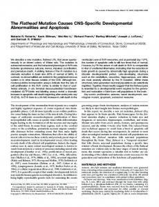
supporting a role for integrins in osteoblast differentiation. Osteoblasts are now known to express several integrin f fE DGEA?species, including Up, (X2, C3 Ct4aC5,C 1' (2, and 33 subunits (Clover et al., 1992; Hughes et al., 1993; Grzesik and GehronRobey, 1994; Saito et al., 1994; Sinha and Tuan, 1996). Furthermore, disruption of integrin:ECM interactions, specifically those involving collagen and fibronectin, blocks osteoblast differentiation and mineralization. Thus, addition of antibodies to a2 integrin or a DGEA peptide which is a component of the cell-binding domain of type I collagen (Staatz et al., 1991) blocks AA-dependent induction of alkaline phosphatase and differentiation-dependent down-regulation of TGF-( receptors in MC3T3-E1 cells (Takeuchi et al., 1996). These responses appear to require focal adhesion kinase and the MAP kinase pathway, because selective inhibition of either of these intermediates blocks alkaline phosphatase induction (Takeuchi et al., 1997). In separate studies, a peptide containing the RGD cell-binding domain of fibronectin was shown to inhibit both bone formation and resorption in a mineralizing organ culture system (Gronowicz and Derome, 1994). Similarly, long-term culture of calvarial osteoblasts with antifibronectin antibodies or fibronectin peptides containing the RGD cell-binding domain has also been reported to suppress differentiation (Moursi et at., 1996). Figure 3. Model for extracellular matrix activation of the osteocalcin promoter. Key com- The author's laboratory recently showed ponents of this model are: (1) the interaction of the type collagen matrix with the etA that disruption of CX2 integrin function by integrin, followed by signaling to the nucleus; (2) phosphorylation and activation of both anti-a2 integrin antibody and DGEA Osf2/Cbfal and possibly other transcription factors; and (3) binding of Osf2/Cbfal to peptide also blocks ascorbic-acid-depenAML sites in the osteocalcin promoter and stimulation of transcription. Several known dent induction of the osteocalcin promointegrin-mediated signal transduction pathways are shown (Clark and Brugge, 1995; ter, osteoblast differentiation, and activaGumbiner, 1996), although those relevant to osteoblast function have not been defined. tion of Osf2/Cbfal (Xiao et al., 1998a). The Also indicated is the physical linkage between integrins and the cytoskeleton via cx-actinin same concentrations of anti-(x5 integrin and the actin filaments, which may also control transcription. Not shown is the possible antibody or RGD-containing peptide were role of nuclear organization in the activation of Osf2/Cbfal. It has been proposed that inactive, although the RGD peptide was this factor can associate with the nuclear matrix as well as with AML sites in the osteo- inhibitory at higher concentrations. calcin promoter, thereby bringing the promoter into contact with the basal transcription Interestingly, the dramatic matrix-depeninitiation complex. Abbreviations: ox2p1, integrin subunits known to form a receptor for dent increase in Osf2/Cbfal binding to type collagen; DGEA, Asp-Gly-Glu-Ala sequence of type collagen, reported to inter- OSE2 noted above is not accompanied by act with the ox2P1 integrin; FAK, focal adhesion kinase; Osf2, Osf2/Cbfal transcription parallel changes in the level of transcripfactor; AML, DNA binding site for AML/runt transcription factors (positions in the mouse tion factor mRNA or protein. This suggests osteocalcin gene 2 promoter are indicated); and TFs, components that, together with that the ECM induces a modification in Osf2/Cbfal to increase its activity as a RNA polymerase 11, form the basal transcription initiation complex. transcriptional enhancer rather than ECM
50
Oral Biol Med Grit Rev Crit Rev Oral Biol Med
(1999)
10(l):40-57 ( 1999)
increasing protein levels. In a related study, overexpression of the c.2 integrin subunit in primary calvarial osteoblast cells also greatly increased osteocalcin promoter activity (Zimmerman et al., 1997). Recent preliminary results from the author's laboratory indicate that Osf2/Cbfa I is phosphorylated by MAP kinases and that this modification stimulates transcriptional activity (Xiao et al., 1998b). Taken together, these results strongly support a model in which an CX2031 integrin:type I collagen interaction is necessary for phosphorylation and activation of osteoblastspecific transcription factors like Osf2/Cbfal present in committed osteoprogenitor cells. There are several examples of type I collagen: oL2A integrin interactions being important in the regulation of other cellular activities, such as activation of collagenase (Riikonen et al., 1995), stimulation of collagen gel contraction (Schiro et al., 1991), and kidney tubule formation (Schwimmer and Ojakian, 1995), although in no case has the entire integrin-mediated pathway been delineated. Clearly, a major direction for future research will be to identify the signal transduction pathway linking integrin:ECM binding to changes in osteoblast-specific gene expression and to determine how this pathway is regulated by changes in matrix synthesis and turnover, mechanical loading of bone, and the hormonal milieu. Fig. 3 summarizes our current thinking about how the ECM regulates gene expression in osteoblasts.
teeth-extra teeth in fact-but this may be a consequence of their heterozygous status for the CBFA1 gene. CBFA1 -/mice contain hypoplastic tooth primordia, although the appropriate cell layers of differentiating tooth structures were present (Otto et al., 1997). It was not determined whether these structures express osteocalcin or other matrix proteins. A comparison of the transcriptional regulation of genes like osteocalcin, bone sialoprotein, and dentin sialophosphoprotein in bones with that in teeth may reveal important information about more subtle regulatory mechanisms that distinguish these two similar, yet functionally very different, tissues.
Conclusions and Future Directions The organogenesis of skeletal structures can be divided into two broad phases: the establishment of a morphogenic field in which signals from organizing centers like the ZPA specify in time and space which cells are destined to form bone, followed by a differentiation phase when gene expression patterns are altered in response to coded positional information to produce chondrocytes and osteoblasts at sites of overt bone formation. Although this article focused on gene regulation during osteoblast differentiation, much progress has also been made in our understanding of these early morphogenic signals. The combined use of promoter analysis and transgenic technology has allowed our understanding of gene expression in bone to progress rapidly. A striking example of this rapid pace of discovery was the simultaneous publication of the identification, targeted inactivation, and human genetics of Osf2/Cbfal. Undoubtedly, this rate of progress will only increase in the coming years. Some of the more outstanding research questions to be addressed in the next five years are summarized below:
Is the Tooth a Kind of Bone? At the molecular level, dentin and cementum share many similarities with bone, including the presence of a mineralized type I collagen matrix and noncollagenous proteins such as osteocalcin, bone sialoprotein, alkaline phosphatase, and BMPs (MacNeil and Somerman, 1993; Lesot et al., 1994). In fact, because no unique markers have been identified to distinguish it from bone, it has been proposed that the cementoblast may actually be a specialized type of osteoblast (MacNeil et al., 1998). In contrast, odontoblasts do express at least two unique proteins, dentin sialoprotein and dentin phosphoprotein, which were recently shown to be synthesized as a single transcript (MacDougall et al., 1997). Like osteoblasts, cementoblasts and odontoblasts require ascorbic acid to express a differentiated phenotype (D'Errico et al., 1997; Heimrika-Wagner et al., 1982), and it is likely that cellImatrix interactions shown to be crucial for osteoblast differentiation are also necessary for dentin and cementum formation. A central question concerns the degree to which genetic mechanisms controlling osteoblast differentiation are relevant to our understanding of odontoblasts and cementoblasts. For example, is Osf2/Cbfa 1 an important factor for determination of these cell types, or are other transcription factors involved? Individuals with cleidocranial dyplasia have 10(1)40 57(1999) 1 1999) I10(lI):40-57
(I) How DO EARLY PATTERNING EVENTS INITIATED BY FACTORS LIKE SHH LEAD TO THE FORMATION OF BONE AT SPECIFIC SITES? Hox gene expression is known to precede mesenchymal condensations in regions targeted for future bone formation, but it remains to be determined how these sites are specified and distinguished from areas destined to form non-osseous structures.
(11) IS OSF2/CBFAI THE ONLY TRANSCRIPTION FACTOR REQUIRED FOR OSTEOBLAST-SPECIFIC GENE EXPRESSION, OR IS IT ONLY ONE OF SEVERAL COMPONENTS, PERHAPS AN ENTIRE FAMILY OF OSTEOGENIC NUCLEAR FACTORS, THAT WORK IN A CONCERTED FASHION TO CONTROL TRANSCRIPTION? An understanding of the full range of transcriptional controls necessary for osteoblast-specific gene expresOral
Biol Med
Crit Rev Oral Biol Med
51
Bellahcene A, Kroll M, Liebens F, Castronovo V (1996). Bone sialoprotein expression in primary human breast cancer is associated with bone metastases development. I Bone Miner Res 1 1:665-670. Benezra R, Davis RL, Lockshon D, Turner DL, Weintraub H (1990). The protein Id: a negative regulator of helixloop-helix DNA binding proteins. Cell 61:49-59. Benson M, Decker 1, Cui Y, Tirasophon W, Franceschi R (1995). Osteoblast basic helix-loop-helix proteins: regulation of the Id family and use of the two-hybrid system to identify novel factors (abstract). J Bone Miner Res 10:S314. Benson M, Cui Y, Aubin 1, Franceschi R (1997). Cloning and analysis of a 2.5 kb murine bone sialoprotein promoter fragment exhibiting osteoblast-restricted expression (abstract). J Bone Miner Res 12:S277. Bianco P, Fisher LW, Young MF, Termine ID, GehronRobey P (1991). Expression of bone sialoprotein (BSP) in developing human tissues. Calcif Tissue Int 49:421-426. Bidwell IP, Van Wijnen Al, Fey EG, Dworetzky S, Penman S, Stein IL, et al. (1993). Osteocalcin gene promoterbinding factors are tissue-specific nuclear matrix components. Proc Natl Acad Sci USA 90:3162-3166. Blanco IC, Wang IM, Tsai SY, Tsai MI, O'Malley BW, Jurutka PW, et al. (1995). Transcription factor TFIIB and the vitamin D receptor cooperatively activate liganddependent transcription. Proc Natl Acad Sci USA
sion will require the systematic analysis of multiple gene promoters.
(111) WHAT ARE THE RELEVANT PATHWAYS FOR ACTIVATION OF OSF2/CBFAI? Osteoblast cultures grown in the absence of AA contain Osf2/Cbfa 1, but the transcriptional activity of this factor remains low until cells are allowed to synthesize an ECM. Preliminary studies suggest the involvement of an integrin-mediated event for Osf2/Cbfal activation, but the details of this pathway remain to be determined.
(IV) ARE THE TRANSCRIPTIONAL CONTROLS IDENTIFIED IN BONE FOUND IN OTHER MINERALIZED TISSUES SUCH AS TEETH? Initial reports on tooth formation in Osf2/Cbfal knockout mice suggest the involvement of this factor in tooth morphogenesis, but it remains to be determined if it functions similarly in bones and teeth. Elucidation of the regulatory controls for multiple odontoblast-related genes will be required to discriminate between general and tooth-specific gene regulatory mechanisms.
Acknowledgments This work was supported by National Institute of Dental Research
grants DE-l 1723 and DE- 1 221 1 The author thanks Mr. M. Douglas Benson for the preparation of Figs.
92:1535-1539.
REFERENCES Ahn MY, Bae SC, Maruyama M, Ito Y (1996). Comparison of the human genomic structure of the Runt domainencoding PEBP2/CBFalpha gene family. Gene 168:279280. Asahina 1, Sampath TK, Nishimura 1, Hauschka PV (1993). Human osteogenic protein-i induces both chondroblastic and osteoblastic differentiation of osteoprogenitor cells derived from newborn rat calvaria. I Cell Biol 123:921-933. Aubin J, Lui F (1996). The osteoblast lineage. In: Principles of bone biology. Bilezikian LGRJP, Rodan GA, editors. San Diego, CA: Academic Press, pp. 5167.
Banerjee C, Hiebert SW, Stein IL, Lian JB, Stein GS (1996). An AML-1 consensus sequence binds an osteoblast-specific complex and transcriptionally activates the osteocalcin gene. Proc Natl Acad Sci USA 93:4968-4973. Banerjee C, McCabe LR, Choi JY, Hiebert SW, Stein IL, Stein GS, et al. (1997). Runt homology domain proteins in osteoblast differentiation: AML3/CBFA1 is a major component of a bone-specific complex. I Cell Biochem 66:1-8.
52
Ci
e
Boden SD, Hair G, Titus L, Racine M, McCuaig K, Wozney JM, et al. (1997). Glucocorticoid-induced differentiation of fetal rat calvarial osteoblasts is mediated by bone morphogenetic protein-6. Endocrinology 138:2820-2828. Bornstein P (1996). Regulation of expression of the alpha 1 (I) collagen gene: a critical appraisal of the role of the first intron isee commentsj. Matrix Biol 15:3-10. Bronckers AL, Gay S, Finkelman RD, Butler WT (1987). Immunolocalization of Gla proteins (osteocalcin) in rat tooth germs: comparison between indirect immunofluorescence, peroxidase-antiperoxidase, avidin-biotin-peroxidase complex, and avidin-biotingold complex with silver enhancement. J Histochem Cytochem 35:825-830. Chen J, McCulloch CA, Sodek J (1993). Bone sialoprotein in developing porcine dental tissues: cellular expression and comparison of tissue localization with osteopontin and osteonectin. Arch Oral Biol 38:241249. Chen I, Thomas HF, lin H, Jiang H, Sodek 1 (1996). Expression of rat bone sialoprotein promoter in transgenic mice. J Bone Miner Res 11:654-664. Chen Y, Bei M, Woo I, Satokata 1, Maas R (1996). Msxl controls inductive signaling in mammalian tooth rlBo
e
Crit Rev Oral Biol Med
Ol:05(99
10(1):40-57 (1999)
morphogenesis. Development 122:3035-3044. Clark EA, Brugge JS (1995). Integrins and signal transduction pathways: the road taken. Science 268:233-239. Clemens TL, Tang H, Maeda S, Kesterson RA, DeMayo J, Pike IW, et al. (1997). Analysis of osteocalcin expression in transgenic mice reveals a species difference in vitamin D regulation of mouse and human osteocalcin genes. I Bone Miner Res 12:1570-1576. Clover 1, Dodds R, Gowen M (1992). Integrin subunit expression by human osteoblasts and osteoclasts in situ and in culture. J Cell Sci 103:267-271. Cserjesi P, Brown D, Ligon KL, Lyons GE, Copeland NG, Gilbert Di, et al. (1995). Scleraxis: a basic helix-loophelix protein that prefigures skeletal formation during mouse embryogenesis. Development 121:1099- 1110. D'Errico J, MacNeil R, Takata T, Berry 1, Strayhorn C, Somerman M (1997). Expression of bone associated markers by tooth root lining cells, in situ and in vitro. Bone 20:117-126. Desbois C, Hogue DA, Karsenty G (1994). The mouse osteocalcin gene cluster contains three genes with two separate spatial and temporal patterns of expression. J Biol Chem 269:1183-1190. Dodig M, Kronenberg M, Bedalov A, Kream B, Gronowicz G, Clark S, et al. (1996a). Identifiction of a TAAT-containing motif required for high level expression of the COLIAI promoter in differentiated osteoblasts of transgenic mice. I Biol Chem 271:16422-16429. Dodig M, Kronenberg MS, Bedalov A, Kream BE, Gronowicz G, Clark SH, et al. (1996b). Identification of a TAAT-containing motif required for high level expression of the COLlAl promoter in differentiated osteoblasts of transgenic mice. J Biol Chem 271:1642216429. Ducy P, Karsenty G (1995). Two distinct osteoblast-specific cis-acting elements control expression of a mouse osteocalcin gene. Mol Cell Biol 15:1858-1869. Ducy P, Desbois C, Boyce B, Pinero G, Story B, Dunstan C, et al. (1996). Increased bone formation in osteocalcindeficient mice. Nature 382:448-452. Ducy P, Zhang R, Geoffroy V, Ridall AL, Karsenty G (1997). Osf2/Cbfa 1: a transcriptional activator of osteoblast differentiation. Cell 89:747-754. Erlebacher A, Filvaroff EH, Gitelman SE, Derynck R (1995). Toward a molecular understanding of skeletal development icommentl. Cell 80:371-378. Fedarko NS, Moerike M, Brenner R, Robey PB (1992). Extracellular matrix formation by osteoblasts from patients with osteogenesis imperfecta. J Bone Miner Res 7:921-930. Fedarko NS, Sponseller PD, Shapiro JR (1996). Long-term extracellular matrix metabolism by cultured human osteogenesis imperfecta osteoblasts. I Bone Miner Res 11 800-805. 7(199) 10(140
10(t I):40-57 ( I1999)
Franceschi RT (1992). The role of ascorbic acid in mesenchymal differentiation. Nutr Rev 50:65-70. Franceschi RT, lyer BS (1992). Relationship between collagen synthesis and expression of the osteoblast phenotype in MC3T3-E I cells. I Bone Miner Res 7:235-246. Franceschi RT, Romano PR, Park KY (1988). Regulation of type I collagen synthesis by 1,25-dihydroxyvitamin D3 in human osteosarcoma cells. I Biol Chem 263:1893818945. Franceschi RT, Iyer BS, Cui Y (1994). Effects of ascorbic acid on collagen matrix formation and osteoblast differentiation in murine MC3T3-E 1 cells. I Bone Miner Res 9:843-854. Frendo I-L, Xiao G, Fuchs S, Franceschi RT, Karsenty G, Ducy P (1998). Functional hierarchy between two OSE2 elements in the control of osteocalcin gene expression in vivo. I Biol Chem 273:30509-30516. Friedenstein Al, Chailakhyan RK, Gerasimov UV (1987). Bone marrow osteogenic stem cells: in vitro cultivation and transplantation in diffusion chambers. Cell Tissue Kinet 20:263-272. Gendron-Maguire M, Mallo M, Zhang M, Grindley T (1993). Hoxa-2 mutant mice exhibit homeotic transformation of skeletal elements derived from cranial neural crest. Cell 75:1317-1331. Geoffroy V, Ducy P, Karsenty G (1995). A PEBP2 alpha/AML- 1-related factor increases osteocalcin promoter activity through its binding to an osteoblast-specific cis-acting element. I Biol Chem
270:30973-30979. Gergen 1, Wieschaus E (1985). The localized requirements for a gene affecting segmentation in Drosophila: analysis of larvae mosaic for runt. Dev Biol 109:321335. Gerstenfeld LC, Chipman SD, Glowacki ], Lian IB (1987). Expression of differentiated function by mineralizing cultures of chicken osteoblasts. Dev Biol 122:49-60. Gerstenfeld LC, Zurakowski D, Schaffer IL, Nichols DP, Toma CD, Broess M, et al. (1996). Variable hormone responsiveness of osteoblast populations isolated at different stages of embryogenesis and its relationship to the osteogenic lineage. Endocrinology 137:39573968. Gitelman SE, Kirk M, Ye 1Q, Filvaroff EH, Kahn Al, Derynck R (1995). Vgr-l/BMP-6 induces osteoblastic differentiation of pluripotential mesenchymal cells. Cell Growth Differ 6:827-836. Grigoriadis AE, Heersche IN, Aubin JE (1988). Differentiation of muscle, fat, cartilage, and bone from progenitor cells present in a bone-derived clonal cell population: effect of dexamethasone. Cell Biol 106:2139-2151. Gronowicz GA, Derome ME (1994). Synthetic peptide containing Arg-Gly-Asp inhibits bone formation and
ritRevOralBio Me Crit Rev Oral Biol Med
53
resorption in a mineralizing organ culture system of fetal rat parietal bones. I Bone Miner Res 9:193-20 1. Gronthos S, Graves SE, Ohta S, Simmons PI (1994). The STRO-1+ fraction of adult human bone marrow contains the osteogenic precursors. Blood 84:4164-4173. Grzesik WI, Gehron-Robey P (1994). Bone matrix RGD glycoproteins: immunolocalization and interaction with human primary osteoblastic bone cells in vitro. I Bone Miner Res 9:487-496. Gumbiner BM (1996). Cell adhesion: the molecular basis of tissue architecture and morphogenesis. Cell 84:345-
Proc Natl Acad Sci USA 86:4455-4459. Kerr JM, Fisher LW, Termine ID, Wang MG, McBride OW, Young MF (1993). The human bone sialoprotein gene
(IBSP): genomic localization and characterization. Genomics 17:408-415. Kesterson RA, Stanley L, DeMayo F, Finegold M, Pike IW (1993). The human osteocalcin promoter directs bone-specific vitamin D-regulatable gene expression in transgenic mice. Mol Endo 7:462-467. Kim RH, Shapiro HS, Li IJ, Wrana IL, Sodek 1 (1994). Characterization of the human bone sialoprotein (BSP) gene and its promoter sequence. Matrix Biol 14:31-40. Komori T, Yagi H, Nomura S, Yamaguchi A, Sasaki K, Deguchi K, et al. (1997). Targeted disruption of Cbfal results in a complete lack of bone formation owing to maturational arrest of osteoblasts [see commentsl. Cell 89:755-764. Lee B, Thirunavukkarasu K, Zhou L, Pastore L, Baldini A, Hecht I, et al. (1997). Missense mutations abolishing DNA binding of the osteoblast-specific transcription factor OSF2/CBFA1 in cleidocranial dysplasia. Nat Genet 16:307-310. Lee lE, Hollenberg SM, Snider L, Turner DL, Lipnick N, Weintraub H (1995). Conversion of Xenopus ectoderm into neurons by NeuroD, a basic helix-loophelix protein. Science 268:836-844. Lesot H, Smith A, Tziafas D, Begue-Kirn C, Cassidy N, Ruch 1 (1994). Biologically active molecules and dental tissue repair: a comparative review of reactionary and reparative dentinogenesis with the induction of odontoblast differentiation in vitro. Cells Mater 4:199218. Li I11 Sodek 1 (1993). Cloning and characterization of the rat bone sialoprotein gene promoter. Biochem I 289:625-629. Lian JI Stewart C, Puchacz E, Mackowiak S, Shalhoub V, Collart D, et al. (1989). Structure of the rat osteocalcin gene and regulation of vitamin D-dependent expression. Proc Natl Acad Sci USA 86:1143-1147. Lian IB, Shalhoub V, Aslam F, Frenkel B, Green J, Hamrah M, et al. (1997). Species-specific glucocorticoid and 1 25-dihydroxyvitamin D responsiveness in mouse MC3T3-E 1 osteoblasts: dexamethasone inhibits osteoblast differentiation and vitamin D down-regulates osteocalcin gene expression. Endocrinology 138:2117-2127. Lin TH, Aplin AE, Shen Y, Chen 0, Schaller M, Romer L, et al. (1997). Integrin-mediated activation of MAP kinase is independent of FAK: evidence for dual integrin signaling pathways in fibroblasts. I Cell Biol 136:1385-1395. MacDougall M. Simmons D, Luan X, Nydegger I, Feng 1, Gu TT (1997). Dentin phosphoprotein and dentin
357.
Heimrika-Wagner AM, Bronckers ALII, Woltgens JHM (1982). Ultrastructural changes in developing hamster molars during vitamin C deficiency in vitro. I Biol Buccale 10:163-173.
Heinrichs AA, Bortell R, Bourke M, Lian JB, Stein GS, Stein JL (1995). Proximal promoter binding protein contributes to developmental tissue-restricted expression of the rat osteocalcin gene. J Cell Biochem 57:90-100. Hodgkinson JE, Davidson CL, Beresford J, Sharpe PT (1993). Expression of a human homeobox-containing gene is regulated by 1,25(OH)2D3 in bone cells. Biochim Biophys Acta 1174:11-16 [published erratum appears in Biochim Biophys Acta 1216:173, 19931. Hughes D, Salter S, Dedhar S, Simpson R (1993). Integrin expression in human bone. J Bone Miner Res 8:527-533. Hunter GK, Goldberg HA (1993). Nucleation of hydroxyapatite by bone sialoprotein. Proc Natl Acad Sci USA 90:8562-8565. Ibaraki K, Termine JD, Whitson SW, Young MF (1992). Bone matrix mRNA expression in differentiating fetal bovine osteoblasts. I Bone Miner Res 7:743-754. Ingraham HA, Albert VR, Chen RP, Crenshaw EB 3rd, Elsholtz HP, He X, et al. (1990). A family of POUdomain and Pit-I tissue-specific transcription factors in pituitary and neuroendocrine development. Ann Rev Physiol 52:773-791. labs EW, Muller U, Li X, Ma L, Luo W, Haworth IS, et al. (1993). A mutation in the homeodomain of the human MSX2 gene in a family affected with autosomal dominant craniosynostosis. Cell 75:443-450. Johnson RL, Tabin CJ (1997). Molecular models for vertebrate limb development. Cell 90:979-990. Katagiri T, Yamaguchi A, Komaki M, Abe E, Takahashi N, Ikeda T, et al. (1994). Bone morphogenetic protein-2 converts the differentiation pathway of C2C12 myoblasts into the osteoblast lineage. I Cell Biol 127:1755-1766 Ipublished erratum appears in J Cell Biol 128(4):following 713, 19951. Kerner SA, Scott RA, Pike JW (1989). Sequence elements in the human osteocalcin gene confer basal activation and inducible response to hormonal vitamin D3. 54
Grit Rev
Oral
Biol
Crit Rev Oral Biol Med
10(1):40-57 (1999)
sialoprotein are cleavage products expressed from a single transcript coded by a gene on human chromosome 4. J Biol Chem 272:835-842. MacNeil RL, Somerman Ml (1993). Molecular factors regulating development and regeneration of cementum. I Periodont Res 28:550-559. MacNeil RL, D'Errico IA, Ouyang H, Berry J, Strayhorn C, Somerman MI (1998). Isolation of murine cementoblasts: unique cells or uniquely-positioned osteoblasts? Eur I Oral Sci 106(Suppl 1):350-356. Mahmoodian F, Gosiewska A, Peterkofsky B (1996). Regulation and properties of bone alkaline phosphatase during vitamin C deficiency in guinea pigs. Arch Biochem Biophys 336:86-96. Markose ER, Stein IL, Stein GS, Lian lB (1990). Vitamin D-mediated modifications in protein-DNA interactions at two promoter elements of the osteocalcin gene. Proc Natl Acad Sci USA 87:1701-1705. McCabe LR, Kockx M, Lian 1, Stein 1, Stein G (1995). Selective expression of fos- and jun-related genes during osteoblast proliferation and differentiation. Exp Cell Res 218:255-262. McCauley LK, Koh Al, Beecher CA, Cui Y, Rosol TI, Franceschi RT (1996). PTH/PTHrP receptor is temporally regulated during osteoblast differentiation and is associated with collagen synthesis. J Cell Biochem 6 1:638-647. Merriman HL, van Wijnen Al, Hiebert S, Bidwell IP, Fey E, Lian J, et al. (1995). The tissue-specific nuclear matrix protein, NMP-2, is a member of the AML/CBF/PEBP2/runt domain transcription factor family: interactions with the osteocalcin gene promoter. Biochemistry 34:13125-13132. Meyers S, Downing IR, Hiebert SW (1993). Identification of AML-I and the (8;21) translocation protein (AMLI/ETO) as sequence-specific DNA-binding proteins: the runt homology domain is required for DNA binding and protein-protein interactions. Mol Cell Biol 13:6336-6345. Montecino M, Pockwinse S, Lian 1, Stein G, Stein 1 (1994). DNase I hypersensitive sites in promoter elements associated with basal and vitamin D dependent transcription of the bone-specific osteocalcin gene. Biochemistry 33:348-353. Moursi AM, Damsky CH, Lull I, Zimmerman D, Doty SB, Aota S, et al. (1996). Fibronectin regulates calvarial osteoblast differentiation. I Cell Sci 109:1369-1380. Murray SS, Glackin CA, Winters KA, Gazit D, Kahn Al, Murray El (1992). Expression of helix-loop-helix regulatory genes during differentiation of mouse osteoblastic cells. I Bone Miner Res 7:1131-1138. Murray SS, Murray EJ, Glackin CA, Urist MR (1993). Bone morphogenetic protein inhibits differentiation and affects expression of helix-loop-helix regulatory mol10(1)40-57 1999) 1):.40-57 ((1999) 10(
ecules in myoblastic cells. I Cell Biochem 53:51-60. Nickerson JA, Blencowe BI, Penman S (1995). The architectural organization of nuclear metabolism. Int Rev Cytol 162(A):67-123. Nishiyori A, Tashiro H, Kimura A, Akagi K, Yamamura K, Mori M, et al. (1994). Determination of tissue specificity of the enhancer by combinatorial operation of tissue-enriched transcription factors. Both HNF-4 and C/EBP beta are required for liver-specific activity of the ornithine transcarbamylase enhancer. J Biol Chem 269:1323-1331. Ogata T, Noda M (I1991). Expression of Id, a negative regulator of helix-loop-helix DNA binding proteins, is down-regulated at confluence and enhanced by dexamethasone in a mouse osteoblastic cell line, MC3T3E 1. Biochem Biophys Res Commun 180:1194-1199. Ogata T, Wozney IM, Benezra R, Noda M (1993). Bone morphogenetic protein 2 transiently enhances expression of a gene, Id (inhibitor of differentiation), encoding a helix-loop-helix molecule in osteoblastlike cells. Proc Natl Acad Sci USA 90:9219-9222. Ogata Y, Yamauchi M, Kim RH, Li 11, Freedman LP, Sodek 1 (1995). Glucocorticoid regulation of bone sialoprotein (BSP) gene expression. Identification of a glucocorticoid response element in the bone sialoprotein gene promoter. Eur I Biochem 230:183-192. Oldberg A, Franzen A, Heinegard D (1986). Cloning and sequence analysis of rat bone sialoprotein (osteopontin) cDNA reveals an Arg-Gly-Asp cell-binding sequence. Proc Natl Acad Sci USA 83:8819-8823. Olson EN, Klein WH (1994). bHLH factors in muscle development: dead lines and commitments, what to leave in and what to leave out. Genes Dev 8:1-8. Otto F, Thornell AP, Crompton T, Denzel A, Gilmour KC, Rosewell IR, et al. (1997). Cbfal, a candidate gene for cleidocranial dysplasia syndrome, is essential for osteoblast differentiation and bone development Isee commentsl. Cell 89:765- 771. Owen TA, Aronow M, Shalhoub V, Barone LM, Wilming L, Tassinari MS, et al. (1990). Progressive development of the rat osteoblast phenotype in vitro: reciprocal relationships in expression of genes associated with osteoblast proliferation and differentiation during formation of the bone extracellular matrix. I Cell Physiol 143:420-430. Quarles LD, Yohay DA, Lever LW, Caton R, Wenstrup RJ (1992). Distinct proliferative and differentiated stages of murine MC3T3-E I cells in culture: an in vitro model of osteoblast development. I Bone Miner Res 7:683-692. Quarles LD, Siddhanti SR, Medda S (1997). Developmental regulation of osteocalcin expression in MC3T3-E1 osteoblasts: minimal role of the proximal E-box cis-acting promoter elements. I Cell Biochem 65:11-24.
Oral Biol Med Biol Med Crit Rev Oral Crit
Rev
55
Riikonen T, Westermarck 1, Koivisto L, Broberg A, Kahari VM, Heino 1 (1995). Integrin alpha 2 beta 1 is a positive regulator of collagenase (MMP-1) and collagen alpha 1 (1) gene expression. I Biol Chem 270:1354813552. Roeckel D, Krieg T (1994). Three-dimensional contact with type I collagen mediates tyrosine phosphorylation in primary human fibroblasts. Exp Cell Res 211:4248. Saito T, Albelda SM, Brighton CT (1994). Identification of integrin receptors on cultured human bone cells. I Orthop Res 12:384-394. Sasaki K, Yagi H, Bronson RT, Tominaga K, Matsunashi T, Deguchi K, et al. (1996). Absence of fetal liver hematopoiesis in mice deficient in transcriptional coactivator core binding factor beta. Proc Natl Acad Sci USA 93:12359-12363. Satokata 1, Maas R (1994). Msxl deficient mice exhibit cleft palate and abnormalities of craniofacial and tooth development [see commentsI. Nat Genet 6:348356. Schiro IA, Chan BM, Roswit WT, Kassner PD, Pentland AP, Hemler ME, et al. (1991). Integrin alpha 2 beta 1 (VLA2) mediates reorganization and contraction of collagen matrices by human cells. Cell 67:403-410. Schule R, Umesono K, Mangelsdorf DI, Bolado 1, Pike 1W, Evans RM (1990). Jun-Fos and receptors for vitamins A and D recognize a common response element in the human osteocalcin gene. Cell 61:497-504. Schwimmer R, Ojakian GK (1995). The alpha 2 beta 1 integrin regulates collagen-mediated MDCK epithelial membrane remodeling and tubule formation. I Cell Sci 108:2487-2498. Shi S, Kirk M, Kahn Al (1996). The role of type I collagen in the regulation of the osteoblast phenotype. I Bone Miner Res 11:1139-1145. Sinha R, Tuan R (1996). Regulation of human osteoblast integrin expression by orthopedic implant materials. Bone 18:451-457. Sodek J, Li IJ, Kim RH, Ogata Y, Yamauchi M, Zhang 0, et al. (1994). Steroid hormone regulation of bone sialoprotein gene transcription. In: The biological mechanisms of tooth eruption, resorption and replacement by implants. Davidovitch Z, editor. Boston: Harvard Society for the Advancement of Orthodontics, pp. 2 15-225.
Sodek J, Kim RH, Ogata Y, Li J, Yamauchi M, Zhang 0, et al. (1995). Regulation of bone sialoprotein gene transcription by steroid hormones. Connect Tissue Res 32:209-217. Sodek J, Li JI, Kim RH, Ogata Y, Yamauchi M (1996). Characterization of the bone sialoprotein (BSP) gene promoter. Connect Tissue Res 35:23-3 1. Staatz WD, Fok KF, Zutter MM, Adams SP, Rodriguez BA,
56
Santoro SA (1991). Identification of a tetrapeptide recognition sequence for the alpha 2 beta 1 integrin in collagen. I Biol Chem 266:7363-7367. Stein GS, Lian IB, Stein IL, van Wijnen Al, Frankel B, Montecino M (1996). Mechanisms regulating osteoblast proliferation and differentiation. In: Principles of bone biology. Bilezikian LGRJP, Rodon GA, editors. San Diego, CA: Academic Press, pp. 69-86. Strauss PG, Closs El, Schmidt J, Erfle V (1990). Gene expression during osteogenic differentiation in mandibular condyles in vitro. J Cell Biol 110: 1369-1378. Sudbeck BD, Parks WC, Welgus HG, Pentland AP (1994). Collagen-stimulated induction of keratinocyte collagenase is mediated via tyrosine kinase and protein kinase C activities. I Biol Chem 269:30022-30029. Suva LI, Seedor IG, Endo N, Quartuccio HA, Thompson DD, Bab I, et al. (1993). Pattern of gene expression following rat tibial marrow ablation. J Bone Miner Res 8:379-388. Takeuchi Y, Nakayama K, Matsumoto T (1996). Differentiation and cell surface expression of transforming growth factor-beta receptors are regulated by interaction with matrix collagen in murine osteoblastic cells. J Biol Chem 271:3938-3944. Takeuchi Y, Suzawa M, Kikuchi T, Nishida E, Fujita T, Matsumoto T (1997). Differentiation and transforming growth factor-beta receptor down-regulation by collagen-alpha 2 beta 1 integrin interaction is mediated by focal adhesion kinase and its downstream signals in murine osteoblastic cells. J Biol Chem 272:2930929316. Tamura M, Noda M (1994). Identification of a DNA sequence involved in osteoblast-specific gene expression via interaction with helix-loop-helix (HLH)-type transcription factors. J Cell Biol 126:773-782. Taylor SM, Jones PA (1979). Multiple new phenotypes induced in 10T1/2 and 3T3 cells treated with 5-azacytidine. Cell 17:771-779. Thesleff 1, Vaahtokari A, Partanen AM (1995). Regulation of organogenesis. Common molecular mechanisms regulating the development of teeth and other organs. Int I Dev Biol 39:35-50. Thies RS, Bauduy M, Ashton BA, Kurtzberg L, Wozney JM, Rosen V (1992). Recombinant human bone morphogenetic protein-2 induces osteoblastic differentiation in W-20-17 stromal cells. Endocrinology 130:1318-1324. Thirunavukkarasu K, Mahajan M, McLarren KW, Stifani S, Karsenty G (1998). Two domains unique to osteoblast-specific transcription factor Osf2/Cbfa 1 contribute to its transactivation function and its inability to heterodimerize with Cbf3. Mol Cell Biol 15:1858-1869. Tickle C (1995). Vertebrate limb development. Curr Opin Genet Dev 5:478-484.
Oral Biol Grit Crit Rev Rev Oral Biol Med Med
10(1)40-57
10(tl ):40-57 ( 1999)
Togari A, Arai M, Nakagawa S, Banno A, Aoki M, Matsumoto S (1995). Alteration of bone status with ascorbic acid deficiency in ODS (osteogenic disorder Shionogi) rats. Ipn I Pharmacol 68:255-261. Tontonoz P, Kim IB, Graves RA, Spiegelman BM (1993). ADDI: a novel helix-loop-helix transcription factor associated with adipocyte determination and differentiation. Mol Cell Biol 13:4753-4759. Towler DA, Rutledge SI, Rodan GA (1994). MSX-2/Hox 8.1: a transcriptional regulator of the rat osteocalcin promoter. Mol Endo 8:1484-1493. Triffit 1 (1996). The stem cell of the osteoblasts. In: Principles of bone biology. Bilezikian LGRIP, Rodon GA, editors. San Diego, CA: Academic Press, pp. 539-550. Wang EA, Israel DI, Kelly S, Luxenberg DP (1993). Bone morphogenetic protein-2 causes commitment and differentiation in C3HIOT1/2 and 3T3 cells. Growth Factors 9:57-71.
Weintraub H, Davis R, Tapscott S, Thayer M, Krause M, Benezra R, et al. (1991). The myoD gene family: nodal point during specification of the muscle cell lineage. Science 251 .761-766. Wenstrup RI, Fowlkes JL, Witte DP, Florer 1B (1996). Discordant expression of osteoblast markers in MC3T3-EI cells that synthesize a high turnover matrix. I Biol Chem 271:10271-10276. Xiao G, Cui Y, Ducy P, Karsenty G, Franceschi RT (1997). Ascorbic acid-dependent activation of the osteocalcin promoter in MC3T3-E 1 preosteoblasts: requirement for collagen matrix synthesis and the presence of an intact OSE2 sequence. Mol Endo 11:1103-1113. Xiao G, Wang D, Benson MD, Karsenty G, Franceschi RT
10(1):40-57 (1999)
(1998a). Role of the 2- integrin in osteoblast-specific gene expression and activation of the Osf2 transcription factor. I Biol Chem 273:32988-32994. Xiao G, Thomas PE, Jiang D, Benson MD, Franceschi RT (1998b). MEK/MAPK activates osteoblast-specific transcription factor, Osf2/Cbfal (abstract). Bone 23:S 150. Yang R, Gerstenfeld LC (1996). Signal transduction pathways mediating parathyroid hormone stimulation of bone sialoprotein gene expression in osteoblasts. I Biol Chem 271:29839-29846. Yang R, Gerstenfeld LC (1997). Structural analysis and characterization of tissue and hormonal responsive expression of the avian bone sialoprotein (BSP) gene. I Cell Biochem 64:77-93. Zeng C, van Wijnen Al, Stein IL, Meyers S, Sun W, Shopland L, et al. (1997). Identification of a nuclear matrix targeting signal in the leukemia and bonerelated AML/CBF-alpha transcription factors. Proc Natl Acad Sci USA 94:6746-6751. Zhang H, Hu G, Wang H, Sciavolino P, Iler N, Shen MM, et al. (1997). Heterodimerization of MSX and Dlx homeoproteins results in functional antagonism. Mol Cell Biol 17:2920-2932. Zhang R, Ducy P, Karsenty G (1997). 1,25-dihydroxyvitamin D3 inhibits osteocalcin expression in mouse through an indirect mechanism. I Biol Chem 272:110116. Zimmerman D, Globus R, Damsky C (1997). In vivo and in vitro models of altered integrin function in osteoblasts: effects on osteoblast maturation and bone remodeling (abstract). I Bone Miner Res 12:S 1 54.
Crit Rev Oral Biol Med
57