ORIGINAL RESEARCH published: 15 September 2015 doi: 10.3389/fnhum.2015.00493
The dissociation between command following and communication in disorders of consciousness: an fMRI study in healthy subjects Natalie R. Osborne 1,2 , Adrian M. Owen 1,2 and Davinia Fernández-Espejo 1,2 *† 1
The Brain and Mind Institute, University of Western Ontario, London, ON, Canada, 2 Department of Psychology, University of Western Ontario, London, ON, Canada
Edited by: Xing Tian, New York University Shanghai, China Reviewed by: Antonio Pereira, Federal University of Rio Grande do Norte, Brazil Roel M. Willems, Donders Institute for Brain, Cognition and Behaviour, Netherlands Hasan Ayaz, Drexel University, USA *Correspondence: Davinia Fernández-Espejo, The Brain and Mind Institute, University of Western Ontario, Natural Science Centre, London, ON N6A 5B7, Canada
[email protected] † Present address: Davinia Fernández-Espejo, School of Psychology, University of Birmingham, Birmingham, UK
Received: 19 June 2015 Accepted: 24 August 2015 Published: 15 September 2015 Citation: Osborne NR, Owen AM and Fernández-Espejo D (2015) The dissociation between command following and communication in disorders of consciousness: an fMRI study in healthy subjects. Front. Hum. Neurosci. 9:493. doi: 10.3389/fnhum.2015.00493
Neuroimaging studies have identified a subgroup of patients with a Disorder of Consciousness (DOC) who, while being behaviorally non-responsive, are nevertheless able to follow commands by modulating their brain activity in motor imagery (MI) tasks. These techniques have even allowed for binary communication in a small number of DOC patients. However, the majority of patients who can follow commands are unable to use their responses to communicate. A similar dissociation between present command following (CF) and absent communication abilities has been reported in overt behavioral assessments. However, the neural correlates of this dissociation in both overt and covert modalities are unknown. Here, we used functional magnetic resonance imaging (fMRI) to explore the neural mechanisms underlying CF and selection of responses for binary communication using either executed or imagined movements. Fifteen healthy participants executed or imagined two different types of arm movements that were either pre-determined by the experimenters (CF) or decided by them (action selection, AS). Action selection involved greater activity in high-level associative areas in frontal and parietal regions than CF. Additionally, motor execution (ME), as compared to MI, activated contralateral motor cortex, while the opposite contrast revealed activation in the ipsilateral sensorimotor cortex and the left inferior frontal gyrus. Importantly, there was no interaction between the task (CF/AS) and modality (MI/ME). Our results suggest that the neural processes involved in following a motor command or selecting between two motor actions are not dependent on how the response is expressed (via ME/MI). They also suggest a potential neural basis for the distinction in cognitive abilities seen in DOC patients. Keywords: functional magnetic resonance imaging (fMRI), disorders of consciousness, command following, communication, motor execution, motor imagery
Introduction In recent years, advances in neuroimaging techniques have made it possible to detect signs of covert cognition in patients with a clinical diagnosis of vegetative state (VS; FernándezEspejo and Owen, 2013). VS patients do not show purposeful overt behavior and thus are considered to be entirely unaware of themselves and their environment (Jennett and Plum, 1972).
Frontiers in Human Neuroscience | www.frontiersin.org
1
September 2015 | Volume 9 | Article 493
Osborne et al.
Command following and communication in DOC
The ability to communicate correct answers depends on preservation of a number of high-order cognitive processes, such as autobiographical memory, semantic representations, mental orientation, etc. However, when accuracy is not taken into account (non-functional communication), providing responses to binary questions ultimately requires the ability to select between two alternative behaviors, representing ‘‘yes’’/‘‘no’’. The specific mechanisms underlying the differences between the ability to respond to a command, and the ability to select between two potential responses to answer a binary question (henceforth referred here as ‘‘CF’’ and ‘‘action selection, AS’’ respectively) have not been explored. Furthermore, the relationship between such differences and the type of behavior (mental or behavioral) used to provide the responses is entirely unknown. In order to investigate these questions, we designed an fMRI paradigm where healthy participants were asked to move their right hand (motor execution, ME) or imagine moving their right hand (MI) in response to auditory cues. Such cues instructed them to either voluntarily select an action between two possible alternatives, or perform the one that was dictated to them.
However, it is estimated that around 20% of them may be able to follow commands by willfully modulating their brain activity in mental imagery tasks (Monti et al., 2010; Cruse et al., 2011). Such tasks typically involve instructing the patient to imagine a motor action (e.g., swinging their arm to hit a tennis ball; Owen et al., 2006), while their neural responses are recorded with functional magnetic resonance imaging (fMRI) or electroencephalography (EEG; for a review of these studies, see Fernández-Espejo and Owen, 2013). To date, 34 VS and other non-responsive patients with a disorder of consciousness (DOC) have demonstrated covert command following (CF) in motor imagery (MI) tasks with EEG (Cruse et al., 2011, 2012a; Gibson et al., 2014; Horki et al., 2014; Coyle et al., 2015), or fMRI (Owen et al., 2006; Monti et al., 2010; Bardin et al., 2011; Fernández-Espejo and Owen, 2013; Forgacs et al., 2014; Gibson et al., 2014). Subsequent studies have used selective visual or auditory attention (Schnakers et al., 2008; Lulé et al., 2013; Naci and Owen, 2013; Monti et al., 2014; Pan et al., 2014), as well as attempted movements (Bekinschtein et al., 2011; Cruse et al., 2012b; Horki et al., 2014) to reveal covert awareness in 34 more patients. The reliability of fMRI for detecting when participants are imagining a motor command, or engaged in other mental imagery tasks (e.g., imagining walking around their house) has allowed some of the approaches above to be successfully used as communication tools, by pairing each pattern of activity with ‘‘yes’’ and ‘‘no’’ responses (Fernández-Espejo and Owen, 2013). However, the majority of patients who successfully follow commands are unable to perform communication tasks (Owen, 2011). Indeed, to date only three DOC patients have been able to successfully communicate accurate answers to yes/no questions in the scanner (Monti et al., 2010; Fernández-Espejo and Owen, 2013; Naci and Owen, 2013), while a fourth exhibited communication capabilities but failed to produce correct answers (Bardin et al., 2011). Command following and communication are well-established signs of consciousness (Giacino et al., 2004) and as such, are systematically explored in standard bedside diagnostic assessments. The Coma Recovery Scale-Revised (CRS-R; Giacino et al., 2004), an internationally accepted behavioral diagnostic tool for DOCs, considers reliable behavioral responses to commands one of the key diagnostic criteria to reclassify a patient as being in a minimally conscious state (MCS; Giacino et al., 2002). Moreover, when present, reliable CF guarantees further assessment of communication capabilities. Importantly, only when communication becomes functional (i.e., the patient is able to give accurate answers) is the patient considered to be emerging from the MCS (Giacino et al., 2002). MCS patients are known to be clinically heterogeneous, but very few works have systematically studied the occurrence of behavioral CF or communication. A recent report including a cohort of 52 MCS patients identified CF in 33%, and non-functional communication in 19% of them. Importantly, only 17% of chronic patients who were assessed more than 1 year after the initial injury showed CF abilities, and none were able to communicate (Estraneo et al., 2014).
Frontiers in Human Neuroscience | www.frontiersin.org
Materials and Methods Participants Fifteen right-handed healthy volunteers (ages 19–29, average 24 years; eight females) with no history of neurological or psychiatric disease participated in the study. All volunteers gave written informed consent and were compensated for their participation in the experiment. The Health Sciences Research Ethics Board of the University of Western Ontario provided ethical approval for the study.
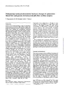
fMRI Paradigm Participants lay supine with their right arm bent at an approximately 90◦ angle so that their forearm rested across their torso. Because movements of the shoulder and upper arm may induce artifacts in the participant’s data (Rossit et al., 2013), a strap around the participant’s chest was used to minimize upper arm and shoulder movements, while allowing for full rotation at the elbow. Figure 1 described the fMRI paradigm used in this experiment. While in the MRI scanner, participants were instructed to either execute or imagine a series of movements involving their right forearm. We used two different arm movements: a ‘‘slide’’, which involved sliding the forearm forward and back; and a ‘‘lift’’, which involved lifting and lowering the forearm. Each sequence involved six movements (combining ‘‘slides’’ and ‘‘lifts’’). Imagery and execution blocks were 20 s long, and were alternated with periods of rest for a total of 8 min. The beginning of each block was cued with the word ‘‘move’’, ‘‘imagine’’ or ‘‘relax’’. Participants also completed blocks where they were instructed to relax while a researcher moved their arm (data not reported here). Within each block (imagery or execution), participants either received a pre-determined sequence (i.e., CF) or were asked to create one by individually choosing one out of the two possible movements at a time (AS), in a 2 × 2 within-subjects factorial
2
September 2015 | Volume 9 | Article 493
Osborne et al.
Command following and communication in DOC
(TR = 2300 ms, TE = 2.32 ms, IT = 900, matrix size = 256 × 256, voxel size 1 × 1 × 1 mm, flip angle = 8◦ ) was also acquired.
fMRI Data Analysis We performed Independent Component Analysis using the FSL MELODIC tool,1 in order to remove motion artifacts (Friston et al., 1996; McKeown and Sejnowski, 1998; Beckmann and Smith, 2004). One of the authors (N.R.O.) visually inspected all the components and identified those that corresponded to headmotion artifacts and were correlated with the execution blocks. An average of 5 ± 2.6 artifactual components were identified per subject and run. Finally, we removed the identified components from the fMRI data. The de-noised data was then pre-processed and analyzed with SPM8.2 After manually AC-PC reorienting the data, the following spatial pre-processing steps were performed: realignment, co-registration of the structural and functional data, spatial normalization to Montreal Neurological Institute (MNI) space, and smoothing with an 8-mm FWHM Gaussian kernel. High-pass filtering with a cut-off period of 128 s was used to remove linear drift. A single subject fixed-effect twoby-two factorial analysis was performed for each subject at the whole-brain level. Factor 1 was defined as ‘‘Task’’ with two levels (motor imagery/motor execution) and Factor 2 was defined as ‘‘Level of selection’’, with two levels (AS/CF). Scans were modeled as belonging to the AS/ME, CF/ME, AS/MI, or CF/MI conditions using the canonical hemodynamic response function (Friston et al., 1995) with the participant’s rest condition used as a baseline. Realignment parameters and passive movement blocks were modeled as effects of noninterest. While all participants reported completing the MI blocks, the nature of MI precludes any observable or external means of confirmation that they did indeed perform the task. However, previous MI studies have demonstrated that activity in the supplementary motor area (SMA) can be used as neural evidence for MI (Owen, 2011). We examined individuals’ whole brain activity during MI conditions compared to rest in order to confirm their completion of the task, and to avoid biases in the analysis from including participants who may not have performed it. 13 out of 15 participants showed significant activity in the SMA (cluster level uncorrected p < 0.001). The remaining two participants were removed from subsequent analyses. Therefore, 13 participants were included in the group analyses, which consisted of one-sample t-tests for each contrast of interest. The statistical threshold was set at a family wise error (FWE) corrected p < 0.05 at the cluster-level. Two additional contrasts, individually comparing ME and MI conditions to rest, were also included to confirm that the task elicited a similar pattern of activation as previously reported paradigms (Owen et al., 2006, 2007; Formaggio et al., 2013; Machado et al., 2013; Fernández-Espejo et al., 2014). The FSL Harvard-Oxford Cortical and Subcortical Structural Atlases (see Acknowledgments) were used for anatomical identification.
FIGURE 1 | fMRI paradigm. This study combined two different arm movements (slide and lift) to create various six-movement long sequences (e.g., “slide lift lift slide lift slide”). Subjects either executed (ME) or imagined (MI) movement sequences that were either pre-determined by experimenters (CF) or chosen by them (AS), in a 2 × 2 within subjects factorial design. Onsets in CF blocks were cued with the words “slide”, or “lift”, while “go” was used to cue onsets in AS blocks. Each block lasted 20 s and consisted of six moves. During rest blocks (dotted), subjects lay still in the scanner. There were four blocks of each condition, which were presented in a pseudorandom order for a total of 20 blocks (in an additional condition not reported here, the experimenter passively moved subjects’ arms). For the CF blocks, subjects were randomly assigned four different movement sequences from a collection of 48 unique, pre-determined movement sequences created for this experiment. CF, command following; AS, action selection; ME, motor execution; MI, motor imagery.
design. During the blocks with pre-determined sequences, each individual action was cued with the word ‘‘slide’’ or ‘‘lift’’. Each participant was randomly assigned four out of a possible 48 unique movement sequences, all four of which were presented pseudorandomly over each experimenter-cued condition. For those where the subject had to create their own sequence, each action was cued with the word ‘‘go’’. There were four blocks of each condition, which were presented in a pseudorandom order for a total of 24 blocks. All participants completed two runs of this task. An infrared MR-compatible camera (MRC Systems GmbH), placed above the participant’s head, was used to record participants’ actions for each run. The recordings were monitored online to confirm that all participants performed all runs with no errors (i.e., they moved their hand to command during ME trials, and remained still during MI trials). In addition to the video monitoring, participants were asked afterwards about their execution of the task. All participants reported performing the imagery task correctly.
Image Acquisition Data was acquired in a 3T Siemens scanner (Magnetom Prisma, Siemens, Germany), with a Siemens 32-channel head-coil, at the Centre for Functional and Metabolic Mapping (CFMM) at Robarts Research Institute. Audio instructions and task cues were presented using Matlab® R2011a on a MacBook Pro laptop (OSX 10.6.8) and an MRI-compatible high-quality digital sound system via noise-attenuated headphones (Sensimetrics, S14). The fMRI protocol included two sessions of 240 volumes each, using echo-planar images (36 axial slices, TR = 2000 ms, TE = 30 ms, matrix size = 70 × 70, slice thickness = 3 mm, in-plane resolution = 3 × 3 mm, flip angle = 78◦ ). A high-resolution T1-weighted MPRAGE structural image
Frontiers in Human Neuroscience | www.frontiersin.org
1 http://www.fmrib.ox.ac.uk./fsl 2 http://www.fil.ion.ucl.ac.uk/spm
3
September 2015 | Volume 9 | Article 493
Osborne et al.
Command following and communication in DOC
FIGURE 3 | Action selection vs. command following. AS (participants selected their movements), as compared to CF (movements were determined by experimenters) elicited greater activity in the middle frontal gyrus, pre-SMA, somatosensory association cortex and insula. Results are thresholded at FWE-corrected p < 0.05 for cluster level activation, and overlaid on an anatomical T1-weighted image.
vs. AS) showed bilateral activation in the lateral occipital cortex (extrastriate visual area) and primary auditory cortices as well as the precuneus cortex. Group activations are shown in Table 2.
FIGURE 2 | Motor execution vs. motor imagery. Group level analysis showed greater activity in contralateral M1/S1 when motor execution was compared to motor imagery (top panel). Conversely, motor imagery, as compared to motor execution, activated the ipsilateral M1, S1 and left inferior frontal gyrus (bottom panel). Results are thresholded at family wise error (FWE)-corrected p < 0.05 for cluster level activation and overlaid on an anatomical T1-weighted image.
Interactions There were no significant interactions between task and level of selection. No activity was observed even when thresholds were lowered to an uncorrected p < 0.01. To increase the sensitivity of our exploration of this interaction, we ran an additional analysis using a mask including all areas active in the main effect of task and level of selection. The result from this additional analysis confirmed no significant effects, even at uncorrected p < 0.01. To further explore the consistency of this (lack of) effect at an individual participant level, we inclusively masked the positive interaction for each individual with their activity from the two main effects. This revealed no significant activity for any participant. Two participants however showed an uncorrected cluster in the left frontal pole, with peak below an uncorrected p < 0.001. Additionally, we calculated the percent signal change for each participant in three 10 mm spherical ROIs (i.e., M1, SMA, and pre-SMA). These were defined using coordinates from the clusters revealed in the group level whole brain analysis for ME vs. rest (coordinates −30 −25 58), MI vs. rest (−3 8 55), and the positive effect of level of selection (3 20 46). Figure 4 shows the percent signal change across all four conditions for each ROI: AS/ME, CF/ME, AS/MI, and CF/MI. Activity in M1 increased during ME conditions compared to imagery, while activity in the SMA followed the opposite pattern, consistently across
Results Motor Imagery vs. Motor Execution The positive effect of task (i.e., ME vs. MI) revealed a significant cluster of activation in the left sensorimotor area, as shown in Figure 2. This included M1, the primary somatosensory cortex (S1), and the superior parietal lobule. The negative effect of task (i.e., MI vs. ME) revealed significant activity in the right S1 and M1, left inferior frontal gyrus and right occipital pole (representing the primary and secondary visual cortices). Group activations are shown in Table 1.
Action Selection vs. Command Following The positive effect of level of selection (i.e., conditions where the participant had to choose between two actions vs. those in which the action was determined by the experimenter) revealed significant activity in frontal regions including bilateral frontal poles and middle frontal gyri, as well as the paracingulate gyrus (including pre-SMA). There was also significant activation in the somatosensory association cortex, specifically the right angular gyrus and the left insular cortex. Group activity for this contrast is shown in Figure 3. The inverse contrast (CF
TABLE 1 | Motor execution vs. motor imagery. Brain structure
Coordinates
Cluster size (k)
T value
p value
x
y
z
Positive effect of task (motor execution > motor imagery) Superior parietal lobule/postcentral gyrus
−24
−43
61
269
6.35
0.01
Negative effect of task (motor imagery > motor execution) Inferior frontal gyrus Postcentral/Precentral gyrus Occipital pole
−57 39 12
20 −25 −88
22 61 28
5731 1680 718
11.44 9.18 7.17