peptides in the salivary glands of the locust, Locusta migratoria. M. Fusé, D.W. Ali, I. Orchard .... pH 7.3 (Sigma, St. Louis, Mo.). ..... Science 197:670â671. Robb S ...
Cell Tissue Res (1996) 284:425–433
© Springer-Verlag 1996
190112.500 220514.200 061318.500 091313.625 121503*500
The distribution and partial characterization of FMRFamide-related peptides in the salivary glands of the locust, Locusta migratoria M. Fusé, D.W. Ali, I. Orchard Department of Zoology, University of Toronto, Toronto, Ontario, M5S 3G5, Canada &misc: Received: 12 August 1995 / Accepted: 4 January 1996
&Abstract. p.1: The distribution and partial characterization of FMRFamide-related peptides in the salivary glands of the locust, Locusta migratoria, were investigated by means of immunohistochemistry, radioimmunoassay and reversed-phase high performance liquid chromatography. Whole-mount preparations of glands stained positively against anti-FMRFamide antisera, and contained the equivalent of 837±80 fmol FMRFamide/gland pair, as determined by radioimmunoassay. FMRFamide-like immunoreactivity occurred in the processes of the transverse nerves of both the prothoracic and mesothoracic ganglia, but was not found in the salivary motoneurons 1 or 2 of the suboesophageal ganglion, both of which directly innervate the salivary glands via the salivary nerve 7b; nor was it found within the salivary nerve 7b itself, leading to the salivary glands. It was, however, found as a superficial nerve plexus on the surface of nerve 7 at the suboesophageal ganglion, but did not appear to extend to the salivary glands. The origin of this staining is unclear. High performance liquid chromatography of salivary gland tissue extracts, monitored by radioimmunoassay, revealed 4 peaks of immunoreactive material, 2 of which co-migrated with AFIRFamide and GQERNFLRFamide, previously isolated from the locust ventral nerve cord. These 2 synthetic peptides did not elevate basal levels of the second messengers cyclic AMP or cyclic GMP in the salivary glands. &Key kwd: words: Salivary glands – Ventral nerve cord – FMRFamide – Immunohistochemistry – Radioimmunoassay – Locusta migratoria (Insecta)
This work was supported by the Natural Sciences and Engineering Research Council of Canada in the form of a research grant to I.O. and a graduate scholarship to D.A., and by Insect Biotech Canada. Correspondence to: M. Fusé&kl / f o n - b:c
Introduction It is becoming increasingly apparent that FMRFamide (Phe-Met-Arg-Phe-amide), and the family of FMRFamide-related peptides (FaRPs), play a large and diverse role in biological processes. The tetrapeptide FMRFamide was originally sequenced from the marine mollusc Macrocallista nimbosa (Price and Greenberg 1977), in which it was found to have cardioexcitatory effects. In recent years the family to which this peptide belongs has grown dramatically and its members have been shown to affect a variety of tissues such as visceral and skeletal muscle (Walther et al. 1984; Cuthbert and Evans 1989; Lange et al. 1991; Peeff et al. 1993), neurons (Walther et al. 1984) and glands (Baines and Tyrer 1989; Yasuyama et al. 1993). Much of the evidence for the presence of FaRPs in insects has been obtained using immunohistochemical and chromatographic techniques. Such studies have shown that FaRPs are associated with both the central and peripheral nervous systems as well as optic lobes, muscle and visceral tissues (Ohlsson et al. 1989; Tsang and Orchard 1991; Schoofs et al. 1993). Furthermore, a number of FaRPs have been sequenced from various insects (Matsumoto et al. 1989; Robb et al. 1989; Fonagy et al. 1992; Nichols 1992; Duve et al. 1992; Schoofs et al. 1993; Lange et al. 1994; Peeff et al. 1994). From these studies, and with the application of available bioassays, the physiological relevance of FaRPs is becoming clearer. It is known for instance, that FaRPs potentiate neurally-evoked contractions of the locust extensor tibiae (Walther et al. 1984; Evans and Myers 1986), inhibit myogenic contractions of locust oviducts (Lange et al. 1991; Peeff et al. 1993) and may modulate the actions of biogenic amines on visceral tissues (Banner and Osborne 1989). Of interest to us is the role that FaRPs may play in the regulation of salivary gland activity in L. migratoria. In Locusta, the aminergic innervation of the salivary glands has been clearly documented (Klemm 1972; Orchard et al. 1992; Ali et al. 1993). The glands are di-
426
rectly innervated via 2 pairs of salivary motoneurons, the SN1 and the SN2, whose cell bodies are located within the suboesophageal ganglion (Altman and Kien 1979). These neurons each send an axon via a branch of nerve 7, salivary nerve 7b, to the salivary glands. The SN1 have been shown to contain dopamine, whereas the SN2 contain serotonin (Gifford et al. 1991; Orchard et al. 1992; Ali et al. 1993). These 2 biogenic amines increase salivation levels (Baines and Tyrer 1989), and are also capable of increasing cAMP levels in dose-dependent manners in isolated salivary gland preparations (Ali et al. 1993). Recent studies have also suggested the presence of FMRFamide-related peptides in the salivary glands (Myers and Evans 1985; Baines et al. 1989; Schoofs et al. 1993). However, the types of FaRPs present and their sites of origin are questionable. Immunohistochemical studies of the locust (Schistocerca gregaria) CNS have indicated that the source of FaRPs innervating the salivary glands originates from a small branch of the posterior transverse nerve from the mesothoracic ganglion (Myers and Evans 1985). Cobalt backfills of nerves in the salivary glands of L. migratoria, on the other hand, show innervation from the prothoracic ganglion (Baines et al. 1989). This has led to the suggestion that FaRPs in the salivary glands of Locusta may originate from the prothoracic ganglion. More recent use of an antibody raised against the sequence for the FaRP, SchistoFLRFamide, has revealed immunoreactivity in the suboesophageal ganglion near the SN2, and in an axon in a nerve along the salivary duct of L. migratoria (Schoofs et al. 1993). This has led to an alternate suggestion, that SchistoFLRFamide is involved in salivary gland function and may be co-localised with serotonin in the SN2 and in the axon of nerve 7b projecting to the salivary glands. However, the high affinity for the C-terminal moiety of FaRPs by this antibody (Schoofs et al. 1993) necessitates stronger confirmation that SchistoFLRFamide is indeed involved. At the very least, these studies indicate the presence of FaRPs and FaRP innervation associated with the salivary glands, suggesting a role for these peptides in the regulation of fluid production and/or secretion. The apparent differences between the studies, however, have necessitated a reappraisal of both the source and distribution of FaRP innervation in the salivary glands, prior to studying their role(s) in fluid secretion. In this study we describe the distribution of, and partially characterize, 4 FaRPs associated with the salivary glands, and determine their sites of origin.
Immunohistochemistry
Materials and methods
Tissue extraction and HPLC analysis
Animals
Ten groups of 10 salivary gland pairs were dissected in physiological saline, transferred to 1 ml of extraction medium (90% methanol;9% glacial acetic acid;1% water), freeze-thawed, sonicated, and centrifuged at 8800×g for 15 min. The supernatant was collected and pooled. The pellet was resuspended in extraction medium and processed as above. All supernatant was pooled and evaporated to dryness using a Speed-Vac concentrator (Savant, Farmingdale, N.Y.), then run through a C18-containing Sep-Pak car-
Locusts were taken from a colony of Locusta migratoria reared as previously described in Peeff et al. (1994). Experiments were performed on adult males, 5–10 days old. A mixture of adult male and female locusts, 5–21 days old, were used for reversedphase high performance liquid chromatography (RP-HPLC) analysis.
Ganglia and salivary glands were dissected under physiological saline (150 mM NaCl; 10 mM KCl; 4 mM CaCl2; 2 mM MgCl2; 4 mM NaHCO3; 5 mM HEPES, pH 7.2; 90 mM sucrose; 5 mM trehalose), and fixed in 2% paraformaldehyde in Millonig’s buffer for 1.5 h. Tissues were washed in phosphate-buffered saline (PBS; 10 mM phosphate buffer, pH 7.2, containing 0.9% NaCl) for 4–5 h. PBS was used to dilute all solutions unless stated otherwise. Tissues were incubated in 4% TritonX-100 (2% normal serum; 2% BSA) for 1 h. This step was carried out to enhance penetration of the antibodies and reduce non-specific binding. Tissues were then processed for either FMRFamide-like, serotonin-like or tyrosine hydroxylase-like immunoreactivity (FMRFa-ir, serotonin-ir, TH-ir, respectively) by the methods described below. The primary antibodies were pre-absorbed in 0.4% TritonX-100 (2% normal serum; 2% BSA) for 18 h to occupy non-specific antigenic sites. Two independently produced polyclonal rabbit anti-FMRFamide antisera were used (antiserum1: Incstar, Stillwater, Mn; antiserum2: Peninsula, Belmont, Calif., USA). Anti-serotonin and anti-TH antisera were purchased from Incstar. For FMRFa-ir, the tissues were incubated for 48 h at 8°C in a 1:1000 dilution of either of the 2 anti-FMRFamide antisera, in 0.4% TritonX-100 (2% normal goat serum; 2% BSA). This was followed by a 5–8 h wash in cold PBS prior to incubation in the secondary antiserum for 18 h at 8°C. The secondary antibody was fluorescein isothyocyanate (FITC)-conjugated goat anti-rabbit immunoglobulin G (IgG) diluted 1:200 (10% normal goat serum). Tissues were washed in PBS and subsequently cleared and mounted on depression slides in 5% n-propyl gallate in 80% glycerol, pH 7.3 (Sigma, St. Louis, Mo.). The commercially available antiFMRFamide antisera both show high specificity for RFamide, and antiserum1 has been shown quantitatively to have similar affinities for FLRFamide, FMRFamide and SchistoFLRFamide (Tsang and Orchard 1991). Double-labelling immunohistochemistry, using either antiFMRFamide and anti-serotonin antisera, or anti-FMRFamide and anti-TH antisera, was performed in a manner similar to that described above. The 2 primary antisera were applied together, whereas the secondary antisera were applied sequentially. The primary and secondary antibodies used, and any changes in their concentrations, are listed below. For FMRFamide/serotonin double-labelling, rabbit anti-FMRFamide antiserum with FITC-conjugated donkey anti-rabbit IgG were used. Goat anti-serotonin antiserum (1:500) was used with Texas Red-conjugated donkey anti-goat IgG. Normal donkey serum was used for pre-absorption. For FMRFamide/TH double-labelling, goat anti-FMRFamide antiserum with Texas Red-conjugated donkey anti-goat IgG were used. Monoclonal mouse anti-TH antiserum (1:400) was used with FITC-conjugated donkey anti-mouse IgG. Normal donkey serum was used for pre-absorption. Goat secondary antisera were purchased from Daymar Inc. (Toronto, Ontario), while donkey secondary antisera were purchased from Jackson Immunoresearch (West Grove, Pa.). Bovine pancreatic polypeptide and FMRFamide were both used for blocking anti-FMRFamide antibodies, and were purchased from Peninsula.
Fig. 1. A–E FMRFa-ir in whole-mount preparations of salivary glands of the locust, Locusta migratoria. FMRFa-ir axons (arrows) follow the length of the salivary duct (sd), and lie on the acini (ac). The salivary duct is highlighted due to autofluorescence of the tissue. A The posterior region of the salivary glands possesses numerous branching FMRFa-ir axons, often following the salivary duct. Scale bar: 60 µm. B FMRFa-ir neurohaemal regions (white arrows) are found predominantly where multiple axons meet. Scale bar: 40 µm. C Only single FMRFa-ir axons are found
in nerves of the anterior portion of the salivary glands. Scale bar: 40 µm. D, E FMRFa-ir axons lie over top of the acini of the salivary glands (E scale bar: 25 µm), with blunt-ended terminals (D scale bar: 40 µm). F–G Double-labelling of nerve 7 exiting the suboesophageal ganglion (asterisk), revealing FMRFa-ir and THir, respectively. Solid arrows denote immunoreactive axons. Clear arrows denote nerve 7 surface. Note the FMRFa-ir on the membrane in (F). Scale bar: 40 µm&.i c/ :gf
428
Fig. 2A, B. Camera lucida reconstructions of FMRFa-ir axons and cells of the salivary glands, ventral nerve cord and suboesophageal ganglion of Locusta migratoria. A The prothoracic (PRO) and mesothoracic ganglia (MESO) and transverse nerves (tn) projecting to the acini (ac) of the salivary glands are depicted. FMRFa-ir axons often follow the salivary duct (sd), eventually joining salivary nerve 7b (n7b). Only FMRFa-ir cell bodies and axons are shown. B The suboesophageal ganglion (SOG) and FMRFa-ir cell bodies (filled cells) are depicted. The SN1 (dopaminergic) and SN2 (serotonergic) motoneurons (clear cells) and their axons exiting via nerve 7 (n7) are also shown. Nerve 7 branches as salivary nerve 7b (n7b, seen in A) and travels to the salivary glands. The SN1 and SN2 cells do not possess FMRFa-ir. FMRFa-ir on nerve 7 is not depicted for clarity. Scale bars: 400 µm&.i c/ :gf
tridge, and prepared for HPLC analysis as described by Lange et al. (1994). Extracts were resuspended in HPLC buffer and chromatographed sequentially on 2 different HPLC systems using various acetonitrile gradients: System 1. A Brownlee RP-C18 Spheri-5 column (46 mm× 25 cm) was used with a linear acetonitrile gradient (containing 0.1% TFA) of 9–60% acetonitrile, run at 1 ml/min. This gradient was run over 34 min, 2 min after injecting the sample. Selected fractions were pooled, evaporated to dryness and redissolved in HPLC solvent to be run on System 2. System 2. A Brownlee Phenyl Spheri-5 column (46 mm× 25 cm) was used with 2 different linear acetonitrile gradients (containing 0.1% TFA), run at 1 ml/min. The first gradient ran from 18–60% acetonitrile, over 60 min, 2 min after injecting the sample. Selected fractions were processed individually, as described for System 1, using the same Phenyl column with a slower acetonitrile gradient. The slower gradient ran from 20–29% acetonitrile, over 60 min, 2 min after injecting the sample. Selected fractions were aliquoted, evaporated to dryness, and resuspended in HPLC buffer alone or spiked with synthetic FaRPs (AFIRFamide, GQERNFLRFamide and ADVGHVFLRFamide from Queen’s University Core Facility, Kingston, Ontario; PDVDHVFLRFamide from Peninsula) previously isolated and sequenced from the locust CNS (Peeff et al. 1994; Lange et al. 1994). These aliquots were rerun on the second Phenyl system. The radioimmunoassay for FMRFamide-like peptides was carried out as described previously by Peeff et al. (1994).
Cyclic AMP and cyclic GMP measurements Salivary glands were dissected under physiological saline and assayed for the effects of FaRPs on cAMP and cGMP levels. Additionally, 5×10⁻⁷ M serotonin was tested on glands, as a positive control for cAMP elevation. Tissues were incubated in physiological saline containing 0.5 mM 3-isobutyl-1-methyl-xanthine (IBMX), along with various concentrations of synthetic AFIRFamide or GQERNFLRFamide for 10 min at room temperature. The reaction was terminated by addition of 500 µl boiling 0.5 M sodium acetate buffer, pH 6.2, followed by 5 min of boiling. The samples were stored for up to 24 h at –20°C. The samples were sonicated, centrifuged at 8800×g for 10 min and the supernatant
removed for cAMP and cGMP determination. The pellet was dissolved in 50 µl 0.5 N sodium hydroxide for protein determination. Cyclic AMP and cGMP levels were measured by radioimmunoassay using a commercially available kit (New England Nuclear, Lachine, PQ). The protein content of the salivary glands was determined using the BioRad protein assay (Melville, NY) based upon Bradford (1976) using γ-globulin as the standard. Chemicals other than the synthetic FaRPs were obtained from Sigma.
Results Immunohistochemistry Salivary glands. The & p . 1 : glands stained positively against the 2 different rabbit polyclonal anti-FMRFamide antisera. Staining patterns were similar for both antisera. The FMRFa-ir was found as an elaborate plexus of immunoreactive processes. In most cases, the processes followed the path of the extensively branched salivary duct (Fig. 1a–c). Branches containing FMRFa-ir processes only joined the salivary nerve 7b midway through the glands. This was apparent from double-labelling experiments revealing FMRFa-ir axons alongside the dopaminergic or serotonergic axons in nerve 7b within the salivary glands (Fig. 2), but not in nerve 7 at the suboesophageal ganglion (Fig. 1f, g). The FMRFa-ir axons often possessed large varicosities (Fig. 1b; arrows) diagnostic of neurohaemal storage and release sites. In contrast to the finer and extensively branched dopaminergic SN1 and serotonergic SN2 processes, which were deeply embedded within the acini (Ali et al. 1993), the FMRFa-ir processes in the salivary glands were anchored as a superficial network of terminal varicosities over the acini (Fig. 1d, e). Nerve branches of the posterior region of the salivary glands exhibited the greatest distribution and intensity of FMRFa-ir staining, and contained many FMRFa-ir pro-
429
Fig. 3A, B. FMRFa-ir in whole-mount preparations of 2 thoracic ganglia and transverse nerve (TN) branches associated with the salivary glands (SG). A FMRFa-ir is traced through the TN (arrows) of the posterior median nerve (MN), from the prothoracic
ganglion (PRO), and enters the salivary glands. Scale bar: 150 µm. B Similar FMRFa-ir is found in the TN of the mesothoracic ganglion (MESO). Scale bar: 90 µm&.i c/ :gf
cesses (Fig. 1b). Very little FMRFa-ir was found in the anterior third of the glands. Only single FMRFa-ir axons were found in anterior nerve branches (Fig. 1c; arrows), with at most 1 or 2 branches staining positively in each salivary gland preparation. These anterior fibres contained FMRFa-ir axons alone, with no TH-ir or seroto-
nin-ir, suggesting that they were not nerve fibres of the salivary nerve 7b. Suboesophageal ganglion. The & p . 1 : suboesophageal ganglion (SOG) is the origin of the 2 pairs of salivary motoneurons, the SN1 (dopaminergic) and the SN2 (serotoner-
430
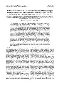
Fig. 4A, B. Fractionation of FMRFa-ir from extracts of 100 adult Locusta salivary glands using 2 RP-HPLC columns with different acetonitrile gradients. A RP-HPLC separation on a C18 column (System 1) revealed 1 broad peak of FMRFa-ir material. The values have been converted from subsamples of 100 salivary glands to 1 salivary gland pair (SG pair). B RP-HPLC separation of pooled fractions 21 to 27 from (A) on a Phenyl column with a fast acetonitrile gradient (System 2A). Four FMRFa-ir peaks were detected (filled bars) and rerun individually with a slower acetonitrile gradient (System 2B). Histogram fills correspond to appropriate fills in individual runs C through F. Arrows denote elution times of labelled peptides&.i c/ :gf
gic). They directly innervate the salivary glands via a branch of nerve 7, the salivary nerve 7b (Gifford et al. 1991; Orchard et al. 1992; Ali et al. 1993). The SOG was therefore double-labelled with anti-FMRFamide and anti-TH (an indication of the presence of dopamine), or anti-FMRFamide and anti-serotonin, to map the locations of FMRFa-ir cells and axons relative to the 2 motoneurons. FMRFa-ir was not found in the SN1 or SN2 of the SOG (Fig. 2b). Instead, 2 FMRFa-ir cell bodies were located directly posterior to the SN2, but did not send projections to the salivary glands. Moreover, FMRFa-ir axons were not present within nerve 7 at the SOG, but were found in processes of the transverse nerves joining the salivary nerve 7b well within the salivary glands. However, FMRFa-ir was apparent as a nerve plexus on the surface of nerve 7 at the SOG (compare Fig. 1f, g; clear arrows). This superficial staining never appeared to extend to the salivary glands. For clarity, this staining is not shown in the composite camera lucida drawing of the SOG (Fig. 2b). Thoracic ganglia. The & p . 1 : 3 thoracic ganglia of the ventral nerve cord were dissected while still attached to the salivary glands via branches of their transverse nerves (TN). These preparations were stained with anti-FMRFamide
antiserum to determine the origins of FMRFa-ir in the salivary glands (Figs. 2, 3). One small axonal tract entering the anterior region of the salivary glands stained positively for FMRFa-ir and was traced to the posterior TN of the prothoracic ganglion (Figs. 2a, 3a). Staining of the TN in the salivary glands was limited to a few branches extending anteriorly. Another FMRFa-ir axon entering the mid-portion of the glands was traced to the posterior TN of the mesothoracic ganglion, with many FMRFa-ir processes projecting in all directions in the salivary glands (Figs. 2a, 3b). The staining pattern suggested that most FMRFamide-like material originated from the mesothoracic ganglia. The branches of the TN from the metathoracic ganglion, which entered the salivary glands, did not stain positively for FMRFa-ir at the level of the salivary glands (data not shown). Controls. Staining &p.1: was almost entirely eliminated in the salivary glands when the primary antiserum was preabsorbed overnight with 50 µg/ml FMRFamide. In contrast, the most typically cross-reactive non-related peptide, bovine pancreatic polypeptide, at a concentration of 50 µg/ml did not visibly alter the intensity or the distribution of staining. No staining was visible when primary
431
or secondary antisera were eliminated in the incubation steps. Radioimmunoassay Quantification of FMRFamide-like material. Anti-FMR&p.1: Famide antiserum 1 was used for the radioimmunoassay (RIA), to quantify the FMRFamide-like peptides in the salivary glands. The amount of FMRFamide-like material in salivary gland tissue extracts was expressed as FMRFamide-like equivalents (FLE), as described by Tsang and Orchard (1991). L. migratoria contained 837±80 fmol FLE/gland pair (n=10). This amount was reduced proportionately when single glands were assayed (420±44 fmol FLE/single gland, n=12), demonstrating that the amount of tissue present did not interfere with the RIA. Reversed-phase high performance liquid chromatography. The & p . 1 : pooled material run through System 1 (C18 column), resulted in the resolution of 1 large peak of FMRFa-ir material over a 3 min period (Fig. 4a). This fraction, and adjacent fractions (20–27), were pooled, evaporated to dryness and run through System 2 (Fig. 4b). The 4 groups of FMRFa-ir fractions (shaded bars) were then run individually through System 2 at a slower gradient. The slower acetonitrile gradient of each individual fraction (Fig. 4c–f) did not appear to resolve any further FaRP peaks. The elution profiles of both systems were remarkably similar to those previously determined from locust ventral nerve cord extracts (Lange et al. 1994), except for the absence of a peak where SchistoFLRFamide and ADVGHVFLRFamide eluted. Individual elution profiles of the peptides isolated and sequenced from ventral nerve cord tissue (Lange et al. 1994), were then compared with the salivary gland profiles. Of the 4 resolved peaks, fractions 26/27 co-eluted with synthetic AFIRFamide, fractions 30/31 co-eluted with synthetic GQERNFLRFamide, while fractions 35/36 and 47/48 did not co-elute with any known FaRPs from the CNS (Fig. 4c–f, respectively). The elution profile of fractions 35/36, however, did match the profile of another FaRP also isolated from the ventral nerve cord, which is not yet fully sequenced (AXXRNFIRFamide; Lange et al. 1994). FaRPs co-eluting with synthetic SchistoFLRFamide (PDVDHVFLRFamide) or ADVGHVFLRFamide were not apparent in the salivary glands (see Fig. 4f; arrows). From these separations, it appears that the salivary glands of L. migratoria contain at least 4 different FaRPs, 2 of which are likely extended FIRFamides and 1 which is an extended FLRFamide. SchistoFLRFamide and ADVGHVFLRFamide do not appear to be present. Effects of FMRFamide-like peptides on cyclic AMP/GMP levels Both dopamine and serotonin are known to induce increases in basal levels of cAMP in a dose-dependent
Fig. 5. Increases in cAMP content/mg protein of salivary glands (SG) of adult male Locusta following incubation for 10 min in 5×10⁻⁷ M serotonin (///), or different concentrations of AFIRFamide (\\\) or GQERNFLRFamide (XXX), with 0.5 mM IBMX. The values reflect increases over IBMX control levels, and are represented as means±SE (n>5). Asterisk represents significant difference (P