2797
The Journal of Experimental Biology 200, 2797–2805 (1997) Printed in Great Britain © The Company of Biologists Limited 1997 JEB1132
THE EFFECTS OF EXPOSURE TO AMMONIA ON AMMONIA AND TAURINE POOLS OF THE SYMBIOTIC CLAM SOLEMYA REIDI RAYMOND W. LEE*, JAMES J. CHILDRESS AND NICOLE T. DESAULNIERS Marine Science Institute and Department of Biological Sciences, University of California, Santa Barbara, CA 93106, USA Accepted 18 August 1997 Summary The nutrition of the gutless clam Solemya reidi is of ammonia incorporation into organic matter supported by the activity of intracellular (assimilation) were determined using 15N as a tracer. 15Nchemoautotrophic bacteria housed in its gill filaments. labeled ammonia assimilation was higher in gill than in Ammonia (the sum of NH3 and NH4+) is utilized as a foot and increased as a function of 15N-labeled ammonia nitrogen source by the association and is abundant in the concentration in the medium. The size of the free amino clam’s environment. In the present study, clams were acid (FAA) pool in the gill also increased as a function of exposed to 0.01–1.3 mmol l−1 ammonia for 22–23 h in the ammonia concentration in the medium. This entire presence of thiosulfate as a sulfur substrate. Ammonia increase was accounted for by a single amino acid, exposure increased the ammonia concentration in the taurine, which was the predominant FAA in both gill and tissue pools of the gill, foot and visceral mass from 0.5 to foot tissue. Aspartate, glutamate, arginine and alanine 2 µmol g−1 wet mass, without added ammonia, to as much were also abundant but their levels were not influenced as 12 µmol g−1 wet mass in the presence of 0.7 and by external ammonia concentration. Ammonia 1.3 mmol l−1 external ammonia. Gill tissue ammonia assimilation appeared to occur at rates sufficient to concentrations were consistently higher than those in the account for the observed increase in taurine level. These foot and visceral mass. The elevation of tissue ammonia findings suggest that taurine is a major product of concentration compared with the medium may be due in ammonia assimilation. part to an ammonia trapping mechanism resulting from a lower intracellular pH compared with sea water and Key words: ammonia, taurine, clam, symbiosis, Solemya reidi, nitrogen metabolism. greater permeability to NH3 compared with NH4+. Rates
Introduction Solemya reidi is a gutless protobranch clam that inhabits burrows in sulfidic sediments on the Pacific coast of the United States and Canada (Bernard, 1980). Reduction or absence of structures associated with particulate feeding is characteristic of clams of the genus Solemya (Reid and Bernard, 1980, and references cited therein). The discovery of chemoautotrophic bacteria–invertebrate symbiosis at deep-sea hydrothermal vents in the Pacific led to the subsequent discovery of this type of symbiosis in S. reidi (Cavanaugh et al. 1981; Felbeck et al. 1981). High densities of chemoautotrophic intracellular symbiotic bacteria are found in the gills of this species as well as in two other species, S. velum and S. borealis (Cavanaugh, 1983; Conway et al. 1992; Felbeck, 1983; Felbeck et al. 1981). Chemosynthetic bacterial symbionts have been documented in over 100 invertebrate species from at least five phyla (Cavanaugh, 1994; Polz and Cavanaugh, 1995). In general, these associations are found in areas where reduced chemical
species are abundant. Sulfide, and in some cases methane, is oxidized as an energy source by the intracellular bacteria, which are then believed to provide organic compounds for host catabolism and biosynthesis. The importance of sulfur-based autotrophy in Solemya reidi is well documented. Sulfide concentration ranges from 0.4 to 1.9 mmol l−1 in the porewater of the sediment from which clams are collected (Lee et al. 1992a) and it can be oxidized as an energy source by both the symbionts and host mitochondria (Powell and Somero, 1986). Symbiont sulfur oxidation results in net assimilation of CO2 by the Calvin–Benson cycle (Anderson et al. 1987). Organic compounds are then translocated to the host tissues (Fisher and Childress, 1986). Utilization of dissolved organic compounds may also be important since free amino acids (FAAs) can be taken up and are present in sediments where S. reidi are found (Felbeck, 1983; Lee et al. 1992a). However, assimilation of
*Present address: Department of Organismic and Evolutionary Biology, Biolabs, 16 Divinity Avenue, Harvard University, Cambridge, MA 02138, USA (e-mail:
[email protected]).
2798 R. W. LEE, J. J. CHILDRESS
AND
N. T. DESAULNIERS
inorganic compounds (autotrophy) is probably the main source of organic carbon and nitrogen in S. reidi. The overall contribution of autotrophy can be inferred from natural abundance stable carbon and nitrogen isotope studies of S. velum and S. borealis (Conway et al. 1989, 1992). The tissues of these associations are highly depleted of 13C and 15N (δ13C=−31 to −35 ‰; δ15N=+4 to −10 ‰) compared with bivalves that rely on particulate feeding. The δ13C and δ15N values of purified symbionts do not differ from those of the host tissues, and this is evidence that the carbon and nitrogen used in biosynthesis are primarily derived from autotrophy (Conway et al. 1989). Assimilation of nitrogen is an important and potentially complex physiological capability of marine symbioses. Nitrogen is often a limiting nutrient for marine autotrophic organisms. The ability to assimilate inorganic nitrogen compounds facilitates the recycling of waste ammonia from amino acid catabolism and the utilization of inorganic nitrogen from the environment in autotrophic marine symbioses such as algal–invertebrate associations (Muscatine, 1980). The metabolism of ammonia and nitrate in symbiotic associations is complicated by the possibility that both host and symbiont are involved in assimilation. Ammonium assimilation by algal–invertebrate associations, which was once thought to involve primarily the algal symbiont, is apparently facilitated in part by the invertebrate host (McAuley, 1995; Rees, 1987; Rees et al. 1994). Since the ‘essential’ amino acids (those that cannot be synthesized and must be obtained from the diet) of invertebrates are probably the same as those of other metazoans (Bishop et al. 1983), even if the host can assimilate ammonia into amino acids, the symbiont may be required for synthesis of essential amino acids. This is particularly applicable to S. reidi, since these clams cannot obtain amino acids from particulate food. In earlier studies, we documented that Solemya reidi can take up and assimilate ammonia as well as nitrate (Lee and Childress, 1994; Lee et al. 1992a). Ammonia is the most abundant dissolved nitrogen source in the sewage sludge outfall environment where clams were collected. Porewater ammonia concentrations are around 50–60 µmol l−1 compared with 1–11 µmol l−1 nitrate and 3–15 µmol l−1 total FAA (Lee and Childress, 1994; Lee et al. 1992a). Ammonia assimilation appears to be dependent on conditions favoring sulfide-based chemoautotrophy. Sulfide stimulates ammonia uptake, and detectable ammonia excretion is only observed after prolonged maintenance in the laboratory or maintenance in sulfide-free sea water (Lee et al. 1992a). Ammonia is incorporated into organic compounds, and the highest rates of incorporation are in the gills (Lee and Childress, 1994). In the present study, we investigated the fate of ammonia within the symbiotic association by measuring tissue ammonia pools and 15N-labeled ammonia assimilation. Biosynthesis of amino acids was investigated by measuring changes in tissue FAA pools in response to increased ammonia availability.
Materials and methods Clam collection and maintenance Solemya reidi Bernard were collected by Van Veen grab from depths of approximately 100 m in Santa Monica Bay, California, near the Hyperion sewage sludge outfall and maintained in laboratory mudtanks at 5–9 °C as described previously (Lee et al. 1992a). 15N-labeled
ammonia incubations All ammonia incubations involved the addition of 100 % 15N-labeled ammonia to facilitate measurement of assimilation rates. Solemya reidi, maintained for 3 days in laboratory mudtanks, were removed from their burrows, rinsed with sea water, then placed in filtered sea water for 5 h. Clams were exposed to 100 % 15N-labeled ammonia (0.01–1.3 mmol l−1; five treatments) for 22–23 h. Incubations consisted of 2–3 clams in 0.5–1.0 l of filtered sea water at 5 °C containing 500 µmol l−1 sodium thiosulfate. ΣNH3 (the sum of [NH3]+[NH4+] measured in our analyses) concentration was determined by flow-injection analysis (FIA; Willason and Johnson, 1986). Following exposure to ammonia, clams were removed from the incubation medium and separated into gill, foot and visceral mass. In our sampling, visceral mass refers to the soft body parts remaining after the removal of the gill and foot. Excised tissues were frozen in liquid nitrogen then stored at −80 °C until analyses of ΣNH3, FAAs and 15N incorporation were made. Tissue extracts and ΣNH3 and FAA determinations Frozen tissue samples were homogenized in nine volumes of 50 % ethanol using a ground-glass homogenizer and then centrifuged (Millipore microfuge, 6400 revs min−1). Tissue extracts were analyzed for ΣNH3 by FIA. Free amino acid analyses were performed by high-pressure liquid chromatography (HPLC) and precolumn fluorimetric derivatization with o-phthalaldehyde (OPA; Lindroth and Mopper, 1979; Mopper and Lindroth, 1982). Derivatized amino acids were separated on a Beckman C-18 column using a methanol–acetate buffer gradient and detected fluorometrically as described previously (Lee et al. 1992a,b). These tissue values are probably below true intracellular ΣNH3 and FAA concentrations owing to dilution by hemolymph in the tissues (discussed in more detail below in Discussion). Results are presented as µmol g−1 wet mass. 15N
determinations Subsamples of frozen tissues were dried at 60 °C, then ground to a fine powder. A portion of the ground sample was treated with 2 mol l−1 NaOH to remove ammonia quantitatively (Lee and Childress, 1996). Treated and untreated samples were analyzed for 15N/14N by continuous-flow isotope ratio mass spectrometry (CF-IRMS) using a Europa Scientific RoboprepCN/Tracermass instrument. Operating conditions were as described previously (Lee and Childress, 1995). In the present paper, ‘assimilation’ refers to incorporation observed in
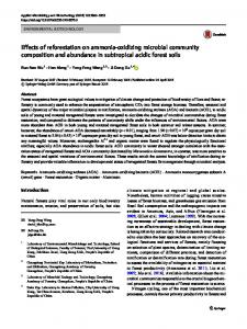
Ammonia metabolism of a symbiotic clam 2799 samples after treatment with NaOH. ‘Σ15NH3’ refers to the amount of 15N-labeled total ammonia (15NH3 and 15NH4+) present in samples determined from the difference in 15N content between NaOH-treated and untreated samples. Results Tissue ΣNH3 Concentrations of ΣNH3 in Solemya reidi tissues ranged from 0.5 to 12 µmol g−1 (Fig. 1A–C). Tissue ΣNH3 concentrations correlated with external ΣNH3 concentration in all tissues tested (Fig. 1A–C). ΣNH3 concentration was clearly elevated in the tissues compared with the medium (Fig. 1A–C). Foot and visceral mass ΣNH3 concentrations were generally lower than gill ΣNH3 concentration. Hemolymph ΣNH3 concentration was lower than that in tissues and was also dependent on external ΣNH3 (Fig. 1D). ΣNH3 concentrations measured from tissues frozen at −80 °C may be slight overestimates, since ΣNH3 concentration can increase in frozen biological samples. Although there are conflicting reports, one study shows an increase of 5–7 µmol l−1 in human blood samples stored at −70 °C (Howanitz et al. 1984). Tissues of a deep-sea mussel symbiotic
14
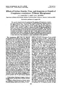
with methanotrophic bacteria (seep mytilid Ia) exhibited gill ΣNH3 concentrations in samples stored at −80 °C of 1.46±0.14 µmol g−1 (mean ± S.D., N=5) compared with 1.22±0.17 µmol g−1 (mean ± S.D, N=5) in samples that were extracted and analyzed immediately. The ΣNH3 concentrations of these fresh and frozen samples were not significantly different (analysis of variance, P>0.05; R. W. Lee, unpublished observations). Thus, storage at −80 °C may have resulted in increased ΣNH3 concentrations in S. reidi tissue samples, but these changes are probably negligible compared with the absolute concentrations and large increases observed as a function of external ΣNH3 concentration (Fig. 1A–D). Effect of external ammonia concentration on 15N assimilation and isotope dilution of 15NH3 Assimilation of 15N was greatest in gill tissue although label was also detected in the visceral mass (Fig. 2). The rate of 15N assimilation increased as a function of external ΣNH3 concentration (Fig. 2). Although clams were exposed to 100 % 15N-labeled ammonia, only part of the gill ΣNH3 was 15N-labeled (Fig. 3). The amount of 15N-labeled ammonia (Σ15NH3) in some gill tissue samples was determined by quantifying 15N lost
14
A pHi=6.5
10 8 6
pHi=7.3
4
Gill
2
8 6
pHi=7.3
4
0 0
0.2
0.4
0.6
0.8
1
1.2
1.4
0.2
0.4
0.6
0.8
1
1.2
1.4
1.2
1.4
D
Visceral mass ∑NH3 (mmol l )
pHi=6.5
10
0 2
C
12 ∑NH3 ( mol g )
Foot
pHi=6.5
10
2
0
14
B
12 ∑NH3 ( mol g )
∑NH3 ( mol g )
12
8 pHi=7.3
6 4
1.6 1.2 0.8 0.4
2 0
Hemolymph
0 0
0.2
0.4
0.6
0.8
1
1.2
1.4
0
0.2
0.4
0.6
0.8
1
External ∑NH3 (mmol l ) Fig. 1. Tissue and hemolymph total ammonia (ΣNH3) concentration of Solemya reidi exposed for 22–23 h to ammonia-enriched sea water with thiosulfate as sulfur substrate. ΣNH3 values are for tissue (A–C) and hemolymph (D) samples from single individuals and are expressed per gram wet mass. The solid line is the isoline for tissue [ΣNH3] = external [ΣNH3]. Broken lines are isolines for tissue [NH3] = external [NH3] for estimated intracellular pH values of 7.3 and 6.5.
N. T. DESAULNIERS 14
12
12
2
[Ammonia] ( mol g )
14 assimilation ( mol g )
AND
15N
2800 R. W. LEE, J. J. CHILDRESS
10 Gill
8 6 4
Visceral mass
∑15NH3 + ∑14NH3
10 8 6 4
∑15NH3
2 0
0 0
0.2
0.4
0.6
0.8
1
1.2
0
1.4
0.2
0.4
0.6
0.8
1
1.2
1.4
External ∑NH3 (mmol l )
External ∑NH3 (mmol l ) Fig. 2. Incorporation of 15N label into Solemya reidi from ammonia exposure experiments. Ammonia added to the medium was 100 % 15N-labeled ammonia. Data points represent determinations made on tissue samples from a single individual following treatment with NaOH to remove label present as 15NH3. Filled circles, gill; open circles, visceral mass.
Fig. 3. 15N-labeled and total ammonia concentration in Solemya reidi gills. Open circles, labeled and unlabeled ammonia as measured by flow-injection analysis (see Fig. 1A). Filled circles, 15N-labeled ammonia determined by quantifying 15N lost by treatment of tissue samples with NaOH. Each data point represents a determination from the gill of a single individual.
following treatment with 2 mol l−1 NaOH. Σ15NH3 concentration was as high as 2 µmol g−1 in gill exposed to 0.7 and 1.3 mmol l−1 external ammonia. The percentage of ΣNH3 present as Σ15NH3 (%Σ15NH3) was variable, with a mean of 19.4±8.8 % (S.D., N=12). At higher external Σ15NH3 concentrations, unlabeled ΣNH3 concentration as well as Σ15NH3 concentration increased in gills.
exposed to varying ammonia concentrations are given in Table 1. The most abundant FAAs were taurine, aspartate, glutamate, alanine and arginine. Taurine concentration was conspicuously elevated compared with the concentrations of all other FAAs measured. Ammonia exposure resulted in an increase in taurine and total FAA concentrations (Fig. 4A,B). No increase in total non-taurine FAA concentration was observed (Fig. 4B). Two extracts of foot tissue were also analyzed (from 0.06 and 0.19 mmol l−1 ΣNH3 treatments). The dominant FAAs were similar to those observed in gill tissue, with taurine present at a higher concentration than all other FAAs.
Tissue free amino acid composition and response to ammonia FAA compositions of gill tissue from individual clams
Table 1. Free amino acid concentrations in gill tissue of individual Solemya reidi exposed to various external ∑NH3 concentrations External ∑NH3 concentration (mmol l−1) Amino acid
0.005
0.005
0.012
0.012
0.012
0.061
0.061
0.061
0.187
0.687
0.687
1.299
1.299
1.299
Aspartate Glutamate Glutamine Glycine Threonine Arginine Taurine Alanine Tyrosine Methionine Valine Isoleucine Leucine
24.2 11.2 2.3 2.9 2 4.7 58.4 5.5 0.9 − 1.5 0.9 1.2
30.5 14 4.3 4 2.8 3.6 84.4 8.1 1.5 − 2.6 1.3 2.3
20.7 6.7 1.6 2.4 1.6 4.6 51.1 4.3 − 0.5 4.2 0.5 1
16.2 10 1.2 2.3 1.6 2.2 83.8 3.7 0.7 − 0.6 0.5 0.8
13.6 15.9 3.3 − 1.9 5.9 106.8 7.7 − − − − 0.6
26.3 13.3 1.3 − 1.5 2.9 99.7 4.6 − − 0.8 − −
20.9 15.1 0.8 − 1.1 1.2 80.5 6.2 0.1 − 1 − −
7.7 25.9 1.1 2.8 1.2 2.6 44.3 10.6 0.8 − 1.3 0.9 1.4
22.9 11.3 1.4 0.8 1.5 1.9 128.4 3.1 0.7 − 1.1 0.6 1
19.9 12.1 0.4 − 1.7 3.8 126.2 5.7 − − 1.3 − 0.4
24.2 5.6 0.9 1.3 0.1 4.7 153.1 1.7 − − 2 4.3 0.8
25.9 20 − − 3.4 2.7 129.4 9.4 0.7 − 1.7 0.5 0.9
18.4 12.2 − 1.4 1.7 3.5 155.3 3.2 0.5 − 1.4 0.3 0.5
44.5 14.2 2.3 2.3 2.2 5.2 182.2 3.4 0.2 − 1.5 0.9 1.5
115.6
159.3
99.2
123.6
155.7
150.5
127
100.4
174.6
171.5
198.8
194.7
198.4
260.4
Total
Free amino acid concentrations are presented as µmol g−1 wet mass. Dashes denote concentrations too low to quantify reliably (less than approximately 0.3 µmol g−1) in 1:10 diluted ethanol extracts.
Ammonia metabolism of a symbiotic clam 2801 300
were probably kept to a minimum between samples of the same tissue type. Differences in hemolymph content could explain why gill ΣNH3 concentration was consistently higher than that in the foot or visceral mass. In S. reidi, internal ΣNH3 concentrations were elevated compared with levels in the medium across a wide range of external ΣNH3 concentrations (Fig. 1A–C). This elevation may be accounted for in part by the acidic intracellular pH (pHi) compared with that of the medium (pHe) and by the greater permeability to NH3 compared with NH4+. If only NH3 is permeable, and if NH3 is in equilibrium between the internal and external compartments, the relationship between internal and external ΣNH3 concentration is (Roos and Boron, 1981):
y=(69±29)x+131.1; r=0.83
A 250
Total FAAs 200
[FAA] ( mol g )
150 100 50 y=(63±28)x+80; r=0.81 y=(6±13)x+51; r=0.27
B 150
[∑NH3]i
Taurine
[∑NH3]e
100 50
Non-taurine
0 0
0.2
0.4
0.6
0.8
1
1.2
1.4
External ∑NH3 (mmol l ) Fig. 4. Free amino acid (FAA) concentrations in Solemya reidi gill. Single determinations (expressed per gram wet mass) from individual clams exposed for 22–23 h to ammonia-enriched sea water with thiosulfate as sulfur substrate. Slopes of regression equations are given ±95 % confidence intervals. (A) Total identified FAAs (see Table 1) measured by OPA derivatization and HPLC. (B) FAAs separated into taurine (d) and total non-taurine (s) FAAs.
Aspartate, glutamate, arginine and taurine concentrations were higher in the 0.19 mmol l−1 external ΣNH3 treatment. Taurine concentration was 73.5 µmol g−1 in the 0.06 mmol l−1 ΣNH3 treatment and 111.9 µmol g−1 in the 0.19 mmol l−1 ΣNH3 treatment. Four additional OPA reactive compounds, that did not correspond to standards used in our analyses, were consistently detected in gill. The concentrations of these compounds did not change as a function ammonia concentration. Discussion Because whole tissues were used in determinations of ΣNH3 and FAA concentrations in Solemya reidi, our values reflect intracellular as well as extracellular concentrations. Extracellular FAA concentrations are generally low (0.2–5 mmol l−1; Bishop et al. 1983) compared with intracellular FAA concentrations, and hemolymph ΣNH3 concentrations were lower than concentrations measured in tissues (Fig. 1). Thus, variation in the amount of extracellular fluid present in these samples is a source of variability in our results. By dissecting and treating samples as consistently as possible, differences in the proportion of extracellular fluid
=
10pK−pHi + 1 10pK−pHe + 1
,
It follows that, since intracellular pH is generally lower than that of sea water (pH 8), internal ΣNH3 concentration will be greater than external ΣNH3 concentration. Two values of intracellular pH were used to calculate the relationship between internal and external ΣNH3 concentration (see Fig. 1): the pHi reported for S. reidi in the literature and a low estimate based on hemolymph measurements. The intracellular pH of excised gill filaments of S. reidi is 7.3 (Kraus et al. 1996), which is in the expected range for a marine mollusc at 5–9 °C (Hochachka and Somero, 1984). However, the intracellular pH of tissues from intact clams under some conditions may be lower than 7.3. Hemolymph draining from incisions in the mantle and visceral mass was taken up into a syringe and analyzed immediately using a water-jacketed (10 °C) microvolume cell and double-junction pH electrode. The hemolymph pH of sulfide-incubated clams ([sulfide] up to 150 µmol l−1; [O2] between 160 and 211 µmol l−1; 9 °C) averaged 6.8 (range 6.6–7.2), and that of clams incubated in sea water averaged 7.1 (range 6.9–7.3; Lee et al. 1992a). The hemolymph pH of three S. reidi measured immediately following collection ranged from 7.2 to 7.5 (R. W. Lee, unpublished data). Assuming that intracellular pH is 0.4 units lower than hemolymph pH (Hochachka and Somero, 1984), and a low hemolymph pH value of 6.9, intracellular pH may be as low as 6.5. Using a pK value (5 °C; 35 ‰ salinity) of 9.99 (Whitfield, 1974), it is predicted that intracellular ΣNH3 concentration would be five times greater than external ΣNH3 concentration for a pHi value of 7.3 and 31 times greater for a pHi value of 6.5. Such predicted values are as high as those observed for whole gill tissue (Fig. 1A–C). Although we cannot distinguish between intracellular and extracellular ammonia in our measurements and values of pHi are estimates, these findings are consistent with a mechanism whereby internal ammonia concentration is elevated compared with that in the medium owing to the relatively acidic intracellular pH and the higher permeability of NH3. Because ammonia is potentially an important nitrogen source for autotrophic symbionts, the finding of millimolar concentrations of ammonia in Solemya reidi gill tissue
2802 R. W. LEE, J. J. CHILDRESS
AND
N. T. DESAULNIERS
suggests that the symbionts encounter high nitrogen availability. In contrast, low ammonia concentrations (µmol l−1) are present in symbioses between cnidarians and algae (Crossland and Barnes, 1977; Falkowski et al. 1993; Wilkerson and Muscatine, 1984). The concentrations of ammonia in S. reidi appear to be typical of other bivalves. Ammonia concentrations of 12 µmol g−1 wet mass are reported for mantle tissue of Crassostrea virginica (Heavers and Hammen, 1985) and 21 µmol g−1 dry mass for Mytilus edulis (Livingstone et al. 1979). Similarly, S. reidi hemolymph ammonia concentrations (0.06–0.21 mmol l−1) from low external ammonia (0.05–0.06 mmol l−1) treatments are within the range of hemolymph ammonia concentrations observed in the clam Rangia cuneata (Henry and Mangum, 1980). High ammonia concentrations in clam tissues may enhance autotrophy by the chemoautotrophic symbionts. Sources of nitrogen are often limiting to marine autotrophs, and ammonia concentrations in sea water are generally in the low micromolar range. Therefore, compared with free-living bacteria living in the water column, the symbionts encounter abundant ammonia. High nitrogen availability may have effects on symbiont assimilation pathways and their regulation. In bacteria, ammonia assimilation is catalyzed by either glutamine synthetase (GS), which has a high affinity for ammonia, or glutamate dehydrogenase (GDH), which has a low affinity for ammonia (Reitzer and Magasanik, 1987). In free-living bacteria, GS is believed to be the primary enzyme involved in ammonia assimilation (Merrick, 1988; Reitzer and Magasanik, 1987), but in the symbiotic bacteria GDH may function in assimilation since ammonia concentrations are potentially high. High ammonia concentrations, which would act to promote symbiont growth, may exacerbate the problems of maintaining stable symbiont populations. The host may possibly regulate symbiont nitrogen assimilation or restrict symbiont access to ammonia. Free amino acid pools Free amino acid levels in the gill tissue of Solemya reidi are in the low range reported for marine molluscs (50–400 µmol g−1 wet mass; reviewed in Bishop et al. 1983). The total concentration of amino acids measured was 99–159 µmol g−1 wet mass in clams exposed to external ammonia concentrations of 0.005–0.06 mmol l−1. These concentrations are similar to total FAA concentrations reported for Crassostrea virginica of 146 µmol g−1 wet mass (Heavers and Hammen, 1985) and Mytilus edulis of 70–200 µmol g−1 wet mass (Zurburg and De Zwaan, 1981). It is well documented that FAAs are important in marine invertebrates as intracellular osmolytes (Pierce, 1982; Somero and Bowlus, 1983). In the present study, environmental salinity was not altered, and the expansion of the total FAA pool of S. reidi gills from 100–150 µmol g−1 to up to 260 µmol g−1 in response to an increased external ΣNH3 concentration could potentially result in cellular swelling. This may have been avoided by a compensatory loss of an amino acid osmolyte not measured in the present study or by the loss of other organic
osmolytes such as methylammonium compounds, e.g. glycine betaine, which are important for osmoregulation in other marine invertebrates (Pierce et al. 1992; Schoffeniels, 1976; Somero and Bowlus, 1983). The effects of increased taurine levels on intracellular osmolarity and the role of other amino acids, related compounds and ions as osmolytes are clearly areas for further investigation. Taurine is the dominant FAA in all clams of the genus Solemya that have been investigated. In S. velum and S. borealis, which also have chemoautotrophic symbionts, taurine constitutes 63–74 % of the FAA pool. As in S. reidi, other abundant FAAs are glutamate, alanine and (in S. velum) aspartate. The total concentration of non-taurine FAAs in gill tissue is similar between species: 83 µmol g−1 in S. velum (Conway, 1990; Conway and McDowell Capuzzo, 1992), 60 µmol g−1 in S. borealis (Conway et al. 1992) and 53±12 µmol g−1 (mean ± S.D., N=14; from all treatments) in S. reidi. However, taurine levels differ greatly between species: 235 µmol g−1 in S. velum gill and 100 µmol g−1 in S. borealis gill. The taurine levels of S. velum exceed the concentrations observed in S. reidi even at high external ammonia concentrations, whereas S. borealis levels are comparable to levels in S. reidi exposed to low to moderate external ammonia concentrations (0.005–0.06 mmol l−1; Table 1). Taurine is a common FAA in other chemoautotrophic symbioses as well as in some (but not all) non-symbiotic marine invertebrates (reviewed in Conway and McDowell Capuzzo, 1992). In the symbiotic seep mytilid Ia from the Gulf of Mexico, taurine and glycine were the dominant FAAs, with taurine levels of approximately 40–50 µmol g−1 (Lee et al. 1992a). Taurine, glycine and alanine were the dominant FAAs in symbiotic deep-sea mussels of the genus Bathymodiolus collected in the South Pacific (Pranal et al. 1995). Taurine synthesis The increase in taurine concentration observed in response to ammonia in S. reidi is not easily explained by host or symbiont metabolism alone. Although biosynthesis of taurine is well documented in animals, there do not appear to be reports of taurine biosynthesis by bacteria. Like vertebrates, marine molluscs can apparently synthesize taurine from cysteine and methionine (Bishop et al. 1983). Recently, a high capacity for taurine synthesis has been demonstrated in bivalve larvae (Welborn and Manahan, 1995). However, methionine, which is the precursor for cysteine, is an essential amino acid that cannot be synthesized by animals. Methionine concentrations were low to undetectable in the FAA pool of S. reidi, and cysteine concentrations (not tested in S. reidi) are low in the FAA pool of other Solemya clams (Conway et al. 1992; Conway and McDowell Capuzzo, 1992). Therefore, a source of cysteine or methionine is needed for the host to synthesize taurine. Cysteine may be provided by the symbiotic bacteria since it is well documented that cysteine synthesis is the predominant way in which bacteria incorporate inorganic sulfur, such as sulfide and thiosulfate, into organic compounds (Kredich, 1996). If sulfide is the sulfur source, cysteine
Ammonia metabolism of a symbiotic clam 2803 biosynthesis involves a two-step process in which serine is converted to O-acetylserine which then reacts with sulfide to form cysteine (Kredich, 1996). Taurine ( mol g )
Role of taurine Increased taurine concentration in response to an increased ammonia supply is unprecedented in symbiotic invertebrates. It is not clear what the functional significance of this observed increase is since no functions for taurine, other than as an osmolyte, have been identified in marine invertebrates. The dramatic increase observed in the present study is suggestive of hitherto unrecognized roles for taurine in symbiotic invertebrates. Since there is a relationship between taurine levels and ammonia availability, taurine may be involved in nitrogen storage and transport. Taurine is rich in N (C:N=2) and can be maintained at a high concentration in the cytosol. Not all amino acids can be present at high concentration without affecting protein function (Somero and Bowlus, 1983). Amino acids such as taurine, glycine, alanine and proline do not affect enzyme Km or Vmax and have favorable effects on protein structural stability (Somero and Bowlus, 1983). In addition to being inert with regard to protein function, taurine is highly soluble and zwitterionic over the physiological pH range. As a zwitterionic compound, taurine can be accumulated without perturbing membrane potential and has low lipophilicity, so it is not readily lost by diffusion (Huxtable, 1992). The finding that rates of taurine production were comparable to rates of total ammonia assimilation is consistent with the incorporation of ammonia into taurine. Total ammonia assimilation (15N-labeled and unlabeled) is greater than 15N assimilation when isotope dilution is taken into account. Σ15NH3 averaged 19.4 % in gill tissue and, assuming that gill ammonia is a single pool, total ammonia assimilation is 5.2 times greater than 15N assimilation. The regression coefficient for the relationship between taurine concentration (µmol g−1) and total ammonia assimilation (µmol g−1) was 1.17±0.60 (95 % confidence interval; Fig. 5). Therefore, ammonia assimilation can account for the increase in taurine concentration. Since the lower 95 % confidence interval limit was 0.58, at least 58 % of the increase in taurine concentration may be due to ammonia assimilation. Further 15NH3 tracer studies, in which 15N can be detected in individual amino amino acids, are needed to gain direct evidence that nitrogen from ammonia is incorporated into taurine. To determine whether taurine can function as a nitrogen storage compound, further studies are needed to document whether taurine can be catabolized by either host or symbiont. The ability to use taurine as a nitrogen source is not universal. Mammals cannot catabolize taurine (Huxtable, 1992), and although marine molluscs appear to have a modest capacity for taurine catabolism, the products and potential involvement of associated bacteria are not known (Bishop et al. 1983). The metabolism of taurine as a source of energy, carbon and nitrogen, and the possible metabolic pathways, have only been documented in bacteria (reviewed in Huxtable, 1992). The
250
y=1.2x+72.9; r=0.78
200 150 100 50 0 0
20
40
60
80
100
120
Total ammonia assimilation ( mol g ) Fig. 5. Gill tissue taurine concentration and calculated total ammonia assimilation based on 15N tracer incorporation and %Σ15NH3 in the gill tissue ammonia pool (see text) in Solemya reidi. Single determinations (expressed per gram wet mass) from individual clams exposed for 22–23 h to ammonia-enriched sea water with thiosulfate as sulfur substrate.
taurine catabolism capabilities of Solemya reidi remain an open question that merits further investigation. If a high capacity for taurine catabolism can be demonstrated, then the role of taurine as a nitrogen (and carbon) storage compound will be supported. We thank the captain and crew of the R.V. Robert Gordon Sproul for assistance in animal collection, A. Seitz for discussions of bacterial sulfur metabolism, T. Garcia for assistance in sample preparation, and D. Manahan for suggestions on the interpretation of some of the results. This work was supported by NSF grants OCE-9301374, OCE9632861 and DIR-901674 and an Office of Naval Research grant NOOO14-92-J-11290 to J.J.C. References ANDERSON, A. E., CHILDRESS, J. J. AND FAVUZZI, J. (1987). Net uptake of CO2 driven by sulfide and thiosulfate oxidation in the bacterial symbiont-containing clam Solemya reidi. J. exp. Biol. 133, 1–31. BERNARD, F. R. (1980). A new Solemya s. str. from the Northeastern Pacific (Bivalvia: Cryptodonta). Jap. J. Malac. 39, 17–23. BISHOP, S. H., ELLIS, L. L. AND BURCHAM, J. M. (1983). Amino acid metabolism in molluscs. In The Mollusca. Metabolic Biochemistry and Molecular Biomechanics (ed. P. W. Hochachka), pp. 243–327. New York: Academic Press. CAVANAUGH, C. (1983). Symbiotic chemoautotrophic bacteria in marine invertebrates from sulphide-rich habitats. Nature 302, 58–61. CAVANAUGH, C. M. (1994). Microbial symbiosis: patterns of diversity in the marine environment. Am. Zool. 34, 79–89. CAVANAUGH, C. M., GARDINER, S. L., JONES, M. L., JANNASCH, H. W. AND WATERBURY, J. B. (1981). Prokaryotic cells in the hydrothermal vent tube worm Riftia pachyptila: Possible chemoautotrophic symbionts. Science 213, 340–342. CONWAY, N. M. (1990). The nutritional role of endo symbiotic
2804 R. W. LEE, J. J. CHILDRESS
AND
N. T. DESAULNIERS
bacteria in animal-bacteria symbioses: Solemya velum, a case study. Ph.D. thesis. Woods Hole Oceanographic Institution/Massachusetts Institute of Technology. 390pp. CONWAY, N., CAPUZZO, J. M. AND FRY, B. (1989). The role of endosymbiotic bacteria in the nutrition of Solemya velum: Evidence from a stable isotope analysis of endosymbionts and host. Limnol. Oceanogr. 34, 249–255. CONWAY, N. M., HOWES, B. L., MCDOWELL CAPUZZO, J. E., TURNER, R. D. AND CAVANAUGH, C. M. (1992). Characterization and site description of Solemya borealis (Bivalvia; Solemyidae), another bivalve–bacteria symbiosis. Mar. Biol. 112, 601–613. CONWAY, N. M. AND MCDOWELL CAPUZZO, J. E. (1992). High taurine levels in the Solemya velum symbiosis. Comp. Biochem. Physiol. 102B, 175–185. CROSSLAND, C. J. AND BARNES, D. J. (1977). Nitrate assimilation enzymes from two hard corals Acropora acuminata and Goniastrea australensis. Comp. Biochem. Physiol. 57B, 151–157. FALKOWSKI, P. G., DUBINSKY, Z., MUSCATINE, L. AND MCCLOSKEY, L. (1993). Population control in symbiotic corals. BioScience 43, 606–611. FELBECK, H. (1983). Sulfide oxidation and carbon fixation by the gutless clam Solemya reidi: an animal–bacterial symbiosis. J. comp. Physiol. 152, 3–11. FELBECK, H., SOMERO, G. N. AND CHILDRESS, J. J. (1981). Calvin–Benson cycle sulphide oxidation enzymes in animals from sulphide rich habitats. Nature 293, 291–293. FISHER, C. R. AND CHILDRESS, J. J. (1986). Translocation of fixed carbon from symbiotic bacteria to host tissues in the gutless bivalve, Solemya reidi. Mar. Biol. 93, 59–68. HEAVERS, B. W. AND HAMMEN, C. S. (1985). Fate of endogenous free amino acids in osmotic adjustment of Crassostrea virginica (Gmelin). Comp. Biochem. Physiol. 82A, 571–576. HENRY, R. P. AND MANGUM, C. P. (1980). Salt and water balance in the oligohaline clam, Rangia cuneata. III. Reduction of the free amino acid pool during low salinity adaptation. J. exp. Zool. 211, 25–32. HOCHACHKA, P. W. AND SOMERO, G. N. (1984). Biochemical Adaptation. Princeton, NJ: Princeton University Press. 537pp. HOWANITZ, J. H., HOWANITZ, P. J., SKRODZKI, C. A. AND IWANSKI, J. A. (1984). Influences of specimen processing and storage conditions on results for plasma ammonia. Clin. Chem. 30, 906–908. HUXTABLE, R. J. (1992). Physiological actions of taurine. Physiol. Rev. 72, 101–163. KRAUS, D. W., DOELLER, J. E. AND POWELL, C. S. (1996). Sulfide may directly modify cytoplasmic hemoglobin deoxygenation in Solemya reidi gills. J. exp. Biol. 199, 1343–1352. KREDICH, N. M. (1996). Biosynthesis of cysteine. In Escherichia coli and Salmonella typhimurium Cellular and Molecular Biology (ed. F. C. Neidhardt, R. Curtiss III, J. L. Ingraham, E. C. C. Lin, K. B. Low, B. Magasanik, W. S. Reznikoff, M. Riley, M. Schaechter, H. E. Umbarger), pp. 514–527. Washington, DC: ASM Press. LEE, R. W. AND CHILDRESS, J. J. (1994). Assimilation of inorganic nitrogen by chemoautotrophic and methanotrophic symbioses. Appl. env. Microbiol. 60, 1852–1858. LEE, R. W. AND CHILDRESS, J. J. (1995). Assimilation of inorganic nitrogen by seep mytilid Ia, an undescribed deep-sea mussel containing methanotrophic endosymbionts: Fate of assimilated nitrogen and the relation between methane and nitrogen assimilation. Mar. Ecol. Prog. Ser. 123, 137–148. LEE, R. W. AND CHILDRESS, J. J. (1996). Inorganic N assimilation and
ammonium pools in a deep-sea mussel containing methanotrophic endosymbionts. Biol. Bull. mar. biol. Lab., Woods Hole 190, 367–372. LEE, R. W., THUESEN, E. V. AND CHILDRESS, J. J. (1992a). Ammonium and free amino acids as nitrogen sources for the chemoautotrophic clam symbiosis Solemya reidi Bernard (Bivalvia: Protobranchia). J. exp. mar. Biol. Ecol. 158, 75–91. LEE, R. W., THUESEN, E. V., CHILDRESS, J. J. AND FISHER, C. R. (1992b). Ammonium and free amino acid uptake by a deep-sea mussel containing methanotrophic bacterial symbionts. Mar. Biol. 113, 99–106. LINDROTH, P. AND MOPPER, K. (1979). High performance liquid chromatographic determination of subpicomole amounts of amino acids by precolumn fluorescence derivitization. Analyt. Chem. 51, 1667–1674. LIVINGSTONE, D. R., WIDDOWS, J. AND FIETH, P. (1979). Aspects of nitrogen metabolism of the common mussel Mytilus edulis: Adaptation to abrupt and fluctuating changes in salinity. Mar. Biol. 53, 41–55. MCAULEY, P. J. (1995). Ammonium metabolism in the green hydra symbiosis. Biol. Bull. mar. biol. Lab., Woods Hole 188, 210–218. MERRICK, M. J. (1988). Regulation of nitrogen assimilation by bacteria. In The Nitrogen and Sulphur Cycles (ed. J. A. Cole and S. J. Ferguson), pp. 331–361. Cambridge: Cambridge University Press. MOPPER, K. AND LINDROTH, P. (1982). Diel and depth variations in dissolved free amino acids and ammonium in the Baltic Sea determined by shipboard HPLC analysis. Limnol. Oceanogr. 27, 336–347. MUSCATINE, L. (1980). Uptake, retention and release of dissolved inorganic nutrients by marine algal–invertebrate associations. In Cellular Interactions in Symbiosis and Parasitism (ed. C. B. Cook, P. W. Pappas and E. D. Rudolph), pp. 229–244. Columbus: Ohio State University Press. PIERCE, S. K. (1982). Invertebrate cell volume control mechanisms: a coordinated use of intracellular amino acids and inorganic ions as osmotic solute. Biol. Bull. mar. biol. Lab., Woods Hole 163, 405–419. PIERCE, S. K., ROWLAND-FAUX, L. M. AND O’BRIEN, S. M. (1992). Different salinity tolerance mechanisms in Atlantic and Chesapeake Bay conspecific oysters: glycine betaine and amino acid pool variations. Mar. Biol. 113, 107–113. POLZ, M. F. AND CAVANAUGH, C. M. (1995). Dominance of one bacterial phylotype at a Mid-Atlantic Ridge hydrothermal vent site. Proc. natn. Acad. Sci. U.S.A. 92, 7232–7236. POWELL, M. A. AND SOMERO, G. N. (1986). Hydrogen sulfide oxidation is coupled to oxidative phosphorylation in mitochondria of Solemya reidi. Science 233, 563–566. PRANAL, V., FIALA-MÉDIONI, A. AND COLOMINES, J. C. (1995). Amino acid and related compound composition in two symbiotic mytilid species from hydrothermal vents. Mar. Ecol. Prog. Ser. 119, 155–166. REES, T. A. V. (1987). The green hydra symbiosis and ammonium. I. The role of the host in ammonium assimilation and its possible regulatory significance. Proc. R. Soc. Lond. B 229, 299–314. REES, T. A. V., FITT, W. K. AND YELLOWLEES, D. (1994). Host glutamine synthetase activities in the giant clam–zooxanthellae symbiosis: effects of clam size, elevated ammonia and continuous darkness. Mar. Biol. 118, 681–685. REID, R. G. B. AND BERNARD, F. R. (1980). Gutless bivalves. Science 208, 609–610. REITZER, L. J. AND MAGASANIK, B. (1987). Ammonia assimilation and the biosynthesis of glutamine, glutamate, aspartate,
Ammonia metabolism of a symbiotic clam 2805 asparagine, L-alanine and D-alanine. In Escherichia coli and Salmonella typhimurium, Cellular and Molecular Biology (ed. F. C. Neidhardt, R. Curtiss III, J. L. Ingraham, E. C. C. Lin, K. B. Low, B. Magasanik, W. S. Reznikoff, M. Riley, M. Schaechter, H. E. Umbarger), pp. 302–320. Washington, DC: American Society for Microbiology. ROOS, A. AND BORON, W. F. (1981). Intracellular pH. Physiol. Rev. 61, 296–434. SCHOFFENIELS, E. (1976). Adaptations with respect to salinity. Biochem. Soc. Symp. 41, 179–204. SOMERO, G. N. AND BOWLUS, R. D. (1983). Osmolytes and metabolic end products of molluscs: The design of compatible solute systems. In The Mollusca. Environmental Biochemistry and Physiology (ed. P. W. Hochachka), pp. 77–100. New York: Academic Press.
WELBORN, J. R. AND MANAHAN, D. T. (1995). Taurine metabolism in larvae of marine molluscs (Bivalvia, Gastropoda). J. exp. Biol. 198, 1791–1799. WHITFIELD, M. (1974). The hydrolysis of ammonium ions in seawater – a theoretical study. J. mar. biol. Ass. U.K. 54, 565–580. WILKERSON, F. P. AND MUSCATINE, L. (1984). Uptake and assimilation of dissolved inorganic nitrogen by a symbiotic sea anemone. Proc. R. Soc. Lond. B 221, 71–86. WILLASON, S. W. AND JOHNSON, K. S. (1986). A rapid, highly sensitive technique for the determination of ammonia in seawater. Mar. Biol. 91, 285–290. ZURBURG, W. AND DE ZWAAN, A. (1981). The role of amino acids in anaerobiosis and osmoregulation in bivalves. J. exp. Zool. 215, 315–325.