J Pathol Inform
Editor-in-Chief: Anil V. Parwani , Liron Pantanowitz, Pittsburgh, PA, USA Pittsburgh, PA, USA
OPEN ACCESS HTML format
For entire Editorial Board visit : www.jpathinformatics.org/editorialboard.asp
Technical Note
The feasibility of using natural language processing to extract clinical information from breast pathology reports Julliette M. Buckley, Suzanne B. Coopey, John Sharko, Fernanda Polubriaginof, Brian Drohan, Ahmet K. Belli, Elizabeth M. H. Kim, Judy E. Garber1, Barbara L. Smith, Michele A. Gadd, Michelle C. Specht, Constance A. Roche, Thomas M. Gudewicz2, Kevin S. Hughes Departments of Surgical Oncology and 2Surgical Pathology, Massachusetts General Hospital, 1Department of Surgical Oncology, Dana Farber Cancer Institute, Boston, Massachusetts, USA E-mail: *Kevin S. Hughes -
[email protected] *Corresponding author Received: 20 December 11
Accepted: 22 May 12
Published: 30 June 12
This article may be cited as: Buckley JM, Coopey SB, Sharko J, Polubriaginof F, Drohan B, Belli AK, et al. The feasibility of using natural language processing to extract clinical information from breast pathology reports. J Pathol Inform 2012;3:23. Available FREE in open access from: http://www.jpathinformatics.org/text.asp?2012/3/1/23/97788 Copyright: © 2012 Buckley JM. This is an open-access article distributed under the terms of the Creative Commons Attribution License, which permits unrestricted use, distribution, and reproduction in any medium, provided the original author and source are credited.
Abstract Objective: The opportunity to integrate clinical decision support systems into clinical practice is limited due to the lack of structured, machine readable data in the current format of the electronic health record. Natural language processing has been designed to convert free text into machine readable data. The aim of the current study was to ascertain the feasibility of using natural language processing to extract clinical information from >76,000 breast pathology reports. Approach and Procedure: Breast pathology reports from three institutions were analyzed using natural language processing software (Clearforest, Waltham, MA) to extract information on a variety of pathologic diagnoses of interest. Data tables were created from the extracted information according to date of surgery, side of surgery, and medical record number. The variety of ways in which each diagnosis could be represented was recorded, as a means of demonstrating the complexity of machine interpretation of free text. Results: There was widespread variation in how pathologists reported common pathologic diagnoses. We report, for example, 124 ways of saying invasive ductal carcinoma and 95 ways of saying invasive lobular carcinoma. There were >4000 ways of saying invasive ductal carcinoma was not present. Natural language processor sensitivity and specificity were 99.1% and 96.5% when compared to expert human coders. Conclusion: We have demonstrated how a large body of free text medical information such as seen in breast pathology reports, can be converted to a machine readable format using natural language processing, and described the inherent complexities of the task.
Access this article online Website: www.jpathinformatics.org DOI: 10.4103/2153-3539.97788 Quick Response Code:
Key words: Breast pathology reports, clinical decision support, natural language processing
BACKGROUND AND SIGNIFICANCE The promise that the Electronic Health Record (EHR) will increase quality while decreasing cost is largely
dependent on widespread Clinical Decision Support algorithms and guidelines determine the diagnosis
integration of computerized (CDS). CDS systems apply to the patient data to help and/or the best course of
J Pathol Inform 2012, 3:23 http://www.jpathinformatics.org/content/3/1/23
action, and then present that result to the clinician and the patient in a visualization that makes it easy to understand and that stimulates action.[1] The caveat is that CDS systems require data that are both structured and machine readable. As the vast majority of data in the EHR are free text there is currently little opportunity to institute CDS systems into clinical practice. The simplest, but most time consuming approach to unlocking the data in free text is to have an expert read and interpret each report. While this approach works relatively well in the day-to-day care of individual patients, it is impractical when attempting real-time CDS on all patients seen at an institution or when undertaking a retrospective review of tens of thousands of cases. To consider one such situation, pathology reports contain tremendously valuable data regarding the clinical situation of the patient. These reports are almost always written in a free text format. While synoptic reporting in some anatomic pathology systems have made an effort in the right direction to provide discreet data elements, there are still comment/note sections that allow result verbiage with free text. Natural language processing (NLP) software has been designed to convert free text into machine readable, structured data. While NLP has been touted as a solution to the problem, this approach is not nearly as simple or effective as it may sound. The inherent linguistic and structural variability within any body of free text poses a significant challenge to efficient retrieval of data.
OBJECTIVE As a proof of principle of the utility, but also of the difficulty, of using NLP to decipher breast pathology reports, we undertook the creation of a database of results from breast pathology reports at the Massachusetts General Hospital (MGH), The Brigham and Women’s Hospital (BWH) and Newton-Wellesley Hospital (NWH). Our goal was to identify which specimens had evidence of any, or all of a number of diagnoses of interest.
APPROACH AND PROCEDURE With the approval of the Partners Institutional Review Board (IRB), all electronically available pathology reports from MGH, BWH, and NWH between 1987 and 2010 that involved breast tissue were identified from the Research Data Repository which holds pathology report data from all institutions. International Classification of Diseases -9 (ICD-9) and Current Procedural Terminology (CPT) codes were used to identify those
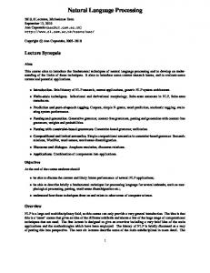
reports pertaining to breast. We determined that the most important diagnoses for our study that might be found, either alone or in combination, within a pathology report were invasive ductal cancer (IDC); invasive lobular cancer (ILC); invasive cancer NOS; ductal carcinoma in situ (DCIS); severe atypical ductal hyperplasia (severe ADH); lobular carcinoma in situ (LCIS); atypical lobular hyperplasia (ALH); atypical ductal hyperplasia (ADH); and benign. As a preparatory step, a folder or “bucket” was then created for each diagnosis within the NLP software (Clearforest, Waltham, MA). A “bucket” would eventually hold a set of words and/or phrases that denoted a specific pathology report. Next, the layout of the pathology report was analyzed. The most important information pertaining to the diagnosis was contained in a section labeled “Final Diagnosis,” which was present for each distinct specimen (a report might have an excision and four shaved margins as five distinct specimens for the same side with the potential for different diagnoses by specimen) [Figure 1]. Thus there was often more than one final diagnosis in a single pathology report on a single day. We identified both the start and the end of the final diagnosis section for each specimen and these sections were parsed out and associated with a Medical Record Number (MRN), Date, and Side [Figures 1 and 2]. Parsing techniques varied by institution, due to the unique, institution specific report layouts. Using NLP software (Clearforest, Waltham, MA), the “Final Diagnosis” section of a test set of 500 reports from each institution was processed. The NLP software displayed all words and phrases in these reports, and the number of times each was used in this set of reports, and provided an interface to associate each word or phrase with one or more of the “buckets.” Each entity generated by the software was then associated with the “bucket” it represented. For example, the entities “infiltrating ductal carcinoma,” “invasive cancer with ductal features,” “invasive cancer, ductal type,” etc. all went into the “invasive ductal cancer” bucket. Some entities went into more than one bucket, such as “invasive carcinoma with both ductal and lobular features,” which was both IDC and ILC. This approach was then applied to the larger data set, to test its functionality and to identify words or phrases missed in the test set. It was also identified that an entity may be negated, and the negation might lie either before or after the text. For example, a report may state that there was “no evidence of invasive carcinoma,” or “residual DCIS was not seen.” All words and phrases that denoted negation and their order in the sentence (before or after the diagnostic entity) were identified and placed in pre- and postnegation categories. A pattern was then created to recognize negation. If an entity was negated, it was not
J Pathol Inform 2012, 3:23 http://www.jpathinformatics.org/content/3/1/23
recorded in the final data set for that record.
This initial table had a row for each MRN, date, side, and specimen and denoted all diagnoses present in that specimen on that date [Figure 2].
The multiple ways of saying each entity were counted as well as the multiple ways of stating negation.
Each of these “final diagnoses” from a single date and side were amalgamated into a single row in a second table that denoted an MRN, date, side, and all diagnoses on that date.
A single row in an Access (Microsoft) table was created for each specimen, where the presence of each entity in that specimen was recorded in the appropriate column.
We then identified a “maximum diagnosis” on each date by establishing a trumping order (an “order of significance”), such that IDC, ILC, or invasive cancer NOS, would outweigh DCIS, which would outweigh severe ADH, which would outweigh LCIS, which would outweigh ALH, which would outweigh ADH, which would outweigh benign.
Accession Number: Report Status: Updated Type: Surgical Pathology Pathology Report: CASE: PATIENT: Date Taken : 2/12/2008 Source Care Unit: Same Day Surgery Unit Path Subspecialty Service:Breast-1 ResultsTo: Signed Out by: Results CLINICAL DATA right breast CA FINAL DIAGNOSIS: BREAST (RIGHT), EXCISION: 1. INVASIVE DUCTAL CARCINOMA WITH CALCIFICATIONS, SEE TABLE #1 2. DUCTAL CARCINOMA IN-SITU WITH CALCIFICATIONS 3. LOBULAR NEOPLASIA (ATYPICAL LOBULAR HYPERPLASIA) 4. HEALING BIOPSY SITE
Where multiple surgeries occurred in the course of treating the same problem on a given side, such as reexcisions for positive margins, we considered these a single “episode of care” for that patient; thus all pathology results from a single side within a 6-month time frame were amalgamated into a third Table organized by MRN, Date, Side, and Episode. Pathology reports outside this 6-month period or from the opposite side were considered as separate episodes.
TABLE OF PATHOLOGICAL FINDINGS #1 INVASIVE CARCINOMA TUMOR SIZE: 1.2 x 1.1 x 0.9 cm (gross measurement) GRADE: 2 LYMPHATIC VESSEL INVASION: Not identified BLOOD VESSEL INVASION: Notidentified MARGIN OF INVASIVE CARCINOMA: Focally positive at the anterior and medial margins of the specimen LOCATION OF DUCTAL CARCINOMA IN-SITU: Within and beyond the region of the mass GRADE OF DUCTAL CARCINOMA IN-SITU:2 MARGIN OF DUCTAL CARCINOMA IN-SITU: The distances to all margins measure 0.2 cm or greater STAINS FOR RECEPTORS: Requested on block B3
The most significant or “maximum” diagnosis was taken as the primary diagnosis, with the others listed as secondary diagnoses in each Table.
BREAST (RIGHT, FINAL INFERIOR MARGIN), EXCISION: LOBULAR NEOPLASIA (ATYPICAL LOBULAR HYPERPLASIA) (SEE NOTE) NOTE: The atypical lobular cells are negative for e-cadherin. BREAST (RIGHT, FINAL MEDIAL MARGIN), EXCISION: LOBULAR NEOPLASIA (ATYPICAL LOBULAR HYPERPLASIA)
Thus, NLP created three data Tables, MRN, Date, Side, Specimen, which separately listed all diagnoses from each specimen on a given day, “MRN, Date, Side, Summary Diagnoses” which summarized all diagnoses from a given day and an “MRN, Side, Episode of Care, Diagnoses” Table which summarized all diagnoses from a given episode.
BREAST (RIGHT, FINAL SUPERIOR MARGIN), EXCISION: THERE IS NO EVIDENCE OF MALIGNANCY BREAST (RIGHT, FINAL LATERAL MARGIN), EXCISION: THERE IS NO EVIDENCE OF MALIGNANCY
GROSS DESCRIPTION:
Report Text
LOBULAR NEOPLASIA
IDC
ILC
DCIS
LCIS
ADH
ALH
Specimen
Side
Date
MRN
Figure 1: Sample pathology report showing the fields extracted (highlighted in bold type). Each specimen was parsed separately and generated its own “final diagnosis”
12/02/2008 Right 1
1
0
0
1
0
1
1
BREAST (RIGHT), EXCISION: 1. INVASIVE DUCTAL CARCINOMA WITH CALCIFICATIONS, SEE TABLE #1 2. DUCTAL CARCINOMA IN-SITU WITH CALCIFICATIONS 3. LOBULAR NEOPLASIA (ATYPICAL LOBULAR HYPERPLASIA) 4. HEALING BIOPSY SITE BREAST (RIGHT, FINAL INFERIOR MARGIN), EXCISION: LOBULAR NEOPLASIA (ATYPICAL LOBULAR HYPERPLASIA) (SEE
12/02/2008 Right 2
1
0
0
0
0
0
1
12/02/2008 Right 3
1
0
0
0
0
0
1
BREAST (RIGHT, FINAL MEDIAL MARGIN), EXCISION: (ATYPICAL LOBULAR HYPERPLASIA)
12/02/2008 Right 4
0
0
0
0
0
0
0
BREAST (RIGHT, FINAL SUPERIOR MARGIN), EXCISION: OF MALIGNANCY
12/02/2008 Right 5
0
0
0
0
0
0
0
BREAST (RIGHT, FINAL LATERAL MARGIN), EXCISION: OF MALIGNANCY
LOBULAR NEOPLASIA
THERE IS NO EVIDENCE
THERE IS NO EVIDENCE
Figure 2: Sample datasheet displaying extracted diagnostic information from the sample report shown in Figure 1. As each specimen generated its own “final diagnosis,” a single row was created for each specimen by MRN, date, side and specimen in the first of three databases created
J Pathol Inform 2012, 3:23 http://www.jpathinformatics.org/content/3/1/23
As our first study was conducted to identify patients with high risk lesions, we opted to review a nonrandom sample of 6,711 pathology reports which were identified in patients who had a diagnosis of severe ADH, LCIS, ALH or ADH, without prior or concurrent cancer. These NLP results were reviewed by human coders who compared the result to the free text report to determine the accuracy of the NLP. The accuracy for the maximum diagnosis and the accuracy for all diagnoses were recorded separately.
Table 1:The number of ways in which each diagnosis was said in pathology reports
RESULTS
Table 2: Different ways in which pathologists describe the presence of atypical ductal hyperplasia
In 76,333 breast pathology reports, multiple entities were identified that represented each of the significant buckets. Excluding typographical errors and spacing errors, we identified 124 ways of saying invasive ductal cancer; 95 ways of saying invasive lobular cancer; 52 ways of saying DCIS; 14 ways of saying severe ADH; 53 ways of saying lobular carcinoma in situ; 17 ways of saying atypical lobular hyperplasia and 14 ways of saying atypical ductal hyperplasia [Table 1]. Examples of ways to describe ADH and invasive carcinoma are shown in Tables 2 and 3. In addition, we identified 21 ways of negating a diagnosis when the words appeared before the diagnosis (e.g., No evidence of invasive ductal carcinoma), and an additional 12 ways of negating the diagnosis when the words fell after the diagnosis (e.g., ADH was not seen). As each entity can potentially be negated by a pre- or postnegative one, must multiply the number of ways of stating the negation by the number of ways of describing that particular diagnostic entity. For example, with invasive ductal cancer; that means 124 ways of saying IDC multiplied by 33 ways of saying “not” gives a total of 4092 potential ways to say IDC was not present. When the processor output was compared to reports as reviewed by expert human coders, 97% of reports were correct for all diagnoses and 97.8% were correct for the maximum diagnosis. Figure 3 demonstrates examples of incorrect diagnoses, where the software did not identify diagnoses that were present in the report. Most commonly this occurred because the diagnosis was written in a pattern not recognized by the software, or simply a typographical error in the report. To calculate the sensitivity, specificity, and predictive value of NLP, we considered “all diagnoses.” A true positive was defined as atypia present, correctly identified with NLP. A false positive was defined as atypia identified by NLP that was not present on the report. A true negative was defined as a benign diagnosis, correctly identified with NLP, and a false negative was defined as atypia present, but not identified by NLP. The sensitivity of NLP to correctly identify all diagnoses
Diagnosis Invasive ductal carcinoma Invasive lobular carcinoma Ductal carcinoma in situ Lobular carcinoma in situ Atypical lobular hyperplasia Atypical ductal hyperplasia
Ways to say 124 95 52 53 17 14
Atypical ductal hyperplasia Atypical duct hyperplasia Atypical intraductal hyperplasia Atypical ductal epithelial hyperplasia Atypical hyperplasia with ductal and lobular features Atypical hyperplasia with mixed ductal and lobular features Atypical lobular and ductal hyperplasia Atypical ductal and lobular hyperplasia Atypical hyperplasia, both typical, and atypical Atypical hyperplasia is present with pagetoid spread in to ducts Papillary duct hyperplasia with atypia
Table 3: Some examples of the 95 ways of saying “invasive lobular carcinoma” Invasive carcinoma consistent with lobular Invasive carcinoma displays both ductal and lobular features Invasive carcinoma displaying both ductal and lobular features Invasive carcinoma has both ductal and lobular features Invasive carcinoma showing ductal and lobular features Invasive carcinoma showing both ductal and lobular features Invasive carcinoma with ductal and lobular features Invasive carcinoma with both ductal and lobular features Invasive carcinoma with mixed ductal and lobular features Invasive carcinoma with features of lobular and ductal Invasive carcinoma with features of both ductal and lobular Invasive carcinoma with predominantly lobular features Invasive carcinoma with primarily lobular features Invasive carcinoma, favor lobular type (Note that some of these phrases would also be categorized as being consistent with “invasive ductal carcinoma”
was 99.1%, with a specificity of 96.53%. The positive predictive value of NLP was 98.63% and the negative predictive value was 97.73%.
DISCUSSION This study highlights one of the principal difficulties encountered in utilizing electronic data beyond its specific context. While a breast pathology report written in free text is easily read and interpreted on an
Side
Specimen
ALH
ADH
LCIS
DCIS
ILC
IDC
LOBULAR NEOPLASIA
23/03/2001
Right
1
0
0
0
0
0
0
0
BREAST (RIGHT), RE -EXCISON: A SINGLE MICROSCOPIC FOCUS OF ATYPICAL HYPERPLASIA (0.1 CM), LOCATED