The FlyCatwalk: A high throughput feature‐based sorting system for artificial selection in Drosophila Vasco Medici*, §, †, Sibylle Chantal Vonesch*, †, Steven N. Fry§, Ernst Hafen*,2 * Institute of Molecular Systems Biology, ETH Zurich, 8093 Zurich, Switzerland § SciTrackSs GmbH, Pfaffhausen, Switzerland † These authors contributed equally
2
Corresponding author: ETH Zurich, Institute of Molecular Systems Biology, HPT E72, Auguste‐Piccard‐Hof 1, 8093 Zurich, Switzerland,
[email protected], +41 44 633 36 88
DOI: 10.1534/g3.114.013664
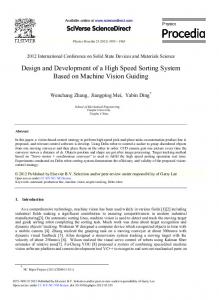
Figure S1 Body segmentation. A: Single complement image frames of the blue channel luminance after background subtraction. Background subtraction results in an image of a bright fly against a dark background. This image is inverted to gain the complement image, a black fly on white background. B: Image after leg deletion. The valid complement frames in the sequence are rotated until they are all aligned and the 95th percentile image is computed to remove the legs, which is possible because their position varies between frames. C: Body segmentation performed using the watershed algorithm (Red= head, yellow = thorax, blue = abdomen). 2 SI
V. Medici et al.
File S1 Supporting Methods Wing extraction Different approaches are applied for wing regions depending on whether they overlap with the body or not. For the regions without overlap mostly the blue channel is used (names of matlab functions in italics): 1. Image adjustment: image adjustment (imadjust) and 2D adaptive noise removal filtering (wiener2), 2. Body detection: thresholding followed by image filling and closing to delete holes in the body mask (imfill and imclose), 3. Wing detection: High pass filtering of the image to cancel out luminance gradients followed by detection of the wing mask (i.e. selecting a region of interest (mask) in which to apply the image processing). Mask definition is performed using adaptive thresholding techniques and the body masked computed in the preceding step and if present, holes are filled (imfill). Legs are detected in the wing mask by additionally including the red channel, in which the legs appear bright, and these regions are removed. This is necessary because legs might get mixed up with veins. 4. Vein detection: A combination of thresholding and edge detection (edge, method option ‘canny’) is used to binarize the image and small objects are deleted (bwareaopen). A skeletonization algorithm is applied to extract the structure of the veins (anaskel) For regions with overlap the red channel is used: 1. Selection of the region of interest from the body mask, 2. Image adjustment: Detection of high spatial frequency content using top‐hat filtering (imtophat) followed by contrast enhancement using contrast‐limited adaptive histogram equalization (CLAHE, adapthisteq), 3. Binarization and cleaning: Thresholding, image closing to join adjacent blobs and deletion of small objects (bwareaopen). Objects with high eccentricity (i. e. they are line‐like) are selected. 4. Vein detection: A skeletonization algorithm is applied to extract the structure of the veins (anaskel). File S2 Supporting Video The FlyCatwalk setup and workflow. Individual steps of the automated phenotyping and detailed parts of the setup are shown. File S2 is available for download as an AVI file at http://www.g3journal.org/lookup/suppl/doi:10.1534/g3.114.013664/‐/DC1
V. Medici et al.
3 SI