REVIEW
The formation of neuromuscular synapses Steven J. Burden1 Molecular Neurobiology Program, Skirball Institute, New York University Medical Center, New York, New York 10016 USA
The formation of synapses requires a series of steps including the generation of neurons and their target cells, the guidance of axons to their targets, and the induction of a specialized presynaptic terminal and postsynaptic membrane. This review is primarily concerned with the inductive events that lead to postsynaptic differentiation. Because this phase of synapse formation begins only after motor neurons have navigated to their target muscle cells, the first section of this review summarizes our current understanding of the principles that govern the guidance of motor axons to skeletal muscle cells. The second section will review our current knowledge of signaling molecules and mechanisms that are critical for inducing postsynaptic differentiation. Although the principles and mechanisms of synapse formation are likely to be similar in the PNS and the CNS, much of our understanding about the mechanisms of synapse formation arises from studies of the vertebrate neuromuscular synapse. Motor axons project along a stereotyped pathway from spinal cord to muscle Much of the evidence for accurate and stereotyped pathfinding arises from studies of the developing chick embryo (Landmesser 1992). The growth cones of chick motor neurons, which are destined to innervate the limb, exit the spinal cord, extend across the rostral half of developing somites, and converge upon a plexus where they intermingle with growth cones that are destined to innervate different muscles. Recent studies suggest that T-cadherin (Fredette et al. 1996), collagen IX (Ring et al. 1996), and the Eph family ligands, HtkL/Lerk2 (Wang and Anderson 1997), which are each expressed preferentially in the caudal half of the somite, repel these motor axons from the caudal half somite and guide them across the rostral somite. Furthermore, because HtkL/Lerk2 (Wang and Anderson 1997), as well as collapsin-1 (Shepherd et al. 1996), are expressed in the dermatome, repulsive signals acting in three dimensions may guide motor axons toward the plexus. In the plexus the growth cones begin to sort and to associate with growth cones of axons that will innervate the same target, indicating that signals for sorting and pathfinding are associated with cellular and/or extracel1
E-MAIL
[email protected]; FAX (212) 263-8214.
lular structures in the plexus (Lance-Jones and Landmesser 1981). After motor axons leave the plexus, they project to either the ventral or dorsal premuscle mass in the limb and ultimately to the appropriate individual muscle (Landmesser 1978). The projection of motor axons to muscle appears to be accurate from the outset, and there are few pathfinding errors that are eliminated by editing (Landmesser 1992). The idea that motor axons are guided to their appropriate targets by navigational clues presented on the pathway is supported further by experiments in which motor neurons and/or their targets are displaced by transplantation (Lance-Jones and Landmesser 1980). These studies demonstrate that axons of displaced motor neurons, which arrive at the plexus from an aberrant location, are able to reorient and emerge from the plexus to innervate their appropriate targets. The directional signals are apparently not supplied by muscle, as motor axons project accurately to the appropriate region of the developing limb even in the absence of muscle cells (Lewis et al. 1981). Thus, mesenchymal cells in the plexus region are thought to have an important role in providing clues that guide and direct motor axons to the appropriate region of the developing limb (Lance-Jones and Dias 1991). The navigating signals that these cells may provide to developing motor neurons are not known, but recent studies indicate that hepatocyte growth factor is expressed in the sclerotome and the limb mesenchyme and can guide motor axons in vitro (Ebens et al. 1996). Guidance clues are provided by cells present on the pathway Precedent for the idea that guidance information can be provided by cells situated along a pathway comes from studies in grasshoppers, where developing neurons, termed guidepost cells, have a critical role in directing sensory neurons to their appropriate targets in the CNS (Bentley and Caudy 1983). In vertebrates, floor plate cells appear to serve a similar function for the axons of spinal commisural neurons (Colamarino and Tessier-Lavigne 1995b). These specialized glial-like cells in the extreme ventral region of the neural tube synthesize and secrete netrin, a protein that stimulates and orients the growth of commisural axons that extend from the dorsal spinal cord to the floor plate and across the midline. Although
GENES & DEVELOPMENT 12:133–148 © 1998 by Cold Spring Harbor Laboratory Press ISSN 0890-9369/98 $5.00; www.genesdev.org
133
Burden
netrin does not guide the axons of spinal cord motor neurons, netrin can act as a repellent for trochlear motor neurons (Colamarino and Tessier-Lavigne 1995a) and for Caenorhabditis elegans motor neurons (Hedgecock et al. 1990). The axons of spinal motor neurons may nevertheless be guided by factors expressed in the floor plate, as the floor plate does contain an activity(s) that is repulsive for developing motor axons (Guthrie and Pini 1995). Axons defasciculate as they turn toward their targets Axons associate, or fasciculate, with one another at certain points during pathfinding and diverge from one another as they grow to distinct targets. Thus, mechanisms that control fasciculation and defasciculation are likely to be highly regulated and to have important consequences for accurate pathfinding (Goodman 1996). Consistent with this idea, mutation of certain receptor tyrosine phosphatases (RTPs) in Drosophila results in poor defasciculation and an inability of motor axons to branch and readily locate appropriate muscles (Desai et al. 1996; Goodman 1996; Krueger et al. 1996). Thus, signaling through receptor tyrosine kinases (RTKs) (Elkins et al. 1990; Callahan et al. 1995) and RTPs is likely to regulate adhesion between motor axons. Laminin, N-cadherin, NCAM, polysialic acid, and fibronectin can promote or retard axon growth and have a role in fasciculation. Nevertheless, these adhesion molecules alone may not have a role in directing different axons to particular pathways. Thus, it is possible that RTKs and RTPs control fasciculation and steering by regulating the strength of adhesion between axons or between axons and substrates. Motor neurons projecting to different muscles express different homeodomain proteins Because different motor axons must decipher and respond differently to navigational clues, motor neurons are presumed to be distinct from one another. Recent studies demonstrate that motor neurons can be distinguished from one another by expression of a unique combination of LIM homeodomain proteins (Tsuchida et al. 1994; Tanabe and Jessell 1996). For example, motor neurons that are positioned in the medial ventral spinal cord project to ventral limb muscle and express Isl-1 and Isl-2, whereas motor neurons that are positioned in the lateral ventral spinal cord project to dorsal limb muscle and express Lim-1 and Isl-2 but not Lim-2. Moreover, motor neurons projecting to axial muscle express Isl-1, Isl-2, and Lim-3. Thus, it would not be surprising to find that the subpopulations of motor neurons, distinguished by their combination of LIM homeodomain proteins, also express different cell surface proteins that have a role in guiding motor neuron growth cones to their appropriate target. Muscle cells may provide pathfinding clues to motor axons once they approach their target A first step in accurate pathfinding is to direct motor axons toward their target, and this is likely to be
134
GENES & DEVELOPMENT
achieved by providing navigational clues along growth pathways and by regulating fasciculation as described above. Once motor axons approach their target, however, additional directional clues are likely to be required, as motor axons must ultimately choose among many target muscles. Motor axons enter the dorsal or ventral region of the developing limb before muscles have individualized; shortly after individual muscles begin to form, axons branch from the main limb nerves and grow toward their appropriate muscle (Landmesser 1992). Because motor axons in muscle-less limbs enter the appropriate dorsal or ventral region of the limb and form primary but not secondary branches (Lewis et al. 1981), muscle cells may have a role in directing motor axons toward the appropriate muscle once the axons are within striking distance of their target. Studies in Drosophila have identified molecules expressed by muscle cells that are important for target selection. Fasciclin III is a cell surface protein expressed by a subset of motor neurons and their target muscle cells. Fasciclin III-expressing motor neurons normally innervate only fasciclin III-expressing muscle cells but will innervate inappropriate muscle cells that have been forced to express fasciclin III (Chiba et al. 1995). Other adhesive molecules, such as fasciclin II (Lin and Goodman 1994) and connectin (Nose et al. 1992), as well as repulsive molecules, such as the semaphorins (Matthes et al. 1995), may have similar roles in regulating target selection (Tessier-Lavigne and Goodman 1996). Schwann cells may have a role in synapse formation Current ideas about pathfinding suggest that the growth cones decipher guidance signals. Studies of regenerating synapses, however, indicate that Schwann cells have an important role in reinnervation and may guide regenerating motor axons to original synaptic sites (Son and Thompson 1995). Thus, although Schwann cells lag behind the growth cones of motor axons during development, making it unlikely that they would have a primary guidance role, they do not lag far behind motor neuron growth cones and could have a role in stabilizing or maintaining newly formed synapses (see below). The neuromuscular synapse is favorable for studying synapse formation Shortly after contact between a growing motor axon and a differentiating myotube is established, signals are exchanged between nerve and muscle that initiate the formation and assembly of a highly differentiated presynaptic nerve terminal and a highly specialized postsynaptic apparatus (Dennis 1981; Hall and Sanes 1993; Jennings and Burden 1993). Muscle differentiation and synapse formation occur concomitantly during development. In vertebrates, motor neurons do not prefer or select a predetermined site on the developing myotube; rather, it appears that synapses can form on most if not all of the myotube surface. Although functional synapses form within minutes to hours after contact between de-
Neuromuscular synapse formation
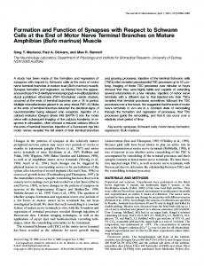
veloping motor nerves and myotubes (Fischbach 1973; Anderson and Cohen 1977), mature and fully differentiated synapses are not evident, at least in mammals, until several weeks after the first contacts are made. The formation of a mature synapse requires further arborization of nerve terminals, withdrawal and editing of synaptic connections, an increase in the efficiency of acetylcholine release, and additional modifications of the postsynaptic membrane. A mature skeletal muscle fiber, a syncitial cell containing several hundred to several thousand nuclei, will ultimately be innervated by a single motor axon that terminates and arborizes over ∼0.1% of the muscle fiber’s cell surface. The neurotransmitter receptors, acetylcholine receptors (AChRs), are localized to this small patch of the muscle fiber membrane, and their localization to synaptic sites during development is a hallmark of the inductive events of synapse formation. Although other proteins are likewise concentrated at synaptic sites, much of our knowledge about the signaling mechanisms responsible for synaptic differentiation have come from studies aimed at understanding how AChRs accumulate at synaptic sites. The neuromuscular synapse is a particularly favorable synapse for studying synapse formation: (1) the synapse is geometrically simple; (2) developing and regenerating synapses can be experimentally manipulated in vivo with relative ease; (3) synapses between motor neurons and muscle cells can be studied in cell culture; (4) synaptic proteins can be readily purified from Torpedo electric organ, an abundant and homogeneous source of neuromuscular-like synapses; and (5) gene expression can be altered and studied in detail in transgenic and mutant mice. Consequently, we have a good understanding of how the neuromuscular synapse is built and an incomplete but growing understanding of the critical genes involved in synaptogenesis. Substructural organization of the neuromuscular synapse The precise organization of molecules in pre- and postsynaptic membranes belies the idea that the neuromuscular synapse is a simple synapse. Rather, the substructure of pre- and postsynaptic membranes suggests that complex mechanisms are required to assemble the synapse and to coordinate pre- and postsynaptic differentiation. Nerve terminals are situated in shallow depressions of the muscle cell membrane, which is invaginated further into deep and regular folds, termed postjunctional folds (Fig. 1). AChRs and additional proteins are localized to the crests of these postjunctional folds, whereas other proteins, including sodium channels, are enriched in the troughs of the postjunctional folds (Hall and Sanes 1993). The mechanisms that lead to this spatial segregation of proteins within the postsynaptic membrane are not known. Clustering of AChRs in the postsynaptic membrane, however, is critical for synaptic function, as a high density of synaptic AChRs is required to generate a syn-
Figure 1. Presynaptic and postsynaptic membranes are highly specialized at neuromuscular synapses. (A) A light microscopic view of a cross section of a muscle fiber shows that nerve terminals, which are capped by Schwann cells, are situated in shallow depressions of an adult muscle fiber. AChRs, stained with horseradish peroxidase-labeled a-bungarotoxin (HRP–a-BGT), are highly concentrated in the muscle membrane at synaptic sites. (B) An electron microscopic view of a neuromuscular synapse shows that synaptic vesicles are abundant in the region of the nerve terminal facing the muscle fiber, where they are clustered adjacent to active zones. Active zones are organized at regular intervals and are aligned precisely with the mouths of the postjunctional folds in the muscle fiber. AChRs, stained with HRP–a-BGT, appear as a dense band at the tops and along the sides of the postjunctional folds; the stain that fills the synaptic cleft is a result of diffusion of the reaction product from the postsynaptic membrane (B, modified from Burden et al. 1979, with permission of The Rockefeller University Press). Bars, 0.5 µm.
aptic potential of sufficient magnitude to initiate an action potential in the myofiber. The nerve terminal is likewise spatially organized, and its substructural organization reflects that of the postsynaptic membrane (Fig. 1) (Hall and Sanes 1993). Synaptic vesicles are abundant in the region of the nerve terminal facing the muscle fiber, where they are clustered adjacent to poorly characterized specializations of the presynaptic membrane, termed active zones, which are the sites of synaptic vesicle fusion. Active zones are organized at regular intervals and are aligned precisely with the mouths of the postjunctional folds. This precise registration of active zones and postjunctional folds ensures that acetylcholine encounters a high concentration
GENES & DEVELOPMENT
135
Burden
of AChRs within microseconds after release, thereby facilitating synaptic transmission. This alignment of structural specializations in pre- and postsynaptic membranes, separated by a 50-nm synaptic cleft, indicates that spatially restricted signaling between pre- and postsynaptic cells is important to coordinate pre- and postsynaptic differentiation. The synaptic basal lamina contains signals for synaptic differentiation Two signaling molecules that regulate postsynaptic differentiation at developing and adult neuromuscular synapses are located in the synaptic basal lamina (Jennings and Burden 1993). The idea that the synaptic basal lamina contains an activity that stimulates postsynaptic differentiation originated from studies of synaptic differentiation in developing (Anderson and Cohen 1977) and regenerating muscle (Burden et al. 1979; McMahan 1990). These studies demonstrated that motor neurons cause a redistribution of previously unlocalized AChRs to synaptic sites and that a signal(s) for AChR clustering is contained in the synaptic basal lamina. Although there is evidence that the synaptic basal lamina also contains signals that direct presynaptic differentiation (Sanes et al. 1978), it has been difficult to establish a simple and reliable cell culture assay for identifying and purifying signals that regulate presynaptic differentiation. Thus, we know little about the nature of the signals for presynaptic differentiation. The following section will summarize our current understanding of the signaling mechanisms that lead to differentiation of the postsynaptic membrane. Agrin induces postsynaptic differentiation Because clustering of AChRs can be readily studied in cell culture, it was possible to devise a straightforward assay for identifying signals that induce this aspect of postsynaptic differentiation. Although several factors can induce clustering of AChRs in cultured muscle cells, the evidence that the relevant signals are located in the synaptic basal lamina provided important insight that allowed for purification of the appropriate in vivo activity. McMahan and colleagues found that extracellular matrix from Torpedo electric organ, a tissue that is homologous to skeletal muscle but more densely innervated, contains an activity that stimulates AChR clustering in cultured myotubes and that this activity, like the signal at developing neuromuscular synapses, triggers clustering of AChRs by post-translational mechanisms (Godfrey et al. 1984; Nitkin et al. 1987). The electric organ activity was purified and termed agrin (McMahan 1990). Agrin is a ∼200-kD protein containing multiple epidermal growth factor (EGF)-like signaling domains, two different laminin-like domains, and multiple follistatin-like repeats. The four EGF-like domains and three laminin-like G domains are contained in the carboxy-terminal region, which is sufficient for inducing AChR clusters in cultured myotubes; sequences in the
136
GENES & DEVELOPMENT
amino-terminal region are thought to be responsible for the association of agrin with the extracellular matrix. Agrin is transported in the axons of motor neurons and released into the synaptic basal lamina, where it is highly concentrated near sites of synaptic vesicle fusion (M. Schwarz, P. Theodosopoulos, R. Marshall, and U.J. McMahan, unpubl.). Two lines of evidence indicate that agrin is necessary for clustering of AChRs at synaptic sites and for inducing postsynaptic differentiation. First, antibodies against agrin block AChR clustering at nerve– muscle synapses that form in cell culture (Reist et al. 1992), and second, mice lacking agrin lack normal synapses (see below) (Gautam et al. 1996). Agrin also regulates the distribution of other synaptic proteins, including acetylcholinesterase (AChE), rapsyn, utrophin, neuregulin (NRG), and NRG receptors (ErbBs) (see below), indicating that agrin has a central role in synaptic differentiation (McMahan 1990; Apel et al. 1995; Rimer et al. 1996; Meier et al. 1997). The agrin gene is expressed in a variety of cell types. Alternative splicing results in multiple agrin isoforms that differ in their AChR clustering efficiency (Ferns et al. 1992, 1993; Ruegg et al. 1992; Hoch et al. 1993). The isoform that appears to be the most active in clustering AChRs is expressed in neurons, whereas other agrin isoforms are expressed in additional cell types, including skeletal muscle cells (Fallon and Gelfman 1989; Ferns et al. 1992; Ruegg et al. 1992). Although agrin has been shown to have a critical role in the formation of neuromuscular synapses, the role of agrin in the CNS and in non-neural cells is currently not known. The active, neuronal-specific isoforms of agrin contain 8 or 19 amino acids at a splice site, referred to as the Z site in rat agrin (Ferns et al. 1992; Hoch et al. 1993) and the B site in chick agrin (Ruegg et al. 1992). A ∼40-kD carboxy-terminal fragment of agrin, containing two EGF-like domains, two laminin-like G domains, and the B site, clusters AChRs with the same potency as full-length agrin (Hoch et al. 1994), whereas a carboxy-terminal 21-kD fragment, containing a single EGF-like domain, a single lamininlike G domain, and the B site, is ∼200-fold less active than full-length agrin (Gesemann et al. 1995). Experiments with chimeric synapses between different frog species indicate that nerve-derived agrin is present at synaptic sites from the earliest stages of synapse formation (Cohen and Godfrey 1992). Furthermore, experiments with chimeric synapses between chick and rat demonstrate that blocking antibodies to nerve-derived but not to muscle-derived agrin inhibit AChR clustering (Reist et al. 1992). Although muscle-derived agrin may have a role in synapse formation, these results demonstrate that muscle-derived agrin cannot substitute for nerve-derived agrin in clustering AChRs, at least in cell culture. Several lines of evidence, however, indicate that muscle agrin could have a role in synapse formation. First, nerve and muscle agrin, when attached to cells or extracellular matrix, can be similarly active in clustering AChRs (Ferns et al. 1992, 1993). Second, muscle-derived agrin can serve as a stop, or growth-arrest signal, for mo-
Neuromuscular synapse formation
tor axons (Campagna et al. 1995). Third, muscle-derived agrin can serve as a transcriptional signal that induces expression of AChR genes (Jones et al. 1996). Selective restoration of agrin expression in motor neurons or in skeletal muscle from agrin mutant mice may provide important insight into the potential role of skeletal muscle-derived agrin (Gautam et al. 1996). MuSK is required for agrin-mediated signaling and synapse formation The mechanism of agrin-mediated AChR clustering is not known, but a RTK, termed MuSK, appears to be a critical component of an agrin receptor complex. MuSK was discovered using a strategy designed to identify tyrosine kinases, which are abundant in the synapse-rich Torpedo electric organ and which might therefore be involved in synaptic differentiation (Jennings et al. 1993). The cytoplasmic domain of MuSK, including sequences outside the tyrosine kinase domain, is similar to that of neurotrophin receptors, whereas the extracellular domain of MuSK, which contains four immunoglobulinlike domains, similar to those in a variety of receptors and adhesion molecules, is divergent from that of the neurotrophin receptors (Jennings et al. 1993). MuSK is highly expressed in Torpedo electric organ and in the skeletal muscle of Torpedo and higher mammals, where it is concentrated in the postsynaptic membrane (Jennings et al. 1993; Valenzuela et al. 1995). Mice lacking MuSK expression (DeChiara et al. 1996), like agrin mutant mice (Gautam et al. 1996), lack normal neuromuscular synapses. Indeed, the similar phenotype of agrin and MuSK mutant mice is consistent with the idea that MuSK is a component of an agrin receptor complex (see below). Both agrin and MuSK mutant mice are immobile, cannot breathe, and die shortly after birth. Muscle differentiation is largely normal in agrin and MuSK mutant mice, but MuSK mutant muscle fibers lack all known features of postsynaptic differentiation. Muscle-derived proteins, including AChRs, AChE, rapsyn, and NRG receptors, which are concentrated at synapses in normal mice, are uniformly distributed in MuSK mutant myofibers. In addition, AChR genes, which are normally transcribed selectively in synaptic nuclei of normal muscle fibers (see below), are transcribed at similar rates in synaptic and nonsynaptic nuclei of muscle fibers from agrin and MuSK mutant mice. These studies show that MuSK is required for neuromuscular synapse formation (DeChiara et al. 1996). Five lines of evidence indicate that MuSK is required for agrin-mediated signaling and is a component of the agrin receptor complex (Glass et al. 1996): (1) Cultured MuSK mutant muscle cells, unlike normal muscle cells, do not cluster AChRs in response to agrin; (2) cultured muscle cells expressing a dominant-negative form of MuSK likewise fail to cluster AChRs in response to agrin; (3) a recombinant, soluble extracellular fragment of MuSK inhibits agrin-induced AChR clustering in cultured muscle cells; (4) agrin induces rapid tyrosine phosphorylation of MuSK; and (5) agrin can be chemically
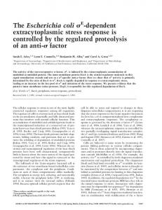
cross-linked to MuSK. MuSK, which has been force-expressed in cells other than myotubes, however, cannot be activated by agrin, indicating that other activities or additional proteins that are myotube-specific are required for agrin-mediated signaling (Glass et al. 1996, 1997). In principle, the additional myotube-specific activity/activities could be a coreceptor (Jing et al. 1996), a coligand (Klagsbrun and Baird 1991; Lopez-Casillas 1991; Rapraeger et al. 1991; Ferns et al. 1993; Aviezer et al. 1994), or post-translational modifications (Fig. 2) (Binari et al. 1997).
Figure 2. Agrin activation of MuSK leads to clustering of synaptic proteins, including AChRs, rapsyn, ErbBs, and musclederived NRG. Current data indicate that a myotube-specific activity/activities is required for agrin to activate MuSK; this activity/activities may be a coreceptor, a coligand, and/or posttranslational modifications. Because staurosporine blocks agrin-stimulated AChR clustering and AChR tyrosine phosphorylation but does not inhibit MuSK tyrosine phosphorylation, a kinase(s) downstream of MuSK is important for agrin-mediated signaling; this kinase(s) may or may not be activated by agrin.
GENES & DEVELOPMENT
137
Burden
agrin and MuSK mutant mice have similar but distinguishable phenotypes, as AChR clusters are entirely absent in skeletal muscle of MuSK mutant mice, whereas AChR clusters, though infrequent, can be detected in agrin mutant mice. Furthermore, motor axons terminate and arborize at some of the AChR clusters in agrin mutant mice. These results raise the possibility that a second signal, which is independent of agrin but dependent on MuSK, can initiate postsynaptic differentiation (Gautam et al. 1996). Alternatively, the high level of MuSK expression in muscle that is deprived of functional innervation (Valenzuela et al. 1995) might result in spontaneous dimerization and ligand-independent activation of MuSK, leading to stimulation of downstream signaling. a-Dystroglycan binds agrin a-Dystroglycan binds agrin (Bowe et al. 1994; Campanelli et al. 1994; Gee et al. 1994; Sugiyama et al. 1994), raising the possibility that dystroglycan collaborates with MuSK to mediate agrin signaling. a-Dystroglycan is a peripheral membrane protein that binds laminin in the extracellular matrix and is covalently linked to b-dystroglycan, an integral membrane protein that associates intracellularly with dystrophin (Ervasti and Campbell 1991) or utrophin (Matsumura et al. 1992), a dystrophinrelated protein that is highly concentrated at nerve– muscle synapses (Ohlendieck et al. 1991). Nevertheless, the role of a-dystroglycan as an agrin receptor remains unclear. Although antibodies to a-dystroglycan have been reported to perturb the formation of AChR clusters induced by agrin (Campanelli et al. 1994; Gee et al. 1994), others have reported the same antibodies to be ineffective in altering the response to agrin (Sugiyama et al. 1994). Moreover, soluble muscle agrin binds dystroglycan well but neither stimulates nor antagonizes AChR clustering by active, neural agrin (Hoch et al. 1994). In addition, recent studies indicate that the dystroglycan-binding sequences in agrin are not necessary for AChR clustering (Gesemann et al. 1995). Furthermore, because AChRs and rapsyn are clustered normally in mice that lack both dystrophin and utrophin and that have markedly reduced levels of dystroglycan (Deconinck et al. 1997; Grady et al. 1997), it seems unlikely that dystroglycan has a critical role in initiating AChR clustering in response to agrin. It remains possible, however, that dystroglycan is a receptor for laminin at neuromuscular synapses and serves to consolidate clustering of AChRs initiated by agrin (Sugiyama et al. 1997; S. Carbonetto, C. Jacobson, F. Montanaro, M. Lindenbau, and M. Ferns, unpubl.). Agrin and MuSK are required for retrograde signaling and presynaptic differentiation Although pathfinding of motor axons to muscle is normal in mice lacking agrin or MuSK, agrin and MuSK mutant mice lack normal nerve terminals (DeChiara et al. 1996; Gautam et al. 1996). Branches of the main in-
138
GENES & DEVELOPMENT
tramuscular nerve in the mutant mice neither establish normal contacts with the muscle nor form correctly positioned or specialized nerve terminals. These results suggest that motor axons in agrin and MuSK mutant mice and are not provided with appropriate stop signals, and in the absence of termination signals, axons wander across the muscle surface. Because MuSK is expressed in skeletal muscle and not in motor neurons (Jennings et al. 1993; Valenzuela et al. 1995), it seems likely that the aberrant behavior of presynaptic terminals in MuSK mutant mice is due to indirect actions of agrin/MuSK signaling. These results raise the possibility that agrin released from nerve terminals causes the muscle cell, via MuSK activation, to reciprocally release a recognition signal back to the nerve, or to Schwann cells, to indicate that a functional contact has occurred. In response to this muscle-derived signal, the nerve would stop growing and undergo presynaptic differentiation. Alternatively, the lack of synaptic activity in MuSK mutant mice may result in aberrant retrograde signaling, resulting in exuberant growth of motor axons. In either case, these results demonstrate the importance of the reciprocal signaling relationship between nerve and muscle during development.
Rapsyn is required for postsynaptic differentiation and is downstream of agrin and MuSK Although there is little information regarding the signaling pathways that are activated by agrin, a 43-kD protein, termed rapsyn, is known to have an important role in agrin-mediated signaling. Rapsyn is a myristolated, peripheral membrane protein that is present at 1:1 stoichiometry with AChRs at synaptic site and may interact directly with AChRs and potentially other synaptic proteins (Burden et al. 1983; Froehner 1991; Philips and Merlie 1992). Agrin stimulates clustering of rapsyn in myotubes grown in cell culture, and clustering of rapsyn and AChRs appears to occur coincidentally at developing synapses (Burden 1985; Noakes et al. 1993). We do not know how agrin activation of MuSK leads to clustering of rapsyn and how clustering of rapsyn leads to the aggregation of other synaptic proteins. Rapsyn, however, is critical for synapse formation. Mice lacking rapsyn expression die within hours after birth and have difficulty moving and breathing (Gautam et al. 1995). Importantly, normal clustering of AChRs, NRG receptors, utrophin, and dystroglycan is lacking in rapsyn mutant mice. Other aspects of synaptic differentiation are relatively normal in rapsyn mutant mice: (1) MuSK is localized to rapsyn mutant synaptic sites, demonstrating that clustering of MuSK at synapses is rapsyn-independent (Apel et al. 1997); (2) AChR gene expression is enriched in the central region of rapsyn mutant muscle; and (3) synaptic basal lamina proteins, AChE and s-laminin, are localized, albeit at lower levels, at mutant synaptic sites. Because basal lamina proteins do not diffuse readily from their site of deposition and because AChE and s-laminin
Neuromuscular synapse formation
are encoded by RNAs that are localized to synaptic sites (Jasmin et al. 1993; Moscoso et al. 1995a), the persistence of synapse-specific gene expression in rapsyn mutant mice may explain the localization of AChE and s-laminin proteins at rapsyn mutant synaptic sites. Forced expression of rapsyn in Xenopus oocytes or in a fibroblast-like cell line, QT6 cells, results in clustering of rapsyn, and clustering of rapsyn is necessary and sufficient to cluster AChRs in these cells (Froehner et al. 1990; Philips et al. 1991). Rapsyn can also cluster MuSK in QT6 cells that are forced to express both rapsyn and MuSK, indicating that rapsyn and MuSK associate either directly or indirectly (Gillespie et al. 1996). Downstream of MuSK The extracellular domain of MuSK appears to have an important role in clustering rapsyn, as mutant or chimeric forms of MuSK that contain only the ectodomain of MuSK can be clustered by rapsyn in QT6 cells (Apel et al. 1997). Because rapsyn is a peripheral membrane protein associated with the cytoplasmic surface of the plasma membrane, these experiments suggest that rapsyn associates with an unknown protein, which is expressed in QT6 cells as well as in muscle cells, that interacts directly or indirectly with the extracellular domain of MuSK. Because MuSK is clustered at synapses in rapsyn mutant mice (Apel et al. 1997), these results suggest a model in which matrix-bound agrin engages and clusters MuSK, MuSK clusters an unknown linker protein(s), the linker protein(s) clusters rapsyn, and rapsyn clusters AChRs. This model, however, does not take into account the kinase activity of MuSK, which is required for agrin to stimulate clustering of AChRs (DeChiara et al. 1996; Glass et al. 1997). Moreover, because staurosporine inhibits AChR clustering without blocking agrinstimulated tyrosine phosphorylation of MuSK (Wallace 1994; Ferns et al. 1996; Fuhrer et al. 1997), there appears to be at least one kinase in addition to MuSK that is necessary for AChR clustering (Fig. 2). Clearly, a better understanding of the agrin-stimulated clustering pathway will require identification of the substrates that are tyrosine phosphorylated by MuSK and are necessary for clustering rapsyn and AChRs. Models for MuSK-mediated clustering must also be consistent with the stoichiometry of MuSK, rapsyn, and AChRs. Although rapsyn and AChR are present at equal stochiometry at synapses (Burden et al. 1983), MuSK is likely to be present at synapses at substantially lower levels: AChR and rapsyn mRNAs are present at similar levels in Torpedo electric organ, whereas MuSK mRNA is ∼60-fold less abundant than rapsyn or AChR (Jennings et al. 1993). Thus, it seems likely that MuSK regulates clustering of synaptic proteins by serving as a catalyst for assembly rather than by participating as a stochiometric and stable component of a rapsyn/AChR complex. Much of our current thinking about agrin/MuSK/rapsyn/AChR interactions is predicated on results obtained in QT6 cells. Interpretation from these studies presumes that mechanisms that regulate the distribu-
tion of rapsyn, MuSK, and AChRs in transfected QT6 cells resemble that in muscle cells. Because clustering of rapsyn in muscle cells is dependent on agrin and MuSK (DeChiara et al. 1996; Gautam et al. 1996), whereas rapsyn clusters ‘‘spontaneously’’ in QT6 cells (Philips et al. 1991), it appears that QT6 cells have substituted or bypassed mechanisms used in muscle that require agrin/ MuSK signaling to cluster rapsyn. Consequently, the steps that follow agrin/MuSK signaling and that lead to rapsyn clustering have not been amenable to study in QT6 cells. Biochemical evidence demonstrating association, direct or indirect, between MuSK and rapsyn either in QT6 cells or muscle cells is currently lacking. Recent studies, however, demonstrate that MuSK and AChRs can be coimmunoprecipitated from muscle cells and that agrin stimulation increases the amount of MuSK that is associated with AChRs (Fuhrer et al. 1997). The complex containing MuSK and AChRs is likely to contain additional proteins (see below), and identification of these proteins is likely to provide insight into mechanisms of agrin-induced AChR clustering.
Agrin stimulates tyrosine phosphorylation of AChRs Agrin stimulates tyrosine phosphorylation of the AChR b- and d-subunits, and the time course of AChR phosphorylation precedes AChR clustering (Wallace et al. 1991; Qu and Huganir 1994; Ferns et al. 1996). AChR phosphorylation and clustering are relatively slow [AChR tyrosine phosphorylation is detectable by 15 min after agrin treatment, and AChR clustering is first detectable ∼3 hr after agrin treatment (Ferns et al. 1996)], indicating that multiple steps are likely to lie between agrin activation of MuSK and AChR phosphorylation and clustering. The function of AChR tyrosine phosphorylation is unclear, but it is insufficient to cluster AChRs, as AChR tyrosine phosphorylation but not AChR clustering is stimulated in muscle cells that are transfected with a trkC/MuSK chimera and treated with NT3 (Glass et al. 1997). The kinase that stimulates AChR tyrosine phosphorylation appears to be distinct from MuSK, as staurosporine blocks agrin-stimulated AChR tyrosine phosphorylation without inhibiting MuSK phosphorylation (Fuhrer et al. 1997). These results indicate that MuSK activation is either required to activate the downstream kinase(s) and/or to recruit AChRs into a complex containing an active kinase(s). Although others have suggested that this downstream kinase(s) may be a Src-like kinase, possibly Src and/or Fyn (Apel et al. 1997; Fuhrer et al. 1997), we have found that AChRs are clustered normally at neuromuscular synapses in mice lacking both Src and Fyn (C.L. Smith, E.D. Prescott, and S.J. Burden, unpubl.). Further studies will be required to determine whether Src-like kinases are required for tyrosine phosphorylation of AChRs and whether tyrosine phosphorylation of AChRs is required for clustering AChRs or other aspects of synaptic function.
GENES & DEVELOPMENT
139
Burden
Certain genes are expressed selectively in synaptic nuclei of myofibers Agrin-mediated signaling results in a reorganization of proteins associated with the muscle cell membrane. A second signaling pathway regulates synaptic differentiation by controlling expression of genes encoding synaptic proteins. Like AChR protein, the mRNAs encoding the different subunits (a, b, g or e, and d) of the AChR are concentrated at synaptic sites (Merlie and Sanes 1985; Fontaine and Changeux 1989; Goldman and Staple 1989). Studies with transgenic mice that harbor gene fusions between regulatory regions of AChR subunit genes and reporter genes have shown that AChR mRNA becomes localized at synaptic sites, at least in part, because AChR subunit genes are transcribed at a higher rate in myofiber nuclei positioned near the synaptic site than in nuclei in nonsynaptic regions of the myofiber (Klarsfeld et al. 1991; Sanes et al. 1991; Simon et al. 1992). At present, AChR subunits are the only genes known to be selectively transcribed in the synaptic nuclei of skeletal myofibers. RNAs encoding other synaptic proteins, including AChE, MuSK, rapsyn, s-laminin, NCAM, utrophin, and the regulatory subunit of protein kinase A, however, are also concentrated at synaptic sites (Jasmin et al. 1993; Moscoso et al. 1995a; Valenzuela et al. 1995; Imaizumi-Scherrer et al. 1996; Gramolini et al. 1997), and these results raise the possibility that these genes are also transcribed preferentially in synaptic nuclei. In support of this idea a recent study reports that the utrophin gene is expressed preferentially in synaptic nuclei (Gramolini et al. 1997). Thus, synapse-specific gene expression may be a general and important mechanism for clustering proteins at developing and adult neuromuscular synapses (Burden 1993; Chu et al. 1995b; Duclert and Changeux 1995). Because proteins at the neuromuscular synapse turn over while the synapse remains stable, signaling mechanisms that maintain the appropriate number and distribution of synaptic proteins must function throughout the life of a synapse. Thus, although signaling pathways that regulate cellular differentiation during early development are not often studied in the context of an adult organism, it is not surprising that agrin-mediated signaling and synapse-specific transcription function at adult as well as at developing neuromuscular synapses.
NRG activates AChR gene expression and is concentrated at synaptic sites Studies of regenerating muscle have shown that signals for synapse-specific transcription are located in the synaptic basal lamina (Goldman et al. 1991; Brenner et al. 1992; Jo and Burden 1992). Thus, signals in the synaptic basal lamina regulate the structure of the postsynaptic membrane not only by post-translational mechanisms by an agrin-dependent pathway, but also by regulating transcription of genes encoding synaptic proteins. Several lines of evidence support the idea that NRG, a
140
GENES & DEVELOPMENT
growth/differentiation factor (see below), may be the basal lamina signal that activates synapse-specific transcription (Fig. 3). First, NRG activates AChR gene expression in cultured muscle cells, and the NRG response element is contained in the same cis-acting region that confers synapse-specific expression in mice (Gundersen et al. 1993; Tang et al. 1994; Jo et al. 1995; Chu et al. 1995a). Second, NRG is concentrated at synaptic sites (Chu et al. 1995a; Jo et al. 1995; Goodearl et al. 1995), and like the signal that activates synapse-specific gene expression, NRG is present in the synaptic basal lamina (Goodearl et al. 1995; Jo et al. 1995). Third, ErbB3 and ErbB4, two members of the EGF receptor family, are receptors for NRG, and both ErbB3 and ErbB4 are concentrated in the postsynaptic membrane at neuromuscular synapses (Alitok et al. 1995; Moscoso et al. 1995b; Zhu et al. 1995). NRG was initially purified on the basis of its activity as a growth factor stimulating tyrosine phosphorylation of ErbB2 (neu), a member of the EGF receptor family, and was termed NDF (neu differentiation factor) or HRG (heregulin) (Holmes et al. 1992; Peles et al. 1992; Wen et al. 1992). Independent studies that led to the purification and cloning of glial growth factor (GGF), a series of ligands that regulate Schwann cell survival and proliferation (Marchionni et al. 1993), and acetylcholine receptorinducing activity (ARIA), a factor that stimulates AChR synthesis in muscle cells (Falls et al. 1993), subsequently revealed that the NRG/NDF gene encodes GGF and ARIA. Because of their potential contribution to neuronal regulation, as well as their ability to stimulate the phosphorylation of ErbB2 (Neu), the various members of the NDF/HRG/ARIA/GGF family have been dubbed
Figure 3. NRG is currently the best candidate for a signal that activates synapse-specific transcription. It is synthesized by motor neurons and muscle fibers and is found associated with the postsynaptic membrane and the synaptic basal lamina. Current evidence suggests that motor neurons synthesize a secreted form of NRG that is released into the synaptic basal lamina and that muscle fibers synthesize a membrane-attached form that is processed to yield a soluble signaling fragment that can associate with the synaptic basal lamina. Two NRG receptors, ErbB3 and ErbB4, as well as ErbB2, are concentrated in the postsynaptic membrane. Activation of these receptors by NRG is thought to stimulate signaling pathways that activate transcription of certain genes in nuclei positioned near the activated receptors.
Neuromuscular synapse formation
NRGs (Peles and Yarden 1993; Carraway and Burden 1995). The NRG gene encodes more than one dozen alternatively spliced products. Most NRG isoforms are transmembrane proteins, which may be active on the cell surface or processed, like EGF, to yield a soluble signaling fragment. At least one alternative splice form has a signal sequence, lacks a transmembrane domain, and is secreted. All NRGs contain a single EGF-like domain, which alone is sufficient for cell signaling. Alternative spicing within the EGF-like domain yields two types of isoforms, a- or b-type, which differ in their affinity for the different NRG receptors. The cytoplasmic domain of NRG is significantly longer than the analogous domain in other growth factors (Wen et al. 1992). Moreover, the NRG cytoplasmic sequence is highly conserved in human, rodent, and chicken NRGs. These results raise the possibility that the NRG cytoplasmic domain has an important function, possibly in signaling or in cytoskeletal association. Alternative splicing results in isoforms containing either an immunoglobulin or cysteine-rich domain. Because NRG isoforms containing the immunoglobulin domain bind heparin (Loeb and Fischbach 1995), secreted and/or processed forms containing the immunoglobulin domain might associate with the extracellular matrix, restricting the growth factor to the cell surface. There is evidence that developing motor neurons first express NRG isoforms containing the cysteine-rich domain and subsequently express both immunoglobulin-containing and cysteine-rich isoforms (Ho et al. 1995; Y. Kuo and L. Role, unpubl.). Thus, it is possible that NRG is a soluble signal at embryonic neuromuscular synapses and becomes localized to the synaptic basal lamina as the synapse matures. Little is known about the role of alternative splicing in other domains of NRG. It is not known whether NRG or agrin must be associated with the basal lamina or whether either could function as a membrane-associated or secreted molecule. The precise arrangement of proteins in the postsynaptic membrane of the neuromuscular synapse, however, suggests that it may be important to restrict these signaling molecules to the synaptic site and possibly even to subdomains within the synaptic site, making it less likely that agrin or NRG could function as soluble signaling molecules. Because membrane proteins that are anchored to the cytoskeleton or to the extracellular matrix have limited lateral diffusion, anchored membrane ligands have the potential of providing the required spatially restricted signaling; nevertheless, because a 50-nm synaptic cleft separates nerve terminal and muscle fiber plasma membranes, it is unlikely that signaling proteins attached to the plasma membrane of one cell could extend across the synaptic cleft to interact with a receptor in the adjacent cell. Because the basal lamina is situated between nerve and muscle and because molecules in the basal lamina do not diffuse laterally, molecules in the synaptic basal lamina are particularly well-suited for providing spatially restricted signaling at neuromuscular synapses.
Autocrine, paracrine, and retrograde signaling Motor neurons express NRG, indicating that motor neurons may supply NRG to the synapse (Falls et al. 1993; Marchionni et al. 1993). Consistent with this idea, NRG can be detected in preterminal axons (Jo et al. 1995; Goodearl et al. 1995), and NRG immunostaining at the synapse decreases following denervation (Jo et al. 1995; Moscoso et al. 1995b; Goodearl et al. 1995). Skeletal muscle fibers, however, also express (Moscoso et al. 1995b; M. Rimer, I. Cohen, T. Lomo, S.J. Burden, and U.J. McMahan, unpubl.) and contribute NRG to the synaptic site (M. Rimer, I. Cohen, T. Lomo, S.J. Burden, and U.J. McMahan, unpubl.), indicating that NRG may function as an autocrine and/or paracrine signal at neuromuscular synapses (Fig. 3). Agrin induces NRG and ErbB clustering The localization of ErbBs and muscle-derived NRG at synapses suggests that motor neurons provide signals to the muscle to direct ErbB and NRG clustering in the postsynaptic membrane. The mechanisms that direct their clustering at synaptic sites are not known, but recent studies indicate that agrin has an important role in clustering both muscle-derived NRG and ErbBs (Meier et al. 1997; M. Rimer, I. Cohen, T. Lomo, S.J. Burden, and U.J. McMahan, unpubl.). Injection of an expression vector encoding neural agrin into the nonsynaptic region of adult skeletal muscle results in expression of neural agrin, synthesized by adult myofibers, at nonsynaptic sites. This ectopic neural agrin induces clustering of AChRs (Cohen et al. 1997; Jones et al. 1997), NRG (M. Rimer, I. Cohen, T. Lomo, S.J. Burden, and U.J. McMahan, unpubl.) and NRG receptors (Meier et al. 1997; M. Rimer, I. Cohen, T. Lomo, S.J. Burden, and U.J. McMahan, unpubl.) at the ectopic agrin sites. These results indicate that agrin is sufficient to induce clustering of musclederived NRG and ErbBs. Taken together with experiments showing that mice lacking agrin or MuSK fail to cluster ErbBs at the synapse (DeChiara et al. 1996; Gautam et al. 1996), agrin-mediated signaling appears to have a critical role in organizing both ligand and receptor components of the NRG signaling system at the synapse. Interestingly, MuSK mRNA, like AChR mRNA, is localized to synaptic sites (Valenzuela et al. 1995), and these results raise the possibility that the MuSK gene is activated preferentially in synaptic nuclei. If NRG were to activate the MuSK gene in synaptic nuclei, then NRG would control the expression of a component of the agrin receptor, and the two signaling pathways would crossregulate one another. Such convergent signaling would serve to mutually reinforce signaling pathways leading to synaptic differentiation. Might NRG be a muscle-derived retrograde signal? Because muscle cells express and concentrate NRG at synaptic sites, NRG is a candidate for a muscle-derived retrograde signal that could promote the survival of ter-
GENES & DEVELOPMENT
141
Burden
minal Schwann cells and indirectly regulate the position of nerve terminals in muscle. Evidence supporting this idea comes from two studies. First, mice that lack ErbB3 fail to develop peripheral Schwann cells and subsequently lose motor and sensory neurons (Riethmacher et al. 1997). As Schwann cells, but not motor neurons, are thought to express ErbB3, these results suggest that Schwann cells are required to maintain motor neurons and that motor and sensory neurons degenerate if Schwann cells are absent. Second, NRG can alter the distribution of Schwann cells and motor axons in skeletal muscle (Trachtenberg and Thompson 1997). Schwann cells within skeletal muscle are normally restricted to intramuscular nerves and to synaptic sites. Injection of NRG into skeletal muscle of newborn rats results in the exuberant growth of motor axons, accompanied by Schwann cells, throughout the muscle. As Schwann cells, but not motor neurons, express NRG receptors, the excessive growth of motor axons is likely due to a direct action of NRG on Schwann cells and an indirect action on motor axons. These results, together with prior studies showing that Schwann cells guide regenerating axons (Son and Thompson 1995), raise the possibility that NRG regulates the position of motor axons by regulating the number and position of Schwann cells in muscle. As neural agrin induces the clustering of muscle-derived NRG (see below), agrin may indirectly stabilize the position of initial contacts between nerve and muscle by clustering NRG at synapses where it can serve as a local, retrograde signal for Schwann cells and indirectly for nerve terminals. The idea that Schwann cells might have an important role in synapse formation clearly requires further consideration. Mice lacking NRG and its receptors NRG is widely expressed in all three germ layers, including mesenchymal and neural tissue, in early development. Importantly, NRG is expressed in the endocardium of the developing heart, and NRG receptors are expressed in the adjacent cardiac muscle. In NRG, erbB2, or erbB4 mutant mice, the trabeculae of the myocardium fail to form, demonstrating that NRG has an important role in inducing differentiation of cardiac muscle (Meyer and Birchmeier 1995). Because the myocardial trabeculae are critical for maintaining blood flow during the early stages of heart development, mice lacking NRG, ErbB2, or ErbB4 die at embryonic day 10.5 (E10.5) (Gassmann et al. 1995; Lee et al. 1995; Meyer and Birchmeier 1995). Although ErbB2 and ErbB4 are expressed in the same cardiac cells, inactivation of either gene results in a mutant phenotype, and these results demonstrate that ErbB4 cannot function in heart development without ErbB2 and that ErbB2 and ErbB4 are not redundant. The characterization of erbB3 mutant mice has been reported recently (Riethmacher et al. 1997). A majority of erbB3 mutant mice die between E11.5 and E13.5, possibly owing to a defect in heart development, but ∼20% of erbB3 mutant mice survive until birth. All erbB3 mutant mice lack Schwann cells, consistent with prior stud-
142
GENES & DEVELOPMENT
ies showing that NRG can regulate survival and proliferation of developing Schwann cells and that ErbB3 is the only NRG receptor expressed in Schwann cells. Importantly, at E15.5 neuromuscular synapses in erbB3 mutant mice appear normal: Motor nerve terminals are positioned normally, and AChRs are clustered. These results indicate that ErbB3 is not required to initiate the first steps in neuromuscular synapse formation. Because both ErbB3 and ErbB4 are concentrated at normal neuromuscular synapses, these results raise the possibility that ErbB4 is sufficient to function as a NRG receptor in the postsynaptic membrane, at least during early stages of synapse formation. In support of this idea, ErbB4 can be detected at neuromuscular synapses prior to ErbB3 (Zhu et al. 1995) (see below). It remains unclear whether NRG-mediated signaling is required for synapse-specific gene expression, as mice lacking NRG, ErbB2, or ErbB4 die ∼4 days prior to neuromuscular synapse formation. Recent data, however, support the idea that NRG may have the suspected role at neuromuscular synapses. Adult mice that are heterozygous for the Ig allele of NRG (NRGIg+/−) have a mild deficiency in synaptic transmission and fewer (50%) AChRs at their neuromuscular synapses (Sandrock et al. 1997). The Ig isoform of NRG, however, is not the major NRG isoform in motor neurons, and indeed the spinal cord of NRGIg+/− mice have nearly normal levels of NRG. These results suggest that the immunoglobulinlike domain of NRG may promote accumulation of this isoform in the synaptic basal lamina even at times during development when the abundance of this mRNA is relatively low and/or that muscle fibers, which synthesize largely the Ig isoform of NRG (X. Yang and S.J. Burden, unpubl.), have an important role in supplying NRG to the synaptic site. Further studies will be required to determine whether NRG-mediated signaling has a role in the formation of neuromuscular synapses and whether synaptic differentiation depends on NRG supplied by motor neurons and/or muscle fibers. Multiple ErbBs at neuromuscular synapses The expression of two NRG receptors together with ErbB2 at synapses raises the possibility that activation of different NRG receptors might regulate different aspects of postsynaptic differentiation. Sequences beyond the tyrosine kinase domain are the major sites for tyrosine phosphorylation and recruitment of downstream signaling molecules in ErbBs, and these sequences are poorly conserved (10%–30% identity) among the ErbBs. Thus, there is reason to believe that different ErbBs activate distinct signaling pathways. Consistent with this idea, of the four known EGF receptor family members, only ErbB3 is predicted to couple to the SH2 domain of PI-3 kinase, and experimental evidence supports this notion. As ErbB3 lacks an intrinsic tyrosine kinase activity and requires hetero-oligomerization with other EGF receptor family members for activation, its role in signaling is apparently to serve as a docking site for signaling proteins that are not recruited to the kinase-active recep-
Neuromuscular synapse formation
tors; this could lead to the stimulation of pathways that cannot be triggered in the absence of ErbB3. Thus, activation of different ErbBs, or different pairwise combinations of ErbBs, could lead to stimulation of different downstream pathways and activation of different cellular responses (Peles and Yarden 1993; Carraway and Burden 1995). Interestingly, antibodies to ErbB3 stain synapses weakly in newborn mice, when ErbB4 staining is already robust (Zhu et al. 1995); these results raise the possibility that ErbB4 has an early role in postsynaptic differentiation and that ErbB3 has a distinct, later role. In this regard, synaptic expression of the AChR a, b, g, and dsubunit genes is evident in mice before birth, whereas the AChR ^ subunit gene is not activated until after birth. In addition, other aspects of synaptic maturation, including the formation of postjunctional folds, develop only after birth in mice, raising the possiblity of a second wave of synaptic signaling. Although it is reasonable to suspect that increasing the concentration of ErbBs in the postsynaptic membrane might increase the strength of NRG signaling at the synapse, it is not known whether ErbB clustering is important for synaptic differentiation. It is known, however, that ErbB clustering is not required to localize AChR mRNAs at synaptic sites during early development, as AChR mRNAs are localized at synaptic sites in newborn rapsyn mutant mice that lack clustered ErbBs. These results demonstrate that synapse-specific transcription can be initiated without ErbB clustering and raise the possibility that activation of uniformly distributed ErbBs by neurally supplied NRG is sufficient to selectively activate AChR genes in synaptic nuclei. Alternatively, signals other than NRG may have a role in initiating synapse-specific transcription (Jones et al. 1996). Downstream of ErbBs ErbB activation is known to lead to activation of Ras (Ben Levy et al. 1994; Marte et al. 1995) and PI-3 kinase (Fedi et al. 1994; Soltoff et al. 1994; Carraway et al. 1995; Gamett et al. 1995) signaling pathways. Although there is agreement that Ras activation is necessary for NRGinduced AChR gene expression (Si et al. 1996; Tansey et al. 1996; Altiok et al. 1997), differing results have been reported for the role of PI-3 kinase in induction of AChR genes by NRG (Si et al. 1996; Tansey et al. 1996; Altiok et al. 1997). The sequences in AChR subunit genes that confer synapse-specific gene expression in transgenic mice and NRG responsiveness in cultured muscle cells are contained in