Harald zur Hausen, DKFZ, Germany) and was cloned in-frame with the cDNA encoding the GAL4 DBD(1â147aa) protein within vector pGBT9 (Clontech), ...
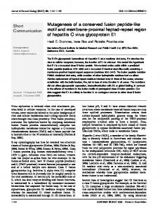
Journal of General Virology (1998), 79, 371–374. Printed in Great Britain ..........................................................................................................................................................................................................
SHORT COMMUNICATION
The identification of a conserved binding motif within human papillomavirus type 16 E6 binding peptides, E6AP and E6BP Robert C. Elston,1 Sawsan Napthine2 and John Doorbar1 1 2
Division of Virology, National Institute for Medical Research, The Ridgeway, Mill Hill, London NW7 1AA, UK Division of Virology, Department of Pathology, University of Cambridge, Tennis Court Road, Cambridge CB2 1QP, UK
A 16-mer peptide library was screened using the yeast two-hybrid system to identify peptides which specifically interact with the human papillomavirus type 16 (HPV-16) E6 protein. Four different peptides were identified, three of which contained an EL-L/V-G motif. A fifth E6 binding peptide, derived from the putative tumour suppressor protein tuberin, was identified during a two-hybrid screen of a HeLa cDNA expression library. This peptide contained a D-I-L-G motif. Homology to the peptides was found within the E6 binding proteins E6AP and E6-BP. A synthetic peptide containing the ELLG motif blocked the interaction of E6 with both E6-AP and E6-BP. The data suggest that E6 interacts through a structurally similar binding domain present within a number of cellular proteins.
The transforming activity of the human papillomavirus type 16 (HPV-16) E6 protein is associated with its interaction with a ubiquitin protein ligase [E6AP (Huibregtse et al., 1991)] and p53 (Werness et al., 1990), resulting in the ubiquitin mediated degradation of p53 (Scheffner et al., 1990). Degradation of p53 fails to explain all the properties associated with the E6 proteins. For example, several HPV-16 E6 mutants lose the ability to transform human embryonic kidney cells, but retain wild-type p53 degradation function (Nakagawa et al., 1995). In addition to E6AP and p53, the HPV-16 E6 protein can interact with several cellular proteins (Keen et al., 1994). Two HPV-16 E6 binding proteins have been identified using the yeast two-hybrid system (Fields & Song, 1989) ; a putative calcium binding protein, E6BP (Chen et al., 1995), and a member of the MCM protein family, hCDC47 (Fujita et al., 1996). A third HPV-16 E6 binding protein, paxillin, has recently been identified (Tong & Howley, 1997). An 18 amino acid region within E6AP was previously reported to be essential for the interaction of E6 (Huibregtse et Author for correspondence : Robert Elston. Fax 44 181 906 4477. e-mail relston!nimr.mrc.ac.uk
0001-5181 # 1998 SGM
al., 1993). No E6 binding domains have been reported for E6BP, hCDC47 or paxillin. To characterize the residues critical for the interaction of E6 within the E6AP domain and to identify other E6 binding specificities, a 16-mer peptide library was screened using the yeast two-hybrid system. This approach has previously been used to identify seven 16-mer peptides fused to the GAL4 activation domain (AD) which interacted with the retinoblastoma protein when fused to the GAL4 DNA binding domain (DBD) (Yang et al., 1995). The peptide library was generously provided by Stanley Fields (State University of New York at Stony Brook, USA). The HPV-16 E6 gene was amplified by PCR from the viral genomic DNA construct (nucleotides 104–559, provided by Harald zur Hausen, DKFZ, Germany) and was cloned in-frame with the cDNA encoding the GAL4 DBD(1–147aa) protein within vector pGBT9 (Clontech), to produce plasmid pGBT916E6 which encoded the protein DBD-16E6. The yeast strain Hfc7 (Clontech) was sequentially transformed with pGBT9-16E6 and the peptide library using the lithium acetate method (Gietz et al., 1992). Of the 3±2¬10' yeast transformants screened, 409 grew at least moderately in the absence of leucine, tryptophan and histidine, but in the presence of 20 mM 3-aminotriazole. Five colonies exhibited activation of the GAL4 inducible β-galactosidase gene consistent with the GAL4 DBD and AD being brought into close proximity through a peptide–E6 interaction. The interactions were E6 specific. No β-galactosidase activity was observed when pGBT9-16E6 was absent, or was replaced by plasmid pVA3 (Clontech) or pLAM5« (Clontech) which encoded the GAL4 DBD fused to murine p53 or lamin C respectively (data not shown). The peptide-encoding plasmids were isolated and sequenced (Hoffman & Winston, 1987). Four different peptides were identified, three of which contained an E-L-L}V-G motif (Fig. 1 a). An additional E6 binding peptide, peptide T27, which was derived from the putative tumour suppressor protein tuberin, was identified from a HeLa-derived cDNA expression library (Clontech) using the yeast two-hybrid system with DBD-16E6 as bait (Fig. 1 b). This peptide contained a D-I-L-G motif. Mutation of the isoleucine residue within the DILG motif to threonine inhibited the interaction of E6 following expression in the yeast two-hybrid system (data not shown). The
DHB
R. C. Elston, S. Napthine and J. Doorbar (a)
(b)
Fig. 1. Sequences of the E6 binding peptides. (a) Peptides 1–4 were isolated from the peptide library screen and are fused to the GAL4 activation domain through the linker peptide LELVDP. (b) Peptide T27 was isolated from the HeLa derived library screen and is fused to the GAL4 activation domain through the linker peptide SARAARA. The E/D-L/IL/V-G motif present within peptides 1, 2, 3 and T27 is shown in bold. Peptide 4 has no homology to this motif.
(a)
(b) Fig. 2. Homology of the identified peptides to E6AP (a) and E6BP (b). Conserved residues are indicated with a shadowed asterisk and semi-conserved residues are indicated with a plain asterisk. The shaded region of E6BP represents the previously identified 204 residue E6 binding domain (Chen et al., 1995). Smaller sized letters represent residues present within the linker sequence between the peptide and the GAL4 activation domain.
interactions were E6 specific using the conditions described above (data not shown). Tuberin is an approximately 180 kDa protein which localizes to the Golgi apparatus, and which specifically stimulates the intrinsic GTPase activity of Rap1a (European Chromosome 16 Tuberous Sclerosis Consortium, 1993 ; Wienecke et al., 1995, 1996). The biological significance of the E6–tuberin interaction is currently being established. Comparison of the peptide sequences with E6AP and E6BP revealed homology within both proteins (Fig. 2 a and 2 b respectively). The region of homology within E6AP corresponded to the 18 amino acid E6 binding domain previously identified by deletion mutagenesis and in vitro binding assays (Huibregtse et al., 1993). Identification of the ELLG motif in a
DHC
different experimental system indicates the significance of this motif and, through comparison of the peptides, permits characterization of the critical residues. The E6 binding domain is defined by the presence of an E}D-L}I}F-L}V-G motif, an upstream requirement for hydrophobic residues, usually containing at least one leucine and one threonine residue, and at least one acidic residue. The complete absence of basic residues, in the seven residues upstream of the ELLG motif, suggests that the presence of basic residues is detrimental to the E6 interaction. Previously, Huibregtse et al. (1993) demonstrated that an 18-mer synthetic peptide corresponding to the E6 binding domain within E6AP would block the interaction between E6
HPV-16 E6 binding motif
(a) Pep-e6ap
(b) Pep-1
(c) Pep-2
(d) Pep-tub
(e) Pep-fib
Fig. 3. Peptide inhibition study. A typical example of a binding experiment between GST–E6AP or GST–E6BP charged glutathione–Sepharose 4B beads and [35S]methionine labelled HPV-16 E6 protein, performed in the presence of increasing concentrations of peptide (0, 1, 10 or 100 µM, left to right). Peptides were supplied by either Genosys Biotechnologies (Pepe6ap, Pep-1), Sigma (Pep-fib) or synthesized in house on a Shimadzu PSSM-8 synthesizer (Pep-2, Pep-tub). All peptides were purified by HPLC to greater than 95 % purity.
and E6AP. To assess the ability of the peptides identified in this study to inhibit the interaction between E6 and GST–E6AP or GST–E6BP, several of the peptides were synthesized. Radioactive HPV-16 E6 protein was produced in vitro following translation in wheat germ extract (Promega) supplemented with -[$&S]methionine. GST fusion proteins of E6AP(aa1– 468) and E6BP were prepared as described previously (Keen et al., 1994). The E6AP-encoding cDNA and the GST–E6BP-expressing construct were generously provided by John Huibregtse (Rutgers University, USA) and Jason J. Chen (Tufts University School of Medicine, USA) respectively. Glutathione–Sepharose 4B beads (50 µl, Pharmacia) were incubated with 1 µM of either GST–E6AP or GST–E6BP. After binding, the beads were incubated for 3 h at 4 °C in LSAB (100 mM NaCl, 100 mM Tris–HCl, pH 8±0, 1 % NP40, 2 mM DTT, 0±1 % nonfat dried milk, 1 mM PMSF) containing equal amounts of [$&S]methionine labelled HPV-16 E6 protein and increasing quantities of peptide (0–100 µM). After washing, bound E6 protein was visualized by 15 % SDS–PAGE and autoradiography. None of the peptides destabilized the interaction between the glutathione–Sepharose 4B beads and the GST fusion proteins (data not shown). Both Pep-e6ap (Fig. 3 a) and Pep-1 (Fig. 3 b) inhibited the interaction between E6 and GST–E6AP or GST–E6BP at 100 µM concentration. Pep-e6ap represents a shortened version of the 18-mer E6AP peptide used in the Huibregtse et al. (1993) study. The third ELLG containing peptide, Pep-2, which has strongest homology to a region within E6BP (Fig. 2 b), inhibited the interaction of E6 to a lesser degree (Fig. 3 c). Peptub inhibited the interaction of E6 with GST–E6BP and to a lesser extent GST–E6AP (Fig. 3 d ). No inhibition was observed with Pep-fib, the fibrinonectin A control peptide (Fig. 3 e). The ability of peptides Pep-e6ap and Pep-1 to efficiently inhibit the interaction of E6 with both E6AP and E6BP
suggests that E6 interacts with both proteins through a structurally similar binding domain. Alternatively, peptide binding may sterically hinder, either directly or through the induction of a conformational change, the interaction of E6 with a second E6 binding domain. The latter possibility seems unlikely given the homology within E6BP to the consensus E6 binding domain. The ability of Pep-tub to inhibit the interaction of E6 with E6BP more efficiently than E6AP suggests that although the binding domains are structurally similar the relative importance of each residue within the domain may differ for each protein. In support of this, type specific differences have been reported for the relative binding of E6 to E6AP and E6BP (Chen et al., 1995). The HPV-16 E6 protein mediates its biological role through protein interactions. The data presented here have identified an E6 binding domain common to E6AP, E6BP and tuberin. This domain is characterized by the presence of an E}D-L}I}F-L}VG motif, together with an upstream requirement for hydrophobic residues, usually containing at least one leucine and threonine residue, the presence of at least one acidic residue and the complete absence of basic residues. Although no ELLG motif is evident within hCDC47 or paxillin, similar motifs have been identified in other E6 binding proteins (L. Banks, personal communication ; P. Howley, personal communication). Furthermore, the identification of a peptide without the ELLG motif indicates that at least one other linear E6 binding domain may exist. The data presented here provide the first molecular explanation of how the E6 protein mediates interaction with a increasing number of cellular proteins. This work was supported by the Imperial Cancer Research Fund, Cambridge University and Glasgow University. We would also like to thank Howard Marsden and Gillian McVey for their assistance with the synthesis and purification of some of the peptides used in this study.
DHD
R. C. Elston, S. Napthine and J. Doorbar
References Chen, J. J., Reid, C. E., Band, V. & Androphy, E. J. (1995). Interaction
of papillomavirus E6 oncoproteins with a putative calcium binding protein. Science 269, 529–531. European Chromosome 16 Tuberous Sclerosis Consortium (1993).
Identification and characterization of the tuberous sclerosis gene on chromosome 16. Cell 75, 1305–1315. Fields, S. & Song, O. (1989). A novel genetic system to detect protein–protein interactions. Nature 340, 245–247. Fujita, M., Kiyono, T., Hayahi, Y. & Ishibashi, M. (1996). HCDC47, a human member of the MCM family. Journal of Biological Chemistry 271, 4349–4354. Gietz, D., Gean, A. St, Woods, R. A. & Schiestl, R. H. (1992). Improved method for high efficiency transformation of intact yeast cells. Nucleic Acids Research 20, 1425. Hoffman, C. S. & Winston, F. (1987). A ten-minute DNA preparation from yeast efficiently releases autonomous plasmids for transformation of Escherichia coli. Gene 57, 267–272. Huibregtse, J. M., Scheffner, M. & Howley, P. M. (1991). A cellular protein mediates association of p53 with the E6 oncoprotein of human papillomavirus types 16 or 18. EMBO Journal 10, 4129–4135. Huibregtse, J. M., Scheffner, M. & Howley, P. M. (1993). Localization of the E6-AP regions that direct human papillomavirus E6 binding, association with p53, and ubiquitination of associated proteins. Molecular Cell Biology 13, 4918–4927. Keen, N., Elston, R. & Crawford, L. (1994). Interaction of the E6 protein of human papillomavirus with cellular proteins. Oncogene 9, 1493–1499.
DHE
Nakagawa, S., Wantanabe, S., Yoshikawa, H., Taketani, Y., Yoshiike, K. & Kanda, T. (1995). Mutational analysis of HPV-16 E6 protein :
transforming function for human cells and degradation of p53 in vitro. Virology 212, 535–542. Scheffner, M., Werness, B. A., Huibregtse, J. M., Levine, A. J. & Howley, P. M. (1990). The E6 oncoprotein encoded by human
papillomavirus types 16 and 18 promotes the degradation of p53. Cell 63, 1129–1136. Tong, X. & Howley, P. M. (1997). The bovine papillomavirus E6 oncoprotein interacts with paxillin and disrupts the actin cytoskeleton. Proceedings of the National Academy of Sciences, USA 94, 4412–4417. Werness, B. A., Levine, A. J. & Howley, P. M. (1990). Association of human papillomavirus types 16 and 18 E6 proteins with p53. Science 248, 76–79. Wienecke, R., Konig, A. & DeClue, J. E. (1995). Identification of tuberin, the tuberous sclerosis-2 product. Journal of Biological Chemistry 270, 16409–16414. Wienecke, R., Maize, J. C., Shoarinejad, F., Vass, W. C., Reed, J., Bonifiacino, J. S., Resau, J. H., Gunzburg, J. D., Yeung, R. S. & DeClue, J. E. (1996). Co-localization of the TSC2 product tuberin with its target
Rap1 in the Golgi apparatus. Oncogene 13, 913–923. Yang, M., Wu, Z. & Fields, S. (1995). Protein–peptide interactions analyzed with the yeast two hybrid system. Nucleic Acids Research 23, 1152–1156.
Received 2 September 1997 ; Accepted 17 October 1997