The Involvement of Posterior Parietal Cortex in Feature and Conjunction Visuomotor Search Alison R. Lane, Daniel T. Smith, Thomas Schenk, and Amanda Ellison
Abstract ■ Successful interaction with the environment often involves the
identification and localization of an item. Right posterior parietal cortex (rPPC) is necessary for the completion of conjunction but not feature visual search, regardless of the attentional requirements. One account for this dissociation is that the rPPC is primarily involved in processing spatial information. For target identification, conjunction tasks require that spatial information is used to determine if features occur at the same location, whereas feature search does not require such a process. This account suggests that if the requirement to localize the target is made explicit, then rPPC may also be necessary for feature search. This was examined using TMS and by manipulating the response mode: Participants were either required to press a button indicat-
INTRODUCTION The posterior parietal cortex (PPC) has been implicated in a multitude of cognitive and behavioral processes. This includes tasks such as selective and sustained attention (Malhotra, Coulthard, & Husain, 2009; Husain & Nachev, 2007; Marois, Leung, & Gore, 2000; Sturm et al., 1999; Coull & Frith, 1998) and the processing of visual, auditory, and somatosensory information such as salience (see e.g., Hodsoll, Mevorach, & Humphreys, 2009; Shomstein & Yantis, 2006; Downar, Crawley, Mikulis, & Davis, 2002). Although the findings indicate that this brain area may be activated in both spatial and nonspatial processes, these may be differentially represented within specific PPC subregions, with a gradient between spatial and nonspatial roles extending from the superior to inferior parietal lobules (Husain & Rorden, 2003). In this article, we investigate a region of PPC known to specialize in visuospatial processing and probe its contribution to visuomotor transformations. The PPC, particularly in the right hemisphere (rPPC), has been identified as a primary component of the frontoparietal network of spatial attention (Corbetta & Shulman, 2002; Gitelman et al., 1999; Corbetta, 1998). Damage to this area can result in hemispatial neglect (Heilman, Watson, Valenstein, & Damasio, 1983), which is characterized by an impairment in directing attention to contralesional Durham University, UK
© 2011 Massachusetts Institute of Technology
ing the presence/absence of the target or else had to point to the target. TMS over rPPC did not disrupt performance of the feature task when a button press was required but significantly increased response time and movement time for the same task in the pointing condition. Conjunction search in both response conditions was significantly impaired by TMS. Performance on a task that required pointing to a target in the absence of distractors and thus did not involve visual search was unaffected by rPPC stimulation. We conclude that rPPC is involved in coding and representing spatial information and is therefore crucial when the task requires determining whether two features spatially co-occur or when search is combined with explicit target localization via a visuomotor transformation. ■
space. There appears to be a right hemisphere dominance for visuospatial attention processing (see, e.g., Pourtois, Vandermeeren, Olivier, & de Gelder, 2001; Vallar, 1993; Heilman & Van Den Abell, 1980), and subsequently it is the rPPC that is of primary interest in this study. However, the precise role of rPPC in mediating spatial attention remains unclear. Visual search is one of the main paradigms used to investigate visuospatial attention (Treisman & Gelade, 1980). In these tasks, participants are required to search a spatial array for a specific target and make a decision regarding the presence of this item. In conjunction search, the target is defined by a combination of features, such as color and form, whereas in feature search the target differs from the distractor items along only one dimension. Although functional imaging has revealed rPPC to be active during both types of visual search (Nobre, Coull, Walsh, & Frith, 2003), it appears from neuropsychological evidence that rPPC is only necessary for tasks involving conjunctions ( Wojciulik & Kanwisher, 1998; Friedman-Hill, Robertson, & Treisman, 1995; Arguin, Cavanagh, & Joanette, 1994). In further support of this, research using the type of visual search detection paradigm described and TMS (a neurodisruptive lesion replacement technique) has shown that rPPC is critically involved in conjunction search but that the same brain region is not necessary for comparable feature tasks (Muggleton, Cowey, & Walsh, 2008; Ellison, Rushworth, & Walsh, 2003). Disruption to the attention system (because of TMS or
Journal of Cognitive Neuroscience 23:8, pp. 1964–1972
lesion) is insufficient to explain this dissociation. Different tasks require varying degrees of attention (as denoted by the seriality of the search rate), with some conjunction searches being performed with more ease than some feature ones, and yet rPPC was found to be critically involved in conjunction but not feature visual search regardless of the attentional task demands (Ellison et al., 2003). The apparent specific involvement of rPPC during conjunction tasks suggests that this area may be primarily involved in the coding and processing of spatial information, in line with the hypothesis proposed by Driver and Vuilleumier (2001). This account can explain the dissociation between the involvement of rPPC in feature and conjunction search because the critical difference between them lies in the requirement to bind visual features in conjunction tasks. A conjunction is only identified if the relevant features are detected at the same location, and it is possible that the binding process involves checking whether features are spatially congruent. On the basis of this account of the role of rPPC, one would predict that if the response is changed from one of simple target detection to one that requires explicit spatial information, then TMS over rPPC would also disrupt feature search. The aim of the present study was to test this prediction and thus also the role of rPPC as a visuospatial processing region. Participants completed feature and conjunction visual search tasks of equal difficulty, using two different response conditions; they either indicated the presence of the target using a manual button press on a response box or touched the target when present (a response requiring accurate target localization). Using this procedure, we could examine whether the same area that is critically involved in the processing of conjunction visual search, namely, rPPC, is also involved in feature search once the requirement to localize the target is incorporated as an essential component of the task. It was predicted that TMS over rPPC would disrupt conjunction search in both response conditions and would impair feature search in the pointing condition but not the button-press version. Evidence from both clinical (Karnath & Perenin, 2005; Rossetti et al., 2005; Mattingley, Husain, Rorden, Kennard, & Driver, 1998; Levine, Kaufman, & Mohr, 1978) and TMS (Rice, Tunik, & Grafton, 2006; Tunik, Frey, & Grafton, 2005; Desmurget et al., 1999) studies suggest that parts of PPC are also involved in visuomotor control. For the purpose of this study, it is important to be able to distinguish between any direct effects of rPPC TMS on motor control and the more indirect effects, which are those that we expect because of the higher spatial requirements introduced in the localization condition. The available evidence suggests that those neuronal collections within PPC that are primarily devoted to processing spatial attributes are found in different PPC subregions to those that are mainly associated with motor control (for more details, see Discussion section; e.g., Rushworth & Taylor, 2006). We would therefore expect that by using a visuospatial task to functionally localize our TMS site, we will mainly interfere with PPCʼs
spatial rather than its visuomotor function. However, to monitor any direct effects of TMS on motor behavior, we also introduced a pure visuomotor control task. This task required the same pointing movements as in the search tasks, but because the visual target was presented on its own, no visual exploration or visual selection was involved.
METHODS Participants Twelve healthy, right-handed participants (7 men and 5 women) aged between 20 and 51 years (median age = 25.5 years) participated in the study. All had normal or corrected-to-normal vision. Participants gave their signed informed consent in accordance with the Declaration of Helsinki and with the approval of Durham University Ethics Advisory Committee and could withdraw at any point. Participant selection complied with the current guidelines for repetitive TMS research (Rossi, Hallett, Rossini, PascualLeone, & The Safety of TMS Consensus Group, 2009). Two of the participants were excluded because rPPC location could not be reliably established, resulting in a final sample size of 10. Visual Stimuli and Tasks The search arrays were programmed using E-Prime (Psychology Software Tools Inc., Sharpsburg, PA), with the program also remotely triggering the TMS and the movement recording systems. The stimuli were presented on a 48-cm (1024 × 768 pixels) Iiyama Prolite touch-screen monitor with a 75-Hz refresh rate. Participants were seated approximately 57 cm away from the centrally aligned monitor (which was within reaching distance for all participants) with the center of the screen at eye level. The room was dimly lit so that participants could see their hands. There were two visual search tasks: feature and conjunction. Participants also had to perform a motor control task. The search display for all of these tasks comprised a 10 × 6 virtual array, which subtended 37° × 27° of visual angle. Items were presented randomly within this array. Visual Search Tasks For the conjunction task, the target was a green forward slash (/ ) and the distractors were green backward slashes ( \) and red forward slashes. All stimuli were presented against a black background and matched for luminance within and between items across the display. The array consisted of 10 randomly presented items: 9 distractors plus the target (target-present condition) or 10 distractors (target-absent condition). Each item in the conjunction search was 3° of visual angle in length. The feature task involved an array of 10 white 2.1° × 2.1° squares (3° diagonally), and the target item was a square with a gap along the upper edge. The size of the gap was determined on an Lane et al.
1965
individual basis such that RT was equivalent to that of the conjunction task (range = 0.1°–0.7°, median gap = 0.2° ). Pilot data (n = 14) using the two search tasks and four different set sizes (4, 8, 10, and 12 items) revealed that both tasks were performed in a serial manner (the mean search functions were 22.77 msec/item and 23.45 msec/item, respectively) and were not significantly different to one another, t(13) = 0.126, p = .901. For both tasks, the target was present on 50% of the trials, and there was never more than one target. On each trial, the search items were equally distributed across the two hemifields, with the target appearing in each hemifield an equal number of times. A white central fixation cross (0.5° × 0.5°) was presented for 500 msec at the start of each trial, which participants were instructed to fixate. This was followed immediately by the presentation of the search array, which remained present until the participant made a response (Figure 1A). Participants were free to move their eyes during the search although eye movements were not recorded. The intertrial interval was 4000 msec, during which time a blank black screen was presented. Both of the visual search tasks were completed under two response conditions. In one condition (button press), participants responded regarding target presence with a manual
button press (left for present, right for absent). In the second condition (pointing), at the beginning of each trial participants had to place their index finger on the start position (which was located 30 cm from the screen). In the targetpresent condition, participants were required to touch the target (aiming for the center of the item) and were instructed to respond with one directed movement. Participants were instructed to press the space bar (which was located at the start position) when the target was absent. Accuracy and speed were stressed as important factors in both conditions, although accuracy was not to be sacrificed for speed. Each of these response conditions was performed in a separate session lasting approximately 1.5 hours, with the session order counterbalanced across participants. The trials were completed in blocks of 40, comprising 20 target-present and 20 target-absent trials. Participants performed 16 blocks of visual search overall. These included 8 blocks of conjunction search trials (4 for each response condition), half of which were completed with TMS delivered to rPPC and the other half were sham-TMS blocks. Participants also completed 8 blocks of feature visual search trials (4 for each response condition), and again 50% of which were TMS and 50% were sham-TMS blocks. The order of blocks was counterbalanced.
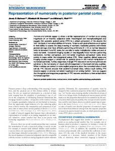
Figure 1. Diagram showing the time sequence of the tasks. (A) Search tasks (feature and conjunction). The search array was presented after a fixation cross, and TMS was delivered for 500 msec from the beginning of array onset. The response could be made at any time after the onset of the array and was either a button press or a pointing movement to the target. (B) Motor control task. A fixation cross was presented, followed by the target. From the beginning of target onset, TMS was delivered for 500 msec and the pointing response could be made at any time.
1966
Journal of Cognitive Neuroscience
Volume 23, Number 8
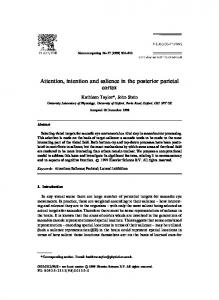
Figure 2. Diagram showing the average stimulated rPPC site (MNI coordinates: x = 50, y = −80, z = 54 mm). The position was verified using each participantsʼ MRI scan coregistered to their skull coordinates using BrainSight software. Scan slices are presented in radiological convention.
Motor Control Task Each trial began with a central 0.5° white fixation cross presented on a black background for 500 msec. This display was followed immediately by the presentation of a white 1° dot at a random location, which was displayed until response (Figure 1B). The dot was presented to each hemifield equally across the trials, of which there were 40 per block. Participants were instructed to fixate the cross at the start of each trial, with their index finger on the start position, and then touch the dot as quickly and as accurately as possible. Five catch trials were also included per block, in which no dot appeared after the fixation cross was extinguished. These trials were included to discourage participants from beginning the movement before target onset. For the catch trials, participants were required to press the space bar. The intertrial interval was 4000 msec. Participants completed two consecutive blocks of trials, one with TMS and one with sham-TMS, the order of which was counterbalanced. The two blocks for the motor control task could be performed at either of the two visual search testing sessions.
Transcranial Magnetic Stimulation and Site Localization Five pulses of repetitive TMS was delivered at 10 Hz to rPPC at visual array onset using a Magstim Rapid (Magstim, Whitland, Carmarthenshire, UK) at 65% of the maximum machine output (i.e., 1.3 T). A 70-mm figure-of-eight coil
was used to deliver stimulation, and the coil was placed tangential to the skull, with the handle pointing backward, parallel to the midsagittal plane. The coil was held in place by the researcher allowing for precise control of the coil position. In the sham-TMS blocks, a discharging coil was placed in proximity to the participant while an inactive coil was positioned over the rPPC site. Therefore, the subjective sensation of coil position and auditory effects were comparable with those experienced in the TMS blocks, but no magnetic stimulation was delivered. Within each session, the order of blocks (TMS/sham-TMS) was counterbalanced between participants. A hunting procedure with the conjunction task, as described in Ashbridge, Walsh, and Cowey (1997) and recently used by Ellison, Lane, and Schenk (2007), was used to identify the location of rPPC. Once the site was established, the position was recorded and marked with a sticker on a tightly fitting lycra swimming cap. The same location was used for the second session. The location of rPPC was verified using each subjectʼs MRI scan and frameless stereotaxy (Brainsight; Rogue Research Inc., Montreal Quebec, Canada), and the critical region was located in the angular gyrus between the intraparietal and the superior temporal sulci (Figure 2).
Movement Recording Physiological data were recorded using an MP35 acquisition unit and BSL Pro 3.7 software (Biopac Systems Inc., Goleta, Lane et al.
1967
CA) to determine the time of movement onset (RT) during the pointing conditions. Surface AgCl electrodes were used for the EMG, which were placed on the right underside forearm. The proximal end electrode was positioned close to the thumb side of the wrist (flexor pollicis longus). The distal end electrode was placed toward the elbow, and the ground electrode was placed close to the wrist on the littlefinger side (flexor profundus digitorum). A Biopac triaxial accelerometer was also placed over the right wrist. The data were sampled using the software channel presets at a rate of 1000 Hz. Data analysis was completed off-line in which the RT was recorded by the researcher, defined as the time between the onset of the search array and the point of the first steep increase relative to the baseline period (fixation) in the EMG record and at least one accelerometer channel.
.265, showing that performance of the two tasks was always matched regardless of the TMS condition or response type. However, the main effect of response, F(1, 9) = 21.91, p = .001, was significant, with RT being longer in the button press relative to the pointing condition (Figure 3). There was also a significant main effect of TMS, F(1, 9) = 20.87, p = .001, whereby RT was increased in the TMS relative to the sham-TMS condition (Figure 3). Furthermore, there was a significant interaction effect between response and TMS, F(1, 9) = 22.33, p = .001, and a significant three-way interaction between task, response, and TMS, F(1, 9) = 9.93, p = .012. This indicates that the effect of the TMS on RT not only differed according to response mode but that this was also related to the type of visual search task. To examine this three-way interaction further, a series of paired-samples t tests were conducted for each task and
Data Analysis RT was defined as the difference between the visual stimulus onset and the pushing of the button for the button-press condition (where movement onset and completion were closely coupled) and the difference between visual stimulus onset and movement onset time in the pointing condition. Movement time (MT) was also calculated for the responses in the pointing condition using the following formula: MT = movement offset − movement onset. RT was subjected to a 2 (Task: feature vs. conjunction) × 2 (Response: button press vs. pointing) × 2 (TMS: TMS vs. sham-TMS) ANOVA. Paired-samples t tests were then conducted to further examine the effects revealed by the ANOVA. These t tests were adjusted for multiple comparisons using a Bonferroni correction, resulting in a corrected alpha level of .013. An additional 2 (Task) × 2 ( TMS) ANOVA was conducted on MT for the pointing response condition. Furthermore, for the motor control task, paired-samples t tests were used to compare RT and MT between TMS and sham-TMS trials.
RESULTS The results from the target-absent trials were not analyzed for simplicity of analysis. Incorrect responses accounted for less than 2% of the data (mean accuracy was 98.02%), and these trials were removed from the RT and MT analyses. Search times for each hemifield were compared for each condition, and no significant differences were found. Therefore, the data from the two hemifields were pooled to increase statistical power. The results of the repeated measures ANOVA conducted using mean RT revealed that there was no significant main effect of task, F(1, 9) = 1.40, p = .268, indicating that both the feature and the conjunction search tasks were matched for difficulty. Furthermore, there was no significant interaction effect between task and response, F(1, 9) = 3.69, p = .087, or between task and TMS, F(1, 9) = 1.42, p = 1968
Journal of Cognitive Neuroscience
Figure 3. Graphs showing the mean RT (in milliseconds) for each search task and each TMS condition. There are separate figures for the button-press condition (A) and the pointing condition (B). Error bars represent the SEM across participants, and an asterisk indicates a significant difference ( p < .05).
Volume 23, Number 8
Table 2. Absolute Movement Error for Each Task TMS
Feature
Conjunction
Motor Control
Sham
15.64 (0.79)
15.66 (0.84)
14.31 (0.65)
TMS
16.33 (1.15)
16.30 (1.31)
15.33 (0.95)
Absolute error (SEM ) between the end point of the movement in the pointing condition and the central position of the target, measured in pixels.
Figure 4. Graphs showing the mean MT (in milliseconds) for the pointing response condition, for each visual search task and each TMS condition separately. Error bars represent the SEM across participants and an asterisk indicates a significant difference ( p < .05).
response condition pair. A significant increase in mean RT with TMS as compared with sham-TMS was found for the conjunction search task for both the button press, t(9) = −4.42, p = .002, and pointing conditions, t(9) = −2.89, p = .018. There was no significant effect of TMS on feature task performance for the button-press condition, t(9) = −0.07, p = .946. Importantly, a significant increase in RT was observed for the feature task in the pointing condition with TMS as compared with sham-TMS, t(9) = −5.92, p < .001. These results indicate that rPPC is critically involved in conjunction visual search with both response types, whereas it only becomes necessary for feature visual search once the requirement to localize the target with a pointing movement is included. MT was calculated for the pointing conditions for each of the two search tasks (Figure 4). These data were subjected to a 2 (Task) × 2 (TMS) ANOVA, which revealed a nonsignificant main effect of task, F(1, 9) = 0.17, p = .693. There was, however, a significant effect of TMS, F(1, 9) = 19.02, p = .002, with mean MT being longer in the TMS condition relative to sham-TMS. There was no significant interaction effect between TMS and task, F(1, 9) = 0.12, p = .740, indicating that for both the conjunction and the feature search tasks, TMS increased the MT of the pointing response. Table 1. Mean RT and MT for the Motor Control Task TMS
RT
MT
Sham
237.19 (18.68)
726.88 (54.65)
TMS
247.88 (23.78)
751.00 (71.16)
The values presented are in milliseconds and the values in parentheses are the SEM across participants. RT was defined as the time between array onset and movement onset.
To examine whether TMS induced a nonspecific delay on pointing movements, the effect of rPPC TMS on performance of a non-search-based visuomotor task (motor control task) was examined (Table 1). For this task, there was no significant effect of TMS on either RT, t(9) = −1.24, p = .245, or MT, t(9) = −1.20, p = .261. This shows that rPPC is not necessary for tasks that require the participant to point at a target when there are no distractors to provide conflicting sources of spatial information, and thus the location is not ambiguous. The effect of the TMS on RT for the pointing condition was not a consequence of a speed-accuracy trade-off effect in relation to movement error (Table 2). The end point of the movement was recorded using the touch-screen monitor, and this was used to calculate the absolute error of the movement (in pixels) relative to the target position, which was defined as the center of the target item (the instructed goal of the movement). Paired-samples t tests revealed that there was no significant effect of the TMS on absolute error for the feature task, t(9) = −1.26, p = .239, conjunction task, t(9) = −0.65, p = .532, or the motor control task, t(9) = −1.39, p = .199.
DISCUSSION It was predicted that TMS over rPPC would not affect performance on a feature visual search task using a standard target detection paradigm but would impair performance for the same task if participants were instructed to localize the target. This prediction was confirmed. The results revealed that although TMS delivered to rPPC did not affect feature search when a button press was required to indicate the presence of the target, TMS over the same area did significantly increase RT relative to the sham-TMS condition when a pointing response was used. The TMS had a disruptive effect on the performance of conjunction visual search irrespective of the response mode in accordance with the prediction made and previous findings (Muggleton et al., 2008; Schindler, Ellison, & Milner, 2008; Fuggetta, Pavone, Walsh, Kiss, & Eimer, 2006; Ellison et al., 2003). The feature and conjunction visual search tasks were of comparable difficulty; they elicited similar serial search behavior and had comparable RTs. Consequently, task type did not have a significant effect on performance. Task difficulty was not previously controlled in experiments dissociating the role of rPPC in feature and conjunction search (e.g., Ellison et al., 2003), this is therefore an important Lane et al.
1969
manipulation. Although the two tasks had comparable attentional demands, there was a dissociation between the involvement of rPPC in conjunction and feature search for the button press condition, suggesting that this area is not primarily concerned with attention processing per se. We hypothesized that the discrepancy between the involvement of rPPC in feature and conjunction search as previously reported (Muggleton et al., 2008; Ellison et al., 2003) is associated with the differential spatial requirements of the two tasks, in accordance with the characterization of rPPC as an area that is predominantly involved in coding the location of visual stimuli (Driver & Vuilleumier, 2001). Accordingly, conjunction search involves the process of feature binding because only if the defining features coincide at the same position will they be seen as part of one item and thus identified as the target. A vital component of conjunction analysis is therefore the matching of the spatial location of independent feature maps, and the evidence suggests that this function is performed by rPPC. If the conjunction task involves only one item that appears at a predictable location, then spatial position does not need to be resolved when determining if the features co-occur, which could explain why rPPC is not necessary for conjunction tasks where there is no spatial ambiguity (Ellison et al., 2003). Feature search tasks (even attentionally demanding ones) do not require the binding process, and this could explain why under standard identification situations (button-press condition) TMS over rPPC does not affect performance on such tasks (an area attributed to be involved in the processing of such a task is right superior temporal gyrus according to its advanced identification speciality; see Ellison, Schindler, Pattison, & Milner, 2004). However, once the spatial aspect of the task is made more explicit, for example, by making target localization part of the response, then rPPC becomes necessary because spatial information has to be used. We propose that the impaired performance induced by rPPC TMS for the conjunction search in both response conditions and for feature search with pointing is caused by disruption of the same underlying process, namely, identifying the spatial location of the visual target. However, an alternative explanation could be that the impairments for each of the tasks are due to the disruption of two separable processes. For conjunction tasks, this would involve interference with the visuospatial attentional processes required for feature binding, as outlined earlier. For the feature search task in the localization condition, the TMS may instead disrupt the motor control processes needed to initiate and execute the pointing behavior. In principle, such a motor explanation is possible, although as the following will explain, we do not believe it to be the most likely explanation for our findings. There is converging evidence for the involvement of PPC in visuomotor control from studies using physiological recordings (Andersen & Buneo, 2002; Snyder, Batista, & Andersen, 1997), functional neuroimaging (Culham, Cavina-Pratesi, & Singhal, 2006; Astafiev et al., 2003; Clower 1970
Journal of Cognitive Neuroscience
et al., 1996), and TMS (Rice et al., 2006; Tunik et al., 2005; Desmurget et al., 1999). Furthermore, neuropsychological studies (Karnath & Perenin, 2005; Rossetti et al., 2005; Mattingley et al., 1998; Perenin & Vighetto, 1988; Levine et al., 1978) have also revealed the importance of PPC for the control of movements. However, it appears that the specific neuronal subregions of PPC that are highlighted as relevant in paradigms using spatial attention (Rushworth & Taylor, 2006; Nobre et al., 2003; Donner et al., 2002; Corbetta, 1998) are anatomically distinct from those predominantly implicated in visuomotor control (Rushworth & Taylor, 2006; Karnath & Perenin, 2005; Desmurget et al., 1999). With specific reference to previous TMS studies investigating PPC and motor control, these have predominantly focused on the left hemisphere, whereas it is the right hemisphere that appears dominant for spatial processing (Pourtois et al., 2001; Vallar, 1993; Heilman & Van Den Abell, 1980) and that was thus the locus of interest in the present study. The procedure we used to localize the stimulation site (i.e., hunting with a visual search paradigm) biased us toward a neuronal subregion of PPC that is specifically involved in spatial attentional processes. Furthermore, the visuomotor paradigms that have demonstrated a necessary involvement of PPC in motor control are different in numerous respects from the simple pointing response required in the tasks of the present study. For example, optic ataxia primarily presents as inaccuracy in rapid pointing using tasks with a strict fixation control and a peripherally presented target (Perenin & Vighetto, 1983; Levine et al., 1978), in contrast to the free-viewing condition that we used. Also, the TMS studies examining PPC and motor control have used target perturbation paradigms (Rice et al., 2006; Desmurget et al., 1999). This is in comparison with the simplistic pointing movement that was required in this study, whereby the required end point of the movement did not change during the trial. Deficits in reaching also appear to be hard to replicate using TMS over PPC ( Johnson & Haggard, 2005). This suggests that the addition of such simplistic pointing as we used during the search tasks is unlikely to have been sufficient to produce the subsequent TMS-induced disruption of performance. Most importantly, we tried to control for visuomotor deficits by also incorporating a pure visually guided pointing control task. The results revealed that the TMS did not significantly affect performance for this task. This indicates that the selected subregion of PPC that was stimulated in our experiment is not involved in the simple visuomotor transformation process that is used to guide a pointing response to a single target. One could argue that this does not exclude the possibility that the TMS has disrupted a more specific visuomotor process, namely, one which is only involved when visual distractors are present, and thus some process of selecting one spatial location and suppressing others is required (Fischer & Adam, 2001; Meegan & Tipper, 1999; Tipper, Lortie, & Baylis, 1992). Subsequently, this suggests that the disruption to the motor Volume 23, Number 8
control processes is in fact an impairment in the ability to resolve the spatial ambiguity caused by the presence of additional visual stimuli, and this could be described as a spatial impairment (albeit one which impacts on motor control processes). This specific visuomotor characterization of the TMS-induced deficit is therefore not substantially different from the spatial interpretation that we proposed at the beginning of the discussion. The results of the present study did reveal a significant TMS effect on MT as well as RT for the feature and the conjunction visual search tasks. However, it is well known that the programming of movements does not end with movement onset and instead the motor program is continuously updated using the sensory information available (Desmurget et al., 1999; Pelisson, Prablanc, Goodale, & Jeannerod, 1986). Consequently, any effects that are reported for MT can reflect processes associated with both movement execution and motor programming (Desmurget & Grafton, 2000). The effects that we observed could thus reflect the fact that TMS over rPPC interfered with the spatial disambiguation of the motor target, which slowed down both the initial and the on-line motor programming processes and subsequently affected both RT and MT. In this instance, it is perhaps more likely that the effects reported for MT reflect ongoing disruption to the movement planning processes as a consequence of the requirement for spatial selection because the same effects were not observed when a pure pointing response was required in the absence of visual search.
Conclusion The present results support the hypothesis that the contribution of rPPC to visual search lies in its ability to select the location of the visual target. We argued that in typical visual search paradigms where the goal is target detection, spatial coding of features is only necessary for conjunction but not for feature searches. On the basis of this, we predicted that if location becomes critical to the successful completion of a feature search, then rPPC will also become essential. To achieve this, the response was changed in one condition from pressing a button to pointing to the location of the visual target. It was found that in this condition, TMS over rPPC did also impair feature search performance. The findings support the account of visual binding as a process of matching the spatial position of the features of the target and furthermore suggest that the critical role of rPPC in visual search is to code the location of visual stimulus features.
Acknowledgments The work was supported by a grant from the Dr. Hadwen Trust for Humane Research—the UKʼs leading medical research charity funding exclusively nonanimal research techniques to replace animal experiments.
Reprint requests should be sent to Alison R. Lane, CNRU, Wolfson Research Unit, Durham University Queenʼs Campus, Stocktonon-Tees, TS17 6BH, UK, or via e-mail:
[email protected].
REFERENCES Andersen, R. A., & Buneo, C. A. (2002). Intentional maps in posterior parietal cortex. Annual Review of Neuroscience, 25, 189–220. Arguin, M., Cavanagh, P., & Joanette, Y. (1994). Visual feature integration with an attention deficit. Brain and Cognition, 24, 44–56. Ashbridge, E., Walsh, V., & Cowey, A. (1997). Temporal aspects of visual search studied by transcranial magnetic stimulation. Neuropsychologia, 35, 1121–1131. Astafiev, S. V., Shulman, G. L., Stanley, C. M., Snyder, A. Z., Van Essen, D. C., & Corbetta, M. (2003). Functional organization of human intraparietal and frontal cortex for attending, looking and pointing. Journal of Neuroscience, 23, 4689–4699. Clower, D. M., Hoffman, J. M., Votaw, J. R., Faber, T. L., Woods, R. P., & Alexander, G. E. (1996). Role of posterior parietal cortex in the recalibration of visually guided reaching. Nature, 383, 618–621. Corbetta, M. (1998). Frontoparietal cortical networks for directing attention and the eye to visual locations: Identical, independent, or overlapping neural systems? Proceedings of the National Academy of Sciences, U.S.A., 95, 831–838. Corbetta, M., & Shulman, G. L. (2002). Control of goal-directed and stimulus-driven attention in the brain. Nature Reviews Neuroscience, 3, 201–215. Coull, J. T., & Frith, C. D. (1998). Differential activation of right superior parietal cortex and intraparietal sulcus by spatial and nonspatial attention. Neuroimage, 8, 176–187. Culham, J. C., Cavina-Pratesi, C., & Singhal, A. (2006). The role of parietal cortex in visuomotor control: What have we learned from neuroimaging? Neuropsychologia, 44, 2668–2684. Desmurget, M., Epstein, C. M., Turner, R. S., Prablanc, C., Alexander, G. E., & Grafton, S. T. (1999). Role of the posterior parietal cortex in updating reaching movements to a visual target. Nature Neuroscience, 2, 563–567. Desmurget, M., & Grafton, S. (2000). Forward modelling allows feedback control for fast reaching movements. Trends in Cognitive Sciences, 4, 423–431. Donner, T. H., Kettermann, A., Diesch, E., Ostendorf, F., Villringer, A., & Brandt, S. A. (2002). Visual feature and conjunction searches of equal difficulty engage only partially overlapping frontoparietal networks. Neuroimage, 15, 16–25. Downar, J., Crawley, A. P., Mikulis, D. J., & Davis, K. D. (2002). A cortical network sensitive to stimulus salience in a neutral behavioral context across multiple sensory modalities. Journal of Neurophysiology, 87, 615–620. Driver, J., & Vuilleumier, P. (2001). Perceptual awareness and its loss in unilateral neglect and extinction. Cognition, 79, 39–88. Ellison, A., Lane, A. R., & Schenk, T. (2007). The interaction of brain regions during visual search processing as revealed by transcranial magnetic stimulation. Cerebral Cortex, 17, 2579–2584. Ellison, A., Rushworth, M., & Walsh, V. (2003). The parietal cortex in visual search: A visuomotor hypothesis. Clinical Neurophysiology, Supplements, 56, 321–330.
Lane et al.
1971
Ellison, A., Schindler, I., Pattison, L. L., & Milner, A. D. (2004). An exploration of the role of the superior temporal gyrus in visual search and spatial perception using TMS. Brain, 127, 2307–2315. Fischer, M. H., & Adam, J. J. (2001). Distractor effects on pointing: The role of spatial layout. Experimental Brain Research, 136, 507–513. Friedman-Hill, S. R., Robertson, L. C., & Treisman, A. (1995). Parietal contributions to visual feature binding: Evidence from a patient with bilateral lesions. Science, 269, 853–855. Fuggetta, G., Pavone, E., Walsh, V., Kiss, M., & Eimer, M. (2006). Cortico-cortical interactions in spatial attention: A combined ERP/TMS study. Journal of Neurophysiology, 95, 3277–3280. Gitelman, D. R., Nobre, A. C., Parrish, T. B., LaBar, K. S., Kim, Y.-H., Meyer, J. R., et al. (1999). A large-scale distributed network for covert spatial attention: Further anatomical delineation based on stringent behavioural and cognitive controls. Brain, 122, 1093–1106. Heilman, K. M., & Van Den Abell, T. (1980). Right hemisphere dominance for attention: The mechanism underlying hemispheric asymmetries of inattention (neglect). Neurology, 30, 327–330. Heilman, K. M., Watson, R. T., Valenstein, E., & Damasio, A. R. (1983). Localization of lesions in neglect. In A. Kertesz (Ed.), Localization in neuropsychology. New York: Academic Press. Hodsoll, J., Mevorach, C., & Humphreys, G. (2009). Driven to less distraction: rTMS of the right parietal cortex reduces attentional capture in visual search. Cerebral Cortex, 19, 106–114. Husain, M., & Nachev, P. (2007). Space and the parietal cortex. Trends in Cognitive Sciences, 11, 30–36. Husain, M., & Rorden, C. (2003). Non-spatially lateralized mechanisms in hemispatial neglect. Nature Reviews Neuroscience, 4, 26–36. Johnson, H., & Haggard, P. (2005). Motor awareness without perceptual awareness. Neuropsychologia, 43, 227–237. Karnath, H.-O., & Perenin, M.-T. (2005). Cortical control of visually guided reaching: Evidence from patients with optic ataxia. Cerebral Cortex, 15, 1561–1569. Levine, D. N., Kaufman, K. J., & Mohr, J. P. (1978). Inaccurate reaching associated with a superior parietal lobe tumor. Neurology, 28, 556–561. Malhotra, P., Coulthard, E. J., & Husain, M. (2009). Role of right posterior parietal cortex in maintain attention to spatial locations over time. Brain, 132, 645–660. Marois, R., Leung, H. C., & Gore, J. C. (2000). A stimulus-driven approach to object identity and location processing in the human brain. Neuron, 25, 717–728. Mattingley, J. B., Husain, M., Rorden, C., Kennard, C., & Driver, J. (1998). Motor role of human inferior parietal lobe revealed in unilateral neglect patients. Nature, 392, 179–182. Meegan, D., & Tipper, S. P. (1999). Visual search and target-directed action. Journal of Experimental Psychology: Human Perception and Performance, 25, 1347–1362. Muggleton, N., Cowey, A., & Walsh, V. (2008). The role of the angular gyrus in visual conjunction search investigated using signal detection analysis and transcranial magnetic stimulation. Neuropsychologia, 46, 2198–2202. Nobre, A., Coull, J., Walsh, V., & Frith, C. (2003). Brain activations during visual search: Contributions of search efficiency versus feature binding. Neuroimage, 18, 91–103.
1972
Journal of Cognitive Neuroscience
Pelisson, D., Prablanc, C., Goodale, M. A., & Jeannerod, M. (1986). Visual control of reaching movement without vision of the limb. II. Evidence of fast unconscious processes correcting the trajectory of the hand to the final position of a double-step stimulus. Experimental Brain Research, 62, 303–311. Perenin, M.-T., & Vighetto, A. (1983). Optic ataxia: A specific disorder in visuomotor coordination. In A. Hein & M. Jeannerod (Eds.), Spatially oriented behavior (pp. 305–326). New York: Springer. Perenin, M.-T., & Vighetto, A. (1988). Visuomotor mechanisms: I. Different aspects of the deficit in reaching for objects. Brain, 111, 643–674. Pourtois, G., Vandermeeren, Y., Olivier, E., & de Gelder, B. (2001). Event-related TMS over the right posterior parietal cortex induces ipsilateral visuo-spatial interference. NeuroReport, 12, 2369–2374. Rice, N. J., Tunik, E., & Grafton, S. (2006). The anterior intraparietal sulcus mediates grasp execution, independent of requirement to update: New insights from transcranial magnetic stimulation. Journal of Neuroscience, 26, 8176–8182. Rossetti, Y., Revol, P., McIntosh, R., Pisella, L., Rode, G., Danckert, J., et al. (2005). Visually guided reaching: Bilateral posterior parietal lesions cause a switch from fast visuomotor to slow cognitive control. Neuropsychologia, 43, 162–177. Rossi, S., Hallett, M., Rossini, P. M., Pascual-Leone, A., & The Safety of TMS Consensus Group. (2009). Safety, ethical considerations, and application guidelines for the use of transcranial magnetic stimulation in clinical practice and research. Clinical Neurophysiology, 120, 2008–2039. Rushworth, M. F. S., & Taylor, P. C. J. (2006). TMS in the parietal cortex: Updating representations for attention and action. Neuropsychologia, 44, 2700–2716. Schindler, I., Ellison, A., & Milner, A. (2008). Contralateral visual search deficits following TMS. Journal of Neuropsychology, 2, 501–508. Shomstein, S., & Yantis, S. (2006). Parietal cortex mediates voluntary control of spatial and non-spatial auditory attention. Journal of Neuroscience, 26, 435–439. Snyder, L. H., Batista, A. P., & Andersen, R. A. (1997). Coding of intention in the posterior parietal cortex. Nature, 386, 167–170. Sturm, W., de Simone, A., Krause, B. J., Specht, K., Hesselmann, V., Radermacher, I., et al. (1999). Functional anatomy of intrinsic alertness: Evidence for a fronto-parietal-thalamic-brainstem network in the right hemisphere. Neuropsychologia, 37, 797–805. Tipper, S. P., Lortie, C., & Baylis, G. C. (1992). Selective reaching: Evidence for action-centred attention. Journal of Experimental Psychology: Human Perception and Performance, 18, 891–905. Treisman, A., & Gelade, G. (1980). A feature integration theory of attention. Cognitive Psychology, 12, 97–136. Tunik, E., Frey, S. H., & Grafton, S. T. (2005). Virtual lesions of the anterior intraparietal area disrupt goal-dependent on-line adjustments of grasp. Nature Neuroscience, 8, 505–511. Vallar, G. (1993). The anatomical basis of spatial hemineglect in humans. In I. H. Robertson & J. C. Marhsall (Eds.), Unilateral neglect: Clinical and experimental studies. Hove: Lawrence Erlbaum Associates. Wojciulik, E., & Kanwisher, N. (1998). Implicit but not explicit feature binding in a Balintʼs patient. Visual Cognition, 5, 157–181.
Volume 23, Number 8