Houston, Texas 77030. HP- 1 is a 30-residue ... which we call HP1-56. The third is likely to .... Anderson Cancer Center, Houston, TX, unless otherwise stated in.
THEJOURNAL OF BIOLOGICAL CHEMISTRY 8 1991 by The American Society for Biochemistry and Molecular Biology, Inc,
Vol. 266, No. 12, Issue of April 25, pp. 7524-7530, 1991 Printed in U.S. A.
The Isolation andIdentification of Multiple Forms of the Neutrophil Granule Peptides from Human Leukemic Cells* (Received for publication, October 10, 1990)
Andrew Bateman$, Ava Singhj, Chaim Shustikll, Wendy M. Mars[[, and Samuel Solomon From the Endocrine Laboratory, Department of Medicine, Obstetrics, and Gynecology, and the YDivisionof Hematology, Department of Medicine, Royal Victoria Hospital/McGill University, Montreal, Quebec H3A lA1, Canada and the IIDepartment of Biochemistry, M. D. AndersonHospital and Tumor Cancer Center, Universityof Texas System Cancer Center, Houston, Texas 77030
HP- 1is a 30-residue cysteine- and arginine-rich pep- Among the more abundant neutrophil granule components tide of the human neutrophil primary granule and is are the recently characterized cystine and arginine rich pepthe most abundant human representativeof the family tides variously known as defensins or corticostatins. These of peptides variously called defensins and corticosta- are peptidesof between 29 and 33 amino acids in length that tins. Peptides belonging to this family have many bio- share a common cystinebackboneandarecationic (1-4). logical activities including the non-oxidative destruc- They are capable of killing a variety of bacterial and fungal tion of ingested microorganisms, the inhibition of ad- species (5, 6) as well as lysis of mammalian cells (1, 2, 7). renocorticotropin-stimulated synthesis of glucocortic- Their mode of action most likely involves the formation of oids, monocyte chemotaxis, the non-cytolytic inhibivoltage-dependent ion permeable channels in the target memtion of [3H]thymidine incorporation in HL-60 promyelocyte-like cells and the stimulation of nifedipine-sen- brane (8).Other biological activities of these peptides are the sitive calcium channels. Using a combination of re- inhibition of the secretion of the glucocorticoids (9, 10) and versed-phaseand size-exclusionhigh performance as monocyte chemotactic agents(11). In humans four neutrohave been liquid chromatography and an HP1 radio-immunoas- phil granule cystine- and arginine-rich peptides say, three immunoreactive peptides were detected andcharacterized. Of these HP-1, HP-2, and HP-3 are identical isolated from the promyelocyte-like cell line, HL-60, except for the amino-terminalresidue (4). Recently, we have and from leukocytes of patients with chronic myelog- isolated and sequenced a fourth human analog, HP-4 which configuration as HP-1 etc. but differs enous and chronic lymphocytic leukemias. Oneof these shares the same cystine peptides was HP-1 itself. A second was identified by considerably in the interveningsequences (2). The structures gas-phase Edman microsequencing as an amino-ter- of these four peptides are asfollows. minallyextendedfragment of theHP-1precursor HP-1 ACYCRIPACIAGERRYGTCIYQGRLWAFCC which we call HP1-56. The third is likely to arise from HP-2 CYCRIPACIAGERRYGTCIYQGRLWAFCC enzymatic cleavage of the precursor ata dibasic site. Of the leukemic cells the greatest amount of HP1-56 HP-3 DCYCRIPACIAGERRYGTCIYQGRLWAFCC relative to HP-1 was found incells from a patient in HP-4 VCSCRLVFCRRTELRVGNCLIGGVSFTYCCTRV myeloblastic crisis but overall the richest source of HP1-56 relative to HP-1 was found to be in fetal lung We have also recently shown that in addition to the activitissue. HP1-56 is difficult to detect in normal periphties discussed above, HP-1 inhibits [3H]thymidine incorpoeral neutrophils and its presence in cells that are acin HL-60 promyelocyte-like cells a t nanomolar tively biosynthesizing primary granule components ration concentrations’ and that HP-4, but not HP-1, inhibits nifesuch as HL-60 may make it useful for studying the biosynthesis of granule polypeptides, their ontogeny, dipine-sensitive calcium channels in vitro (27). A cDNA that encodes the HP-1precursor has beencloned and possibly as a marker protein for leukemic diseases. and sequenced (12). It was identified as an mRNA species that is overexpressed in chronic myelogenous leukemia (13). Another group has isolated andsequenced a cDNA encoding Neutrophil granules contain a variety of antimicrobial pro- HP-3 from human CML’ cells (14). Thenucleotide sequence teins and polypeptides including lysozyme, elastase, cathep- of these two mRNAs differs only by a single base. HP-2 is sins, myeloperoxidase, and lactoferrin. Disorders character- probably generated by exoprotease digestion of HP-1 and HPized by an increased susceptibility to infection are oftendue 3 since it lacks the amino-terminal residue, and no mRNA t o failure in the biosynthesis andpackaging of these proteins. specifically corresponding to HP-2 has been detected. HP-1 and HP-3 cDNAs have also been isolated from the HL-60 * This work was supported in parby Medical Research Council of promyelocytic cell (15).The nucleotide sequence of the HP-1 Canada Grants MT-1658 and MA-6733, National Institute of Child mRNA encodes a 94-residue protein (including the initiator Health and HumanDevelopment Grant 3 R 0 1 HD04365, andby the methionine). The peptide HP-1 is located at the extreme Fonds pour la Formation de Chercheurs et l’aide a la Recherche du carboxyl terminus of this precursor. QuBbec. The costs of publication of this articlewere defrayed in part The aim of the studies reported here was to structurally by the paymentof page charges.This article must therefore hereby be marked “advertisement” in accordance with 18 U.S.C. Section 1734 solely to indicate this fact. $ Receipient of a Fellowship from the Fonds de la Recherche en Santb du QuGbec. § Received a Fellowship from the Cedar Cancer Fund, Royal Victoria Hospital.
A. Bateman, A. Singh, L. F. Congote, and S. Solomon, submitted for publication. * The abbreviationsused are:CML, chronicmyelogenous leukemia; CLL, chronic lymphocytic leukemia; HPLC, high performanceliquid chromatography.
7524
HP-1 and HPI-56 in Leukemic Cells
7525
centrifuging a t 2000 X g for 15 min.The supernatantwas then passed through 10 C-18 Sep-Paks in series to concentrate the peptide. These were eluted and lyophilized as described above. Solid tissue (fetal lungs a t between 16 and 19 weeksgestation and adult lung fragments removed during biopsy)was extracted by homogenization with a Polytron homogenizer using approximately 10 ml of extraction medium/g of tissue. The homogenate was centrifuged at 2000 X g for 15 min and the pellet re-extracted. The pooled supernatants were passed through C-18 Sep-Paks in series as above and the eluates dried. To extract peptides from medium that had been conditioned with HL-60 cells the medium was first acidified using an equal volume of double-strengthextraction medium (16). This was then passed through Sep-Pak cartridges as described above for the cell extracts, MATERIALSANDMETHODS eluted in 80% acetonitrile, 0.1% trifluoroacetic acid, lyophilized, and Antibodies-The antibodies were prepared in rabbits using purified then purified in the same way as the cell extracts. HPLC Purification Procedures-The Sep-Pak eluatewas fractionHP-1 conjugated to keyhole limpet hemocyanin using glutaraldehyde ated by reversed-phase HPLC using a Waters (3-18pBondapak colas the conjugating agent. Details of the inoculation procedure and the isolation of HP-1 have been discussed elsewhere (10). The radio- umn (7.8 mm X 30 cm) eluted over a 3-h period using a gradient of immunoassay was performedinphosphate-bufferedsaline buffer, 0-80% acetonitrilein 0.1% trifluoroacetic acid throughout at an 0.5% bovine serum albumin, pH 7.4, with radioiodinated tracer HP- elution rate of1.5 ml rnin". Aliquots of the eluted fractions were assayed by HP-1 radioimmunoassay in duplicate toidentify fractions 1 at 10,000-15,000 cpm, a t a final volume of 300 pl, and incubated of interest. All samples were subjected to this initial phaseof purifiovernight a t 4 "C. The bound and free tracers were separated using cation. Subsequent purifications were by size-exclusion HPLC using the second antibody method. The antibodies raised against HP-1 do two Waters 1-125 Protein Pak columns linked in series and eluted in not distinguishbetween HP-1, -2, and-3, and do not cross-react with 40% acetonitrile in0.1% trifluoroacetic acid throughout.The column HP-4 or with the major rabbit homologs. Based on the method of was eluted at 1 ml rnin". HP-1 immunoreactive components were production of these antibodies they should be directed to COOHfurther purified using a second reversed-phase gradient of20-80% terminal or mid-portionregions of the peptide. acetonitrile in 0.1% trifluoroacetic acid over 90 min a t 1.5 ml min" Radioiodinated peptide tracerwas prepared using the chloramine- using a Waters C-18 gBondapakcolumn (3.9 mm X 30 cm). T method. A total of 2 pg of HP-1 were dissolved in 10 pl of 2 M Amino Acid Analysis and Microsequencing-For amino acid analysodium phosphate buffer, pH 7.4, with 1 mCi of '2,51-labeledsodium sis aliquots of purified peptides were lyophilized in borosilicate glass iodide (Amersham Corp.) in a 1.5-ml polypropylene microcentrifuge hydrolysis tubes and hydrolyzed for 16 h at 105 "C with 6 N HCl in a tube. The reaction was initiated by the additionof 2 pg of chloramine- vacuum. Amino acid analysis was performed using a model 6300A T in 20 pl of phosphate-buffered saline and terminated 15s later by Analyzer (Beckman). Purified peptides were then subjected to gasthe addition of 25 pg of sodium metabisulfite in 20 p1 of phosphate- phase Edman microsequencing on an Applied Biosystems 470A sebuffered saline buffer. Free iodide was removed by batch extraction quenator asdescribed previously (17). The samples were not reduced on a Waters C-18 Sep-Pakusing 50 ml of 0.1% trifluoroaceticacid to oralkylatedpriorto sequencing. Fastatombombardmentmass wash off the excess iodide and 5 ml of 80% acetonitrile in 0.1% spectrometry of isolated peptides was performed using a VG Analyttrifluoroacetic acid to elute the iodinated peptide. ical ZAB RF mass spectrometer asdescribed previously (18). HL-60 Cells-HL-60 cellswere obtained fromAmerican Type Culture Collection. They were grown continuously in RPMI 1640 RESULTS medium supplemented with 20% fetal bovine serum at 37 "C in a 5% CO,, 95% 0, atmosphere a t a maximum density of approximately 1.5 Specificity of the Antisera-The anti-HP-1 antibodies were million cells/ml. Preparation of Blood Samples-Blood was separated into erythro- tested for specificity using HP-4 and the rabbit corticostatins (9). Over the concentrations used the antibodies only recogcyte and leukocyte fractions by sedimentation in 1% dextran T500 (Pharmacia, Uppsala, Sweden) and contaminating redbloodcells nized HP-1 (Fig. 1). (Note thatHP-1, HP-2, and HP-3 coelute were removed by lysis in 0.15 M ammonium chloride. Purified white in the HPLC systems used in these experiments so that the blood cells were separated into mononuclear and granulocyte fracpeaks designated HP-1 are mixtures of all three peptides with tions by Ficoll-Paque(Pharmacia)densitygradientcentrifugation HP-1 being the most abundant component.) The glutaraldeaccording to the manufacturer's recommendations. Differential cell hyde conjugation method links peptides to thecarrier protein counts were then performed usinga Coulter Counter (Technicon, through their amino termini. The antisera generated from Tarrytown, NY). The chronic myelogenous leukemic samples were therapeutic cell these conjugates should therefore recognize mid-portion or depletionsobtainedfromthe leukophoresisservice of the M. D. carboxyl-terminal regions of HP-1. Anderson Cancer Center, Houston, TX, unless otherwise stated in Multiple Forms of HP-1 Immunoreactivity in HL-60 Cellsthe appropriate figure legend. The cells were submitted to a density When extracts of HL-60 cells were fractionated by reversedgradientcentrifugation usingFicoll-Hypaque gradients(density 1.077) and centrifuged at 700 X g. Separated cells were washed with saline and residual red blood cells removed from leukocytes by lysis with ammonium chloride. The cells were stored in 40% glycerol and frozen at -80 "C. The blastic crisis sample was treated in a similar mannerexceptthatthedensitygradientcentrifugationstep was omitted. Tissue Extraction-The extractions of cells grown in culture and cells from blood samples were performed in the sameway. The cells were washed in saline, and then resuspended in a high salt acidic extraction medium as described elsewhere (16). The cells were extracted by sonication using an UltrasonicHomogenizer (Cole Palmer, Chicago, IL). Aliquots of the cells were examined microscopically to ensurethatthe cells had been disrupted. Theextract was then 50 ! I I centrifuged a t 2000 X g for 15 min and the pellet re-extracted. The 0 1 2 3 4 pooled supernatants were then desalted and concentrated using SepLog dose [nM] Pak C-18 cartridges (WatersAssociates, Milford, MA) and eluted in 10 ml of 80% acetonitrile in 0.1% trifluoroacetic acid as previously FIG. 1. The cross-reactivity of HP-1, HP-4, and the rabbit described (16), and the eluatewas lyophilized. Peptides were isolated analogs CS-I and CS-I1 (9) with a polyclonal antibody raised from conditionedmedium byadding equalvolumes of ice-cold double- against HP-1. The y axis is bound uersus unbound iodinated tracer, strengthextraction medium, incubating at 4 "C for 15 min and and thex axis is the log concentration of unlabeled peptide.
characterize potential post-translational intermediatesin the processing of the HP-1 precursor. Using a specific HP-1 RIA and HPLC methodologies we have isolated and characterized multiple forms of HP-1 from HL-60 cells and from circulating leukocytes of leukemic patients. These polypeptides may provide information about the processing pathways of an important neutrophil granule component. The larger form of HP-1 is not found in normal circulating neutrophils but is found in leukemic cells and thus itmay provide a potential marker for leukemic diseases.
HP-1 and HPl-56 in Leukemic Cells
7526
phase HPLC, using a 3-h linear gradient, three HP-1 immunoreactive components could be distinguished (Fig. 2). Component I1 had the same elution position as HP-1isolated from human neutrophils. When passed through an HPLC sizeexclusion column, it also eluted in the position of purified neutrophil HP-1. After size-exclusion chromatography component I11 eluted between two and threemin earlier than HP1. Size markers gave this peptide an approximate mass of 300
E
150
Ile-Pro-Glu-Val-Val-Val-Ser-Leu-Ala-Trp-Asp-Glu-Ser-LeuAla-Pro-Lys-His-Pro-Gly-Serwas obtained. The initial yield
a
0-
-
9 0.81.23,- 0.4-
]I
c
d. I
6000 daltons (Fig. 3). Component I11 is therefore larger than HP-1,butitisnot sufficiently large to be the predicted precursor peptide derived from removal of the signal peptide at residue 20 of the prepropeptide. Component I elutes in the same position as HP-1 after the size-exclusion HPLC chromatography. The Identification of Immunoreactive Component IZZ-Immunoreactive component I11 was partially purified using sizeexclusion chromatography (Fig. 3). This peptide was then further purified by using a second step of reversed-phase HPLC on a C-18 pBondapak column with a gradient of 2580% acetonitrile in 0.1% trifluoroacetic acid throughout and a 90-min linear gradient. The peptide was then subjected to automated gas-phase Edman sequencing without prior reduction andalkylation of the cystine residues. The sequence Asp-
01
O
I
r
n L.
50
I
150
100
TIME (inin)
FIG. 2. An analysis of HP-1-related peptides extracted from HL-60 cells and fractionated by reversed-phase HPLC. The gradient that was used was from 0.1% trifluoroacetic acid to 80% acetonitrile in 0.1% trifluoroacetic acid over 3 h at 1.5 ml/min. The upper panel represents the UV absorbance a t 215 nm, and thelower panel is the correspondingHP-1 immunoreactivity. 24000
4500
2500
TIME (inin)
FIG.3. Size-exclusion HPLC of HP- 1 immunoreactive component 111 from HL-60 cells (see Fig. 2). The upperpanel represents the UV absorbance a t 215 nm, and thelower panel shows the corresponding HP-1 immunoreactivity. Size markers are shown with arrows in the upper panel and are chymotrypsinogen (24,000), thymosin-P-4 (5,000), adrenocorticotropin (4,500), corticotropin-like intermediate lobe peptide (2,500),and substance P (1,350).
on the sequenator was 80 pmol. When this sequence is compared with the sequence for the HP-1precursor deduced from cDNAs (Fig. 4), it is clear that this peptide corresponds to a pro-HP-1 cleavage product originating from cleavage of proHP-1 between residues 38 and 39 of the precursor taking the initiator methionine as residue 1. We have called this bigger form of HP-1 immunoreactivity HP1-56 because cleavage of the predicted HP-1 precursor at this point yields a 56-residue COOH-terminal fragment. Amino acidcompositional data are consistent with component I11 being HP-1-56 (Table I). The cleavage site is between an Ala-Asp bond. The Identification of Immunoreactive Component I-Immunoreactive component I elutes with the same molecular weight as HP-1itself on the size exclusion columns (data not shown). It was possible therefore that itrepresents one of the minor forms of the human peptide, namely HP-2 or -3. This is unlikely because in extracts of human neutrophils the elution positions of these peptides overlap. The elution positions of HP-2 and HP-3 were established using fast atom bombardment mass spectrometry (Fig. 5), andmass ions from all three peptides, HP-1, -2, and -3, were obtained from the same HPLC fraction using the same 3-h gradient as in Fig. 2. Attempts to isolate and sequence component I have not been successful. A less direct approach suggests that component I is a proteolytic intermediate originating by the cleavage of pro-HP-1 at the dibasic site in positions 61 and 62. HP1-56 was purified and subjected to tryptic digestion for 3 h at a 1:100 ratio of trypsin to peptide byweight. The digestion products were then acidified and rerun on the same HPLC gradient that was used to separate the initial crude extract of HL-60 cells (Fig. 6). The main tryptic digestion product of HP1-56 coelutes with component I (Fig. 6B). HP-1 contains many potential trypticcleavage sites; however, we have shown previously that nonreduced HP-1 is not cleaved by trypsin under the conditions used, presumably because the high degree of disulfide bridging sterically hinders the access of the enzyme to the HP-1 arginyl residues. The major tryptic cleav10
1
Met arg
thr
leu ala ire leu
ala ala
ile
20
leu leu val ala leu
gln ala gln ala glu
r
FIG.4. The amino acid sequence 30 of the prepropeptide for HP-1 pre- pro leu gtn ala arg ala asp glu vat ala ala ala pro glu gln ile ala ala asp ile 40 dicted from the cDNA nucleotide se60 50 quence (13). The position of HP-2 is vat ser leu ala trp asp glu ser leu ala pro lys his pro gly ser underlined. HP-1 is extended by a single pro glu val vat residue, the alanine at position 65. The 70 80 position of the cleavage that would give ;arg lys asn met alacys tyr cys arg ile pro ala cys ile ala gly glu arg arg tyr rise to HP1-56 is shown by the arrow. 94
gly thr
cys ile
90
tyr gln gly
arg leu
trp
ala phe
cys cys
HP-1 and HP1-56 in Leukemic Cells age product of HP1-56 is therefore HP-1 extended by 2 residues, i.e. Asn-Met-HP-1. In the absence of sequence data it appears likely that theimmunoreactive component I is also Asn-Met-HP-1. T h e MajorForm of HP-1Immunoreactivity in Medium Conditioned by HL-60 Cells Is Not HP-I-Cell culture medium conditioned for 3 days was extracted as discussed under “Materials andMethods” and fractionated on reversed-phase HPLC. Themajor immunoreactive component in the medium had the same retention time as HP1-56 (Fig. 7). This experiment was repeated three times and on each occasion little or no HP-1 was detected in the medium. Multiple Forms of HP-1 Immunoactivity in Blood h u k o cytes from Patients with Leukemia-The big form of HP-1, HP1-56, is present at very low levels in peripheral blood leukocytes from healthy donors and is difficult to detect. We have investigated the forms of HP-1 immunoreactivity found in peripheral leukocytes from patients with leukemia. An HPLC extractof cells from a patientwith chronic lymphocytic leukemia is shown in Fig. 8. It is clear that two HP-1 immunoreactive peaks are present. The earlier eluting component corresponds to HP-1. The later eluting peptide has the same elution time as HP1-56. This component was analyzed by HPLC size-exclusion chromatography and was again found to elute in the same position as HP1-56. Big HP-1 immunoreactivity was purified from several CLL samples and pooled. It was subjected to gas-phased Edman degradation and gave TABLE I The amino acid composition of “big” HP-1 isolated from HL-60 cells Approximately 70 pmol were hydrolyzed and analyzed as discussed under “Materials and Methods.” For calculating the ratios of amino acids 1was taken as (Thr+ Met + Phe)/3. The ratiosexpected were calculated from the sequence for HP1-56 deduced from cDNA (1315). The picomole value of each amino acid has not been corrected for background contaminationor oxidative losses. Cysteine completely oxidized and was not quantitated. Amino Picomoles Ratio Ratio acid detected expected obtained Asx 3.1 225 3 Thr 81 1.1 1 Ser 248 3.4 3 Glx 302 4.1 4 207 Pro 2.9 4 339 4.6 4 G~Y Ala 363 5.02 5 Val 162 2.2 3 38 Met 0.5 1 Ile 236 3.2 4 Leu 185 2.6 3 138 1.9 3 TY r Phe 69 0.95 1 His 67 0.92 1 156 2.1 2 LYS 358 4.9 5 Arr!
the sequence Asp-Ile-Pro-Glu-Val-Val-Val-Ser-Leu-Ala-TrpAsp-Glu-Ser-Leu-Ala-Pro-Lys-His-Pro-Gly-. . . . This is identical to the sequence obtained for HP1-56 from HL-60 cells, confirming that HP1-56 can be extracted from cells obtained ex vivo and is not an artifact of the cell culture conditions. All CLL samples contained HP1-56, although the levels varied widely (Table 11). The levels of HP1-56 exceed 1ng/million cells in four out of twenty samples, and theratios of HP-1 toHP1-56 could be as low as 4:l. Samples P1 to P5 were taken from the same patient over a 2-month period the ratios of HP-l:HP1-56 remained consistent between extracts demonstrating thatthe variations seen between patients of (Table 11) reflect real differences andarenotartifacts sample handling or preparation. Samples taken from patients in the chronic phase of CML showed little HP1-56 relative to HP-1. Several HP-1 immunoreactivity HPLC profiles for chronic phase CML samples are shown in Fig. 9, D-F. We have been able to examine two patients in blast crisis and found there were large amounts of HP-1 immunoreactivity eluting in the position of HP1-56 (Fig. 9, B and C ) . This material shown in Fig. 9 has been rechromatographed on size-exclusion HPLC and also coelutes with HP1-56 in this system (data not shown). The sample used in Fig. 9A was from a patient in the chronic phase of CML and also has large amounts of HP1-56 relative to HP1. This sample was obtained from the second leukapheresis bag of the patient, and a differential count revealed most of the cells were mononuclear. They represent therefore a different cell population than thechronic leukemic samples used in Fig. 9, D-F. Multiple Formsof HP-1 in Fetal Tissue-Fig. 10 shows that multiple forms of HP-1 occur in human fetal lung extracts. The earlier eluting peak of HP-1 immunoreactivity corresponds to HP-1itself. The later eluting peak of HP-1 immunoreactivity coelutes with HP1-56 when refractionated by size-exclusion HPLC. Adult lung tissue was subjected to the same extraction andpurification procedures and revealed one major peak of HP-1 immunoreactivity that coeluted on reversed-phase HPLC with HP-1 (data not shown). DISCUSSION
The nucleotide sequence of the cDNA for HP-1 (12) predicts a 94-residue prepropeptide. If Von Heijnes’ rules apply (19), the signal sequence should extend to residue 19 thereby generating a 74-residue HP-1 precursor peptide. Using antiHP-1 antibodies and HPLCchromatography we have isolated and partially sequenced a large HP-1 molecule that is present in HL-60 promyelocytes but virtually absent from peripheral neutrophils. This peptide corresponds to the carboxyl-terminal56 residues of the HP-1precursor peptide predicted from mRNA studies. We have been unable to detect the larger 74residue peptide predicted from the nucleotide sequence. The
HP-2 3 368
7527
HP- I 3483
H P- 3
3 439
FIG. 5. The coelution of HP-1, -2, and -3.A humanneutrophilextract was passed through the same reversed-phase HPLC steps as the extractsof HL-60 cells. The HP region was subjected to fast atom bombardment mass spectrometry. The presence of three molecular ions corresponding to HP-1, -2, and -3 in the same fraction confirms that these three peptides coelute and suggests that HP-1 immunoreactive component I seen in the HL60 extracts (Fig. 2) is not any one of these peptides. The masses of the ions were assigned using cesium iodide cluster ions as standards, asdescribed elsewhere (18).
HP-1 and HPl-56 in Leukemic Cells
7528 HP-1-56 HP-1
400
8
200
0
50
150
100
TIME (min)
TIME (mid
FIG. 6. The likely identity of HP-1 immunoreactive component I. A shows an HP-1 radioimmunoassay across the HP-1/ HP1-56 regions of an HL-60 cell extract using the same HPLC gradient as in Fig. 2. The HP1-56 material was pooled and subjected to tryptic digestion, usinga 1:lOO ratio of the enzyme (L-l-tosylamide2-phenyl chloromethyl ketone-treated, Sigma) to the substrate, in 50 mM ammonium bicarbonate buffer, pH 8.0, for 3 h. The digested material was reinjected and run on the same gradient, and an HP-1 radioimmunoassay was performed ( B ) . The HP-1 tryptic fragment of HP1-56 eluted in thesame position as HP-1 immunoreactive component I.
*O
1
UP-1
HP-1-50
t
t
FIG. 8. Peripheral mononuclear cells from a patient with chronic lymphocytic leukemia were extracted and fractionated using the same HPLC gradient as used in Fig. 2. The upper panel shows the UV absorbance, and thelower panel shows the corresponding HP-1 immunoreactivity. The differential cell count was lymphocytes, 98.1%;neutrophils, 1.4%;and basophils, 0.5%. The total cell count was 2.4 X 10'.
TABLE I1 The amounts of HP-1 and HP1-56 detected by HP-1 radioimmunoassay in a series of CLL samples expressed as nanograms/million cells P1 and P5were from the same patient over a 2-month period and are listed chronologically. Sample
nglcell
A B C
D E F
80
90
100
HP-1
110
TIME (mid
FIG. 7. HP1-56 but not HP-1 is found in medium conditioned by HL-60 cells (approximately 150 ml conditioned for 3 days). The medium was extracted and fractionated on the same HPLC gradient used to analyze the HL-60 cell extracts (Fig. 2).
possibility that thispeptide was not extracted inthe high salt acidic extraction medium we employed was examined by repeating the extractions in 5 M acetic acid with phenylmethanesulfonyl fluoride. This extraction protocol is known to quantitatively solubilize peptide precursors including those considerably larger than theHP-1 precursor. No evidence for a large form of HP-1 other thanHP1-56 was obtained in this experiment. It is possible that the predicted 74-amino acid precursor exists too transiently toaccumulate in cells. The nucleotide sequence for the HP-1 precursor reveals two potential initiation sites (i.e. at the two methionines in Fig. 4). Both sites fulfill the Kozak consensus requirements for initiation sites. The second potential initiation site occurs immediately before the HP-1 sequence. Although it would be unusual for a peptide to be synthesized directly from the initiation site with no intervening precursor, this cannot be ruled out. Examples of peptides synthesized in thisway occur in neutrophils and HL-60 cells, notably prothymosin-a (20). The existence of HP1-56 proves, however, that the HP-1 precusor is initiated at thefirst methionine part andpossibly all of the time.
G H I J K
L M N 0 P1 P2 P3 P4 P5
X
1.03 0.62 3.07 14.1 9.9 18.5 3.1 5.1 4.8 52.0 6.2 35.1 116 2.3 1.2 2.79 1.01 4.15 4.15 3.7
Ratio
HP1-56
(HP-l:HP1-56)
nglcell x
0.05 0.05 0.12 0.4 0.4 0.45 0.62 0.2 0.7 4.1 1.3 1.2 4.6 1.12 0.19 0.66 0.14 0.41 0.4 0.46
20 12 25.6 34 24.7 41.0 50.0 25.6 6.8 12.0 4.6 28.7 25.2 19.0 6.1 4.2 6.9 10.1 10.4 8.0
Neutrophil extracts contain several proteolytic agents and the possibility exists that HP1-56 is an artifactual digestion product of the predicted larger precursor. This is unlikely for several reasons. First the same HPLC profiles are obtained from several different sources. Samples from cells grown in culture were extracted immediately after harvesting, whereas cells obtained from human patients underwent more prolonged preparation. If the cleavage generating HP1-56 is a digestion artifact, it should be more apparent in the latter samples but this was not observed. Second, artifactual proteolysis would be likely to generate several cleavage products, whereas we detect only one major form. We have isolated and identified several polypeptides from human neutrophils and HL-60 cells including lysozyme, eosinophil cationic protein, thymosin-@-4,and prothymosin-a, but in no case were proteolytic modifications to these proteins detected. Finally, we
HP-1 and HPl-56 in Leukemic Cells
7529
FIG. 9. HP-1 and HP1-56 in leukocytes from patients with chronic myelogenous leukemia. B and C are HPLC fractions of extracts of cells from patients with blast crisis. The other panels arechromatograms from patients whowere in the chronic phase of the disease. All samples were fractionated by Ficoll-Hypaque and are extracts of the sedimenting cell fraction, except B which was not fractionated, and F which is from the nonsedimenting interface. Note that the analyses were performed over a period of more than a year and the variability in the elution positions of the peptides between chromatograms reflects deterioration in the HPLC columns and the use of more than one column over this period. All samples were from leukophoresis except sample C.
70
eo
90
100
110
120
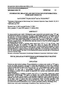
have isolated an HP1-56-like peptide from the medium conditioned by HL-60 cells. Thus HP1-56 can be isolated in the I absence of the cellular material that is the major source of potential proteolytic activity. eo0 The presence of this material in the conditioned medium is of interest. At nanomolar concentrations HP-1 and HP1-56, n a but not HP-4, are inhibitors of [3H]thymidine incorporation P 800 in HL-60 cells.' The apparent secretion of HP1-56 by HL-60 I cells may thus be biologicallysignificant. The virtual absence n I of HP-1 suggests that thismaterial does not arise from granule 300 release or cell lysis. Conditioned medium from peptide-synthesizing cells often contains more of the unprocessed precursor polypeptide than its mature product. It has been shown 0 120 00 20 40 TIME80 (mh) that thisis material that has not enteredthe selection process for translocationinto vesicular organelles but has passed FIG. 10. HP- 1immunoreactivity in human fetal lungs. Nine insteadinto the bulk flow constitutive secretion pathway lungs were used between 16 and 19 weeks gestational age (2 at 16 during segregation in the Golgi apparatus (21). weeks, 1 at 17 weeks, 4 at 18 weeks, and 2 at 19 weeks). The lungs In other peptide biosynthetic systems, cells grown in vitro were pooled and extracted as described under "Materials and Methods" and the extract fractionated on a Vydac (3-18 reversed-phase have not always yieldedthe same peptide end products as the HPLC column eluted at 3 ml/min from 0 to 60% of 80% acetonitrile equivalent cell type in vivo (22). It was therefore important in 0.1% trifluoroacetic acid throughout. to prove that theputative intermediate HP1-56occurs in cells obtained ex uiuo. Initial experiments with human bone marrow provided low amounts of HPl-56 whichcould not be structurally characterized. We therefore examined leukocytes from patients with leukemia and found that some of these extracts contained apeptide that had the same retention time as HP1-56 on reversed-phase and size-exclusion HPLC. This peptide was purified to homogeneity and partially sequenced by gas-phase Edman degradation. The results are consistent with it being identical to HP1-56, demonstrating that the putative precursor can be isolated from cells generated i n uiuo. We have shown that there is likely to be cleavage of the HP-1 precursor at a dibasic site to generate HP-1 immuno0.5 I reactive component I. Cleavageof peptides from their precur0 10 20 30 40 50 AMINO ACID RESIDLE IWIWOW 61 sors at dibasic and less frequently at monobasic residues is FIG. 11. The predicted &turn score for the peptide H P l - 5 6 the common mode of peptide biosynthesis. Experiments using calculated using the parameters of Chou and Fasman (26).A synthetic substrates have shown that the precursor cleavage six-amino acid window was used. enzymes require specific secondary structural elements in 1200
.
w
r
'L4.44 4
7530
HP-1 and HPl-56 in Leukemic Cells
addition to thedibasic site to be optimally active. In particular A smaller fragment can also be detected which is chromatothe dibasic sequence should be close to a @-turn(23). Second- graphically distinct from HP-1 and which may correspond to ary structure calculations of the HP-1 precursor show that cleavage of the precursor at a dibasic sequence. The larger the sequence His-Pro-Gly-Ser immediately preceding the di- HP-1 immunoreactive peptide can be isolated from medium basic site has the highest propensity to form a @-turn in the conditioned with HL-60 cells. HP-1 is not present in this HP-1 precursor sequence. This is consistent with it being a medium. HP1-56and HP-1 therefore belong to different site of peptide precursor cleavage. Fig.11 shows the predicted pools. One possible explanation is that HP1-56 is found as an intermediate in the Golgi apparatus, whereas HP-1 is a fully @-turnplot for the HP-1 prepropeptide. Much interest has recently focused on the presence of neuropeptides in the processed peptide found in the primary granules. HP1-56 is immune system (reviewed in Ref.24). The results above found inthe HL-60 promyelocytic cell line and in some suggest that some peptides that areendogenous to theimmune leukemias but not in mature circulating neutrophils. HL-60 system, such as HP-1, may have processing steps in common cells are known to be actively involved in biosynthesizing other primary granule components such as myeloperoxidase with neuroendocrine peptides. The HP-1 cDNA wasoriginally isolated as a potential stage-(reviewed in Ref. 25), supporting the hypothesis that HP1-56 is a biosynthetic intermediate inthe synthesis of HP-1. Using specific marker for leukemia (13, 14). In the present study HP1-56 was found in all CLL samples examined, although HP1-56 to study granule peptide biosynthesis at the protein the levels varied widely. Elevated levels were found in ap- level is attractive, because HP1-56 caneasily be distinguished proximately 20% of the samples, which is consistent with from the final product, HP-1, andbecause it is readily detectprevious studies on the mRNA levels. Samples from patients able and quantifiable in both simple isolated cell systems such with CML,however, did not conform to our expectations as HL-60 cells and in complex tissue samples such as thefetal based on mRNA studies. CML consists of at least two recog- lung. The otherpolypeptides of the neutrophil primary grannizable phases; an initial chronic phase but which is followed ule are large proteins. In contrast HP-1 and itsbiosynthetic by the stage known as “blast crisis” where cells at multiple intermediates arerelatively simple molecules, and theyshould stages of maturity arefound. This ischaracterized by increas- therefore provide a useful system in which to study the ing numbers of undifferentiated blast cells. Blast crisis is molecular basis of granule protein and peptide biosynthesis. usually fatal. At the cDNA level, cells from the peripheral Acknowledgments-The excellent technical assistance of Nathalie blood of patients in the chronic phase of CML expressed the Croteau and Neola Matusiewicz is gratefully acknowledged. highest levels of HP-1 message(13). The levels of HP-1 REFERENCES message fell in the lymphoblastic crisis of CML and were 1. Zhu, Q., Singh, A. V., Bateman, A., Esch, F., and Solomon, S. (1987) J. essentially absent during the myeloblastic crisis. In contrast Steroid Biochem. 27, 1017-1022 2. Singh, A. V., Bateman, A., Zhu, Q., Shimasaki, S., Esch, F., and Solomon, the ratios of the putative precursor peptide HP1-56 to the S. (1988) Biochem. Biophys. Res. Commun. 155,524-529 mature peptide HP-1 were higher in a myeloblastic crisis 3. Selsted, M. E., Brown, D. M., DeLange, R. J., and Lehrer, R. I. (1983) J. Bid. Chem. 2 5 8 , 14485-14489 CML sample than in thechronic phase of CML. The reason 4. Ganz, T., Selsted,M. E., Szklareg, D., Hanvig, S. S. L., Daher, K., Bainton, for this apparent disassociation in the expression of HP-1 D. F., and Lehrer, R. I. (1985) J. Clin. Inuest. 7 6 , 1427-1434 5. Selsted, M. E., Szklarek, D., and Lehrer, R. I. (1984) Inject. Immun. 4 5 , mRNA and the putative precursor peptide is not yet clear. 150-154 One possible explanation is that the cellular mechanisms for 6. Lehrer, R. I., Ganz, T., Selsted, M. E., Babior, B. M., and Curnette, J. T. (1988) Ann. Intern. Med. 1 0 9 , 127-142 converting the large form of HP-1 into the 30-residue HP-1 7. Lichtenstein, A,, Ganz, T., Selsted, M. E., and Lehrer, R. I. (1986) Blood peptide are not fully developed in blast cells or in the HL-60 6 9 , 1607-1611 8. Kagan, B. L., Selsted, M. E., Ganz, T.,and Lehrer, R. I. (1990) Proc. Natl. cell line, leading to theaccumulation of the larger peptides. A d . Sci. U. S. A. 87, 210-214 The only tissue we have examined in which HP1-56 occurs 9. Zhu, Q., Hu, J., Mulay, S., Esch, F., Shimasaki, S., and Solomon, S. (1988) Proc. Natl. Acad. Scr. U. S. A. 85,592-596 to a greater extent than HP-1 is infetal the lung, demonstrat- 10. Zhu, Q., Bateman, A,, Singh, A. V., and Solomon, S. (1989) Endocr. Res. 15,129-149 ing that HP1-56 can be extracted from both transformed and 11. Territo. M. C.. Ganz. T.. Selsted. M. E.. and Lehrer. R. I. (1989) . , J. Clin. nontransformed cells and tissue. In this connection we have &we.&. 84,2017-2020’ found that adult humanlungs contain much less HP1-56 than 12. Mars, W. M., van Tuinen, P., Drahkin, H., White, J., and Saunders, G. (1988) Blood 71,1713-1719 HP-1 (data not shown) and that the related rabbit peptide 13. Mars, W. M., Florine, D. L., Talpaz, M., and Saunders, G. F. (1985) Blood 6 5 , 1218-1225 CS-I (9) exists predominantly as the fully processed peptide 14. Wiedemann, L. M., Francis, G. E., Lamb, R. F.,Burn,.J. H., Winnie, J. N., in late gestational and post-natal rabbit tissue^.^ At present Mackenzie, E. D., and Birnie, G. D. (1989) Leukemca 3 , 227-234 we do not know if the HP-1 bearing cells of the immature 15. Daher K. A., Lehrer, R. I., Ganz, T., and Kronenberg,M. (1988) Proc. Natl. A c ~Sei. . U. S. A. 8 5 , 7327-7331 lung are derived from bloodcells or not (immunohistochemis- 16. Bennett, H. P. J., Browne, C. A., and Solomon, S. (1981) Biochemistry 20, 4530-4538 try with our anti-HP-1 antisera cannot distinguish between 17. Esch, F. (1984) Anal. Biochem. 126,39-49 HP-1 and HP1-56 in tissue sections); however, the presence 18. Ba-teean, A., Dell, A,, and Morris, H. R. (1985) J. Appl. Biochem. 7 , 126of HP1-56 in fetal tissues, but not in the equivalent adult 19. von15LHeijne, G. (1986) Nucleic Acid Res. 14,4683-4690 tissue, provides the opportunity for future ontogenic studies. 20. Goodall, G. J., Dominguez, F., and Horecker, B. L. (1986) Proc. Natl. Acad. Sci. U. S. A. 83,8926-8928 The structural data presented here allow us to reconstruct 21. Moore, H-P., Gumbiner, B., and Kelly, R. B. (1983) Nature 302,434-436 22. Eipper, B. A,, Clembotski, C. C., and Mains, R. E. (1983) J. Biol. Chem. a possible biosynthetic pathway. HP-1 is synthesized as a942 5 8 , 7292-7298 residue prepropeptide (12, 14, 15). The largest intermediate 23. Cohen, P. (1987) Biochimie 69,87-89 that we have detected in the processing of this precursor is a 24. Bateman, A., Singh, A,, Kral, T., and Solomon, S. (1989) Endocrine Reu. 10,92-112 fragment whose amino acid sequence is identical tothe 25. Collins, S. J. (1987) Blood 7 0 , 1233-1244 Chou, P. Y., and Fasman, G. D. (1974) Biochemistry 13,211-222 COOH-terminal 56 residues of the predicted prepropeptide. 26. 27. Macleod, R. J., Hamilton, J. R., Bateman, A,, Belcourt, D., Hu, J., Bennett, J. Hu and S. Solomon, manuscript in preparation.
H. P. J., and Solomon, S. (1991) Proc. Natl. Acad. Sei. U S A . 88, 552556