The IUPHAR Compendium of Basic Principles For Pharmacological Research in Humans Patrick du Souich, Michael Orme and Sergio Erill, eds. Contributors Bo Abrahamsen, MD, Ph.D. Santiago Arroyo, M.D., Ph.D. Ettore Beghi, M.D. Yoav Ben-Shlomo, MBBS, Ph.D. Donald Birkett, Ph.D. Kim Brixen, M.D., Ph.D. Kim Brøsen, M.D., Ph.D. Rosario Calvo, Ph.D. David W. Chadwick, M.D. Piu Chan, M.D., Ph.D. Lara Chayab Imti Choonara, M.D. Michel Clanet, M.D. Giancarlo Comi, M.D. Per Damkier, M.D., Ph.D. Simon de Denus, B. Pharm, M.Sc. Patrick du Souich, M.D., Ph.D. Anique Ducharme, M.D., M.Sc. Robert Dufour, M.D. Kim Fisher, Ph.D.
Jacqueline French, M.D. Christopher G. Goetz, M.D. Marie-Claude Guertin, Ph.D. Neil Hagen, M.D. Boulos Haraoui, M.D. Anh Ho Ngoc, Ph.D. Moustapha Kassem, M.D., Ph.D. Amos Korczyn, M.D., M.Sc. Teresa Kus, M.D., Ph.D. Maxime Lamarre-Cliche, M.D. Pierre Larochelle, M.D., Ph.D. Nerea Leal, Ph.D. Jacques LeLorier, M.D., Ph.D. Giancarlo Logroscino, M.D., Ph.D. Fred Lublin, M.D. John C. Lukas, M.S., Ph.D. Johanne Martel-Pelletier, Ph.D. Vittorio Martinelli, M.D. Richard Mattson, M.D. Douglas C. McCrory, M.D.
Claudio A. Naranjo, M.D. John Oldenhof, Ph.D. Wendy Parulekar, M.D. Jean-Pierre Pelletier, M.D. Emilio Perucca, M.D., Ph.D. Chris H. Polman, M.D. Jean-Pierre Raynauld, M.D. Monica Rodriguez, Ph.D. Myroslava Romach, M.D. Edward M. Sellers, M.D., Ph.D. Folke Sjöqvist, M.D., Ph.D. Per Solberg Sorensen, M.D. T.J. Steiner, Ph.D. Elena Suarez, Ph.D. Caroline M. Tanner M.D., Ph.D. Jean-Claude Tardif, M.D. Peer Tfelt-Hansen, M.D., Ph.D. Jake J. Thiessen, Ph.D. Bozidar Vrhovac, M.D. Tom Walley, M.D.
Published by IUPHAR Administrative Office Department of Pharmacology College of Medicine University of California, Irvine Irvine, CA 92697-4625 www.iuphar.org Cover design by Lindsay Hart © International Union of Basic and Clinical Pharmacology, 2004 All rights reserved. No part of this publication may be reproduced, stored in a retrieval system, or transmitted in any form or by any means, electronic, mechanical, photocopying, recording or otherwise, without the prior permission of the publishers. ISBN 0-9533510-6-X
1
Patrick du Souich Professor and chairman of the Department of Pharmacology of the Medical School at the University of Montréal, Québec. Vice-chairman of the Clinical Division of IUPHAR.
Sergio Erill Director of the Esteve Foundation, a nonprofit institution aimed at fostering communication among scientists. He has been Professor and Chairman of the Department of Pharmacology at two different Spanish universities, and Visiting Professor at the Université de Montreal. He has been president of the Spanish Society for Pharmacology and served in the Selection Committee for Merck Sharp and Dohme International Fellowships in Clinical Pharmacology.
Michael Orme Emeritus Professor of the University of Liverpool, United Kingdom. He was Professor of Pharmacology and Therapeutics there from 1984 to 2001 during which time he was secretary of the Clinical Section of IUPHAR from 1987 - 1993. He was dean of the faculty of medicine at Liverpool University from 1991 1996. He was honorary secretary of the European Association for Clinical Pharmacology and Therapeutics (EACPT) from 1993 - 2003 and is currently chairman of EACPT.
2
The IUPHAR Compendium of Basic Principles for Pharmacological Research in Humans Introduction ...............................................................................................................................................4 Chapter 1. Ethical Considerations ...............................................................................................................5 Chapter 2. Good Clinical Practice .............................................................................................................11 Chapter 3. Assessment of Endpoints: Kinetics and/or Dynamics .............................................................16 Chapter 4. Pharmacogenetics and Pharmacogenomics .............................................................................27 Chapter 5. Paediatric Drug Research ........................................................................................................34 Chapter 6. Phase I (Human Pharmacology) ..............................................................................................40 Chapter 7. Follow-Up of Drugs After Market Entry .................................................................................50 Chapter 8. Bioavailability and Bioequivalence .........................................................................................55 Chapter 9. Pharmacoeconomics and Economic Evaluation of Drug Therapies........................................67 Chapter 10. Drug Utilization .....................................................................................................................76 Pharmacological Research in Cardiovascular Disorders ....................................................................86 Chapter 11. Hypertensive Vascular Disease .............................................................................................87 Chapter 12. Lipid Lowering Agents........................................................................................................106 Chapter 13. Anti-atherosclerotic Drugs...................................................................................................118 Chapter 14. Heart Failure ........................................................................................................................132 Chapter 15. Arrhythmias .........................................................................................................................143 Pharmacological Research in Neurologic Disorders ..........................................................................164 Chapter 16. Epilepsies and Convulsive Disorders .................................................................................165 Chapter 17. Headache Disorders .............................................................................................................183 Chapter 18. Alzheimer’s Disease and Other Dementias .........................................................................212 Chapter 19. Parkinson’s Disease and Other Extrapyramidal Disorders ..................................................228 Chapter 20. Multiple Sclerosis and Other Demyelinating Diseases........................................................248 Pharmacological Research in Mental Disorders ................................................................................263 Chapter 21. Mood Disorders ...................................................................................................................264 Chapter 22. Anxiety Disorders ...............................................................................................................272 Chapter 23. Schizophrenic Disorders ......................................................................................................278 Chapter 24. Alcoholism and Nicotine Addiction ....................................................................................285 Pharmacological Research in Joint Disorders ....................................................................................293 Chapter 25. Osteoarthritis/arthrosis Short Term Studies.........................................................................294 Chapter 26. Osteoarthritis/arthrosis Long Term Studies .........................................................................308 Chapter 27. Rheumatoid Arthritis ..........................................................................................................318 Pharmacological Research in Other Disorders ..................................................................................337 Chapter 28. Neoplastic Diseases .............................................................................................................338 Chapter 29. Analgesic Drugs for Cancer Pain Management...................................................................352 Chapter 30. Drugs Used in Osteoporosis ................................................................................................366
3
Introduction One of the first recorded human trials was conducted by reverend Edward Stone who found in 50 patients that 1.8 g of powder of willow bark cured their fever, results that were published in 1763 (1). The active compound, salicylic acid, was synthesized only in 1860. Since then, innumerable compounds have been used to cure almost any ailment without evidence of activity. Evidence of drug efficacy was initially required in 1962 with the passing of the Food, Drug and Cosmetic Act by the United States Congress. Currently, in all countries, development and approval of new pharmaceutical entities requires controlled trials proving efficacy. In order to standardize drug registration and approval of drugs, the first International Conference on Harmonization (ICH) was held in 1990. Even if a tremendous progress has been achieved by using ICH guidelines, many aspects of human research remain controversial (2), and even for theoretically rather simple trials, such as those aiming at proving bioequivalence, specifications and study methods differ slightly from one to another in different countries (3). Should we be concerned with refining the methodology of clinical trials? The answer is yes. Let us consider digitalis. William Withering transformed digitalis from a folk remedy to a modern drug when he transformed a "family receipt for dropsy" that contained more than 20 substances, to a single substance by assuming that foxglove was the active ingredient. Clinical observations enabled Withering to recognize the plant’s narrow margin of safety and the importance of dose: just enough foxglove to cause diuresis, but not enough to cause vomiting or very slow pulse. With these observations, Withering introduced foxglove to the medical profession in 1785 (4). Despite many small trials, it took two centuries to clearly demonstrate the benefits of digoxin in heart failure, and we know now that these benefits include reduction of symptoms, improvement in NYHA class, increased exercise time, modest increased in left ventricular ejection force, enhanced cardiac output, and decreased hospitalizations, and that digoxin does not reduce overall mortality but reduces the rate of hospitalization (5,6). How to conduct trials to demonstrate drug efficacy? Despite the fact that guidelines for drug development are rather standardized, there is less information about the design of a clinical trial. The objective of this Compendium is to provide the scientific community interested in human research with an easy-to-use reference on how to design a research protocol to assess the effectiveness of a drug in a series of pathological conditions. The Compendium cannot cover every class of drug and condition, and thus it has primarily focused on cardiovascular and nervous system drugs. The section dealing specifically with the design of clinical trials, chapters 11 to 30, is presented according to a common template to facilitate its consultation. This section is preceded by shorter chapters dealing with general concepts that are applied to the development of almost any drug. The Compendium does not intend to constitute a guideline, but rather an easy source of information on how to design and conduct a clinical trial aiming at demonstrate drug efficacy. The Editors 1. Stone E. An account of the success of the bark of the willow in the cure of agues. Philosophical Transactions of the Royal Society 1763;53:195-200. 2. Rockhold FW. Industry perspectives on ICH guidelines. Stat Med 2002;21:2949-2957. 3. Nakai K, Fujita M, Ogata H. International harmonization of bioequivalence studies and issues shared in common. Yakugaku Zasshi 2000;120:1193-1200. 4. Withering W. An Account of the Foxglove and Some of Its Medical Uses. Birmingham. United Kingdom: M. Swinney; 1785:2. 5. Tauke J, Goldstein S, Gheorghiade M. Digoxin for chronic heart failure: a review of the randomized controlled trials with special attention to the PROVED and RADIANCE trials. Prog Cardiovasc Dis 1994;37: 49–58. 6. Digitalis Investigation Group, The effect of digoxin on mortality and morbidity in patients with heart failure. N Engl J Med 1997;336:525–533.
4
Chapter 1. Ethical Considerations
Bozidar Vrhovac, M.D. Professor Emeritus Medical School Zagreb Zagreb CROATIA and Co-chairman National Bioethics Committee for Medicine of Croatia and Corresponding Member Croatian Academy of Arts and Sciences
5
I. INTRODUCTORY REMARKS One of many characteristics of modern society is a pronounced interest in ethical questions. Medicine, especially research on humans, is expectedly, at the top of the list. Why is that so? In spite of the fact that many patients receive therapeutic benefits from participating in clinical trials, benefits that may even be greater than those of standard medical care, randomized clinical trials differ from standard medical treatment in their purpose, characteristics, justification of risks and allocations of interventions according to chance. The research based on various interventions potentially poses risks to the participants that are not always compensated for by medical benefits but that are justified by the potential scientific value of the knowledge which will be got from the trial. The history of international instruments on ethics is not long. Already before World War II, use of controlled clinical trials was proposed and accepted as the scientific, reliable way of proving efficacy and safety of new therapeutic agents. The atrocious experiments performed by nazi physicians during World War II led, almost immediately (1947) after the war, to the preparation of the Nuremberg Code on ethics of medical research. The Helsinki Declaration followed in 1964 and is now (sixth revision in 2000) taken as the gold standard for research ethics, intending to provide a universal set of principles, which direct the ethical conduct of clinical medical research involving human subjects throughout the world. This is still true in spite of several weaknesses which are at the moment of writing these lines intensively discussed. Other instruments must be mentioned, such as the UN General Assembly Universal Declaration of Human Rights in 1948 and the International Covenant on Civil and Political Rights in 1966. The Belmont Report, elaborated in the US in 1979, is in this country very important for developing new drugs. The Belmont report is, in the US, almost better known than the Helsinki Declaration, and, with its legislative revisions performed later, it is still a very comprehensive instrument. Recent and very important instruments are the documents issued by the 1990 founded International Conferences of Harmonization (ICH) founded in 1990. Originally the basic aim of ICH was to harmonize the requirements for new drugs in the three biggest drug developers, namely US, European Union and Japan. Later, its guidelines and consensus documents have been accepted by the «rest of the world» most probably because of the importance of the pharmaceutical markets in these countries rather than because of other less materialistic reasons. The first principle of ICH (taken from WHO GCP in 1995) states: ”Clinical trials should be conducted in accordance with the ethical principles that have the origin in the Declaration of Helsinki, and that are consistent with Good Clinical Practice (GCP) and the applicable regulatory requirement(s)”. The booklet prepared by the Council of International Organizations of Medical Sciences (CIOMS) in 1982, in 1993 and in 2002 is today the most informative and comprehensive source of information for ethical research, trying to correct the inconsistencies of the latest revisions of the Helsinki Declaration. These inconsistencies perhaps explain why »studies that breach the provisions of the Helsinki Declaration are still commonly conducted, with the full knowledge of regulatory agencies and institutional review boards». Of the realistic and justified aspects of the Helsinki Declaration, the most important is the respect for the person’s rights e.g. personhood of subjects, followed by investigators beneficence for subjects participating in the trial and distributive justice in distribution of risk and benefit associated with medical research. The growing importance of persons rights (patients and healthy volunteers) is illustrated by the special attention given to trials in vulnerable and socially unprivileged patients.
6
Issues of conflict of interest, of transparency and of publishing negative trials are closely linked with ethics as well. II. THE ETHICS COMMITTEE The guarantee for the ethical conduct of the study should be a multidisciplinary ethical body called in various countries the Ethics committee, institutional review board, independent ethical committee (EC/IRB). Its size is according to many documents of “at least five” members. This important detail is not mentioned in the Helsinki Declaration. With a small number of members it cannot be expected that an institutional review board will be independent when it decides about resources brought by the sponsor to the institution and its investigators. The number of members must be large enough to ensure that besides the layman, the nurse, the ethicist, and the statistician (who are often named as useful non-scientific members of EC/IRB), at least some members must be experts in the medical and scientific aspects of the clinical trial. Scientific and ethical review cannot be separated. How can someone discuss the ethics of a clinical trial without knowing in detail all facts about the disease in question and its standard treatment? Only medically and scientifically competent members of the ethical committee can safeguard the rights, safety and well -being of the research subjects. Is it optimal that the same ethics committee, and this is often the case, evaluates research projects and other relevant ethical questions which are constantly present in a health institution, such as artificial prolongation of life of irreversibly sick patients, abortions, unethical behaviour of medical staff, to name only a few. The EC/IRB should be an independent body, either regional or (for smaller countries) central-national and should discuss research projects only. Such development goes in the described direction and many (even bigger) countries already have central ethics research committees or institutions (US Office for Human Research Protection, U.K. Central Office for Research Ethics Committees, Canada National Council on Ethics in Human Research). In the EU for multicentre trials one member country must give one opinion. This is achieved in various ways one of them being that the central committee delegates the decision to a regional one. II.1. Informed Consent Document (ICD) How does the EC/IRB functions and what are the foci of their activity? The most important ethical aspect of the clinical trial is the Informed Consent Document (ICD). The already mentioned International Convenant accepted by the United Nations Assembly in 1966 stresses that « no one shall be subjected without his free consent to medical or scientific experimentation ». A number of documents, meetings and discussions have been written and organized about the optimal format of this important ethical aspect of the clinical trial documents. It is of the utmost importance that the subject participating in a scientific research project, for instance a clinical trial, understands all details of the planned experiment. To reach this aim the investigator must ensure that the prospective subject has got all the necessary information on the basis of which he reaches at the decision to take part in the trial without having been subjected to coercion, undue influence or inducement, or intimidation. The ICD should contain a statement indicating that the study involves research, should describe the purpose of the research, the expected duration of the subject’s participation, should contain the description of the procedures to be followed (with the indication of which are experimental), of the study treatments, and, if applicable, the nature of random assignment. Moreover the ICD should contain a description of the foreseeable risks or discomforts, of the benefits that may be expected for the subject or for others and disclosure of appropriate alternative procedures or courses of treatment, if any, that might be advantageous to the subject. In addition the ICD will include a statement
7
describing the extent to which confidentiality and privacy (of records identifying the subject) will be maintained, a statement that participation is voluntary and that the refusal to participate involves no penalty or loss of benefits. It must describe the actions foreseen in the case of injuries (compensation, medical treatment), explanation on whom to contact for additional questions, and, if applicable, any other necessary detail which ensures complete comprehension of above mentioned points by the participant. Whenever it is not possible to obtain the written ICD, the non-written consent should be documented and witnessed. The problems of obtaining a written ICD for trials in patients with decisional (cognitive) impairment such as patients who are mentally ill, those with Alzheimer’s disease, acutely ill subjects (head trauma, cardiopulmonary arrest, and stroke) or children (no uniform criteria for assent and dissent exist) are considerable. In these cases a legally authorised representative, a proxy or an advanced ICD (in the case of anaesthesia for example) should be used. In addition, financial details of the trial should be disclosed. Transparency of financial arrangements encourages people to do the right thing. The individual investigator should not stand to benefit personally in financial terms from their involvement in the study. II.2. Analysis of Details Another function of the EC/IRB is to conduct a careful analysis of all details, which could influence the reliability of the trial results. The new sentence in the latest revision of the Helsinki declaration must be mentioned here (articles 19 and 20): “Medical research is only justified if there is a reasonable likelihood that populations in which research is carried out stand to benefit from the results of the research”. The analysis should begin with the investigator and their team, their potential to recruit subjects without aggressive behaviour, the likelihood of a conflict of interest when trying to serve both the best interests of the patients and the best interests of the research. The analysis should also consider the remuneration and other advantages for both the investigator and their institution. The patient selection, the planned measurements (frequency, justified invasiveness), concomitant and rescue therapy, monitoring of adverse events (especially those which will indicate the need to stop the trial) and comparator therapy must be analyzed. II.3. Placebo The function of the EC/IRB is to analyze the need to use placebo. The use of placebo has been considered by many as non-realistic and unjustified. The most controversial item of the last two revisions of the Helsinki declaration (article 29) states: “The benefits, risks, burdens and effectiveness of a new method should be tested against those of the best current prophylactic, diagnostic and therapeutic methods. This does not exclude the use of placebo, or no treatment, in studies where no proven prophylactic, diagnostic or therapeutic method exists”. Strictly interpreted, this article would rule out the use of placebo controlled clinical trials e.g. a dummy treatment administered to the control group in a controlled clinical trial in order that the specific and non-specific effects of the experimental treatment be distinguished whenever licensed therapeutic method already exists. Active controls (which the investigators are keen for) cannot, in many circumstances, provide reliable evidence of efficacy and safety of the new drug, except by showing the non-inferiority of the new drug. There are many groups of therapeutic agents where placebo controls are justified or even mandatory: analgesics, many psychopharmacologicals, antihypertensives, antianginals, antiarrhythmics and drugs used in primary prevention to name only a few. It is essential that the use of placebo does not pose a risk of serious discomfort, irreversible harm or death or that existing therapy improves survival or decreases serious morbidity.
8
The sixth revised version of the Helsinki Declaration raised a number of discussions, many more than after the fifth version in which the same proposal was already present, with the result that the World Medical Association prepared a special footnote which states: “The WMA hereby reaffirms its position that extreme care must be taken in making use of placebo-controlled trials and that in general this methodology should only be used in the absence of existing proven therapy. However placebo-controlled trials may be ethically acceptable, even when proven therapy is available, under the following circumstances: - where for compelling and scientifically sound methodological reasons its use is necessary to determine the efficacy and safety of prophylactic, diagnostic or therapeutic method; or - where a prophylactic, diagnostic or therapeutic method is being investigated for a minor condition and the patients who receive placebo will not be subject to any additional risk of serious irreversible harm”. Beside WMA, many meetings in various international organizations like the European Medicines Evaluation Agency (EMEA), the Food and Drug Administration (FDA), the Pharmaceutical Research and Manufacturers of America (PhRMA), the International Federation of Pharmaceutical Manufacturers Associations (IFPMA) and the European Forum for Good Clinical Practice (EFGCP) have discussed the need for a new revision of the Helsinki declaration because of the presence of other conflicting articles. For example, article 30 states that “every patient should be assured of access to the best proven method identified by the study”. This is not acceptable because of the widely known fact that one study cannot identify “the best proven method”. Therefore, a new version of the Declaration of Helsinki is to be expected. In September 2003 the General Assembly of WMA founded a Working group (its deadline is May 2004) with this goal. II.4. Payment Payment in the form of money, gifts, and privileges can only be offered as a recruitment incentive not as a benefit for participation. In clinical trials the prospects of benefit from an experimental treatment and the provision of free ancillary care are viewed as compensation for participation. Healthy volunteers do not need treatment and care. So payment is justified as an incentive for participation. There are many other ethically sensible areas of clinical research. The examples are, beside those already mentioned, the need to withhold treatment, wash out periods, research involving foetuses and in vitro fertilization, involving pregnant women, children, college students and prisoners. In each of these cases the local EC/IRB has to adapt its decisions according to the local legislation, uses and environment. In conclusion, clinical trials have numerous ethical aspects. A scientifically and medically well planed clinical trial is ethical and represents the only way for obtaining reliable results which will help in better treatment of a wide circle of patients. Local EC/IRBs have to define what is methodologically essential and ethically appropriate and these aspects are still the subject of intense debate. Ethical committees structured as proposed in this chapter guarantee that ethical principles, accepted to day as appropriate, are observed. III. SUGGESTED READINGS 1. 2. 3.
Council of International Organizations of Medical Sciences, International Ethical Guidelines for Biomedical Research Involving Human Subjects, Geneva 2002. Amdur RJ, Bankert EA, Institutional Review Board, Management and Function, Jones and Bartlett Pubs, Boston 2002. Vrhovac B, Placebo and Helsinki Declaration, What to Do? Sci Engeen Ethics ( in press 2003).
9
4. 5. 6. 7. 8. 9. 10. 11. 12. 13.
Miller, FG, Rosenstein DL, The Therapeutic Orientation to Clinical Trials, N Engl J Med. 2003;348:1383-86. World Medical Association Declaration of Helsinki: Ethical principles for Medical Research Involving Human Subjects, Edinburgh Scotland, World Medical Association, October 2000: Accessed Aug 10,2003 at http://www.wma.net/e/policy/b3.htm Diamant JC, The revised Declaration of Helsinki-Is Justice Served? Int J Clin Pharmacol Ther 2002;40:76-83. European Union’s Clinical Trials Directive, Accessed Aug 10, 2003 at http://www.barnettinternational.com/RSC_PDFUploads/nda%20eu%20toc.rtb Good Clinical Practice Consolidated Guideline, Accessed Aug 10, 2003 at http://www.hcsc.gc.ca/hpfb-dgpsa/tpd-dpt/goodclin_main_e.html European Forum for Good Clinical Practice, European Guidelines for Auditing Independent Ethics Committees, Geneva 2002. World Health Organization (TDR/CDS/WHO) Operational Guidelines for Ethics Committees That Review Biomedical Research, WHO, Geneva 2000. The European Agency for Evaluation of Medicinal Products: EMEA/CPMP, Position Statement on the USE of Placebo in Clinical Trials with regard to the revised Declaration of Helsinki, EMEA/17424/01. International Conference on Harmonization (ICH) Good Clinical Practice Guidelines Accessed Aug 10, 2003 at http://www.ifpma.org/ich5e.html National Council on ethics in human research-Canada Accessed Aug 19, 2003 at http://www.ncehrcnerh.org/english/mstr_frm.html
10
Chapter 2. Good Clinical Practice
Per Damkier, M.D., Ph.D. Clinical Pharmacology Institute of Public Health University of Southern Denmark Odense University Winslewparken 19 5000 Odense C DENMARK
11
I. INTRODUCTORY REMARKS GCP, Good Clinical Practice, is an international set of ethical and quality standards that applies to medicinal trials in humans. Compliance to these standards provides the health authorities as well as the general population with assurance of the integrity of trial subjects and the validity of the data generated. It is important to stress that these standards de facto relate to research; in fact a more appropriate title would be Good Clinical Research Practice. These standards have been in effect for many years pertaining to all trials intended to generate data for marketing authorisation procedures whether it be new medicinal products or a line extension for an already marketed product. As a general rule, academic research involving already marketed products and not intended to generate results for marketing authorization purposes has been exempt from these rules. As more general attention and political focus is given to quality assurance and to the autonomy of the individual within health care systems, these standards are getting more attention world-wide; Within the EU, these rules, as of May 1st 2004, pertain to any medicinal trials in humans, including those involving already marketed drugs and trials performed without industry engagement. This represents a serious challenge to the academic independent drug related research, as systems to assure GCP compliance must be developed, which in turn requires allocation of appropriate resources. I.1 History The first documents describing some quality recommendations for the design of clinical trials in humans can be dated back to USA, where Harry Gold from Cornell University Medical School published two influential papers Conference on Therapy in 1946 and again in 1954. The emergence of the thalidomide disaster around 1960 further served to justify the need for formal guidelines and procedures for clinical trials with new medicinal products. Throughout the 60’s and 70’s guidelines were refined and implemented throughout the world. These were, unfortunately, far from being easily comparable due to differences in approaches to clinical trials, mainly between USA, Japan and Europe. It became obvious that some sort of international consensus on this issue was overdue in order to promote mutual recognition of clinical trials and marketing authorization procedures. The result was the birth of the current guidelines that in effect are known as ICH (International Conference on Harmonization)-GCP. I.2. GCP concepts In order to give a better understanding of the principal GCP concepts, some key definitions are given below: Sponsor A person, institution, company or organization which takes responsibility for the initiation, management and/or financing of a clinical trial. Note that the sponsor does not necessarily finance the study. Sponsor and investigator may be identical in which case the term (surprise) sponsor-investigator is used. Investigator A person who is responsible for the trial conduct at the trial site. If multiple investigators are involved at a trial site, a principal investigator must me appointed. Monitoring The act of overseeing a clinical trial to ensure that it is conducted recorded and analysed according to the trial study plan, standard operating procedures, GCP and regulatory requirements. SOP, Standard Operating Procedure A set of written detailed instructions to achieve uniformity of the performance of a given function.
12
Audit An independent examination of all trial related activities and documents in order to determine if the trial was conducted, recorded and analysed according to the trial study plan, standard operating procedures GCP and regulatory requirements. The audit procedure is independent of the monitoring procedure. II. GCP IN CLINICAL RESEARCH The ICH-GCP guideline specifies, in rather general terms, how to design, conduct, record and report a clinical trial in accordance with GCP standards. In order to comply with this, it is the responsibility of the sponsor, or sponsor-investigator, to ensure that a set of standard operating procedures is written. An important point is that the guideline explicitly states that the level and intensity of the GCP monitoring process is specified by the sponsor according to the complexity of the study: size, purpose, design, blinding, outcome measures etc. These standard operating procedures form the basis of the central element of GCP-compliance: the monitoring process. The monitoring process must be conducted by individuals possessing documented skills of GCP monitoring, and, obviously, the monitors cannot be directly involved in the study otherwise. II.1. The monitoring process The monitoring process is the fundamental aspect of GCP compliance. This systematic process is based on study-specific SOP’s, and serves to document that the study complies with GCP standards. There are three phases of this process some core issues of which are listed below. Please note that this is not an exhaustive listing. Before study initiation the monitor visits each investigation site to verify that • Sponsor and investigator responsibilities are properly described • Relevant authorizations are present • Written informed consent is acceptable • The investigation site realistically can provide the specified number of subjects within the specified time-frame • Procedures for handling of trial medicine and laboratory tests are present • Source documents are specified During the trial, the monitor visits the trial sites to verify that • The trial is performed and documented as planned • Informed consent is given from every participant • In- and exclusion criteria are fulfilled • Data are correctly recorded and are in accordance with the specified source documents • Corrections in the case report form (CRF) are properly performed and documented • Serious adverse event are handled correctly After the trial is completed the monitor verifies that • Trial medicine is accounted for in detail • The trial database is properly secured and validated • Trial documents are filed and stored properly In effects this means that quite a number of SOP’s must be developed and maintained. A standard operating procedure must exist for all items and procedures below: • Protocol • Informed consent • Investigators brochure
13
• • • • • • • • • • •
Case Report Form (CRF) Trial medication Adverse events Protocol amendments Monitoring Monitor’s report Monitor qualification Filing Audit Handling of documents Structure and approval of SOP’s
All monitor’s visits must be accompanied by a written report describing what was monitored, the outcome of the monitoring process, including errors and deviations, and initiatives to correct the latter. For the individual researcher this likely represents an insurmountable task and flexible systems to handle GCP in independent academic drug research must be developed. II.2. A Danish example In Denmark a public GCP monitoring service has been organised, and a brief overview is given here for inspiration: The first public GCP initiative was taken back in 1995 at the University of Aarhus. Anticipating the implementation of the aforementioned EU directive, it was estimated that about 80 independent academic drug trials in humans were initiated every year in Denmark. A coordinated activity and initiative has resulted in the presence of three GCP-units in Denmark, which are all situated around medical schools and universities. The GCP-units are partly funded by the Government; we are estimating that, having reached steady-state, about 20 full time monitors will be employed, and that a total budget would linger around USD 2 million. The three units work closely together and have agreed upon identical SOP systems, and apply identical principles of services: Smaller trials, typically trials in Ph.D. projects, are monitored free of charge, while larger trials must account for factual GCP-costs, if more than 100 hours of service is required. Despite initial skepticism from researchers this system has so far proven manageable, but the full scale test still waits, at the time of writing, the implementation of the EU directive. II.3. Challenges and perspectives for independent academic drug research The challenge is to assure compliance with GCP standards, while not consuming an insurmountable amount of resources, which would cripple the independent academic clinical drug research. It is here that one must recall that the guidelines are general specifications of GCP standards. However, the translation into factual procedures, and the specific way these are implemented leave some room for breathing and interpretation. The approach that the pharmaceutical industry and most CRO organisations have taken is meticulously detailed and very resource consuming. This is understandable, as they cannot afford to have a large pivotal phase III trial be subjected to scrutiny, or even rejection, by health authorities due to a GCP related issue. Hence, the industry developed GCP concept is designed to account for a worst case scenario. So as GCP seems to enter an era were the principle is likely to become applicable to any drug related research in humans, it is really up to all of us, as independent academic clinical researchers, to influence the way that GCP is implemented. There is no denying that implementation of GCP in academic research programs will assimilate some of our scarce funding. However, it is the opinion of this author, that we should welcome many of the principles covered by GCP. Quality in performance and respect for patient’s integrity is imperative for the thrust on which the very existence of any health care system relies, and there are simply no valid arguments to exclude research related activities from these principles. And, if we make our opinion and experience heard it is my belief that it is possible to reach an interpretation of the
14
GCP guidelines which satisfies this principle and still allows for a fruitful continuation of independent drug research. III. REFERENCES The current ICH-GCP guideline can be viewed (among many sites) at the European Medicines Agency’s homepage: www.emea.eu.int/pdfs/human/ich/013595en.pdf. The FDA has an entire homepage with plenty of easily accessible information: http://www.fda.gov/oc/ gcp/default.htm.
15
Chapter 3. Assessment of Endpoints: Kinetics and/or Dynamics
Rosario Calvo, Ph.D.1 John C Lukas, M.S., Ph.D. 2 Elena Suarez, Ph.D. 3 Monica Rodriguez, Ph.D. 3 Nerea Leal, Ph.D. 3
1
Department of Pharmacology University of the Basque Country Leioa, Vizcaya 48940 SPAIN Tel + 34 94 6012761 Fax + 34 94 4800128 Email
[email protected]
2
Department of Pharmacy and Pharmaceutical Technology University of Salamanca Salamanca 37001 SPAIN
3
Department of Pharmacology University of the Basque Country Leioa, Vizcaya 48940 SPAIN
Corresponding Author: Rosario Calvo, Ph.D.
16
I. INTRODUCTORY REMARKS A crucial, yet often limiting, factor in the advance of the majority of sciences is the ability to measure and analyze those variables that are truly relevant to a particular field, as for example, in pharmaceutical science, the concentrations in plasma and the effect of a drug. These measurements would be of little use were it not for the development and progress of collateral disciplines such as pharmacokinetics (PK) or pharmacodynamics (PD). The PK examines the relation between the dose administered and the achieved concentration in blood or at the biophase, or “what the body does to the drug”, and the PD deals with the relation between drug amount and effect, or “what the drug does to the body”. In its turn, progress in PK or PD is due mostly to the availability of sensitive and specific analytical methods in order to determine the evolution of the levels of drug and its metabolites in biological fluids and tissues as well as to quantify the drug´s therapeutic effect. In parallel, the mathematical techniques and methods used in pharmaceutical science to characterize the kinetic processes have also developed considerably, mostly borrowing from other disciplines such as chemistry, physiology or enzymology. The application of these analytical and mathematical techniques to specific studies has enhanced the knowledge of the PK behavior of many drugs, constituting an important advance in adjustment of the dose. But, unpredictability remains a problem since the kinetics usually show large interindividual variation, mainly due to genetic, environmental and pathophysiological factors. For this purpose, state of the art statistical methods generally denominated as “population” PK/PD models have been introduced in the field aiming to evaluate the between (inter) and within (intra) individual differences in the PK/PD in medicated populations of subjects. The dose is related to the effect through the PK and also through the PD which relates the concentration with the effect. Comprehension of these relations is truly essential for the rational development of therapeutics because the PD determines the target concentration required to produce a specific effect or endpoint, while the PK specifies the appropriate dose regimen to reach that target. This constitutes the basis of what in therapeutics is called “Target Concentration Intervention” (TCI), in contrast to the therapeutic drug monitoring (TDM) methods based solely on a single concentration and Bayes informed PK. As in the case of PK, methods have also been developed to assess the pharmacological effect in vivo and provide biomathematical model descriptions of the PD. It is already well known that the interindividual differences in PD are numerous and are related to factors such as age, race or disease. Therefore, future efforts must lead to the exploration of covariate models for predicting individual PD parameters. This knowledge together with its PK counterpart could constitute the basis for rational individualized therapy. The combination of PK and PD models permits predicting the temporal evolution of the effect at any dose or regimen. Also, PK/PD analysis of concentration – time – effect data assists in detecting a series of underlying complications. For example, tolerance development, the formation of active metabolites or the desynchronization between the evolution of the drug concentration and that of the effect. The latter can be due either to drug distributing into an effect compartment or because of an indirect effect mechanism. In all these conditions the plasma concentrations could not be used directly as “targets” (TDM) but integrated PK/PD models would permit estimation of the drug concentration at the effect site as well as the relationship between them. II. OBJECTIVE The main objective of therapy is to achieve maximum efficacy avoiding the risk of toxicity. This can be achieved via empirical adjustment of the dose, based either on observations of effects (effect – time
17
evolution) and/or concentrations (concentration – time), or optimally based on knowledge of the pharmacokinetic (PK) and pharmacodynamic (PD) parameters. II.1. Effect – time evolution The measurement of the pharmacological effect and the adjustment of the dose as a function of the effect appears, in principle, as the most sensible and intuitive approach. However, there are several problems. The observed pharmacological effect, as well as the time it takes to achieve it and its duration, are measurements that change with the dose and the mode of administration, therefore they are variables. This implies that they cannot be used to make predictions in other situations, different from those of the observations at hand. Additionally, from the knowledge of the effect vs time evolution there can be no direct deduction of parameters, thus having to employ integrated PK/PD analyses as will be discussed below. Parameters are considered those characteristics of the drug that do not change with time, dose or administration route and therefore can be used to adjust or predict adequately any therapeutic regimen. For example, the classical dose - effect relationship provides a useful estimate, that of the dose producing 50% of the maximal effect, ED50. Yet, this is not a true parameter since it depends on the time post-dose when the effect was measured. Dealing with the pharmacological effect is complicated further because of the difficulty to obtain a precise, objective, and continuous measurement of the effect. The use of biomarkers attempts to relieve this problem although we are still far from having the ideal (and validated) biomarker for the majority of drugs. The use of biomarkers in drug development and clinical practice was revised in the Ninth European Federation of Pharmaceutical Science Conference on Optimizing Drug Development held in Basel 2001 (1). The terminology has been put up to date and the differences between the terms “end point”, “surrogate endpoint” and “biomarker” clarified. Biomarkers are now defined as “physical signs or laboratory measurements that may be detected in association with a pathologic process and that have putative diagnostic and or prognostic utility”. They are, therefore, factors which can be measured objectively and are evaluated as indicators of biological or pathological processes and/or indicators of the response to a therapeutic intervention. In general, biomarkers have a much wider range than surrogate endpoints. We understand as endpoints those variables which can be used to measure how a patient feels after a specific treatment or how a specific body function evolves, “the clinical impact of therapeutic intervention”. The term surrogate endpoint implies that some variables related to the endpoint or the clinical response have been used as biomarkers but are not the final response of the drug (2,3). For example, blood pressure is a biomarker for prevention of hypertension and, at the same time, a surrogate endpoint for the prevention of myocardial infarction and stroke. The measurement of the degree or percentage of prevention would be the endpoint. This example serves also to demonstrate the difficulty in selecting adequate biomarkers since it is still discussed whether the systolic, diastolic or mean blood pressure is the one of interest. The most widely used biomarkers are plasma concentrations of drugs that are used as guides to dosage in clinical practice (e.g. TDM). Biochemical and molecular biomarkers, such as leukotrienes, angiotensin I and II, or CD4 cell count are of great utility but are hampered by complexity in their mechanism of action which is widely interconnected to other processes. Consequently, no single such biomarker can predict a significant proportion of the observed clinical endpoint. For example, CD4 cell count explains 30% of the survival to HIV and CD4 count plus viral load explains 70%. Something similar occurs with the gene biomarker products which are also under rapid development. Genes and their function are identified in the genome. Then the proteome is used to identify proteins from
18
selected genes. Evaluating how mutations cause disease as a result of protein differences, between healthy and diseased subjects, appears to provide candidate gene biomarkers. However, the complexity of the genome or of the pathway from expression to phenotype to macroscopic reality has deflated initial hope. Adversities aside, the development of adequate biomarkers for a drug apart from better characterization of its PK/PD for analysis or prediction, now appears crucial for the drug development effort. Valid biomarkers help completing the proof-of-concept in the early phases, facilitating decision on continuation with the new drug. Eventually, biomarkers permit reduction of the number of patients in later phases (II or III) and adjustment of the dose in specific populations or in individuals receiving the drug through different routes or dose regimen. Recall however, that even when the ideal biomarker is known, it is necessary to have the PK/PD parameters permitting to make predictions. II.2. Concentration – time evolution (PK) The observations of plasma or blood concentration are also non generalizable in their pure form. The maximum concentration reached after a specific dose or the peak or trough after repeated dosing, are measurements useful exclusively for adjusting the dose in situations reproducing the one where they were obtained (body – drug whole). Nevertheless, the measurement of the concentration evolution with time is advantageous, compared to the evolution of the effect, since from this kinetics the PK parameters can be derived that are of great use in adjustment of the dose. Pharmacokinetics has advanced considerably with the use of mathematical models and computer packages which aid in the analysis and processing of the information generated in clinical practice. The PK also permits the simulation of conditions affecting a particular patient. These models and accessory packages are tools which assist in obtaining parameters but add no scientific surplus value to the information than that input by the experiment, including the user. Modelling will simply reflect the knowledge of the physicochemical characteristics of the drug, the precision of the analytical techniques for drug assays. With more complex models, the qualifications of the user also become more important. The basic or primary PK parameters are apparent volume of distribution (V) and clearance (CL). V is defined as the relation between dose and initial concentration (V = Dose/ Co). CL is a relation between elimination rate (or distribution rate, for intercompartmental clearances) and concentration in blood or plasma. With passive phenomena (or first order kinetics), which are the most usual in the body, a larger dose implies a larger concentration in blood or plasma and consequently larger elimination rate, thus V and CL are always constant. They are therefore considered parameters and permit prediction of the dose which would be necessary in a patient to produce a specific effective concentration level. For example, knowing V we could predict the dose needed to reach a specific target (loading dose = target concentration x V). Additionally, CL is a parameter independent of the complexity of the kinetic model (mono, bi or tricompartmental) and is calculated simply as Dose / AUC (assuming complete absorption of the drug). In steady state, after an infusion or multiple dosing, CL is related to the steady state concentration (Css) or average Css (Css = infusion rate / CL), so this parameter can be used to predict the dose regimen (maintenance dose = target Css x CL). Both parameters, CL and V, are primary parameters and are directly related to the physiological processes of the organism. They give us an idea of the relative importance of the space where the drug is distributed and of the organs which eliminate it. Another PK parameter, commonly employed in dose adjustment, is the elimination rate constant (Kel). It is a mixed parameter that depends on CL and V (Kel = CL / V) and is often recast in the form of the halflife parameter as t1/2 = ln (2) / Kel, now with units of time. This parameter gives an idea of when the steady state is reached and can be used to predict when a Css monitoring sample can be taken.
19
A facet which should not be neglected when performing a kinetic study or using the concentrations as markers is to know a priori what needs to be finally estimated from the observations. The parameters obtained depend on what has been measured, for example, metabolites, unbound or total drug, enantiomers or racemic mixture. Another frequently encountered issue is the necessity or not to measure unbound drug (not bound to plasma proteins). It is important to recall that on some occasions the unbound concentration in plasma should be considered since binding in plasma and tissues (the effect site) may not be equal. Nevertheless, if the binding is linear in the range of therapeutic or toxic concentrations, the free and total concentration are simple ratios one of the other and in these cases it is not necessary to measure the free concentration. In contrast, nonlinear plasma protein binding (free drug concentrations increase disproportionately with increasing total drug concentrations) can create havoc in analysis unless free drug concentrations are measured. Perhaps an advantage of parameter estimation as a function of free concentration is that it supplies information about the (intrinsic) behavior of the drug excluding possible differences in the degree of binding. For example, the V of unbound drug, Vu, corrected for the weight, can be extrapolated from animal to man permitting estimation of the first dose in humans. An approximate value of CL for the unbound drug, Clu, can be obtained from “in vitro” studies with microsomes in the initial stages of development. In conclusion, V and CL are fundamental parameters for establishing a dosing regimen, but, depending on the physicochemical characteristics of the drug, the models for the distribution can become complicated and the number of parameters may increase, particularly there may now be more than one volume of distribution. In this case, the volume used to adjust the therapeutic dose, should be the one closer to the effect site (and could also be the steady state volume, Vss) (see Propofol example below). It is important to remember that the basic concepts reflected in V and CL are always applicable independently of the complexity of the complete model. II.3. Programs for data analysis Depending on the experiment, the intentions may range from obtaining estimates of PK (PD) model parameters in a single subject or the mean for multiple subjects (a population), or up to the rigorous resolution of the inter and intra subject variability in a population as reflected into statistical distributions of the parameters (Bayesian priors). A subsequent, yet very important task, is usually that of relating the parameters, or their inter subject variability with individual specific covariates, so that a priori prediction of the individual PK (PD) characteristics, and hence the dose, can be improved. Several software packages have been designed and marketed, mainly in the last 20 years and can be distinguished as single subject analysis programs, or population analysis programs, although both can be used, with the exceptions of some occasions, to perform single or multiple subject analyses. The decision about which approach to use depends, in order of significance, on the density of the observations (rich or sparse data), the scope of the study (e.g. obtaining estimates for a single subject only, or Bayesian priors for a population), and ad hoc criteria regarding modelling. Rich sampling is when there are several drug concentration observations per individual, necessarily more than or equal to the parameters in the model and well distributed in time (e.g. phase I). Sparse sampling is when there are fewer data points per subject than parameters in the PK model to be estimated. Sparse data occur frequently, e.g. in the clinic, due to monitoring restrictions or in phases II, III or IV, due to logistical or ethical considerations. It is also frequent in dose escalation experiments with drug levels below the quantification limit, particularly in small animals, and also in toxicological single point per animal studies.
20
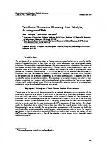
With rich sampling, individual fitting programs can always be used in single or multiple subject problems, in addition to population specific packages for the latter case (Fig. 1). In sparsely sampled designs, some knowledge about the population at large is always required for estimation of the individual PK model. In single subject sparse PK model estimates, a Bayesian prior from the population has to be introduced at some point in order to inform or support the algorithm on the data gaps. Single subject PK/PD modelling packages are WINNONLIN (Pharsight Corp., Mountain View, CA) and SAAM (Saam Institute Inc., Seattle, WA), this latter permitting the introduction of Bayesian priors from the literature or earlier studies when the data is sparse (e.g. a monitored concentration for each subject). When many individuals are treated as one within a single subject fit, the analysis performed is known as naive and produces estimates of the mean parameter without any indication of statistical spread. If that information is desired, with rich data and for more than one individual the single subject model fitting run can be simply repeated or iterated for each case. An improvement is a population – like analysis known as standard two stage (STS), because in the first stage all subject specific PK or PD parameters are obtained and in the latter, their centering and dispersion (mean, standard deviation) are simply calculated. With STS, relationships with covariates (e.g. age, weight, sex, and creatinine) can be assessed with standard commercial statistical analysis packages (SPSS, SAS, S-PLUS etc). Figure 1. Schematic of decision tree for use of population methods in data analysis.
MULTIPLE SUBJECTS (POPULATION)
SINGLE SUBJECT
NO
NO Rich data
Rich data
YES
WINNONLIN SAAM, etc
YES
WINNONLIN STS SAAM NONMEM, etc
SAAM NONMEM
NONMEM NPEM, etc
For sparse data and multiple subject experiments, the use of population or population modelling approach is most appropriate. Belonging to the well known statistical problem of mixed effects, population algorithms use the information from the remaining population to complete the model of each subject in an iterative process of enriching a prior parameter distribution at each step. Population methods eventually produce estimates of the complete distribution for the PK model parameters (population mean, standard deviation), useful as Bayesian priors, as well as the distribution of the residual error: measures of the inter- and intraindividual variability respectively. Population analysis programs are NONMEM (nonlinear mixed effect modelling, NONMEM Project Group, UCSF, CA) (4) or NPEM (non parametric expectation maximization, Laboratory of Applied Pharmacokinetics, USC, CA) (5) and have been validated and compared (6,7). Recently, a package with a more user friendly interface has been introduced (WINNONMIX, Pharsight Corp., Mountain View, CA), and although not widely used so far, it is lately gaining ground (8,9).
21
Population fits are usually followed by a maximum a posteriori (MAP) Bayesian estimation step, where the PK (PD) model parameters are obtained for each individual based on the just obtained population priors. Generally, population analyses are far more complex than single subject approaches, in terms of expertise required and man hours and computer time invested, mainly because there is no single pharmacostatistical model solving the parameter estimation problem. Neither is there a single best fit criterion and the solution process includes visual inspection of residuals or evaluation of confidence intervals. Thus the analyst becomes part of a loop in successive model improvement steps. “Turn of the crank” modelling is impossible with population methods. A simple PK model which may take minutes in a computer and a single subject algorithm to solve may require days or weeks for its population counterpart, even excluding computer time delays due to the size of the sample. The time and knowledge invested is largely extended when multiple occasions of the same subjects and covariate models for the PK parameters are created within the package. The complete population analysis, beginning with the design of the samples, collection, analysis, covariate modelling and possibly simulation, is a highly demanding task. The PK parameters depend on demographic and physiopathological covariates such as renal insufficiency, diabetes, age, sex and weight and this finally affects the dose. The causality or pathways of such variation are usually not well known and thus, unpredictability remains a problem for most drugs. Covariate model development is a very important effort subsequent to any characterization of the individual kinetics in a population. Some population packages allow introduction of a theoretically unlimited number of covariates in the population fit, thus permitting immediate reduction of the inter-individual variability (NONMEM, WINNONMIX); others allow only a limited number of covariates to be introduced in the fit, thus covariate modelling is performed externally. Even drugs in use for years, like methadone, in clinical practice, show variability in the response (10). In recent experimental work with methadone, it was observed that sex (11), protein binding (12) and Pglycoprotein (13) modify the PK/PD of methadone and it has been suggested that these covariates could be implicated in the variability observed in the clinic. Another example is propofol. Many studies report on the influence of various covariates on the kinetic parameters of propofol, but without a consensus as to the importance of each one (14). Weight, age and formulation have been associated with the variability. Additionally, plasma protein binding, mainly to lipoproteins, is modified in thyroid, diabetic or critical patients, which could finally impinge on the kinetic parameters (15-17). Models have been developed between these variables and the unbound fraction of propofol “in vitro”, which could aid in the inclusion of lipoprotein levels in population PK analyses (17). Immunosuppressant medication also shows worrisome variability in the kinetics which is further complicated by the standard oral administration. Much of the variability described in the parameters appears to be associated with the bioavailability which may also vary with post transplantation time (18). In this situation, in addition to estimating the mean parameters it is important to quantify the variability, which is important in adjusting the dose and the regimen. A variation in CL, for example, would immediately reflect in a change in the required maintenance dose or the dosification rate. Population covariate modelling often deals with inter occasion variation within the same subject, in addition to the inter intra individual variabilities. These concepts are important to the Target Concentration Interval (TCI). In NONMEM, for example, the overall variability for each patient can be summarized in three parts, the between subject variability (BSV) and the inter occasion variability (IOV) for each PK parameter and the within subject variability (WSV) for the concentrations. Then covariate models can be developed to reduce the BSV and IOV. If the dose is to be adjusted between different subjects the BSV and IOV must be treated for the parameter of interest (e.g. CL), but if the adjustment is within the same subject the WSV has to be considered.
22
The population approach has been used for years, and increasingly, in all phases of drug development (19) and in postmarketing studies. Some of the most recent studies with NONMEM, which have implied an advance in the adjustment of the dose in the clinic, are listed in Table 1. Table 1. Some of the latest studies where population methods are employed for dose adjustment. Drug Reference Efavirenz Chantal et al. Clin Pharmacol Ther 2003; 73: 20-30 Galantamine Piotrovsky et al. J Clin Pharmacol 2003; 43: 514-523 Levosimedan Jonsson et al. Br J Clin Pharmacol 2003; 55: 544-551 Mycophenolic acid Shum et al. Br J Clin Pharmacol 2003; 56: 188-197 Ciprofloxacine Payen et al. Antimicrob Agents Chemother 2003; 47:3170-3178 Zidovudine Capparelli et al. J Clin Pharmacol 2003; 43: 133-140 Enoxaparin Bruno R et al. Br J Clin Pharmacol 2003; 56: 407-414 Nedaplatin Ishibashi et al. Br J Clin Pharmacol 2003; 56:205-213 Population methods are intimately tied to simulation, deterministic when random components are absent or stochastic when they are not. The latter is used in drug development for “in silico” dosing regimen and risk assessment, facilitating the reduction of trial patients in the later phases. Most of the above packages can be used to perform simulations in a population setting (20). The applicability of the models, the presentation of concise final reports, and the relevance of the study are factors which should be considered in order for population analyses to be useful in clinical practice and in learning about the behavior of the drug in its development phases (21). In conclusion, dose predictions are usually simple applications of the elementary PK principles discussed above and permit estimating a target concentration after administration of a specific dose or a particular regimen. Nevertheless, the target concentration should not be far removed from knowledge of the concentration – effect relationship because, in fact, that is where the target originates. The selection of a target concentration requires exploration of the PD relation not only for the desired effect but the range from undertherapeutic to toxic effects. In clinical practice, there is debate regarding the utility of two approaches: TCI which is based on the above discussed PK/PD based principles and Therapeutic Drug Monitoring (TDM) which is based on adjusting the dose to maintain a range of drug levels, perhaps loosing from sight the concept of target and its immediate relation to an effect. II.4. Concentration – effect relationship (PD) The concentration – response relationships lead to the estimation of PD parameters, useful in drug development as well as in the clinic. The most common PD parameters are the maximum effect that can be reached (Emax) and the EC50, or concentration in plasma or blood capable of producing 50% of the maximum effect, known as potency in “in vivo” studies. These parameters are independent of the dose or time and therefore allow prediction of the effect at any concentration independently of how it was generated. These parameters, like with the PK, are obtained from models, although these are typically diagnostic at steady state rather than with explicit time dependence. The classical models are the Emax or hyperbolic model and the sigmoid model which ensue from the classical drug – receptor relations. The Emax model is expressed as,
E = E max
C EC 50 + C
23
In this form it is seen that once the parameters are obtained the effect at any concentration (C) can be estimated. Additionally, the parameter EC50, expressed as free (unbound to protein) concentration of drug, offers valuable information since it usually is of the same order of magnitude as the “in vitro” potency of the drug, IC50, obtained from receptor binding studies in early drug development stages. As such, it can be considered as the target for therapy. In a recent study with lerisetron, a new 5HT3 antagonist in phase III development, the PD was measured using its surrogate effect, the Bezold-Jarish reflex. The observed value for the EC50 “in vivo” was in the range of the affinity of lerisetron in binding studies published earlier, which allowed the assumption that the activity of lerisetron was due to the parent product and not to the presence of possibly active metabolites, as had been suggested for other compounds of the same family (22). This approach has also been used for comparing adenosine and other lipophilic derivatives as well as various benzodiazepines (23,24). In spite of the importance of knowing the concentration - effect relationship, for many drugs there is little documentation regarding their PD parameters, including those which are commonly monitored such as aminoglycosides, cyclosporin, phenytoin and digoxin. An exception is theophyline for which the Emax value is known (expiratory flow rate) and as well as the EC50. These parameters have been used successfully to estimate the target concentration in the clinic (25). There is also ample evidence that interindividual differences in PD are sizeable and are associated with variables such as age, race and pathologies. Therefore in the future, effort should focus on resolving the population PD (e.g. with mixed effects approaches) hoping to eventually employ covariates for predicting individual PD characteristics. These studies must be designed in accordance with basic epidemiological principles, i.e. with populations where all the possible covariables (demographic or pathophysiological) can be completed for all subjects. The results from these PD studies together with their PK counterpart would constitute the basis for adequate use of concentrations and effects as therapeutic targets. II.5. Complex PK/PD situations and applicability of integrated models The concentration – effect relations can also be used to detect situations where there is an apparent lack of relation between concentrations and effect, when plasma concentrations can not be used directly as biomarkers Such conditions exist, for example, when there is temporal disequilibrium between plasma and biophase, tolerance, presence of active metabolites, or enantiomers with distinct pharmacological activity, e.g. tramadol (26,27) and methadone (28). But even in these situations, the use of integrated PK/PD models could permit the prediction of the effect, reached after a certain drug dose or through a specific administration route, since the PD diagnoses the concentration necessary for a specific effect and the PK informs us of the dose corresponding to that concentration. Since the kinetic and dynamic processes are intimately related to the temporal evolution of the pharmacological effect, combined PK/PD models have been developed in place of characterizing separately the concentration vs time and effect vs time relations. Of all PK/PD models dealing with complicated conditions, the most developed and amply used is the effect compartment model. It resolves the possible disequilibrium between the central distribution compartment and the effect site via an empirical equilibration rate ke0 in a “link” model which allows the estimation of the concentrations at the effect site. In clinical practice, it is used for dose adjustment, particularly in anesthesia with propofol and phentanyl, since they are drugs with complex kinetics of multiple compartments posing the problem, discussed earlier, of choice of appropriate parameter for dose adjustment. For example, with propofol the effective concentration range for hypnosis is 2 - 3 mg/L. If, for
24
estimation of the therapeutic dose, we were to select the central V in a hypothetical patient of 70 kg weight with V = 10 L, the dose provided would be 30 mg, completely ineffective. If we were to select Vss for the same purpose (Vss = 466 L), the estimate would be 1428 mg, well above the therapeutic dose. The solution lies in performing the same task employing the peak-effect volume (Vpe = 20 L) estimated via the link model. The correct dose would then be 60 mg. This concept is actually programmed into the target controlled infusion pumps used in the operating room for anesthesia with propofol. Another practical example of the importance of PK/PD integration is evident in a study where two oral formulations of ibuprofen were compared. The PK had been studied in healthy volunteers (typical study of bioequivalence) and the evolution of the effects (fever) in children with hyperthermia. At first, the kinetic study appeared to indicate that the two formulations were different in absorption as reflected in the time needed to assess Cmax (Tmax). Nevertheless, observation of the effect - time evolution in the children did not show any difference. Integrated PK/PD analysis of both populations jointly, with the use of NONMEM, allowed the determination of the causes of this discrepancy. Due to the nature of the indirect response mechanism, via which the fever process proceeds, the differences in the plasma concentration were not reflected in the observed therapeutic response. Eventually, the two formulations were bioequivalent (29). The integration of PK and PD is key to understanding the use of TCI as an alternative to TDM. This latter approach, widely used at present, often fails precisely because it does not consider the pharmacological effect. The time seems ripe to start paying attention to the concentration effect relation and to think of strategies to individualize the dose with the help of the concentrations but without loosing from sight the synthesis of the PK/PD concepts. In conclusion, the appropriate combination of biomarker identification and selection, and bioanalytical methods for development and validation and the use of PK/PD models (including population approaches) for fitting data and predicting future clinical endpoints, can provide powerful insights and efficacious guidance for individual patients. III. REFERENCES 1. 2. 3. 4. 5. 6. 7. 8.
Biomarkers Definitions Working Group. Biomarkers and surrogate endpoints: Preferred definitions and conceptual framework. Clin Pharmacol Ther 2001;69:89-95. Colburn WA. Biomarkers in drug discovery and development: From target identification through drug marketing. J Clin Pharmacol 2003;43:329-341. Colburn WA, Lee J. Biomarkers, Validation and pharmacokinetics-pharmacodynamic modelling. Clin Pharmacokin 2003;42:997-1022. Sheiner LB, Beal SL. Pharmacokinetic parameter estimates from several least squares procedures: superiority of extended least squares. J Pharmacokinet Biopharm 1985;13:185-201. D'Argenio DZ, Schumitzky A. A program package for simulation and parameter estimation in pharmacokinetic systems. Comput Programs Biomed 1979;9:115-134. Jelliffe R, Schumitzky A, Van Guilder M. Population pharmacokinetics/ pharmacodynamics modelling: parametric and nonparametric methods. Ther Drug Monit 2000;22:354-65. Vermes A, Mathot RA, van der Sijs IH, Dankert J, Guchelaar HJ. Population pharmacokinetics of flucytosine: comparison and validation of three models using STS, NPEM, and NONMEM. Ther Drug Monit 2000;22:676-687. Jauregizar N, Quintana A, Suárez E, Razcka E, de la Fuente L, Calvo R. Age-related changes in pharmacokinetics and pharnacodynamics of lerisetron in the rat: a population pharmacokinetics model. Gerontology 2003;49: 205- 214.
25
9. 10. 11. 12. 13. 14. 15. 16. 17. 18. 19. 20. 21. 22. 23. 24.
25. 26. 27. 28. 29.
Jaureguizar N, Wald J, Astobieta A, Rodríguez Sasiaian JM, Lukas J, Calvo R. Population pharmacokinetics of netilmicin in short-term prophylactic treatment. Br J Clin Pharmacol 2003;55:552-559. Eap CB, Buclin T, Baumann P. Interindividual variability of the clinical pharmacokinetics of methadone. Clin Pharmacokinet 2002;41:1153-1193. Rodriguez M, Carlos MA, Ortega I, Suarez E, Calvo R, Lukas JC. Sex specificity in methadone analgesia in the rat: A population pharmacokinetic and pharmacodynamic approach. Pharm Res 2002;19:858-867. Garrido MJ, Valle M, Calvo R, Troconiz IF. Altered plasma and brain disposition and pharmacodynamics of methadone in abstinent rats. J Pharmacol Exp Ther 1999;288:179-187. Rodriguez M, Ortega I, Soengas I, Suarez E, Lukas JC, Calvo R. Effect of P-glycoprotein inhibition on methadone analgesia and brain distribution in the rat. J Pharm Pharmacol in press. Schüttler J, Ihmsen H. Population pharmacokinetics of propofol. Anesthesiology 2000;92:727-738. Zamacona MK, Suárez E, Aguilera L, Rodriguez Sasiain JM, Aguirre C, Calvo R. Serum protein binding of propofol in critically ill patients. Acta Anaesth Scand 1996;40:741-745. De la Fuente L, Lukas JC, Jaureguizar N, Vazquez JA, Calvo R, Suarez E. Prediction of unbound propofol concentrations in diabetic population. Ther Drug Monitor 2002;24:689-695. De la Fuente L, Lukas JC, Vazquez JA, Jaureguizar N, Calvo R, Suarez E. In vitro binding of propofol lipoproteins in thyroid dysfunction. Eur J Clin Pharmacol 2002;58:615-9. Suarez AM, Lukas JC, Calvo R, Valverde MP, Calvo MV, Lanao JM, Dominguez-Gil A. Population time-dependent pharmacokinetics of cyclosporine in renal transplant patients. PAGE, Switzerland, 2002. FDA guidance for industry, “Population pharmacokinetics” U.S. DHHS, FDA, Center for drug evaluation and research, Rockville, MD, USA. 1999; http:// www.fda.gov/cder/guidance/1852fnl.pdf Holford NHG, Hale M, Ko HC, Steimer JL, Sheiner LB, Peck CC. Simulation in drug development: Good practices. 2000; http:// www.dml.georgetown.edu/cdds Ette E, Williams P, Ho Kim Y, Lane JR, Liu MJ, Capparelli E. Model appropriateness and population pharmacokinetics modelling. J Clin Pharmacol 2003;43:610-623. Jaureguizar N, Calvo R, Suárez E, Quintana A, Razcka E, Lukas J. Pharmacokineticks and pharmacological effect of lerisetron, a new 5-HT3 antagonist in rats. J Pharm Sci 2002;91:41-52. Mandema W, Sansom LH, Dias-Vieitez MC; Hollander-Jansen M, Danhof M. PharmacokineticPharmacodynamic modelling of electroencephalografic effects of benzodiacepines. Correlation with receptor binding and anticonvulsant activity. J Pharmacol Exp Ther 1991;257:472-477. Van Schaick EA, Kulkarni C, von Frijtag Drabbe Kunzel JK, Mathot RAA, Cristalli G, Ijerman AP, Danhof M. Time course of action of three adenosine A1-receptor agonists with differing lipophilicity in rats: comparison of pharmacokinetic, haemodynamic and EEG effects. Arch Pharmacol 1997;356:827-837. Holford NG. Target concentration intervention: beyond Y2K. British J Clin Pharmacol 2001;52:55S59S. Valle M, Garrido MJ, Pavon JM, Calvo R, Troconiz IF. Pharmacokinetic-Pharmacodynamic modelling of the antinociceptive effects of main active metabolites of tramadol, (+)-O-desmethyltramadol and (-)O-desmethyltramadol in rats. J Pharmacol Exp Ther 2000;293:646 – 653. Garrido MJ, Valle M, Campanero MA, Calvo R, Troconiz IF. Modelling of the in vivo antinociceptive interaction between an opioid agonist, (+)-O-desmethyltramadol, and a monoamine reuptake inhibitor (-)-O-desmethyltramadol in rats. J Pharmacol Exp Ther 2000;295:352 – 359. Carlos M, du Souich P, Lukas JC, Carlos R, Suarez E, Calvo R. Effect of omeprazole on oral and i.v. RS-methadone. Pharmacokinetics and pharmacodynamics in the rat. J Pharm Sci 2002;91:16271638. Troconiz IF, Armenteros S, Planelles MV, Benitez J, Calvo R, Dominguez R. PharmacokineticPharmacodynamic modelling of the antipyretic effect of two formulations of ibuprofen. Clin Pharmacokin 2000;38:505- 518.
26
Chapter 4. Pharmacogenetics and Pharmacogenomics
Kim Brøsen, M.D., Ph.D. Institute of Public Health Clinical Pharmacology University of Southern Denmark Winsløwparken 19 DK-5000 Odense DENMARK Tel +45 65 50 37 51 Fax +45 65 91 60 89 Email
[email protected]
27
I. INTRODUCTORY REMARKS It has been recognized for more than 50 years that genetic differences between people contribute to interindividual differences in the response to many commonly used drugs. Pharmacogenetics is the term used for more than 40 years to denote the science about how heritability affects the response to drugs. Pharmacogenomics is an apparently new science about how the systematic identification of all the human genes, their products, interindividual variation, intraindividual variation in expression and function over time may be used both to predict the right treatment in individual patients and to design new drugs. The term pharmacogenomics was coined in connection with the human genome project, but there is no internationally accepted consensus depicting any semantic differences between pharmacogenetics and pharmacogenomics, and in practice the two terms are used interchangeably. However it seems that most would use pharmacogenetics to depict the study of single genes and their effects on interindividual differences in drug metabolising enzymes, and pharmacogenomics to depict the study of not just single genes but the functions and interactions of all genes in the genome in the overall variability of drug response, whether this is caused by pharmacokinetics, pharmacodynamics or both. The human genome is composed of 3.1 billion nucleotide bases, and the number of genes is about 26.000. Alternative splicing is relatively common and it may add to the complexity of the human proteome. The average gene consists of 3000 bases, but sizes vary greatly, with the largest known human gene being dystrophin at 2.4 million bases. The functions are unknown for over 50% of discovered genes. Another characteristic of the human genome is that by chance two unrelated subjects have approximately 99.9 % DNA sequence homology. Still this leaves room for more than 1 million nucleotide bases being different between two randomly selected subjects. Very often the variability in DNA consists in only one nucleotide base change, and if this occurs in more than 100 subjects at a given position in the DNA, it is referred to as a single nucleotide polymorphism, abbreviated SNP and pronounced “snip”. SNPs make up about 90% of all human genetic variation and it occurs at every 100 to 300 bases. Two of every three SNPs involve the replacement of cytosine (C) with thymine (T), and SNPs can occur in both coding (gene) and noncoding regions of the genome. Many SNPs have no effect on cell, organ or whole body function, but some could predispose people to disease or influence their response to a drug. In the classical pharmacogenetics a genetic polymorphism was a monogenic or Mendelian inherited pharmacological trait existing in at least two phenotypes (and presumably in at least two genotypes) the rarest of which exist in at least 1% of the population. The difference between the classical and the modern definition of a genetic polymorphism is among others, that a phenotype caused by homozygosity of an SNP with a frequency of 1 % has a frequency in the population of one in 10.000. Most of the classical pharmacogenetic entities have been discovered initially through an adverse drug event in a single or just a few subjects. This was followed by speculations into the putative pharmacological mechanism and subsequently into the identification of the target protein and eventually the target gene. By sequencing the target gene from subjects with different phenotypes functionally important SNPs could be discovered, and this would lead to a genotype test and eventually the application of genotyping in clinical practice. The modern pharmacogenomic approach is to reverse the sequence and originate from genotypes or haplotypes and subsequently to study if this in any way is related to differences in either efficacy or adverse drug events in individual subjects in regular clinical studies. To summarize, classical pharmacogenetics has searched the gene(s) for an abnormal drug response of proven clinical value whereas pharmacogenomics presently and in the future will search the bearing if any, on drug response of known genes, SNPs or haplotypes. During the past 50 years pharmacogenetics has focussed on drug metabolizing enzymes, and there are a number of reasons why this is so. Firstly, drug metabolizing enzymes may vary more in function and expression than most other pharmacokinetic or pharmacodynamic targets, the reason being, that drug
28
metabolising enzymes as a general rule are not per se related to health or survival. Secondly, provided that the drug is administered intravenously or absorbed completely following oral ingestion which is true for most but not all drugs) the area under the plasma concentration versus time curve (AUC) or the mean steady-state concentration of the drug is a very specific marker of drug elimination, ie. a low concentration means rapid elimination and a high plasma concentration means slow elimination. If in turn the elimination of drug depends on one enzyme, then the drug level is a very specific in vivo marker of the enzyme in question. Finally, the experimental setup, the drug assay technology and enzyme kinetics and pharmacokinetics applied in both in vitro and in vivo drug metabolism research is very much the same irrespective of what drug metabolizing enzyme or what drug is under study, and that creates a big flexibility in research. While modern pharmacogenomics still is a science in the making, the pharmacogenetics of drug metabolising enzymes and particular the cytochrome P450 (CYP) enzymes has been in the focus for almost 30 years. This remainder of this chapter deals with in vivo methodologies and strategies in CYP pharmacogenetics. II. CYP PHARMACOGENETICS II.1. Cytochrome P450 Most drugs are lipophilic compounds that are mainly eliminated by oxidation catalyzed by the cytochrome P450 (CYP) enzyme system in the liver. The total number CYP genes in all species is 270, but the human CYP superfamily consists of 57 CYP genes and 33 pseudogenes organized in 18 families and 42 subfamilies. The CYP play a key role in the metabolism, and the endogenous substrates for CYP include fatty acids, eicosanoids, sterols and steroids, bile acids, vitamin D, retinoids and uroporphyrogens. The CYP also represent the most important way for detoxification of many foreign chemical including drugs. The drug metabolizing CYP belong to families 1, 2 and 3. II.2. CYP2D6 The CYP2D6 is the source of the sparteine/debrisoquine oxidation polymorphism, and in 7-9 % of Caucasian populations referred to as poor metabolizers the enzyme is not expressed due to mutations in the CYP2D6 gene on the long arm of chromosome 22. The frequency of poor metabolizers in Blacks and Orientals is only about 2-3%. CYP2D6 displays a marked allelic heterogeneity, and approximately 80 known variants, mainly in the form of single nucleotide polymorphisms in the gene, have been reported. However, the CYP2D6*3, *4 and *5 together predict about 90% of poor metabolizers. The CYP2D6 oxidizes approximately 60 drugs including all of the tricyclic antidepressants, some antipsychotics, selective serotonin reuptake inhibitors, antiarrhythmics, beta-adrenoceptor blockers and opiates. Separation of individuals into the extensive and poor metabolizer phenotype can be done by either phenotyping or genotyping, and each approach has advantages and disadvantages. The classical model drugs for studying CYP2D6 are sparteine and debrisoquine, and they still are superior to all other probe drugs in this regard. The reason is that they both are almost exclusively oxidized by CYP2D6 and the amount of metabolite appearing in the urine reflects CYP2D6 very precisely, provided urine collection has been complete. The second important feature is that the fraction of the two drugs not metabolized is excreted unchanged in substantial amounts via the kidneys. Thus the metabolic ratios debrisoquine/4hydroxydebrisoquine and sparteine/dehydrosparteines in 8-12 urinary samples following oral ingestion of 10 and 100 mg debrisoquine and sparteine, respectively, provide accurate and very specific measures of CYP2D6 irrespective of any urine loss. The distribution of both metabolic ratios is clearly bimodal, and extensive and poor metabolizers thus are clearly separated. Poor metabolizers have debrisoquine and sparteine metabolic ratios above 12.6 and 20 respectively (antimodes), and extensive metabolizers have
29
metabolic ratios below these values. However both sparteine and debrisoquine are obsolete drugs and hence no longer manufactured. Dextromethorphan is an alternative, and a dextromethorphan/dextrorphan ratio above 0.3 in most population studies has indicated the poor metabolizer phenotype. The problem with most substrates of CYP2D6 apart from sparteine and debrisoquine, in terms of serving as probe drugs, is that in the absence of CYP2D6 their preferred route of elimination is still oxidation via the same route to the same metabolite as that made by CYP2D6 only this being catalyzed by alternatively low affinity CYPs and at a much slower rate. The alternative CYPs also vary in activity, and although the parent compound/metabolite ratio in plasma or urine indeed reflects CYP2D6 very accurately, it does not separate extensive and poor metabolizers completely as do sparteine and debrisoquine metabolic ratios. In Caucasians approximately 1% carries one or several extra copies of CYP2D6 and these individuals are always ultrarapid metabolizers. However only about 15% of phenotypically rapid metabolizers arbitrarily defined as subjects that have a metabolic ratio of sparteine below 0.15 have the duplicated allele. The molecular mechanism of ultrarapid metabolism in the remainder 85% is not known. During the last 20 years the pharmaceutical industry has largely banned the development of CYP2D6 substrates because of the difficulties in handling a polymorphically metabolized drug. However there was a time where it was not common or possible to use in vitro methods (see chapter XX) to detect which CYPs catalyzed a particular drug including CYP2D6. There are three different in vivo methods that can be applied in order to find out if a drug is metabolized by CYP2D6. The phenotyped panel approach implies that the drug in question is given to 6-12 healthy extensive metabolizers and a similar number of poor metabolizers for either sparteine or debrisoquine. The drug under investigation is usually administered as a single oral dose, but sometimes it may be necessary to give it repeatedly in order to measure the steady-state concentration. CYP2D6 substrates are characterized by the fact that the AUC or Css is higher, usually 2-5 times higher in the poor metabolizers compared with the extensive metabolizers and that the total drug clearance is similarly lower in the former compared with the latter. Usually, but not always, the elimination half-life is 2-5 times longer in the poor compared with the extensive metabolizers. An alternative approach is to investigate a randomly selected group of typically 20-30 patients receiving the drug in question for treatment and to correlate their steady-state concentrations with the sparteine, debrisoquine or dextromethorphan metabolic ratios. For CYP2D6 the correlation is positive, i.e. the higher the steady-state concentration the higher the metabolic ratio. The third method is based on the use of a selective potent inhibitor and here quinidine has been the most commonly used. Six to twelve healthy extensive metabolizers take the drug either once or repeatedly and AUC or Css is determined before and during the concomitant intake of quinidine 100-200 mg/day. For a typical CYP2D6 substrate either of these pharmacokinetic parameters increases 2 to 5 times during quinidine. Quinidine is preferred over other inhibitors of CYP2D6 because it is the only one that selectively inhibits CYP2D6 and not other CYPs. The method can be refined by including a group of poor metabolizers as a negative control in whom the plasma concentration does not change during quinidine. Of the three methods, the quinidine inhibition method is the least commonly used because of the risk of proarythmias caused by quinidine. All three methods can be refined to look at individual pathways by detecting partial formation clearances of drugs in relation to the CYP2D6 phenotype. Research not only has identified most of the CYP2D6 substrates but it has also shown that CYP2D6 polymorphism displays marked dose dependent kinetics for most of its substrates and that it is the source of many important drug-drug interactions when two substrates that both are metabolized by CYP2D6 are co-administered. II.3. CYP2C19 Approximately 20 years ago a genetic polymorphism in drug oxidation different from the sparteine/debrisoquine polymorphism was discovered through a bimodal distribution of the aromatic 4hydroxylation of the (S)-enantiomer of the antiepileptic drug mephenytoin. In Caucasians 2-3% are poor
30
metabolizers. The S-mephenytoin oxidation polymorphism displays marked interethnic variability as 1520 % of Orientals are poor metabolizers. About 10 years ago it was reported that the CYP2C19 is the source of the S-mephenytoin oxidation polymorphism. CYP2C19 also displays a considerable allelic heterogeneity, and so far 9 different single nucleotide polymorphisms have been reported in the poor metabolizers. However, the CYP2C19*2 and the CYP2C19*3 mutations reflecting G→A SNPs in exon 5 and 4 respectively still account for approximately 90 % of the poor metabolizers. The CYP2C19 is an important mediator of the biotranformation of tertiary amine tricyclic antidepressants (N-demethylation of amitriptyline, clomipramine, imipramine and trimipramine), all of the proton pump inhibitors, the bioactivation of proguanil, moclobemide and several other drugs. Mephenytoin is the classical model drug used for phenotyping, but the drug is no longer in clinical use and hence it is no longer manufactured. However it is still available for pharmacogenetic research. Following the ingestion of a single oral dose of mephenytoin, urine should be collected for up to 8 or 12 hours. Urine is analysed for (R)- and (S)-mephenytoin, and the ratio between the chromatographic peak areas, the S/R ratio, is determined. The stereoselective metabolism of mephenytoin is abolished in the poor metabolizers and hence the S/R ratio is close to unity. In extensive metabolizers, (S)-mephenytoin is rapidly hydroxylated and (R)-mephenytoin is slowly demethylated. Thus the S/R ratio is less than one in extensive metabolizers and usually is less than 0.1. An acid labile metabolite is formed in extensive metabolizers and this is gradually converted to (S)-mephenytoin even if the urine is kept at -20°C. Thus the longer the sample is kept before the assay the higher the S/R ratio. If the urine sample is treated with acid then the S/R ratio becomes much higher in extensive metabolizers but it does not change in the poor metabolizers. In all samples where the initial S/R ratio is determined to be above 0.5 it is advisable to add acid and repeat the assay. In the poor metabolizers the S/R ratio does not change whereas in the extensive metabolizers it increases by a factor 10 or more. There is certainly a need for a better model drug than mephenytoin, and omeprazole appears to be a candidate. Following a single oral dose of 20 mg of omeprazole a blood sample is drawn after 3 hours. The omeprazole/5-hydroxyomeprazole ratio in plasma has been reported to be above 7 in the poor metabolizers and below 5 in the extensive metabolizers. The in vivo methods for determining if a drug is metabolized are not different from what has been described above for CYP2D6 except that the inhibitor method is not used. Drugs such as fluvoxamine and moclobemide are potent inhibitors of CYP2C19 but they are not selective for this CYP. Thus an increase in plasma concentration of a drug during concomitant intake of either of these drugs does not prove that the drug in question is metabolized by CYP2C19. II.4. CYP2C9 CYP2C9 is a major enzyme catalyzing the biotransformation of warfarin, phenytoin, fluvastatin, several NSAIDs, tolbutamide and other oral antidiabetics. The CYP2C9 also is the source of a genetic polymorphism but contrary to the CYP2D6 and CYP2C19 polymorphism this was not discovered through a bimodal distribution of a metabolic ratio for one of the drugs metabolized by the enzyme. Rather it was discovered by sequencing of the gene and detection of several SNPs. CYP2C9*1 is the wild-type allele, and besides there are two important single nucleotide polymorphisms the CYP2C9*2 associated with a functionally important Arg144Cys substitution and the CYP2C9*3 associated with another important Ile359Leu substitution. In Caucasians the frequencies of the 6 different genotypes are 65-70 %, 15-20 % and 8-10 % for CYP2C9*1/*1, CYP2C9*1/*2 and CYP2C9*1/*3. The poor metabolizer genotypes CYP2C9*2/*2, CYP2C9*3/*3 and CYP2C9*2/*3 each occur in about 1-2%.
31
Tolbutamide has been proposed to be a candidate for a model drug to probe for CYP2C9, but it is not possible to separate the 6 common genotypes completely. In one clinical study by Scordo et al. (1) the average maintenance dose of warfarin [mg/week (±s.d.)] to achieve the desired International Normalized Ratio (INR) (a measure of anticoagulant effect) was: CYP2C9*1/*1: 39 (±15), CYP2C9*1/*2: 28 (±11), CYP2C9*1/*3: 21 (±7), CYP2C9*2/*2: 21 (±4), CYP2C9*2/*3: 18 (±9) and CYP2C9*3/*3: 9 (±4). Although this study differs in that it does not measure the pharmacokinetics but rather the dose required in different genotypes to achieve a desired pharmacodynamic end point, it is still a paradigm for studying the clinical relevance of pharmacogenetics in drug metabolism. It clearly shows that the CYP2C9 oxidation polymorphism is an important determinant of the warfarin dose, but it also shows that within each geno(pheno-) type there is a considerable variability caused by other genes, the constitution of the patient and the environment. CYP2C9 genotyping before treatment with warfarin probably has limited, if any, value in practice. II.5. Pheno- or genotyping for drug metabolizing enzymes Pheno- or genotyping as an aid for choosing the optimal dose from the start in theory should be performed if the drug is exclusively metabolized by a single CYP, if the drug has a low therapeutic index and if clinical dose titration is not feasible. For CYP2D6 the possible candidates include tricyclic antidepressants, some antipsychotics, and some antiarrhythmics and for CYP2C9 the possible candidates could be warfarin and phenytoin. However pheno- or genotyping for CYP enzymes has never achieved widespread use in clinical practice because, as explained above, the response is not determined alone by a singe enzyme or gene. The advantage of phenotyping compared with genotyping is, that phenotyping gives an up-to-the-minute account of the CYP in question integrating the result of the genetic, constitutional and environmental influences. The disadvantage is that it involves the ingestion of a model drug which is often obsolete as a therapeutic agent and hence no longer manufactured and also that it is necessary to collect urine or to draw a blood sample. Genotyping separates patients into categories, it is not influenced by environmental or constitutional factors and it only has to be performed once in the life time of the patient. III. FUTURE DIRECTIONS On the basis of the last 25 years of intensive studying the pharmacogenomics/-genetics of CYP enzymes a number of general statements regarding the role of genetic factors for variation in drug response can probably be formulated. First until proven otherwise the response to any drug is always determined to some extent by genetic factors. However drug response is never determined by a single gene or by a group of genes alone. It is rather determined by several interacting genes and with important influences from the environment and from the constitution of the patient. Now that the human genome is known in practically all detail there is widespread optimism, that in the near future, it will be possible to tailor the treatment to the individual patient on the basis of the patient’s genotype. This author does not entirely share this optimism. Genotyping as an aid to select the right dose of the right drug in the individual patient would be of theoretical use if the response is mainly determined by a single gene or a limited group of genes, and if all of the environmental and constitutional influences have a more limited influence, and besides are known in detail and can be measured in the individual patient. It is anticipated that less than 10 % of drugs in 10 years from now will be prescribed following a pharmacogenetic test. However by extrapolation from the 25 or more years of experience in the CYP field studying pharmacogenetics/-genomics will lead to new important insights and discoveries that will ultimately lead to the development of new and better drugs and to the rational use of drugs that are already on the market. According to this author’s view it is here that the true importance and benefits of pharmacogenetics lies.
32
IV. REFERENCES 1.
2. 3.
Scordo MG, Pengo V, Spina E, Dahl ML, Gusella M, Padrini R. Influence of CYP2C9 and CYP2C19 genetic polymorphisms on warfarin maintenance dose and metabolic clearance. Clin Pharmacol Ther 2002;72:702-710. This chapter was written on the basis of numerous studies that have not been cited in the text. As a textbook that gives a comprehensive overview and is reasonably well updated the following can be recommended: Levy RH, Thummel KE, Trager WF, Hansten PD, Eichelbuam M, eds. Metabolic Drug Interactions. Philadelphia: Lippincott Williams & Wilkins, 2000. There is a website with a constant update and comprehensive overview: Oscarsson M., Ingelman-Sundberg M., Daly A.K., Nebert D.W. (Eds.). Human cytochrome P450 (CYP). Available at http://www.imm.ki.se/CYPalleles.
33
Chapter 5. Paediatric Drug Research
Imti Choonara, M.D. Professor in Child Health Academic Division of Child Health (University of Nottingham) Derbyshire Children’s Hospital Derby DE22 3DT UK Tel 01332 724693 Fax 01332 724697 Email
[email protected]
34
I. INTRODUCTORY REMARKS Studies in Europe (1) and Australia (2) have shown that a significant number of children, both in hospital and the community, receive unlicensed and off-label medicines. Unlicensed medicines are where the medicine has been modified from that specified in its product license (3). This may involve crushing a tablet in order to make it into a suspension so that a young child can take the medicine orally. Off-label medicines are those that are used in a different manner than recommended in the product license. This may involve using the medicine in a different age group, for a different indication, at a different dose or by a different route to that recommended (3). II. PAEDIATRIC DRUG RESEARCH II.1. Why? The licensing process was introduced in response to drug toxicity that affected children and adults. In particular, the Medicines Act (UK 1968) and the Kefauver-Harris amendment (USA 1962) were passed following 1) drug toxicity in the newborn infant – e.g. the grey baby syndrome due to chloramphenicol and 2) drug toxicity in the developing fetus – e.g. phocomelia due to thalidomide. It is ironic that legislation introduced following drug toxicity in the newborn infant and the developing fetus has failed to ensure that medicines used in paediatric patients are fully tested in relation to efficacy and toxicity. The use of unlicensed and off label medicines is thought to be associated with a greater risk of drug toxicity (4). An additional problem associated with the widespread use of off label medicines is the lack of appropriate formulations for young children. Young children cannot swallow tablets and they therefore require liquid formulations. Recent studies have shown that many young children are prescribed tablets or capsules even though they are too young to swallow them (5). Drug toxicity in children is different to that in adults (6). One therefore needs to study medicines in paediatric patients in order to prevent future cases of drug toxicity. Different mechanisms of drug toxicity in children are illustrated in Table 1. II.2. Who? Paediatric drug research needs to involve the pharmaceutical industry working in partnership with paediatric health professionals. The latter group consists of doctors, pharmacists and nurses with paediatric expertise. Ideally paediatric clinical pharmacologists, who have both the expertise of other paediatric health professionals and an understanding of clinical pharmacology should be involved, especially in relation to the design of the clinical trials. The pharmaceutical industry has previously been reluctant to become involved in drug research in children. The legislative changes that have been introduced in the USA (the FDAMA and the Pediatric Rule) have provided a significant financial incentive to study medicines (7). Discussions are currently taking place in Europe with regards to introducing some financial incentive for the European pharmaceutical industry. Table 1 Major adverse drug reactions in paediatric patients Year Drug / Compound Age group ADR 1886 Aniline dye Neonates Methaemoglobinae mia 1956 Sulphisoxazole Neonates Kernicterus
35
Mechanism Percutaneous absorption Protein displacing effect on bilirubin
1959
Chloramphenicol
Neonates
1979
Sodium valproate
1980 1990
Salicylate Propofol
Young children (< 3 years) Children Children
1996
Lamotrigine
Children
Grey baby syndrome Hepatic failure
Impaired metabolism
Reye's syndrome Metabolic acidosis
Unknown Unknown Dose related? Unknown Associated with comedication with sodium valproate
Skin reactions
Abnormal metabolism?
(Reproduced with permission from Paed Perinatal Drug Ther (6))
II.3. Where? One cannot perform a paediatric clinical trial in an adult clinical trials unit. It is accepted that sick children need to be treated by paediatric health professionals. Similarly for clinical trials involving children, paediatric health care professionals need to be involved, ideally within a paediatric unit. It is important to recognise that clinical trials and other aspects of paediatric drug research can be performed in district general hospitals (8). For general paediatric conditions, these units are probably preferable to tertiary centres where one is more likely to see a highly selective patient group that is not representative of children throughout the community. For those clinical trials that involve children as outpatients, it is important that the children are assessed in a child friendly location, i.e. safe with toys and play therapists available. It may also be appropriate to assess the child at home. II.4. Which Medicines? The success of any clinical trial is related to the clinical need for the medicine. Investigators are more likely to participate in a study of a medicine which is likely to result in significant clinical benefit to children than one where there is already satisfactory treatment. The clinical need of the medicine will also be taken into account by the ethics committee. Ethics committees are more likely to recognise that a clinical trial is appropriate in children if there is no current treatment available. This does not mean, however, that the clinical trial will automatically be approved as the design of the study may be inappropriate. The International Conference on Harmonisation, which includes representatives from the European Medicines Evaluation Agency (EMEA), the Food and Drug Administration (FDA) and Japan have issued ICH E11, Clinical Investigation of Medicinal Products in the Paediatric Population (7). This guidance categorises medicinal products and their value in children into three categories. These are shown in Table 2. The aim is to encourage the study of medicines in the first two groups where there is the greatest potential clinical benefit. Table 2 ICH Classification of medicinal products for children Medicinal products for diseases predominantly or exclusively affecting paediatric patients Medicinal products intended to treat serious or life threatening diseases occurring in both adults and paediatric patients for which there are currently no or limited therapeutic options Medicinal products intended to treat other diseases and conditions
36
II.5. When? The timing of studies in children is clearly dependent upon several factors. These include whether one is dealing with a serious or life-threatening disease for which there is currently no or limited treatment available. In this situation early paediatric studies are essential. However, where existing treatment is available then clinical trials in children should only be conducted after initial safety data has been established in adults. II.6. Which paediatric patients? The clinical nature of the medicine will determine whether it needs to be studied in neonates, infants or children. It is important that investigators are aware of the ICH Guidance in relation to the classification of the five different age groups in relation to paediatric patients. These are shown in Table 3. Table 3 ICH Classification of children by age Age Category Preterm neonates 2440 mm×ms) was used to recognize LVH. These composite ECG criteria have about 95% specificity in healthy people and 50% sensitivity in patients with LVH ascertained at necropsy or by echocardiography LVH (5). IV.2.J. Specific criteria for early withdrawal and discontinuation As described for phase II studies in II.2.J. IV.2.K. Data analysis method In one trial, time to event methods (Cox proportional hazards model and Kaplan Meier curves) were used to compare outcomes for participants randomly assigned to the investigational drug and the comparators. Analyses were by modified intent to treat (modified by the exclusion of 2 sites with data integrity concerns), unless otherwise specified, and were stratified by the choice of standard of care and geographic region in which the participant's clinical site was located. Analyses of primary and secondary events considered censoring due to losses to follow-up (e.g., participants for whom the primary event status was unknown on the closing date), non-cardiovascular disease-related deaths (as appropriate), and the closing date of the study. Losses were censored at the date the primary event status was last known (either the date provided by the site during the closeout process; or the date of the last follow-up visit). The proportional hazards assumption was tested by including an interaction term between the randomized treatment indicator and log-transformed follow-up time. Blood pressure changes from baseline were compared between the 2 treatment groups using, the t test. All analyses were performed using SAS statistical software (Version 8.0, SAS Institute Inc, Cary, NC) (6). In the second trial, analysis of all cardiovascular endpoints was by intention to treat; all randomized patients were included in their treatment group, and all available follow-up data were included from
98
randomization to the end of the study. The difference between treatment groups with respect to clinical events was assessed by a Cox regression model with degree of LVH (measured as a continuous variable) and the Framingham risk score defined by baseline characteristics as covariates. Treatment effects were measured by hazard ratios (relative risks) and 95% CIs by Cox regression models. The risk reduction for drug XXX against drug YYY was calculated as 100×(1–relative risk). Event rates over time are presented as Kaplan-Meier curves. Adjustment for blood pressure was derived from Cox regression models with blood pressures throughout the trial as time-varying covariates. Differences between groups in changes in ECG measures of LVH were analysed with the Wilcoxon rank-sum test, and the frequency of adverse experiences with Fisher's exact test (5). V. REFERENCES
1.
2. 3. 4.
5.
6.
Chobanian AV, Bakris GL, Black HR, Cushman WC, Green LA, Izzo JL Jr, Jones DW, Materson BJ, Oparil S, Wright JT Jr, Roccella EJ; Joint National Committee on Prevention, Detection, Evaluation, and Treatment of High Blood Pressure. National Heart, Lung, and Blood Institute; National High Blood Pressure Education Program Coordinating Committee.Seventh report of the Joint National Committee on Prevention, Detection, Evaluation, and Treatment of High Blood Pressure. Hypertension 2003;42:1206-52. Friede T, Kieser M. Blinded sample size reassessment in non-inferiority and equivalence trials. Stat Med. 2003;22:995-1007. Temple R. Policy developments in regulatory approval. Stat Med. 2002;21:2939-48. ALLHAT Officers and Coordinators for the ALLHAT Collaborative Research Group. The Antihypertensive and Lipid-Lowering Treatment to Prevent Heart Attack Trial. Major outcomes in high-risk hypertensive patients randomized to angiotensin-converting enzyme inhibitor or calcium channel blocker vs diuretic: The Antihypertensive and Lipid-Lowering Treatment to Prevent Heart Attack Trial (ALLHAT). JAMA 2002;288:2981-97. Dahlöf B, Devereux RB, Kjeldsen SE, Julius S, Beevers G, de Faire U, Fyhrquist F, Ibsen H, Kristiansson K, Lederballe-Pedersen O, Lindholm LH, Nieminen MS, Omvik P, Oparil S, Wedel H; LIFE Study Group. Cardiovascular morbidity and mortality in the Losartan Intervention For Endpoint reduction in hypertension study (LIFE): a randomised trial against atenolol. Lancet 2002;359:9951003. Black HR, Elliott WJ, Grandits G, Grambsch P, Lucente T, White WB, Neaton JD, Grimm RH Jr, Hansson L, Lacourciere Y, Muller J, Sleight P, Weber MA, Williams G, Wittes J, Zanchetti A, Anders RJ; CONVINCE Research Group. Principal results of the Controlled Onset Verapamil Investigation of Cardiovascular End Points (CONVINCE) trial. JAMA 2003;289:2073-82.
VI. EXAMPLES OF LANDMARK WELL DESIGNED TRIALS 1. Lewis E, Hunsicker L, Clarke W, et al. Renoprotective effect of the angiotensin-receptor antagonist irbesartan in patients with nephropathy due to type 2 diabetes. N Engl J Med 2001;345:851-60. 2. Brenner B, Cooper M, De Zeeuw D, et al. Effects of losartan on renal and cardiovascular outcomes in patients with type 2 diabetes and nephropathy. N Engl J Med 2001:345:861-69. 3. Dahlöf B, Devereux R, Kjeldsen S, et al. Cardiovascular morbidity and mortality in the Losartan Intervention For Endpoint reduction in hypertension study (LIFE): a randomised trial against atenolol. Lancet 2002;359:995-1003. 4. ALLHAT Officers and Coordinators for the ALLHAT Collaborative Research Group. Major cardiovascular events in hypertensive patients randomized to doxazosin vs chlorthalidone: the Antihypertensive and Lipid-Lowering Treatment to Prevent Heart Attack Trial (ALLHAT). JAMA 2000;283:1967-75.
99
5. Hansson L, Zanchetti A, Carruthers S, Dahlöf B, Elmfeldt D, Julius S. Effects of intensive bloodpressure lowering and low-dose aspirin in patients with hypertension: principal results of the Hypertension Optimal Treatment (HOT) randomised trial. Lancet 1998;351:1755-62. 6. Staessen J, Fagard R, Thijs L, et al. Randomised double-blind comparison of placebo and active treatment for older patients with isolated systolic hypertension in Europe. Lancet 1997; 350: 757-64. 7. HOPE (Heart Outcomes Prevention Evaluation) Study Investigators. Effects of an angiotensinconverting-enzyme inhibitor, ramipril, on cardiovascular events in high-risk patients. N Engl J Med 2000;342:145-53. 8. Hansson L, Lindholm L, Ekbom T, et al. Randomised trial of old and new antihypertensive drugs in elderly patients: cardiovascular mortality and morbidity the Swedish Trial in Old Patients with Hypertension-2 study. Lancet 1999;354:1751-56. 9. Blood Pressure Lowering Treatment Trialists’Collaboration. Effects of different blood-pressurelowering regimens on major cardiovascular events: results of prospectively-designed overviews of randomised trials. Lancet 2003;362:1527-35.
100
APPENDIX 1 – Canadian Hypertension Recommendations Recommended techniques for measuring blood pressure 1. Measurements should be taken with a sphygmomanometer that is known to be accurate. Although a mercury manometer may be preferable, a recently calibrated aneroid, or a validated and recently calibrated electronic device, can be used. Aneroid devices and mercury columns need to be clearly visible at eye level. 2. Choose a cuff with an appropriate bladder width matched to the size of the arm. The optimal bladder width equals the arm circumference/2.5, with an acceptable range of 80% to 100% of the arm circumference. 3. Place the cuff so that the lower edge is 3 cm above the elbow crease and the bladder is centred over the brachial artery. The patient should be resting comfortably for 5 min. in the seated position with back support. The arm should be bare and supported with the antecubital fossa at heart level because a lower position will result in an erroneously higher systolic blood pressure and diastolic blood pressure. There should be no talking, and the patient’s legs should not be crossed. At least two measurements should be taken in the same arm with the patient in the same position. Blood pressure also should be assessed after 2 min. of standing and at times when patients report symptoms suggestive of postural hypotension. Supine blood pressure measurements may also be helpful in assessing elderly and diabetic patients. 4. Increase the pressure rapidly to 30 mmHg above the level at which the radial pulse is extinguished (to exclude the possibility of a systolic auscultatory gap). Continue to auscultate at least 10 mmHg below phase V to exclude a diastolic auscultatory gap. 5. Place the bell or diaphragm of the stethoscope gently and steadily over the brachial artery. 6. Open the control valve so that the rate of deflation of the cuff is approximately 2 mmHg/heart beat. A cuff deflation rate of 2 mmHg/heart beat is necessary for accurate systolic and diastolic estimation. 7. Read the systolic level – the first appearance of a clear tapping sound (phase I Korotkoff) – and the diastolic level B the point at which the sounds disappear (phase V Korotkoff). Record the blood pressure to the closest 2 mmHg on the manometer (or 1 mmHg on electronic devices), as well as on the arm used, and note whether the patient was supine, sitting or standing. Record the patient’s heart rate. The seated blood pressure is used to determine and monitor treatment decisions. The standing blood pressure is used to examine for postural hypotension, if present, which may modify the treatment. 8. If Korotkoff sounds persist as the level approaches 0 mmHg, then the point of muffling of the sound is used (phase IV) to indicate the diastolic pressure. 9. In the case of arrhythmia, additional readings may be required to estimate the average systolic and diastolic pressure. Isolated extra beats should be ignored. Note the rhythm and pulse rate. 10. Leaving the cuff partially inflated for too long will fill the venous system and make the sounds difficult to hear. To avoid venous congestion, it is recommended that at least 1 min. should elapse between readings. 11. Blood pressure should be taken at least once in both arms, and if an arm has a consistently higher pressure, then that arm should be clearly noted and subsequently used for blood pressure measurement and interpretation.
101
APPENDIX 2 - Guidelines for the Measurement of Blood Pressure by ambulatory blood pressure monitor Ambulatory blood pressure measurements will be made using the Spacelabs Model 90207 monitor. On the days that the ABPM equipment will be applied, patients should arrive at approximately 8h00 AM to allow additional time for ABPM procedures such that dosing of medication occurs as close to 9:00 AM as possible. The ABPM monitors will be programmed to measure blood pressure every 30 minutes throughout the day (0500 – 2300) and every 60 minutes at night (2300 – 0500). Patients will be advised not to move the arm during each blood pressure measurement and will also be given instructions concerning interruption of measurement in case of malfunction of the device or repositioning of the cuff if it slips. For each of the two 24-hour ABPM monitoring sessions, the following procedures will be performed: 1. Do not reuse batteries. Always install four (4) fresh AA batteries prior to initializing the monitor. 2. Connect the cable from the modem to the monitor. 3. When attaching the monitor to the patient, first palpate the patient’s brachial artery (you can mark location with felt-tip pen) then apply an appropriate sized ambulatory BP cuff to patient’s non-dominant arm (e.g. if patient is right-handed, apply cuff to left arm). 4. Take up to five correlation readings. Attach T-tube to office sphygmomanometer, monitor and cuff. Allow cuff to inflate and listen to pressure. The monitor must be within 10 mm Hg of the pressure obtained by the column of mercury. If not, adjust cuff and try again. 5. Remove T-tube and attach cuff to monitor. 6. Manually trigger one or two ABPM readings to make sure the monitor is working properly. Give the patient a dose of study medication and then manually trigger another ABPM reading. The official “dose time” for the 24-hour recording will be the time shown on the ABPM clock as you pressed the blue start/stop button for the manual reading. Do not use your watch or a wall clock. Record the noted ABPM clock time on the CRF as the dose time. Dosing must occur at 8:00 AM plus or minus one hour. 7. Just prior to removal of the monitor, take a final manual reading using the start/stop button. The final manual reading should occur as close to 24 hours since prior dosing as recorded on the CRF. (The patient might need to repeat the ABPM if the monitoring is < 24 hours in duration, reference Appendix 11.3.3.) If you see “Ecxx” (i.e. error code) on the ABPM display, take another reading to ensure you have a captured BP reading. Remove the monitor from the patient’s arm. 8. After downloading of your patient’s data is complete, you may initialize the monitor for future use, making certain to first install fresh batteries. Make sure the monitor is turned off for storage. The data collected will be evaluated to determine if it meets the criteria for a successful monitoring session. Operating instructions for ambulatory blood pressure monitoring Refer to the operating manual for a detailed explanation of ABPM operation. HANDLING OF ABPM DATA FOR ANALYSIS All data editing will be performed on blinded data with no further editing performed once patient treatment assignments are known. Screening Rules for Individual Readings The following screening rules will be used by the ABPM vendor to evaluate the validity of the individual readings from a patient’s monitor. Screening Rules For Individual 20-Minute Interval Readings:
102
1. 2. 3. 4.
If the observed systolic blood pressure reading is either 250 mm Hg, then the entire monitoring record will be considered invalid. If the observed diastolic blood pressure reading is either 130 mm Hg, then the entire monitoring record will be considered invalid. If the calculated pulse pressure (i.e. SBP minus DBP) is either 150 mm Hg, then the entire monitoring record will considered invalid. If the observed pulse rate reading is either 200 bpm, then the entire monitoring record will be considered invalid.
An entire monitoring record refers to all readings and/or calculations, e.g. SBP, DBP, mean arterial pressure (MAP), pulse pressure (PP), and pulse rate (PR), for a particular 20-minute interval. In addition, prior to statistical analysis of the ABPM results, the following screening rule will be used to further determine the validity of the individual SBP, DBP, and PR readings from a patient’s monitor. 5.
For each observed reading, the six readings surrounding the observed reading (i.e. three before and three after) will be averaged together (the average will exclude the observed reading). If the observed reading differs by more than three standard deviations of the mean of the six surrounding readings and is outside of the range of values considered plausible for that particular measurement (see below) then the observed reading will be considered invalid. For observed readings that are numbered 1, 2, 3, 3rd to last, 2nd to last, or last, a wrap-around technique will be used to apply this screening rule. Plausible Range of Observed Readings: SBP mean ± 40 mm Hg DBP mean ± 20 mm Hg PR mean ± 20 bpm
Calculation of hourly means Hourly means relative to both the dosing time and clock time will involve only valid 20-minute interval ABPM records. No imputation of missing or invalid readings/records will be performed. Additionally, one valid 20minute interval reading per hour will be adequate for a hourly mean for each of the variables (e.g. SBP, DBP, MAP, PP, and PR). Hourly means relative to dose time Hour relative to dose time will be defined starting at dose time and incremented every 60 minutes. Valid readings collected at dosing and up to (but not including) one hour after dosing will be averaged to yield a 1hour post-dose mean, valid readings collected at one hour post-dose and up to (but not including) two hours post-dose will be averaged to yield a 2-hour mean, etc. Hourly means will then be calculated for each of the hours post-dose that the monitor recorded measurements. For example, if dosing was at 08:14 then records taken at 08:14 to 09:13 will be considered as within dose time hour 1, records taken at 09:14 to 10:13 will be considered as within dose time hour 2, etc., with hourly means relative to dose time calculated accordingly. Hourly means relative to clock time Clock time hour will be defined as given below with hourly means calculated for each clock time hour. Time Interval Clock Time Hour 24:00 – 00:59 0 01:00 – 01:59 1 02:00 – 02:59 2 03:00 – 03:59 3
Time Interval Clock Time Hour 12:00 – 12:59 12 13:00 – 13:59 13 14:00 – 14:59 14 15:00 – 15:59 15
103
04:00 – 04:59 05:00 – 05:59 06:00 – 06:59 07:00 – 07:59 08:00 – 08:59 09:00 – 09:59 10:00 – 10:59 11:00 – 11:59
4 5 6 7 8 9 10 11
16:00 – 16:59 17:00 – 17:59 18:00 – 18:59 19:00 – 19:59 20:00 – 20:59 21:00 – 21:59 22:00 – 22:59 23:00 – 23:59
16 17 18 19 20 21 22 23
Readings of valid records taken at the end of a monitoring that are less than 24 hours since dosing and which would have the same clock time hour as the first clock time hour defined at the beginning of the dosing period will be used. All readings taken at or beyond 24-hours after dosing will not be used in the calculation of hourly means relative to clock time. For example, if dosing was at 08:14 then records taken the following morning from 08:00 to 08:13 will be used in the calculation of the hourly mean for clock time hour 8 while all records taken after 08:13 will not be used. Criteria for a successful monitoring The following rules will be used to evaluate whether the entire 24-hour interval of readings is unsuccessful and may need to be repeated. Criteria for Successful Monitors: The following types of monitors will not be considered successful: 1. those with more than a total of six non-consecutive hourly means missing during the 24-hour dosing period, or 2. those with more than three consecutive hourly means missing during the entire 24-hour dosing period. Although there are potentially inherent differences in the hourly means relative to dose time and relative to clock time, as well as inherent differences due to the method of defining clock time hour, determination of whether or not a monitoring is “successful” and potentially needs to be repeated will be based on the ABPM vendor’s calculations. ABPM Derived Endpoints For all successful monitorings at baseline the ABPM vendor will calculate the 24-hour mean relative to clock time for diastolic blood pressure (DBP) to determine whether the patient qualifies for randomization into the active treatment phase. Additionally, for all successful ABPMs the following endpoints as defined below will be derived: 1. Last 6-hour mean for systolic blood pressure (SBP), diastolic blood pressure (DBP), mean arterial pressure (MAP) and pulse rate (PR), based on the hourly means relative to dose time, 2. 24-hour mean for SBP, DBP, MAP and PR, based on the hourly means relative to dose time, 3. Morning (06:00-11:59) mean for SBP, DBP, MAP and PR, based on the hourly means relative to clock time, 4. Daytime (06:00-21:59) mean for SBP, DBP, MAP and PR, based on the hourly means relative to clock time, 5. Night time (22:00-05:59) mean for SBP, DBP, MAP and PR, based on the hourly means relative to clock time, 6. Systolic and diastolic load (i.e. the overall percentage of valid measurements for SBP above 140 mm Hg during the daytime and above 130 mm Hg during the night time, and the overall percentage of valid measurements for DBP above 90 mm Hg during the daytime and above 7. 85 mm Hg during the night time, respectively).
104
ABPM response rate definitions are: 1) ABPM DBP "control" rate: 24-hour mean DBP < 80 mm Hg 2) ABPM DBP "response" rate: 24-hour mean DBP < 80 mm Hg or a reduction from baseline of > 10 mm Hg 3) ABPM SBP "response" rate: 24-hour mean SBP < 130 mm Hg or a reduction from baseline of > 10 mm Hg. Graphical Presentation of Hourly Means The hourly ABPM means (relative to dosing time) at baseline and at the end of the study will be averaged over all patients to get overall mean blood pressure profiles over the 24 hour post-dose period. These mean profiles will be graphically displayed for all treatment groups. The mean profiles of the change from baseline in hourly means (relative to dose time) will also be graphically displayed for all treatment groups. It is only intended to perform these analyses for the primary ABPM analysis dataset as specified in the STATISTICS section of the protocol.
105
Chapter 12. Lipid Lowering Agents
Robert Dufour, M.D. Directeur associé Unité de recherche en hyperlipidemies et athérosclerose Institut de recherches cliniques de Montréal 120, avenue des Pins Ouest Montréal, Québec H2W 1R7 CANADA Tel 514 987-5758 Email
[email protected]
106
I. INTRODUCTORY REMARKS Atherosclerotic cardiovascular disease is still the number one killer and the main cause of morbidity in the world. The link between high blood cholesterol, atherosclerosis and coronary heart disease (CHD) has been known for decades, but the scientific community has paid attention to this major risk factor only since powerful lipid lowering agents (LLA) were developed and commercialized about 15 years ago. The new statins (HMG Co A reductase inhibitors) can lower total and the most atherogenic LDL-cholesterol by up to 50-55% at maximum dosage. The beneficial effect of statins on plasma lipoproteins is reflected by a significant reduction in lesions progression and major cardiovascular events (by 25-40%) as demonstrated by many prospective primary and secondary prevention trials. The fibrates, another class of LLA that primarily decrease plasma triglycerides and increase the anti-atherogenic HDL-cholesterol, have also shown in angiographic and event trials a 25% reduction of major cardiovascular end points. More recently, the use of a statin to decrease LDL-C in combination with nicotinic acid to increase HDL-C in secondary prevention gave an even more spectacular 90% reduction of recurrent cardiovascular events after three years of treatment. If the question of the benefit of treatment with LLA on cardiovascular disease is no more debatable, many issues regarding the use of LLA in clinical practice are still unresolved. For instance, in vitro and animal studies have documented various pleïotropic beneficial effects of statins on coagulation, fibrinolysis, oxidation and inflammation. Since these pathophysiological mechanisms are fundamental to atherosclerosis progression, it will be of great interest to assess whether the beneficial effect of statins is only related to lipid lowering or it is also mediated by other mechanisms. In patients with low lipid levels who are at risk of CHD because of intravascular inflammation or oxidation it remains unknown whether they would benefit from statins. Another fundamental question regards the optimal level of plasma cholesterol. It is still questioned whether the ¨lower is better¨ and whether there is a threshold effect to the lipid lowering benefit. Obviously many combination therapy trials will have to be conducted to resolve this issue. Concerning the long term safety of LLA and recognition of adverse effects little is known. The longer these agents are used, the more we learn about subtle presentation of side effects. The neuromuscular symptoms associated with statin use or the fibrate induced fatigue are a few examples. These unrecognized side effects are also certainly involved in the problem of poor compliance to LLA. It is well documented, particularly in the elderly population, that after one year of treatment only 25% of the patients are still taking their statin. Even more worrisome is the possibility that a patient may suffer an unrecognized side effect that limits their quality of life. The issue of safety is crucial to permit the use of LLA in the paediatric population. Since statins provide effective prevention of atherosclerosis and CHD to patients with genetic diseases such as familial hypercholesterolemia in which cholesterol deposition in arteries starts in childhood, it is mandatory to establish the safety of statins at younger ages. Some forms of morbid dyslipidemias such as hyperchylomicronemia and secondary pancreatitis are still untreated by drugs. Fortunately microsomal triglyceride transfer protein (MTP) inhibitors are in development and will need to be tested in these severe hypertriglyceridemic conditions. Other agents targeting enzymes and receptors of lipid metabolism such as cholesterol transfer protein (CETP) inhibitors, acyl transfer protein inhibitors, bile acid transporter inhibitors, LDL antioxidants etc. will also increase our arsenal of LLA in the next few years. These drugs will certainly open the way to combination therapies but may be also be the cause of lipid lowering drug interactions. II. PHASE II STUDIES FOR REGISTRATION OF NEW DRUGS II.1. Outline of a typical development plan During this phase the lipid lowering effect of the new drug is compared to placebo in a population of moderately hyperlipidemic patients over a relatively short period of time (12-24 weeks). These controlled trials are initiated once earlier studies in normal volunteers have assessed the pharmacokinetics, optimal and
107
maximally tolerated dosages, dose schedule and interactions with food and other drugs. The Phase II trials are designed to evaluate the efficacy and short- term safety of the new agent. The assessment of efficacy of lipid lowering drugs is not free of ambiguity. LDL-C is an accepted surrogate for coronary heart disease, the ultimate aim of lipid lowering, and thus evidence that a drug lowers LDL-C by at least 15% is adequate for registration. However, for HDL-C raising and TG lowering drugs, not proven surrogates for CHD, it is likely that in addition to showing a benefit on lipoproteins, the demonstration of a benefit on clinical CHD outcomes will also be needed. A multicenter, randomized, parallel group design with 2 to 4 groups receiving several drug doses, and one placebo group is generally used. Regulatory agencies require a minimum of two drug dosages and 12 weeks of active treatment for approval. A minimum of two dosages is necessary for observation of a dose versus effect relationship. The number of patients randomized is dependant upon the magnitude of the lipid lowering effect and the drop out rate expected. Patients with secondary dyslipidemia and a history of cardiovascular disease (secondary prevention) are excluded since the risk of placebo treatment for 3-4 months in this last group, is considered unethical. Because treatment with LLA is always additive to diet and lifestyle modifications, a pre-randomization run-in period of 4-8 weeks on diet and placebo is mandatory. Patients included in these short-term trials are usually allowed to enter subsequent long-term follow-up trials. II.2. Short-term studies Monotherapy trial for treatment of primary dyslipidemia II.2.A. Objectives a. To determine the safety and efficacy as monotherapy for primary hypercholesterolemia. II.2.B. Primary endpoints a. The percent change of LDL-cholesterol from baseline (randomization visit) to the end of the study. b. The incidence and prevalence of adverse events and clinical safety laboratory parameters abnormalities. II.2.C. Secondary endpoints a. Percentage change in lipoprotein cholesterols and triglycerides (total, LDL-C, HDL-C, VLDL-C). b. Percentage change in apolipoproteins (apoA1, apoB, LDL apoB and Lp(a)). c. Percentage of patients meeting the NCEP (National Cholesterol Education Program) guidelines target lipid values at the end of the study. d. Percentage change in fat-soluble vitamin levels (vitamin E, A, D, ß-carotene) and INR as a functional measure of vitamin K status (for drugs affecting lipid absorption). e. Quality of life assessed by the SF-12 Health Survey. II.2.D. Study design A multicenter, randomized, double-blind, parallel group, placebo controlled design is generally used. Study begins with a screening phase using patient’s file, questionnaire, physical examination and blood tests to identify those meeting the clinical and lipids inclusion/exclusion criteria. Potential candidates then enter a 4-8 weeks lead-in, stabilization period, during which they will receive single-blind placebo medication, lifestyle and diet counselling according to NCEP guidelines. A minimum of six weeks without LLA is needed before randomization. The lead-in period is also used to monitor drug compliance. Those not taking 75-80% of their medication are usually excluded. At this point patients fulfilling the inclusion/exclusion criteria are randomized to placebo or one of two active treatment arms (with one or up to four different doses of study medication). Sometimes a short titration phase may be used within the active treatment arms. During the treatment period lasting generally 12 to 24 weeks, the dosage remains stable until the end of the study. No other lipid lowering or lipid affecting agents are allowed during the
108
study, and an effort is made to keep the concomitant medications stable. At the end of the study, each participant is usually invited to take part in a long term extension trial with a predetermined randomization to an active drug treatment regimen. II.2.E. Planned sample Assuming a dropout rate of 15 to 25% over a 24 weeks period, approximately 150 patients per group are needed to detect, with 90% power, a significant difference of 15% (p ≤ 0.05) between each treatment arm and placebo. II.2.F. Study population Adults (over 18 years) with primary hypercholesterolemia. II.2.G. Specific inclusion criteria a. Adults over 18 years of age at screening. b. Females must not be pregnant or lactating. Females of childbearing potential, must use a medically acceptable form of contraception at least four weeks before and until four weeks after the end of the study. c. Documented history of hypercholesterolemia with LDL-cholesterol (a mean of the 2 values obtained at the screening and randomization visits) ≥ 3.4 mmol/L and ≤ 6.2 mmol/L and TG ≤ 3.5 mmol/L. d. Have the ability to comply with the NCEP Diet. II.2.H. Specific exclusion criteria a. History of atherosclerotic vascular disease including CABG (Coronary Artery Bypass Graft), PTCR (Percutaneous Transluminal Coronary Revascularisation), clinical or symptomatic angina pectoris, myocardial infarction, stroke, or peripheral vascular disease. b. Uncontrolled primary hypothyroidism (as defined by TSH > 1.5 times the upper limit of normal at the screening visit), nephritic syndrome, or other possible causes of secondary dyslipidemia. c. HemoglobinA1C > 8.0%. d. History of cancer in the past five years (excluding basal cell carcinoma). e. Uncontrolled hypertension as defined by a systolic BP ≥ 160 mmHg or diastolic BP ≥ 100 mmHg. f. Chronic renal failure or serum creatinine > 1.7 times the upper limit of normal at the screening visit. g. Unexplained serum CK > 3 times the upper limit of normal at the screening visit. h. Active hepatitis or cholestasis or ALT > 2 times the upper limit of normal at the screening visit. i. History of drug or alcohol abuse within the last year (more than 21 alcoholic beverages/week). j. Chronic diarrhoea or malabsorption. k. Subjects taking one of the following medications and unable to maintain a stable dose at least four weeks prior to the screening visit and for the duration of the study: tamoxifen, raloxifene, estrogen and or progestins, thiazide diuretics, isotretinoin, ß-blockers, thyroid hormones, androgens, fiber supplements, protease inhibitors. l. Subjects currently taking and unable to discontinue prior within eight weeks prior to randomization and throughout the study: Any lipid lowering agent, cyclosporine, orlistat, systemic corticosteroid, alpha-glucosidase inhibitors, m. Drug compliance lower than 80% of the expected tablet count during the last two weeks of the lead-in period. n. Have received any investigational drug within four weeks prior to the screening visit. II.2.I. Tools for assessing primary endpoints Blood tests.
109
II.2.J. Specific criteria for early withdrawal and discontinuation The criteria for discontinuation from the study are pre-specified: a. Failure to meet randomization criteria; b. Protocol non-compliance; c. Adverse events; d. Investigator judgment; e. Patient withdraws consent; f. Pregnancy; g. ALT ≥ 3 times the upper limit of normal; h. CK ≥ 10 times the upper limit of normal or CK ≥ 5 times the upper limit of normal with clinical signs of myopathy; on two consecutive occasions at least one week apart. II.2.K. Data analysis method Baseline homogeneity of the variables is examined by a one way ANOVA. The analysis of efficacy is done according to the intention-to-treat principle. All statistical tests are two sided and p values ≤0.05 are considered statistically significant. The only multiplicity adjustments are the Bonferroni adjustments for the primary hypothesis tests, one for each of the active treatment arm vs. the placebo. II.3. Long-term studies Following completion of the short term, double blind, placebo controlled, efficacy and safety study, subjects are often offered entry into an open-label, long term (usually 12 months), extension study. The purpose of the trial being to gather data on long-term safety and tolerability of the study drug. In monotherapy longterm trials, during the first few weeks, the lipid lowering drug is usually titrated up (when it is possible), for subjects not attaining at the end of the short-term trial, the LDL-cholesterol goal dictated by NCEP guidelines. In combination trials, it may be the combined agent that is titrated up. After the titration period, usually patients are followed less frequently (every 3 months). Still, if necessary to reach therapeutic goal, other lipid lowering agents may be added during long-term follow-up. To demonstrate safety, the ICH guidelines generally ask that 600 patients be treated for 6 months with the new agent and 100 patients studied for one year. These extension studies offer also the possibility to test the persistence of the lipid lowering effect. III. PHASE III STUDIES FOR REGISTRATION OF NEW DRUGS III.1. Outline of a typical development plan The phase III development plan of a lipid lowering drug must include a double-blind, randomised, placebo-controlled parallel group study which replicate the short-term phase II study, but generally uses only the optimal dosage and recruits a larger number of patients. The study is performed in duplicate usually in different populations; one in North America and one in Europe for example. The U.S. Food and Drug Administration (FDA) requires that phase III studies need to recruit a significant number of women and non-Caucasian participants. The main objective of phase III studies is to test the reproducibility and the expandability of the phase II results to the general population. The number and the nature of the other trials in the development plan will be dictated by the intended use and indication of the drug. For instance, if a strong and well tolerated hypocholesterolemic agent with a new mechanism of action is intended to be used in replacement of statins to decrease LDL-cholesterol lowering, the phase III plan will include: a. Trials comparing the most powerful statins on the market with the new agent, in moderately and severely hypercholesterolemic subjects.
110
b. The same comparison in special populations such as patients with Heterozygous Familial Hypercholesterolemia (FH) adult or children, Homozygous FH, renal failure, sitosterolemia or aortic stenosis. c. Combination trials with fibrates in patients with mixed dyslipidemia. d. Long-term outcome trials (recurrent myocardial infarction or cardiovascular death) in secondary prevention. e. Trials measuring the benefit of the drug on surrogate markers of atherosclerosis such as endothelial dysfunction, intima-media thickness or coronary plaques evaluated by intravascular ultrasound (IVUS) or angiography. Before testing the new drug in children, elderly people or patients with renal failure it is necessary to demonstrate the pharmacokinetic and pharmacodynamic equivalence in a few subjects (8-10). A trial performed in a paediatric population with Familial Hypercholesterolemia is outlined below as an example of a phase III study. III.2. Short-term monotherapy studies Monotherapy trial in children with Heterozygous Familial Hypercholesterolemia III.2.A. Objectives To evaluate the efficacy and safety as a monotherapy agent in adolescents with heterozygous familial hypercholesterolemia (HeFH). III.2.B. Primary endpoints a. Percentage of change of LDL-cholesterol from baseline to six months. b. Incidence of adverse events, ECG and clinical laboratory safety parameters. III.2.C. Secondary endpoints a. Specific apolipoproteins, lipoprotein cholesterol and triglycerides. b. Serum steroid hormone levels c. Serum fat-soluble vitamin levels d. Serum vitamin B-12 and serum/red blood cell folate levels e. INR as a functional measure of vitamin K status f. Linear growth III.2.D. Study design A multi-center, double-blind, placebo-controlled, parallel group, randomized study to evaluate the safety and efficacy in monotherapy. The study consists of a five to eight week drug washout, diet stabilization lead-in period, and a twenty-six (26) weeks double-blind treatment period. At the end of the lead-in period, patients meeting the randomization criteria will be randomized to one of the two arms: Active drug at the optimal dose or placebo. An unbalanced randomization can be used such that the ratio of patients on active drug/placebo will be 2/1. III.2.E. Planned sample Sample size will be similar to what has been described for phase II monotherapy trials. But since the study includes adolescents, it is wise to recruit a larger number of patients to counteract a significant Lost to follow-up. With the same purpose, the number of visits should be kept minimal. III.2.F. Study population Male and female adolescents between 10 and 17 years of age with HeFH.
111
III.2.G. Specific inclusion criteria a. HeFH documented by LDL-cholesterol levels consistent with FH (LDL-C > 4.2 mmol/L) or documentation of LDL-cholesterol receptor gene mutation AND a positive family history of atherosclerosis at or before 50 years of age in males, and before 60 years of age in females, OR documented family history of hyperlipidemia with LDL cholesterol levels above 95th percentile for age and sex before treatment. b. The participant and the parent/legal guardian must provide informed consent prior to the subject undergoing any study-specific screening procedures. III.2.H. Specific exclusion criteria a. As described in the monotherapy phase II trial for treatment of primary dyslipidemia. b. History of homozygous familial hypercholesterolemia. III.2.I. Tools for assessing primary endpoints Blood tests. III.2.J. Specific criteria for early withdrawal and discontinuation As described in the monotherapy phase II trial for treatment of primary dyslipidemia. III.2.K. Data analysis method As described in the monotherapy phase II trial for treatment of primary dyslipidemia. Long-term monotherapy studies As described in the monotherapy phase II trial for treatment of primary dyslipidemia. IV. PHASE III STUDIES FOR REGISTRATION OF NEW DRUGS: ADJUNCTIVE THERAPY INDICATIONS IV.1. Outline of a typical development plan At the present time, the vast majority of patients treated for dyslipidemia receive only one lipid agent (monotherapy). There are numerous reasons for that: First, the NCEP has designated LDL-C reduction as the primary target of treatment to prevent atherosclerosis and statins used in monotherapy can normalize LDL-C for most of the patients. Second, most of the long-term event trials that have proven the benefit of lipid lowering to prevent CVD have been done with monotherapy. Third, combinations of certain lipid lowering agents (i.e gemfibrozil+ statin) have been proven hazardous in the past, with cases of rhabdomyolysis and acute renal failure. Fourth, in patients with mixed dyslipidemia (elevated LDL-C and TG), the hypertriglyceridemia is often secondary to overweight, a poor diet, lack of exercise or alcohol intake, and can be corrected by lifestyle modifications. Fifth, physicians are reluctant to use two different drugs in asymptomatic patients to reduce, at significant costs, a single risk factor of CVD, particularly in primary prevention. Nevertheless, some clinical situations require combinations of lipid lowering agents to effectively treat morbid forms of dyslipidemias. A combination of a full dose statin and ezetimibe is often necessary to normalize LDL-C in HeFH patients. A statin associated with nicotinic acid can effectively take care of high LDL-C and low HDL-C, a form of dyslipidemia often encountered in survivors of myocardial infarction. High risk patients with severe mixed dyslipidemia may need a combination of a fibrate and a statin for optimal therapy.
112
New drugs in development may have relatively modest cholesterol lowering effect but a complementary anti-atherosclerotic mode of action on oxidation, inflammation or thrombosis. The phase III development design for such agents intended to be used as adjunctive therapy must minimally include: a. Two large scale, short-term, parallel group, trials in different populations comparing the effect of the optimal dose of the new drug used in combination with a statin vs the statin alone on lipid levels and surrogate markers of atherosclerosis, inflammation and oxidation. b. Another short-term trial using the new drug in combination with a fibrate compared with the fibrate alone in patients with mixed dyslipidemia. c. A long-term trial comparing in secondary prevention, the same treatment arms over cardiovascular endpoints. d. A long-term trial testing the effect of the combination with a statin on carotid atherosclerosis progression evaluated by measurement of intima-media thickness (the only method approved by FDA). Some studies on LLA intended to be utilised in combination therapy have used a cross-over design where each patient is treated successively with each drug and then with the combination. In such study design each patient serves as his own control, and each of the three treatment periods has to be long enough (2-3 months) to prevent a carry-over effect of the previous treatment. IV.2. Short-term adjunctive therapy studies Combination-therapy trial in subjects with primary hypercholesterolemia at high-risk of cardiovascular disease not controlled by a starting dose of a statin alone IV.2.A. Objectives To evaluate the efficacy and the safety of the investigational drug administered in combination with a statin in subjects with primary hypercholesterolemia and at high risk of cardiovascular disease, who do not reach the target value of LDL-C (2.5 mmol/L) on starting dose of the statin alone. IV.2.B. Primary endpoint a. The proportion of subjects achieving target LDL-C (according to NCEP ATP-III guidelines) after 12 weeks of treatment (the aim is to show that a greater number of patients will reach the LDL-C target by combining the investigational agent to the statin rather than by increasing the dosage of the statin). IV.2.C. Secondary endpoints b. The proportion of subjects achieving target LDL-C at weeks 2, 4 and 8. c. The percent change of LDL-C, TG, VLDL-C, HDL-C, apolipoprotein B, apolipoprotein A1, apolipoprotein C III, apolipoprotein E and Lp(a) after 12 weeks of treatment. d. Change in quality of life, evaluated by the SF-36 questionnaire, after 12 weeks of treatment. e. Adverse events and laboratory abnormalities occurring during the active treatment phase. IV.2.D. Study design A multi-center, randomized, double-blind, parallel group study. Subjects with primary hypercholesterolemia and at high risk of cardiovascular event because of CHD history or diabetes mellitus or presence of other risk factors (absolute risk greater than 20% at 10 years according to Framingham tables) will be recruited. After a four weeks wash-out period of any lipid lowering agent (eight weeks for fibrates and one year for probucol), candidates will receive a starting dose of statin for another four weeks (open-label). At the end of the open-label period, they will be randomized to continue on statin therapy and placebo or to the combination of statin and the investigational drug at a
113
fixed dose for 12 weeks. In each group, the statin dosage is doubled after 4 and 8 weeks of treatment if LDL-C remains greater than 2.5 mmol/L. Diet therapy will be followed for the duration of the study. IV.2.E. Planned sample To show a 15% difference in the percentage of subjects achieving the target LDL-C level, with a power of 90% and a significance level of 0.05; a total sample of approximately 500 participants (250 per treatment arm) is needed. IV.2.F. Study population Men and women with primary hypercholesterolemia as well as with cardiovascular disease or well controlled diabetes mellitus or a cardiovascular risk greater than 20% at 10 years according to Framingham tables (NCEP ATP-III) IV.2.G. Specific inclusion criteria a. Age 30 to 70 years. b. Primary hypercholesterolemia with an LDL-C ≥ 2.5 mmol/L and TG ≤ 3.5 mmol/L after the four weeks run-in period of open label statin therapy. c. Documented history of cardiovascular event (acute coronary syndrome; coronary, cerebral or peripheral revascularization; stroke or transient ischemic attack). AND/OR Well controlled and stable diabetes mellitus (HbA1C ≤ 7.0% at baseline and no change in treatment later than 3 months before screening). AND/OR A risk of CHD greater than 20% at 10 years, according to the modified Framingham tables of the NCEP ATP-III. IV.2.H. Specific exclusion criteria a. Medical condition likely to limit life span to less than one year b. Women of child bearing potential who do not agree to practice an effective method of birth control until one month following study completion c. Lactating women d. Postmenopausal women on hormone replacement therapy e. Patients with homozygous familial hypercholesterolemia f. Uncontrolled endocrine or metabolic disease known to influence serum lipids or lipoproteins (thyroid hormone substitution must be stable for at least 3 months prior to screening) g. Chronic renal insufficiency or nephrotic syndrome h. Active or chronic hepatobiliary or hepatic disease i. Treatment with agents with known interactions with the statin or the investigational drug used in the study j. Any other lipid-altering agents (drug or food supplement) administered during the study IV.2.I. Tools for assessing primary endpoints Blood tests and questionnaire about quality of life (SF-36 Acute, Chapter 25, Appendix B). IV.2.J. Specific criteria for early withdrawal and discontinuation Threatening of health or well-being of a participant by their continuation in the study, in the opinion of the investigator. IV.2.K. Data analysis method The analysis of the primary efficacy variable is based on the intention-to-treat and performed using chisquare test. Analysis at each time point (4, 8 and12 weeks) is provided.
114
IV.3. Long-term adjunctive therapy studies Combination-therapy trial in subjects with mixed dyslipidemia IV.3.A. Objectives To demonstrate that the fixed combination of a new triglyceride lowering agent (TLA) with a cholesterol lowering agent (CLA), titrated to the NCEP ATP-III guidelines, can slow the progression of atherosclerosis when compared to the cholesterol lowering agent alone, in subjects with mixed dyslipidemia after 24 months of treatment. IV.3.B. Primary endpoint a. Annualized rate of change in intima-media thickness (IMT). IV.3.C. Secondary endpoints: b. Annualized rate of change in IMT of each anatomical site (internal carotid, bifurcation and common carotid). c. Change in lipid parameters and biochemical markers of vascular inflammation and insulin resistance. d. Cardiovascular events (combination of coronary death, non-fatal myocardial infarct, stroke and revascularization procedures). e. Adverse events and abnormalities of clinical laboratory parameters. IV.3.D. Study design A multi-center, double-blind, randomized, parallel group, carotid ultrasound study of the fixed combination compared to the cholesterol lowering agent alone administered to subjects with mixed dyslipidemia. Potential subjects will undergo a 4-week lead-in period when they will receive diet and lifestyle counselling only (no LLA), as described in the NCEP ATP-III clinical guidelines or equivalent to establish screening lipid levels. This is especially important in patients with mixed dyslipidemia since diet and lifestyle has great impact on triglyceridemia. At the end of the lead-in period, eligibility for entry into the study is determined. All eligible subjects will enter a run-in period of treatment with the cholesterol lowering agent alone. The dosage will be titrated-up every four weeks to a target LDL-C level as per NCEP ATP-III guidelines. Once the target LDL-C is reached, subjects will then be randomized to continue on the CLA alone or to the combination with the TLA at fixed dose. Carotid ultrasonography will be performed at baseline and every six months for 24 months of treatment. Clinical safety and/or lipid efficacy assessments will be performed at each visit. IV.3.E. Planned sample It is estimated from similar studies that the standard deviation of the annualized rate of IMT change is 0.06 mm/yr. With a sample size of 340 subjects in each treatment group and a two-sided alpha level of 0.05, the study will have 90% power to detect a difference of 0.015 mm/yr. With a dropout rate of 20%, 850 subjects will be randomized. IV.3.F. Study Population Men and women with mixed dyslipidemia persisting after diet and lifestyle modifications. Patients with dysbetalipoproteinemia (Type III) should be excluded because they are particularly responsive to TLA. IV.3.G. Specific inclusion criteria a. Age 30 to 65 years b. Mixed dyslipidemia defined by LDL-C ≥ 4.2 mmol/L and ≤ 6,2 mmol/L AND TG ≥ 2.3 mmol/L and ≤ 6.5 mmol/L.
115
IV.3.H. Specific exclusion criteria a. Women who are pregnant or lactating. b. Patients with dysbetalipoproteinemia (Type III) c. Patients with symptomatic cardiac or cerebrovascular disease (within the last 3 months prior to the screening visit) d. Patients who do not have a satisfactory baseline carotid ultrasound. e. Patients requiring specific lipid lowering therapy different from the CLA used in the study. f. Subjects taking any drugs known to interact with the medications used in the study. g. Subjects with uncontrolled hypertension (>140/90 mmHg), diabetes or hypothyroidism. h. Subjects with history of acute pancreatitis, malignancy, hepatobiliary disease (active), nephrotic syndrome, chronic renal failure or malabsorption. i. Subjects with alcohol consumption of more than 14 alcoholic beverages per week. j. Subjects with severe obesity or body mass index > 35 kg/m2. k. Subjects with poor compliance during the run-in period (< 80%) as assessed by tablet count. IV.3.I. Tool for assessing primary endpoints Quantitative ultrasonography. IV.3.J. Specific criteria for early withdrawal and discontinuation Finding of severe atherosclerotic carotid stenosis requiring surgery or adverse event requiring discontinuation of treatment. IV.3.K. Data analysis method The analysis of safety and efficacy data is done on the intention-to-treat population. But to be included in the efficacy analysis a participant needs to have at least both a valid baseline and a valid follow-up carotid IMT evaluation performed at least one year post baseline. The primary endpoint comparison between the two treatment groups needs to include analysis of time by treatment interaction. V. OTHER STUDIES (SPECIAL INDICATIONS) V.1. Hyperlipidemia in the elderly Since the benefit of treating hyperlipidemia to prevent cardiovascular events in elderly men and women (70-80 years) at high cardiovascular risk has been demonstrated in large scale randomized trials; the population involved in new drug trials should include a significant proportion of older patients. The number of drugs susceptible to be prescribed unilaterally to older people for secondary prevention of CVD is growing every year (aspirin, ACE inhibitors, beta-blockers, clopidogrel, statins, etc.). As a consequence, pharmacokinetic and interaction studies should be performed in elderly subjects. New drug phase III trials should not be restrictive in terms of concomitant medication in order to represent a “real life” situation and obtain valuable information on the safety of the new agent in this segment of the population. It may also be judicious in the future to include systematically in every long term trial involving subjects older than 70 years an evaluation of cognitive functions in order to detect subtle beneficial or deleterious effects. V.2. Hyperlipidemia in dialysis and renal transplant patients Total cholesterol level is inversely associated with survival in dialysis patients, a group at high risk of cardiovascular disease. The nephrotic syndrome is associated with high plasma levels of total and LDL-C, thus can promote atherogenesis and the risk of CHD. Chronic renal failure is characterized by hypertriglyceridemia, and decreased levels of HDL-C in approximately 30% of patients. Patients with kidney transplantation exhibit variable patterns of hyperlipidemia; some patients are purely hypertriglyceridemic while others present with hypercholesterolemia or mixed dyslipidemia. Unfortunately, the risk of myotoxicity and of interaction between cyclosporine and many of the currently available lipid lowering drugs (statins or fibrates), and the lack of large scale trials, has hampered their
116
utilization in this subgroup of patients. Thus, new drug development projects should include phase III short and long term studies to examine the efficacy and safety to reduce hyperlipidemia and CVD in patients with renal disease. VI. EXAMPLES OF LANDMARK WELL DESIGNED TRIALS Monotherapy, short-term trials 1. Nawrocki JW, Weiss SR, Davidson MH, Sprecher DL, Schwartz SL, Lupien PJ, Jones PH, Haber HE, Black DM. Reduction of LDL cholesterol by 25% to 60% in patients with primary hypercholesterolemia by atorvastatin, a new HMG-Co A reductase inhibitor. Arterioscler Thromb Vasc Biol 1995;15:678-682. 2. Holdaas H, Hartmann A, Stenstrom J, Dahl KJ, Borge M, Pfister P. Effect of fluvastatin for safely lowering atherogenic lipids in renal transplant patients receiving cyclosporine. Am J Cardiol 1995;76:102A-106A. Monotherapy, long-term trials 1. Smilde TJ, Trip MD, Wollersheim H, Van Wissen S, Kastelein JJP, Stalenhoef AFH. Rationale, design and baseline characteristics of a clinical trial comparing the effects of robust vs conventional cholesterol lowering and intima media thickness in patients with familial hypercholesterolemia. Clin Drug Invest 2000;20:67-79. 2. Knopp RH, Brown WV, Dujovne CA. Effects of fenofibrate on plasma lipoproteins in hypercholesterolemia and combined hyperlipidemia. Am J Med 1987;83(suppl 5B):50-59. Adjunctive therapy trials 1. Gagné C, Bays HE, Weiss SR, Mata P, Quinto K, Melino M, Cho M, Musliner TA, Gumpiner B. Efficacy and safety of Ezetimibe added to ongoing statin therapy for treatment of patients with primary hypercholesterolemia. Am J Cardiol 2002; 90:1084-1091. 2. Heinonen TM, Schrott H, McKenney JM, Sniderman AD, Broyles FE, Zavoral JH, Kivel F Black DM. Atorvastatin, a new HMG-CoA reductase inhibitor as monotherapy and combined with colestipol. J Cardiovasc Pharmacol Therapeut 1996;1:117-122. 3. Vega GL, Ma PTS, Cater NB, Filipchuk N, Meguro S, Garcia-Garcia AB, Grundy SM. Effects of adding fenofibrate (200mg/day) to Simvastatin (10 mg/day) in patients with combined hyperlipidemia and metabolic syndrome. Am J Cardiol 2003;91:956-960. Clinical events trials 1. Downs JR, Clearfield M, Weis S, et al. for the AFCAPS/TexCAPS Research Group. Primary prevention of acute coronary events with lovastatin in men and women with average cholesterol levels: results of AFCAPS/TexCAPS. JAMA 1998;279:1615-1622 2. Shepherd J, Cobbe SM, Ford I, et al. Prevention of coronary heart disease with pravastatin in men with hypercholesterolemia. N Engl J Med 1995;333:1301-1307 3. The Scandinavian Simvastatin Survival Study Group. Randomized trial of cholesterol lowering in 4444 patients with coronary heart disease: the Scandinavian Survival Study (4S). Lancet 1994; 344: 1383-1389.
117
Chapter 13. Anti-atherosclerotic Drugs
Jean-Claude Tardif, M.D. 1 Therese M. Heinonen, D.V.M. 2 Marie-Claude Guertin, Ph.D. 2
1
Research Center Montreal Heart Institute 5000 Belanger Street Montreal, Quebec H1T 1C8 CANADA Tel 514-376-3330 x 3612 Fax: 514-593-2500 Email
[email protected]
2
Research Center Montreal Heart Institute 5000 Belanger Street Montreal, Quebec H1T 1C8 CANADA
118
I. INTRODUCTORY REMARKS At this moment, atherosclerotic disease places a great burden on society. Not only is atherosclerosis currently one of the leading causes of death around the world, but it also has a major impact on an individual’s quality of life as a result of chronic pain, activity restriction, unemployment and disability. In 2002 in the United States alone, it was estimated that over 62 million individuals had one or more type of cardiovascular disease and that the direct and indirect cost of atherosclerotic coronary artery disease (CAD) was over 110 billion dollars. A variety of risk factors for the development of atherosclerosis have been identified. These risk factors include smoking, high blood pressure, high cholesterol, diabetes, obesity, male gender, genetic predisposition, and physical inactivity. Lifestyle modifications and pharmacological therapy, including cholesterol-lowering medication, have become the standard of care provided to patients with coronary artery disease. While these interventions have been effective, they have failed to keep pace with the expanding “at risk” population. In particular, the increased proportion of elderly individuals and the rise in prevalence of risk factors such as metabolic syndrome, diabetes, and obesity are cause for concern. It is important to recognize that cardiovascular disease not only affects the elderly but is also a leading cause of premature death in individuals under the age of 75. Furthermore, hospitalization rates by age groups indicate that acute myocardial infarction (MI) and ischemic heart disease become important diseases by the fourth and fifth decades of life. As a result of this trend, there has been an increase in the number of deaths due to cardiovascular disease worldwide with the expectation that this trend will continue for the next fifteen years. A complex series of events leads to the formation of atherosclerotic plaques in human arteries. Atherosclerosis is now understood to be a systemic chronic inflammatory disease characterized by the excess accumulation of lipid-laden macrophages (foam cells) within the arterial wall. The steps involved in the development of atherosclerotic lesions include endothelial expression of adhesion molecules, release of cytokines and chemokines, involvement of reactive oxygen species, macrophage accumulation in the arterial wall, and incorporation of oxidized LDL. The resulting atheroma is comprised of cholesterol, inflammatory cells, and matrix. Atherosclerotic lesions are heterogeneous and the clinical manifestation of atherosclerosis is dependent on the lesion composition and affected vascular bed, as well as other factors. When the atherosclerotic plaque ruptures the clinical presentations include MI, unstable angina, stroke, transient ischemic attack (TIA), and cardiac death. The complex pathophysiology of atherosclerotic disease highlights the fact that many processes contribute to lesion development, and suggests that cholesterol modification is not the only mechanism by which the condition may be positively affected. It is well accepted that high serum levels of cholesterol, and particularly low-density lipoprotein cholesterol (LDL-C), increase the risk for CAD. The use of HMGCoA reductase inhibitors (statins) became widespread as data from numerous clinical trials supported their safe and efficacious use in a broad population base. With a reduction in combined cardiovascular morbidity and mortality of 30 – 35% resulting from their use, statin drugs have had a substantial positive benefit on health care. However, the majority of clinical events are not prevented by statins and the persistently high level of cardiovascular disease is far from satisfying. Attention has therefore been focused on non-lipid risk factors of CAD including some of the key systems involved in the formation and progression of atherosclerotic plaque. As a result of the ubiquitous and chronic nature of atherosclerosis, as well as the persistently high incidence of cardiovascular morbidity and mortality, research efforts in this area remain vital. In recent years these efforts have changed our understanding of atherosclerosis significantly. A process which was once thought of as a static accumulation of fat in the arteries is now realized to be a very dynamic process in which numerous environmental, genetic, and individual factors contribute. Research which at one time focused almost exclusively on cholesterol metabolism has now branched out to provide further insight into the role of
119
other processes such as inflammatory contributors. A number of chemokines and cytokines involved in the inflammatory process have now been identified, and potential markers of disease progression and regression are under evaluation. Additionally, our ability to understand the process of atherosclerosis and the causative factors has gained sophistication with the experimental and clinical employment of various high-technology tools. These tools include the use of genomics, proteomics, the identification of soluble plasma markers, as well as imaging technologies such as quantitative coronary angiography, intravascular coronary and peripheral ultrasound, B-mode carotid ultrasound, positron emission tomography, computed tomography, and magnetic resonance imaging. The development of atherosclerotic plaque changes both vascular structure and function. The endothelial lining of the blood vessels is important in the regulation of vascular tone and the accommodation toward greater blood supply in circumstances of greater demand. Modification of endothelial function is one of the earliest clinically demonstrated changes within the vasculature during the course of atherosclerotic progression. Subsequent lesion progression with the accumulation of cholesterol and matrix leads to clinically detectable changes in vascular anatomy and further functional changes. The vascular wall becomes thickened with accumulating debris, and the lumen eventually narrows despite remodelling changes to accommodate expanding plaque volume. The convergence of these factors, along with the inflammatory components, results in supply/demand mismatch as well as plaque instability and rupture – all accumulating in clinical disease. While progress has been made in the field of atherosclerosis, there remain many challenges in developing an agent which will work by a non-lipid anti-atherosclerotic mechanism. There are currently no regulatory guidelines for the development of such drugs and no therapies approved for the treatment of atherosclerotic coronary disease that work by non-lipid mechanisms. Additionally, despite the recent generation of data identifying potential biomarkers such as C-reactive protein, clear and independent links between such markers and coronary artery disease still need to be strengthened in order to maximize their utility in drug development programs. Other developmental hurdles include the chronic nature of CAD, trial duration, ethical considerations, concomitant medications, and plaque heterogeneity. Since atherosclerotic plaques develop slowly over decades, in most instances, clinically meaningful changes using even the most sensitive methods have required relatively long-term therapy of 12-18 months. Due to the effectiveness of current therapy, including statins, at decreasing cardiovascular risk in a wide patient population, true placebo-controlled trials in patients with cardiovascular risk have become unethical. Also due to the demonstrated effectiveness of current therapy, any potential new therapy is expected to be evaluated as an adjunct to standard care. Standard care may involve several medications for lipidlowering, as well as hypertension and glucose control. Care must therefore be given to potential interactions between new agents and these concomitant medications. While the development of new antiatherosclerotic agents that work by non-lipid mechanisms will continue to present a developmental challenge, the new tools available, and the diligence of those undertaking this challenge is expected to make significant inroads in our ability to successfully identify the next wave of effective therapy. II. PHASE II STUDIES FOR REGISTRATION OF NEW ANTI-ATHEROSCLEROTIC DRUGS II.1. Outline of a typical development plan Although atherosclerosis is a systemic condition, which leads to stroke and other serious peripheral circulatory disorders, the development of anti-atherosclerotic agents typically focuses on coronary disease indications. Standard exploratory studies conducted during this stage of development include those designed to assess the maximally tolerated dosages over a short to moderate treatment durations, pharmacokinetics and drug interactions. The length of treatment should be sufficient to obtain the safety and efficacy information needed to determine the feasibility of the long-term evaluations needed in Phase III. Additionally, since these agents work by a non-lipid mechanism, it is important for the early clinical
120
trials to explore the utility of other potential biomarkers. Two types of biomarkers may be included in these trials. The first type of biomarker is one which will provide pharmacodynamic information on the biochemical system targeted. For example, an agent which activates enzyme X should be evaluated at this stage for the degree of activation conferred across an acceptable dose range, if possible. The second type of biomarker is one which will provide a reasonable indication of clinical efficacy (ie. vascular improvement). The selection of potential biomarkers for evaluation should be based on a good understanding of the agent’s mechanism of action, as well as the pathophysiology of atherosclerotic disease. The appropriate set of biomarkers for each evaluated agent may be quite different depending on the mechanism and anticipated response. Therefore the development plan for each agent may also be unique. Additionally, since lipid-lowering therapy has clearly demonstrated effectiveness and safety, it is important to assess at an early stage the influence of the study drug on current therapy through the use of well designed drug interaction studies. The results of these carefully designed early studies are critical for appropriate design of pivotal studies, which may be significant in terms of size, duration, and resource needs. There are several different approaches employed in Phase II to obtain clinical efficacy. One approach is to evaluate a population with a clinical indication for cardiac catheterization in order to enable the use of sensitive but invasive imaging tools such as intravascular ultrasound (IVUS) to detect drug-induced changes. Such an approach has the advantages of directly assessing the coronary circulation, minimizing sample size and detecting small but potentially important changes in plaque burden. However, the obvious disadvantages are the invasiveness of the assessment and the limitations regarding patient selection. Another approach is to conduct initial evaluations in individuals with stable moderate atherosclerotic disease using non-invasive tools such as B-mode ultrasound in peripheral vessels (carotid and brachial). Such an approach has the advantage of non-invasive assessments in a widely available patient population, but relies on correlations between peripheral changes and coronary disease. Additionally, this approach may require a larger sample size to detect drug effect changes. Often, both of these approaches are included in the Phase II development plan. The typical pivotal trial will utilize a double-blind, multi-center, placebo-controlled, randomized, adjunctive therapy, parallel group design. At least 2 or 3 dose levels should be explored, preferably within the same trial. Early proof-of-concept monotherapy studies or single-center studies may be included as part of the phase II development program. Patients included in short-term studies should be allowed to enter long-term open-label follow-up for collection of additional safety information. II.2. Short-term studies II.2.i. Adjunctive therapy trial in patients with clinical indication for cardiac catheterization - IVUS II.2.i.A. Objectives To evaluate short-term efficacy and tolerability during adjunctive therapy use II.2.i.B. Primary endpoints a. The percent change in plaque volume (follow-up – baseline)/baseline x 100) measured by 3D IVUS. II.2.i.C. Secondary endpoints a. Absolute change in plaque volume on three dimensional (3-D) IVUS b. Change in percent plaque volume on 3-D IVUS c. Changes in plaque volume in anatomically comparable 5-mm segments centered on the sites with lowest and highest plaque burden at baseline by 3-D IVUS d. Changes in plaque characterization indices assessed by IVUS
121
e. Coronary score assessed by quantitative coronary angiography (QCA) defined as the per-patient mean of the minimal lumen diameter for all lesions measured. f. Incidence of adverse events II.2.i.D. Exploratory endpoints Changes from baseline in markers of biochemical target activation and markers of atherosclerosis pathophysiology (ie. inflammatory markers). II.2.i.E. Study design A multicenter, randomized, placebo-controlled, parallel-group design is typically used. Inclusion criteria usually require patients to be at least 18 years of age and with a need for diagnostic coronary angiography for clinical indication (with or without percutaneous coronary intervention). Patients are usually required to have stable background treatment following standard care practice. Patients are screened and the initial IVUS examination is performed in a target coronary artery which must not have undergone previous percutaneous coronary intervention (PCI) nor be a candidate for intervention at the time of the baseline catheterization. The baseline phase should be of sufficient duration to allow for scheduling of catheterization procedures within an acceptable time and stabilization of concomitant medication (4 weeks). Patients are then randomized to treatment groups including placebo and typically 2-3 doses of active study medication. A treatment phase of variable length (1-12 months) is initiated based on the characteristics of the drug and the mechanism of action. During the active treatment period, background medication must remain as stable as possible. The follow-up IVUS procedures are performed from 6 weeks to 12 months (see below for longer term IVUS studies) following the baseline IVUS. II.2.i.F. Planned sample Assuming that the standard deviation of the % change in plaque volume is around 6%, a sample size of approximately 65 patients per treatment group will provide 80% power to detect a 3% difference in plaque volume between the placebo group and one of the active treatment groups using a two-sided significance level of 0.05. Approximately 50 patients per treatment group are needed to detect a difference between the placebo group and two combined active groups in a 3-arm trial given the same assumptions. The final sample size must also account for loss-to-follow up of approximately 20 – 25%, given the cardiac catheterization procedures required. II.2.i.G. Study population Adults who require diagnostic coronary angiography for clinical indication, and stabilized to other background medications according to standard of care practices. II.2.i.H. Specific inclusion criteria a. Male or female aged ≥18 years b. Female patients who are not of childbearing potential (at least two years postmenopausal, surgically sterile, or practicing adequate contraception) c. Scheduled for clinically indicated coronary angiography (with or without PCI) d. Presence of at least one luminal diameter stenosis of 20% or more in one coronary artery by visual (angiographic) estimation e. Presence of a non-PCI target coronary artery in which IVUS examination can be performed (target vessel) f. The target vessel must not have undergone previous PCI nor be a candidate for intervention at the time of the baseline catheterization II.2.i.I. Specific exclusion criteria a. Previous or planned coronary artery bypass surgery
122
b. Any medical or psychiatric condition which in the opinion of the investigator makes the patient an unsuitable candidate for the study c. History of alcohol or drug abuse within the past year II.2.i.J. Tools for assessing primary endpoints Assessment of the main outcome measures of atherosclerosis regression is performed by 3-D reconstruction of IVUS images. All the IVUS images should be interpreted by experienced technicians supervised by a cardiologist blinded to treatment assignment. The baseline and end-of-treatment studies should be viewed together. The use of reproducible IVUS landmarks (i.e. aorto-ostial junction, branches) and a known pullback speed (0.5 mm/sec) facilitate comparison of the same 30-mm segment on both studies and permit volumetric (3-D) analysis. Frame-by-frame review of the images is also systematically used to confirm matching of segments. The images are digitized and quantitative analysis performed. The lumen and external elastic membrane (EEM) borders can be traced manually or using an edge detection algorithm if all tracings are visually verified. Plaque, lumen and total vessel volumes are computed by multiplying the corresponding areas of each of the cross-section by the distance between neighbouring slices and by then adding all the products. Cross-sections are analyzed in the 30-mm segment of interest at both baseline and follow-up. Plaque, lumen and total vessel volumes are first computed for the entire length (30 mm) of the analyzed segment. Volumes are also calculated on 5-mm segments centered on the sites with a) smallest plaque burden at baseline and b) largest plaque burden at baseline. In addition to the absolute plaque volume, percent plaque volume is also calculated as plaque volume divided by total vessel volume times 100. Detailed analysis of plaque composition is then also performed with IVUS at baseline and follow-up using plaque characterization indexes. For the quantitative analysis of coronary angiograms (QCA), all angiograms from a given patient should be viewed together and analyzed by experienced technicians supervised by a cardiovascular radiologist blinded to the patients’ treatment assignments. II.2.i.K. Specific criteria for early withdrawal and discontinuation Standard criteria for early withdrawal include withdrawal of consent, or adverse events that render the continued treatment of patient(s) medically unacceptable. II.2.i.L. Data analysis method The data analysis methods used may be dependent on the objective of the trial. One method is to base the analysis of efficacy variables on the intention-to-treat (ITT) population (including all randomized patients who have follow-up data). However, for exploratory purposes, the per-protocol population (patients with no major protocol violations) may also be investigated. Statistical tests are generally two-sided and p values ≤0.05 are often considered statistically significant. Percent change is analyzed using analyses of variance (ANOVA) models. Analyses of covariance (ANCOVA) models adjusting for baseline values are used for absolute change. Coefficients of correlation are computed to assess the relationship between changes in inflammatory markers and primary and secondary endpoints. The safety analysis includes adverse events presented by treatment group using descriptive statistics (frequencies and counts). II.2.ii. Adjunctive therapy trial in patients with stable atherosclerosis – brachial artery reactivity II.2.ii.A. Objectives To evaluate short-term efficacy and tolerability during adjunctive therapy use II.2.ii.B. Primary endpoints a. Change in flow mediated dilation (FMD) defined as the percent change from brachial artery diameter (mm) at rest to maximal brachial artery diameter during reactive hyperemia.
123
II.2.ii.C. Secondary endpoints Secondary endpoints are designed to distinguish between vasodilatory changes resulting from changes in endothelial function as opposed to smooth muscle function. a. Change in nitroglycerin-mediated dilation (percent change from brachial artery diameter before administration to maximal brachial artery diameter after administration of sublingual nitroglycerin, an endothelium-independent vasodilator) b. Change from baseline in normalized brachial artery diameter c. Change from baseline in blood pressure, heart rate and velocity versus time integral II.2.ii.D. Exploratory endpoints a. Changes from baseline in markers of biochemical target activation and markers of atherosclerosis pathophysiology (i.e. inflammatory markers). II.2.ii.E. Study design A randomized, placebo-controlled, parallel-group design is generally used. Single-center or small multicenter studies are often preferable due to inter-operator variability. There are typically three phases, a phase for stabilization of concomitant medication and diet, a single-blind placebo-baseline phase, and a double-blind treatment phase. Inclusion criteria usually require patients to be at least 18 years of age with objective evidence of coronary artery disease, peripheral artery disease or carotid artery disease. At screening, patients are asked to refrain from significant changes in dietary, smoking, and exercise habits for the duration of the trial as these may affect their endothelial function. Following the baseline period (typically 4 weeks), patients have vascular reactivity assessed through ultrasound procedures performed on the brachial artery. Patients are then randomized to treatment groups including placebo and typically 2-3 doses of active study medication. A treatment phase of 8-12 weeks is initiated based on the characteristics of the drug and the mechanism of action. During the active treatment period, background medication must remain as stable as possible. The follow-up brachial assessments are performed at the end of the treatment period. II.2.ii.F. Planned sample A sample size of approximately 45 patients per treatment group will provide 80% power to detect a 2.5% difference in change in FMD between the placebo group and one of the active treatment groups assuming that the standard deviation of the change is 4% and using a two-sided significance level of 0.05. II.2.ii.G. Study population Adults with objective evidence of coronary artery disease, peripheral artery disease or carotid artery disease and stabilized to other background medications according to standard of care practices. II.2.ii.H. Specific inclusion criteria a. Males or females aged ≥18 years b. Female patients who are not of childbearing potential (at least two years postmenopausal) c. Other specific inclusion criteria are based on defining objective evidence of atherosclerotic disease as follows: • Stable angina pectoris for which frequency, severity, duration, time of appearance, and precipitating events have not changed for 60 days prior to screening; • Myocardial ischemia as evidenced by any of the following 9 Stress ECG showing ischemic ST-segment response 9 Stress echocardiography showing myocardial wall motion abnormality, or 9 Myocardial perfusion scan showing a myocardial perfusion defect • At least 50% occlusion of the lumen of one or more coronary arteries, as evidenced by coronary angiography, IVUS, or other methods • ECG evidence of Q-wave myocardial infarction
124
• •
Iliac, femoral or carotid artery atherosclerosis as evidenced by angiography, ultrasound duplex scan, IVUS or other methods Previous carotid endarterectomy, peripheral bypass surgery, or abdominal aneurysm.
II.2.ii.I. Specific exclusion criteria a. History of myocardial infarction or unstable angina within 4 weeks prior to screening b. History of PCI, coronary artery bypass, cerebrovascular accident, or diagnosis of heart failure within 3months prior to screening c. Uncontrolled hypertension d. Uncontrolled diabetes e. Concomitant administration of supplemental antioxidants, L-arginine supplements, dipyridamole, pentoxifylline, angiotensin-converting enzyme (ACE) inhibitors, or ACE receptor inhibitors. f. Any medical or psychiatric condition which in the opinion of the investigator makes the patient an unsuitable candidate for the study g. History of alcohol or drug abuse within the past year II.2.ii.J. Tools for assessing primary endpoints High resolution B-mode ultrasound imaging is used to determine the diameter of the right brachial artery before and immediately after reactive dilation induced by ischemia which is produced by inflating a blood pressure cuff to 200 mmHg or to 50 mmHg greater than the systolic blood pressure (whichever is higher) for 5 minutes. Arterial diameter at baseline and at 60 seconds after cuff deflation is recorded. The relative difference is the measure of reactivity. Image analysis should be performed by a central laboratory with experienced technicians blinded to study treatment. II.2.ii.K. Specific criteria for early withdrawal and discontinuation The investigator will discontinue a patient before completion of the study if consent is withdrawn or if it is medically unacceptable to continue treatment due to adverse events. II.2.ii.L. Data analysis method The analysis of efficacy variables may be based on the intention-to-treat (ITT) population (including all randomized patients who have follow-up data). All statistical tests are two-sided and p values ≤0.05 are considered statistically significant. Analysis of variance or analysis of covariance adjusting for the baseline values could be used to compare the primary and secondary endpoints between groups. In any case, data analysis methods should be planned a priori and chosen to answer the specific objectives of the trial. II.3. Long-term studies Since the accumulation of lipid rich plaque material develops slowly over several decades, it has generally been accepted that chronic therapy would be required in order to detect the effects of these agents on measures of plaque burden or subsequent risk of cardiovascular events. For example, while some recent IVUS trials have demonstrated efficacy of exploratory agents when administered over a period of weeks, approved lipid-lowering agents with a demonstrated ability to decrease cardiovascular morbidity and mortality have generally required treatment durations of 18 months or more in order to demonstrate plaque volume changes detectable with IVUS or carotid ultrasound. Therefore, trials may need to be designed to evaluate long-term administration in order to detect significant treatment effects. The primary objective of these long-term studies is to provide an opportunity for the drug effect to be realized and to obtain data on tolerability and safety during long-term use. The studies described above as short-term studies may also be considered long-term studies depending on the study drug mechanism of action and the anticipated time required for treatment effect. In addition to the use of QCA and IVUS in Phase II long-term studies to detect anti-atherosclerotic effects, carotid imaging is also an effective imaging tool.
125
II.3.i. Adjunctive therapy trial in patients with clinical indication for cardiac catheterization - IVUS II.3.i.A. Objective To evaluate long-term efficacy and tolerability during adjunctive therapy use Study description is similar to short-term study description, except that long-term studies using IVUS generally provide for follow-up evaluations after 12-24 months. II.3.ii. Adjunctive therapy trial in patients with stable atherosclerosis – carotid intima-media thickness (IMT) evaluations II.3.ii.A. Objective To evaluate long-term efficacy and tolerability during adjunctive therapy use II.3.ii.B. Primary endpoints a. The change over time in mean maximum IMT across 12 pre-selected carotid arterial segments. II.3.ii.C. Secondary endpoints a. The difference in the slope of left and right common carotid artery far wall mean IMT progression (up to 200 far-wall measurements over a distance of approximately 2 cm beginning 1 cm below the bifurcation and using a standard angle of imaging) b. The change in mean far wall IMT c. The change in maximum far wall IMT d. The rate of carotid artery progression measured as linear slope over annual ultrasound examinations e. The average of the maximum carotid IMT of the far wall of up to 4 arterial segments (right and left distal common and right and left carotid bulb) f. The rate of progression in the far wall of the left and right carotid bulb IMT g. The rate of progression of the mean of maximum IMTs of the left and right common carotid artery h. Change from baseline in mean IMT of the left and right common carotid arteries i. Change from baseline in the maximum IMT of the left and right common carotid arteries j. Incidence of adverse events. II.3.ii.D. Exploratory endpoints Changes from baseline in markers of biochemical target activation and markers of atherosclerosis pathophysiology (i.e. inflammatory markers) II.3.ii.E. Study design A multicenter, randomized, placebo-controlled, parallel-group design is generally used. Inclusion criteria usually require patients to be at least 18 years of age with evidence of carotid and coronary atherosclerosis. Patients are usually required to have stable background treatment following standard care recommendations. Patients are screened and the initial carotid ultrasound examination is performed bilaterally on the common and internal arteries. Patients are then randomized to treatment groups including placebo and typically 2-3 doses of active study medication. It is generally thought that treatment effects on the common carotid artery mean IMT could be seen after one year of treatment, but an 18-24 month treatment period would enhance the likelihood of this. Due to the non-invasive nature of carotid IMT assessment, serial examinations of the carotid wall are often undertaken. II.3.ii.F. Planned sample The sample size calculation is based on an average expected effect of 0.02 mm/yr change from baseline in carotid IMT measurements in at least one treatment group (for a total change of 0.04 mm in a 24-month
126
study). No change is expected in the placebo group. According to the literature, 0.20 mm would be a reasonable estimate of the standard deviation of the change in IMT from baseline to 24 months. Under these assumptions, a sample size of approximately 400 patients per treatment group would provide 80% power to detect a difference of 0.04 mm in change in IMT between the placebo group and one of the active treatment groups using a two-sided significance level of 0.05. II.3.ii.G. Study population Patients with evidence of atherosclerotic disease II.3.ii.H. Specific inclusion criteria a. Patients may be selected based on a pre-specified minimal baseline IMT measurement (> 0.8 mm), b. Patients may be selected based on other evidence of atherosclerotic cardiovascular disease c. Patients may be selected based on both of these criteria. II.3.ii.I. Specific exclusion criteria a. History of carotid revascularization b. Patients in whom a screening IMT is suboptimal c. Any medical or psychiatric condition which in the opinion of the investigator makes the patient an unsuitable candidate for the study d. History of alcohol or drug abuse within the past year II.3.ii.J. Tools for assessing primary endpoints Images of the right and left common carotid and internal carotid arteries are captured, including images of the near and far wall, using high-resolution B-mode ultrasound. Ultrasound methodology should be specifically designed to include procedures to quality control the critical components of measurement variation including instrumentation, and ultrasound operations. Standardization of ultrasound machines at all sites is optimal, but not necessary. Image analysis is performed centrally at a center with experienced technicians. II.3.ii.K. Data analysis method The analysis of efficacy variables may be based on the intention-to-treat (ITT) population (including all randomized patients who have follow-up data). The continuous efficacy endpoints are analyzed using an analysis of covariance (ANCOVA) model with treatment and center as effect and the parameter’s baseline as covariate. If serial examinations of the carotid wall are undertaken, repeated measures ANOVA could also be conducted to study the way IMT changes over time. Chi-square tests or Cochran-Mantel-Haenszel tests using center as a stratification factor are used for categorical endpoints. Other data analysis methods more suitable for exploratory evaluations may be employed, depending on the objectives of the trial. III. PHASE III STUDIES FOR REGISTRATION OF NEW ANTI-ATHEROSCLEROTIC DRUGS III.1. Outline of a typical development plan Phase III studies are conducted to establish the overall risk-benefit relationship of the drug and to provide adequate information for drug labeling. Therefore, for an atherosclerosis indication, the endpoints, patient population, and patient numbers should be consistent with these goals. It is important to recognize that no anti-atherosclerotic drugs working by a non-lipid mechanism have yet been approved. Additionally, there are no official guidelines written to direct the development of such agents. Three concerns have previously been identified for drugs working through novel, non-LDL cholesterol mechanisms. The first concern is that use of true placebo-controlled trials is not ethical in high-risk patients. Secondly, the administration of background “usual care” consisting of statins and other lipid-lowering therapy may add to the complexity of trial design and interpretation. Finally, with the need to demonstrate the effectiveness
127
of adjunctive therapy, sample size may become prohibitively large. The use of the imaging technologies outlined in Phase II trial descriptions may some day provide sufficient data to support the use of these endpoints as a surrogate for the reduction of cardiovascular risk. However, to date, the ability to rely solely on imaging endpoints in Phase III is yet theoretical. Therefore the development plan outlined in this document consists of a multicenter (usually multi-national), double-blind, randomized, placebocontrolled trial of two parallel groups designed to assess the combined incidence of cardiovascular morbidity and mortality. III.2. Long-term adjunct therapy trial III.2.i. Adjunctive therapy trial in patients at high risk for a major cardiovascular event III.2.i.A. Objectives To assess the effect of the investigational drug versus placebo on the combined incidence of cardiovascular morbidity and mortality The definition of cardiovascular morbidity may include only “hard” endpoints such as non-fatal myocardial infarction, and stroke, or be expanded to include other “soft” endpoints, such as the need for coronary revascularization procedures, worsening angina requiring hospitalization, objective evidence of ischemia, and incidence of peripheral arterial disease. III.2.i.B. Primary endpoint a. Combined incidence of cardiovascular morbidity and mortality III.2.i.C. Secondary endpoints a. Incidence of all cause mortality b. Incidence of cardiovascular mortality c. Incidence of cardiovascular morbidity d. Combined incidence of a subset of the events within the cardiovascular morbidity and mortality definitions. e. Incidence of adverse events. III.2.i.D. Exploratory endpoints Changes from baseline in markers of biochemical target activation and markers of atherosclerosis pathophysiology (ie. inflammatory markers). III.2.i.E. Study design A multicenter, randomized, adjunctive therapy, double-blind, placebo-controlled parallel-group design could be utilized. Patients should receive standard care, including treatment of underlying diabetes, hypercholesterolemia and hypertension, prior to entry into this trial. There are several reasonable approaches to overall study design. One approach is to design the trial so that it will be complete with a fixed number of patients completing a minimal, fixed treatment period. Another approach is to design the trial so that it will complete when a predetermined number of patients have experienced a primary event based on expected control group rates and anticipated treatment effects. III.2.i.F. Planned sample The sample size will be based on the expected event rate in the selected patient population as well as the anticipated magnitude of treatment effect as demonstrated in Phase II trials. Other factors to consider in determining sample size include standard of care in country/region where trial is being conducted, planned trial duration, and time to effect based on an individual agent’s mechanism of action. A sample size of 2,000
128
– 6,000 moderate to high-risk patients per treatment group with minimally 18-month follow up would be a reasonable expectation. III.2.i.G. Study population Patients at moderate to high risk of cardiovascular morbidity and mortality based on the presence of one or more risk factors. Patients should receive treatment for modifiable risk factors prior to entry into trial. III.2.i.H. Specific inclusion criteria a. Specific inclusion criteria are selected based on identification of patients with moderate to high risk of cardiovascular morbidity and/or mortality. b. Inclusion criteria may include one or more of the following major risk factors: • Cigarette smoking • Hypertension (BP > or = 140/90 mm Hg) • Low HDL cholesterol (< 40 mg/dL) • Family history of premature CHD (CHD in male first degree relative < 55 years; CHD in female first degree relative < 65 years) • Age (> 55 years) • Diabetes c. Inclusion criteria may include one or more of the following life-habit risk factors • Obesity • Physical inactivity • Atherogenic diet d. Inclusion criteria may include one or more of the following emerging risk factors • C reactive protein • Lipoprotein (a) • Homocysteine • Prothrombotic and proinflammatory factors • Impaired fasting glucose • Evidence of subclinical atherosclerotic disease III.2.i.I. Specific exclusion criteria a. Any medical or psychiatric condition which in the opinion of the investigator makes the patient an unsuitable candidate for the study b. History of alcohol or drug abuse within the past year III.2.i.J. Tools for assessing primary endpoints An independent Clinical Endpoint Committee (CEC) is established to review and classify all suspected major cardiovascular events according to pre-defined guidelines and definitions. The CEC remains blinded to treatment group assignment for the duration of the trial. The CEC is typically composed of 5 cardiologists experienced in patient care. Reviewers are assigned at random to review each case and classify the event. For the classification to be considered final, consensus between two primary reviewers is required. If consensus between two primary reviewers is not reached, the case is typically decided by simple majority of the entire committee. III.2.i.K. Specific criteria for early withdrawal and discontinuation The investigator will discontinue a patient before completion of the study if consent is withdrawn or if it is medically unacceptable to continue treatment due to adverse events. An independent Data Safety Monitoring Board (DSMB) is typically established and responsible for assessing patient safety during the course of the trial. A trial may be discontinued if such a recommendation is made by the DSMB based on a periodic review of the generated data.
129
III.2.i.L. Data analysis method The analysis of efficacy variables is based on the intention-to-treat (ITT) population. Statistical tests are 2sided at the 0.05 level of significance. Survival analysis including Kaplan-Meier curves and log-rank tests to compare survival curves across groups are used. Multivariate analysis using Cox proportional hazards models may also be performed. IV. OTHER STUDIES (SPECIAL INDICATIONS) Other studies may be considered for inclusion in the development plan for anti-atherosclerotic agents that work by non-lipid mechanisms. Patient populations that may warrant focused evaluation in separate clinical trials include patients with traditional or emerging risk factors, or genetically defined conditions which confer high risk of CAD. Separate trials in patients with diabetes, metabolic syndrome, genetic predisposition to CAD, peripheral arterial disease, etc. may provide focused and useful information for further development. There may be also other imaging technologies that may have an important role in drug development aside from those mentioned previously. Magnetic resonance imaging, computed tomography, positron emission tomography and other technologies may all have an important role in determining the effectiveness of potential anti-atherosclerotic agents. These additional studies may have utility in early stage development to help establish proof of concept and better define the mechanism of action. Alternately further evaluations of specific populations and technologies may have greatest utility in Phase IV trials to further define established efficacy. V. EXAMPLES OF LANDMARK WELL DESIGNED TRIALS Intravascular Ultrasound 1. Tardif JC, Gregoire J, Schwartz L, Title L, Laramee L, Reeves F, Lesperance J, Bourassa MG, L'Allier PL, Glass M, Lambert J, Guertin MC; Canadian Antioxidant Restenosis Trial (CART-1) Investigators. Effects of AGI-1067 and probucol after percutaneous coronary interventions. Circulation 2003;107:552-558. 2. Nissen SE, Tsunoda T, Tuzcu EM, Schoenhagen P, Cooper CJ, Yasin M, Eaton GM, Lauer MA, Sheldon WS, Grines CL, Halpern S, Crowe T, Blankenship JC, Kerensky R. Effect of recombinant ApoA-I Milano on coronary atherosclerosis in patients with acute coronary syndromes: a randomized controlled trial. JAMA 2003;290:2292-2300. 3. Cote G, Tardif JC, Lesperance J, Lambert J, Bourassa M, Bonan R, Gosselin G, Joyal M, Tanguay JF, Nattel S, Gallo R, Crepeau J. Effects of probucol on vascular remodeling after coronary angioplasty. Multivitamins and Protocol Study Group. Circulation. 1999;99:30-35. 4. Tardif JC, Gregoire J, Lesperance J, Lambert J, L'Allier PL, Rodes J, Anderson T, Blue JW, Imus J, Heinonen T. Design features of the Avasimibe and Progression of coronary Lesions assessed by intravascular UltraSound (A-PLUS) clinical trial. Am Heart J 2002;144:589-596. Brachial artery reactivity 1. Cohen JD, Drury JH, Ostdiek J, Finn J, Babu BR, Flaker G, Belew K, Donohue T, Labovitz A. Benefits of lipid lowering on vascular reactivity in patients with coronary artery disease and average cholesterol levels: a mechanism for reducing clinical events? Am Heart J 2000;139:734-738. 2. Dupuis J, Tardif JC, Cernacek P, Theroux P. Cholesterol reduction rapidly improves endothelial function after acute coronary syndromes. The RECIFE (reduction of cholesterol in ischemia and function of the endothelium) trial. Circulation 1999;99:3227-3233. Carotid intima-media thickness (IMT) 1. De Groot, E, Jukema W, Montauban AD, et al. B-mode ultrasound assessment of pravastatin treatment effect on carotid and femoral artery walls and its correlations with coronary arteriographic
130
findings: a report of the regression growth evalution statin study (REGRESS). J Am Coll Cardiol 1998;31:1561-1567. 2. Smilde TJ, van Wissen S, Wollersheim H, Trip MD, Kastelein JJ, Stalenhoef AF.Effect of aggressive versus conventional lipid lowering on atherosclerosis progression in familial hypercholesterolaemia (ASAP): a prospective, randomised, double-blind trial. Lancet 2001;357:577-581. Major cardiovascular morbidity and mortality studies 1. Randomised trial of cholesterol lowering in 4444 patients with coronary heart disease: the Scandinavian Simvastatin Survival Study (4S) Lancet 1994;344:1383-1389. 2. Shepherd J, Cobbe SM, Ford I, Isles CG, Lorimer AR, MacFarlane PW, McKillop JH, Packard CJ. Prevention of coronary heart disease with pravastatin in men with hypercholesterolemia. West of Scotland Coronary Prevention Study Group. N Engl J Med 1995;333:1301-1307. VI. SUGGESTED READINGS 1. Orloff DG. Use of surrogate endpoints: a practical necessity in lipid-altering and antiatherosclerosis drug development. Am J Cardiol 2001;87(4A):35A-41A. 2. Aurecchia S, Orloff D, Sobel S. Regulatory concerns at various phases of drug development. Am J Cardiol 1998;81(8A):2F-4F. 3. Black DM. Documenting regression of atherosclerosis: practical approaches in drug development. Am J Cardiol 2002;89(4A):1B-3B. 4. Feinstein SB, Voci P, Pizzuto F. Noninvasive surrogate markers of atherosclerosis. Am J Cardiol 2002;89(5A):31C-43C. 5. Naghavi M, Libby P, Falk E, et al. From vulnerable plaque to vulnerable patient: a call for new definitions and risk assessment strategies: Part I. Circulation 2003;108:1664-1672. 6. Naghavi M, Libby P, Falk E, et al. From vulnerable plaque to vulnerable patient: a call for new definitions and risk assessment strategies: Part II. Circulation. 2003;108:1772-1778. 7. Heinonen TM. Acyl coenzyme A: cholesterol acyltransferase inhibition: potential atherosclerosis therapy or springboard for other discoveries. Expert Opin Investig Drugs 2002;11:1519-1527.
131
Chapter 14. Drugs for Heart Failure
Anique Ducharme, M.D., M.Sc. 1 Simon de Denus, B. Pharm, M.Sc. 2
1
Montreal Heart Institute 5000 Belanger Street East, Montreal, Quebec HIT 1C8 CANADA Tel 514-376-3330 ext.3927 Fax 514-593-2575 Email
[email protected]
2
Faculty of Pharmacy University of Montreal Montreal, Québec CANADA and Department of Pharmacy Montreal Heart Institute Montreal, Québec CANADA
Corresponding Author: Anique Ducharme, M.D., M.Sc.
132
I. INTRODUCTORY REMARKS Heart failure (HF) has reached epidemic proportions in the U.S. with nearly 5 millions individuals afflicted with this condition. Despite major advances in the treatment of this condition, both pharmacological and non-pharmacological, HF is responsible for more than 250 000 deaths per year in the US alone. Furthermore, this condition is associated with significant morbidity with almost 1 million hospital admissions each year. Because of this, the management of HF is associated with an annual cost of more than 24 billion dollars. These statistics clearly illustrates that novel therapies, both pharmacological and non-pharmacological, are still required to improve the prognosis of these patients. Unfortunately, the development of new HF drugs is complicated by a lack of established surrogate markers as reliable as blood pressure in hypertension or cholesterol concentrations in dyslipidemia. II. PHASE II STUDY TO EVALUATE THE EFFECTIVENESS AND SAFETY OF NEW HEART FAILURE DRUGS II.1. Outline of a typical development plan This phase generally begins with open, single-blind or double-blind studies comparing various dosages of a new agent in addition to or compared to an established treatment. Frequently, these dosages are derived from the hypertension literature, because these dosages have been proven to be safe and effective. These studies are generally conducted in a limited number of centers. Results of these pilot studies are vital for appropriate design of key studies, since these small initial trials then lead to large dose-finding multicenter, controlled trials. These studies are generally conducted using a parallel design and compare the investigational drug with placebo or an active-control, depending if it is expected to be used in addition to current treatment or as an alternative to an established treatment. For example, angiotensin receptor blockers (ARBs) were initially compared to angiotensin-converting enzyme (ACE) inhibitors because they were expected to provide a superior clinical benefit through a more complete inhibition of the effects of the renin-angiotensin-aldosterone system, while being better tolerated because they do not cause an accumulation of bradykinin. Oppositely, when experimental data demonstrated that bradykinin accumulation could positively influence the evolution of HF, the addition of an ARB in patients already receiving an ACE inhibitor was investigated with positive results. These dose-finding studies will generally involve patients with HF of moderate to severe symptoms of New York Heart Association (NYHA) II-IV. Asymptomatic patients are usually excluded (NYHA functional class I). End points commonly consist of vital signs, exercise tolerance, hemodynamic measurements, echocardiographic measurements, neurohormone concentrations and patient’s assessment of well-being. The potential for drug interactions with commonly used cardiovascular drugs with a high potential for interaction (warfarin, digoxin) and other drugs known to affect the metabolism of commonly used drugs (cimetidine, rifampin) may also be investigated, but these investigations are generally conducted in healthy or hypertensive patients and, unfortunately, only rarely in HF patients. Because the pharmacokinetics of some agents vary between healthy individuals and patients with HF, it is possible that the importance of a drug interaction could vary between these two patient populations and it would be therefore preferable to evaluate drug interactions of interest in the HF patient population also. II.2. Short-term studies II.2.A. Objectives Evaluate both the short-term efficacy, generally through hemodynamic monitoring, neurohormonal measurements and functional class, and the safety of the agent.
133
II.2.B. Primary endpoints Because there is no established surrogate marker to evaluate the efficacy of drugs in HF, endpoints vary greatly in short term studies, but generally consist of either: a. Hemodynamic measurements (filling pressures, cardiac index, systemic vascular resistance, blood pressure, heart rate) b. Improvement in symptoms or functional class c. Improvement in exercise capacity (peak oxygen consumption, sub maximal exercise tolerance or 6 minutes walk test). d. Measurements of neurohormones: catecholamines, brain natriuretic peptide (BNP), others. e. Echocardiographic or Magnetic Resonance Imaging (MRI) parameters: left ventricular (LV) dimensions, volumes, LV ejection fraction (LVEF), and degree of mitral regurgitation. f. Patients’ perception of dyspnoea and well-being. II.2.C. Secondary endpoints In these initial phase II studies, secondary endpoints will consist of the incidence of adverse drug effects (safety and tolerability) and mortality, cardiovascular events (CV mortality, myocardial infarction, stroke and causespecific hospitalizations). The impact of the agent under investigation on quality of life may also be evaluated. II.2.D. Exploratory endpoints Relationship between dose, efficacy, and adverse effects. Drug withdrawal and rebound effects. II.2.E. Study design A least one randomized, double-blinded, parallel-group design, using multiple centers is generally conducted. Patients can be randomized to different doses of the investigated drug versus placebo or an active control, usually with a dose-ranging design. Patients should be receiving an optimal medical therapy at proven or maximally tolerated doses at the beginning of the study; they should be maintained on this regimen throughout the duration of the study. The drug titration period will depend on the agent being investigated and the need to monitor for vital signs, electrolytes, renal and liver function tests. Maintenance phase should be of at least 3 months duration, but no longer than 6 months. At the end of the study, the patients should be withdrawn from the investigational drug based on a pre-established schedule. If the drug is planned to be on the market in a near future, an open label extension phase can be offered to the patient. Concomitant therapy Unless contraindicated, all patients should be on optimal medical therapy (both agents and doses) consisting of agents from the following class of agents: a. ACE inhibitors or ARBs b. Beta-blockers c. Spironolactone (in NYHA class III-IV patients) d. Diuretics, usually e. Digoxin II.2.F. Planned sample In these initial trials, the sample size will depend on the end points measured. Typically the smaller initial trials will include approximately 50 to 100 patients, whereas the larger dose-finding trial can include up to a few hundred patients. II.2.G. Study population Patients with symptomatic, chronic HF
134
II.2.H. Specific inclusion criteria a. Adults (at least 18 years of age) b. Left ventricular ejection fraction ≤40% (or 35%), measured in the last 6 months. c. NYHA class II-IV symptoms. d. Stable symptoms: duration of stability varies according to study (4 days-3 months, except studies of decompensated HF) e. Stable medical regimen ≥1 month (except diuretics) II.2.I. Specific exclusion criteria a. Inability to provide informed consent b. Unstable angina, myocardial infarction or coronary revascularisation within the last 6 weeks-3 months. c. Planned cardiac surgery (within 3-6 months). d. Significant valvular disease. e. Systolic blood pressure < 90 mmHg (except in studies of cardiogenic shock). f. Uncontrolled hypertension (blood pressure systolic >170 mm Hg or diastolic >105 mm Hg). g. Heart rate < 60 bpm for agent causing significant bradycardia. h. Advanced heart block without pacemaker. i. Significant renal insufficiency (definition varies between trials and the nature of the drug studied). j. Significant non-cardiac co-morbidities or laboratory abnormalities. k. Pregnancy and women of childbearing potential unless a safe contraception method is used. l. A potentially reversible cause of HF (e.g. thyrotoxicosis or uncontrolled supraventricular arrhythmia). m. Known drug or alcohol misuse, poor compliance with treatment or any other serious systemic disease that might complicate management and reduce life expectancy. n. Administration of any investigational drug within the preceding 30 days. II.2.J. Tools for assessing endpoints Primary endpoints a. Echocardiography or MRI b. Exercise stress test with or without O2 consumption measurements c. Right heart catheter d. Blood samples e. Visual analog scale (for symptoms’ self-evaluation) Secondary endpoints a. Emergency room visits log b. Discharge summary: cause & number of hospital admission c. Death certificate d. Quality of life questionnaire The measure of the endpoints will be performed according: a. Review of the Case Report forms for all clinical events, including queries of the patient’s clinical chart if needed b. Standardized method of evaluation: Core laboratory for consistency of data: echocardiography, MRI, blood samples (neuro-hormones, BNP, etc.), and exercise stress test. II.2.K. Specific criteria for early withdrawal and discontinuation a. Patient’s request/consent withdrawal b. Pregnancy
135
c. d.
Serious adverse effects When, in the opinion of the physician, continuation of the therapy is not in the best interest of the patient.
II.2.L. Data analysis method The analysis of efficacy variables is based on the intention-to-treat principle, which includes all randomized patients. The specific statistical analyses will vary according to the types of variables measured and end points evaluated. All statistical tests are two-sided and statistical significance is generally established with a p value ≤ 0.05. Multiple statistical methods can be used for the primary and secondary end points. II.3. Long-term studies At the end of the randomized phase, it is usual for patients completing short-term studies to be offered an extension phase with open-label follow-up. The objective of this prolongation is to provide data on tolerability and safety during long-term use. The study drug may be continued for as long as it is felt to be clinically beneficial, until the agent is available on the market. III. PHASE III STUDY TO EVALUATE THE EFFECTIVENESS AND SAFETY OF NEW DRUGS FOR HEART FAILURE III.1. Outline of a typical development plan Whereas phase II studies generally focus on surrogate markers of efficacy, phase III trials are specifically designed to evaluate the impact of the drug under investigation on clinical outcomes, mainly death and hospitalization. This phase generally includes at least one large multicenter double-blind, randomized placebo or active control trial of patients with symptomatic HF (NYHA functional class II-IV). Patients with NYHA functional class I are generally excluded because of their lower risk of cardiovascular events. For patients in NYHA II, a criteria for potential instability is often required (ex. hospital admission for HF within 6-12 months or use of diuretics). The follow-up of these studies should be of at least 6 months, preferably 12 months, but can be extended to several years. III.2. Short-term studies The design for phase III trials in HF is similar to that described above for phase II trials. Sometimes, multiple doses or regimens are explored. III.3. Long-term studies III.3.A. Objectives To evaluate the long-term efficacy and safety of a given treatment compared with optimal medical management. III.3.B. Primary endpoints Mortality (total, cardiovascular), or a combination of mortality and morbidity (which generally consists of cardiovascular hospitalizations or episodes of worsening HF) is usually used. III.3.C. Secondary endpoints Secondary endpoints for phase III trials will consist of any of the previously mentioned endpoints not included in the primary endpoints or any components of a combined end point. In addition, drug safety and tolerance is reported as a secondary endpoint. Furthermore, the following can also constitute secondary endpoints: ischemic events (myocardial infarction, angina, need for revascularisation), changes in neurohormones or inflammatory markers levels, exercise tolerance, quality of life or changes in
136
echocardiographic (or MRI) parameters (left ventricular dimensions, LV ejection fraction [LVEF], ventricular volumes, degree of mitral regurgitation, etc…). III.3.D. Exploratory endpoints Exploratory analyses are generally limited to any sub-group analysis not prospectively defined. III.3.E. Study design Phase III study are usually multicenter, randomized, double-blinded placebo-controlled or active-controlled studies. Concomitant therapy Optimal medical treatment as described for the phase II in section II.2.E. III.3.F. Planned sample In phase III trials, the sample size is calculated based on the number of events expected in the population studied during the course of the trial and the expected reduction on these events rate with the new treatment. Usually, several thousand patients are planned to be enrolled. III.3.G. Study population As described for phase II studies in the section II.2.G. III.3.H. Specific inclusion criteria The inclusion criteria are generally the same as those used for the phase II. III.3.I. Specific exclusion criteria The exclusion criteria are generally the same as those used for the phase II. In addition, patients awaiting a cardiac transplantation are often excluded. III.3.J. Tools for assessing endpoints Primary endpoints a. Clinical end-points committee (CEC): an independent CEC is usually formed to review every event occurring during the course of the study, with blind adjudication. Secondary end points a. Discharge summary (cause & duration of hospitalization) b. Worsening HF: chest X-Ray, BNP level, IV diuretic used or number of unplanned HF clinic visits. The measurement of endpoints is performed as described for the phase II studies in the section II.2.J. III.3.K. Specific criteria for early withdrawal and discontinuation These criteria are generally the same as those discussed for phase II trials in section II.2.K. III.3.L. Data analysis method The data analysis is performed in an intention to treat fashion although an “on treatment” analysis is sometimes performed retrospectively in some cases (e.g. high rate of withdrawal). IV. PHASE III STUDY TO EVALUATE THE EFFECTIVENESS AND SAFETY OF NEW DRUGS FOR HEART FAILURE IN SPECIAL POPULATIONS The majority of HF trials have previously focused on patients with depressed left ventricular systolic function (ex. LVEF < 40%), probably because they have a poorer prognosis than patients with symptomatic HF and
137
preserved systolic function. Nevertheless, this preserved HF group represents almost a third of all HF patients, but has not been studied extensively. With the aging of the population and the prevalence of diabetes, which has reached epidemic proportions, this group will increase dramatically. Thus, efforts have been put forward lately to study more carefully HF patients with preserved systolic function. Unfortunately, the diagnosis is often difficult, since dyspnoea is often multifactorial (including participation of cardiac causes, hypertension and pulmonary disease). Furthermore, there is a lack of consensus on the criteria to define and investigate this entity. Fortunately, a potential surrogate marker could help differentiate between shortness of breath of pulmonary or cardiac origin, the BNP, which is a hormone secreted mainly by the ventricle in response to stretch. IV.1. Outline of a typical development plan Similar to phase III study as outlined in section III.1, except for the inclusion criteria. IV.2. Long term studies IV.2.A. Objectives As described for phase III studies in section III.3.A. IV.2.B. Primary endpoints As described for phase III studies in section III.3.B. IV.2.C. Secondary endpoints As described for phase III studies in section III.3.C. IV.2.D. Study design As described for phase III studies in section III.3.E. Concomitant therapy Optimal medical treatment is unknown in this patients’ group, and usually relies on the treatment of concomitant illnesses (ex. ischemia, dyslipidemia, diabetes, hypertension, etc.). IV.2.E. Planned sample As for phase III trials of HF patients with systolic dysfunction, the sample size is calculated based on the number of events expected in the population studied during the course of the study and the expected reduction on these events rate with the new treatment. Unfortunately, since HF with preserved systolic function has not been fully studied, assumptions on prognosis and number of events are currently difficult to assess. IV.2.F. Study population a. Preserved LV systolic function (EF > 40%) b. Others, as outline for phase II studies in section II.2.G. IV.2.G. Specific inclusion criteria The inclusion criteria are similar to those described for phase II studies in section II.2.H. IV.2.H. Specific exclusion criteria The exclusion criteria are similar to those described for phase II studies in section II.2.I. IV.2.I. Tools for assessing endpoints The tools to assess and measure the endpoints are usually the same as those used during the phase III in section III.3.J.
138
IV.2.J. Specific criteria for early withdrawal and discontinuation Usually the same as those discussed for phase II trials in section II.2.K. IV.2.K. Data analysis method Usually the same as those used for the phase III in section III.3.L., but special emphasis should be put on the number of events needed (see above). V. PHASE IV STUDIES Phase IV studies are usually large-scale, open-label postmarketing surveillance trials. These studies take place once the FDA or EMEA has approved the agent. The goal is to evaluate if the safety and efficacy of the agent evaluated in controlled studies is maintained in a large population. These usually include patients from subgroups potentially at higher risk for adverse events. Physicians systematically document their observations concerning patients with HF on case report forms. This trial design is rarely used in the HF population. V.1. Outline of a typical development plan Similar to phase II and III studies outlined above. V.2. Short-term study V.2.A. Objectives The goal is to find if the safety and efficacy of the pharmacological agent as evaluated in controlled studies is maintained in a large population. The duration is usually 6 months. V.2.B. Primary endpoints Similar to phase II and III studies outlined in section II.2.B. and III.3.B. In addition, global tolerability is rated among specific subgroups of patients: genders, age and co-morbidities. Quality of life questionnaire can also be included. V.2.C. Secondary endpoints Similar to phase II and III studies outlined in sections II.2.C. and III.3.C. V.2.D. Study design Randomized, active-controlled, open-label study Concomitant therapy As described in the phase II outline in section II.2.E. V.2.E. Planned sample While for antihypertensive drugs several thousand patients are usually necessary, the study of drugs indicated for HF usually include smaller cohorts. The number of patients enrolled in these trials have been as small as 50 and, generally, is limited to no more than a few hundred patients. V.2.F. Study population As described in the phase II studies outlined in section II.2.G. V.2.G. Specific inclusion criteria As described in the phase II studies outlined in section II.2.H.
139
V.2.H. Specific exclusion criteria As described in the phase II studies outlined in section II.2.I. V.2.I. Tools for assessing endpoints a. Tolerability questionnaire b. Blood samples c. Exercise tolerance test Measure of endpoints As described in the phase II outlined in section II.2.J. V.2.J. Specific criteria for early withdrawal and discontinuation As described in the phase II outlined in section II.2.K. V.2.K. Data analysis method As described in the phase II outlined in section II.2.L. V.3. Long-term studies V.3.A. Objectives The goal is to compare the effects of drug A and drug B on clinical outcome. The duration may be up to 5 years. The only example of a phase IV study in patients with HF is the recently published COMET (Carvedilol Or Metoprolol European Trial). Since beta-blockers have been shown to reduce mortality in HF patients with systolic dysfunction, they aimed to compare the effects of carvedilol and metoprolol on clinical outcome, in patients who were on background treatment with diuretics and ACE inhibitors. The methods of the COMET trial will be used as an example of phase IV study. V.3.B. Primary endpoints The primary end points of the COMET study were all-cause mortality and the composite end point of allcause mortality or all-cause admission. V.3.C. Secondary endpoints Similar to the ones described for phase III trial. V.3.D. Study design Randomized, active-controlled, open-label or double-blind study (COMET). Concomitant therapy a. On stable HF treatment with ACE inhibitors for ≥4 weeks unless contraindicated. b. On treatment with diuretics (≥40 mg of furosemide or equivalent) for at least 2 weeks. c. Digitalis, ARBs, or other vasodilators could be used at the discretion of the investigators. V.3.E. Planned sample The COMET study was planned as an event-driven parallel-group survival study to compare carvedilol and metoprolol with respect to all-cause mortality. A total of 1020 fatal events were needed to detect a risk reduction of 20% with at least 80% power with an overall type I error of 0.05. V.3.F. Study population Eligible patients were men or women with symptomatic chronic HF (New York Heart Association [NYHA] class II–IV),
140
V.3.G. Specific inclusion criteria a. At least one cardiovascular admission during the previous 2 years. b. LV ejection fraction ≤0.35, measured within the previous 3 months or left-ventricular end diastolic diameter ≥6·0 cm and a fractional shortening < 20% measured by echocardiography. V.3.H. Specific exclusion criteria a. A recent change of treatment, current treatment with diltiazem or verapamil, amiodarone (>200 mg per day) or class-I antiarrhythmic drugs. b. Patients with unstable angina, myocardial infarction, or coronary revascularisation or stroke within the previous 2 months. c. Uncontrolled hypertension (blood pressure systolic >170 mm Hg or diastolic >105 mm Hg). d. Hemodynamically significant valvular disease. e. Symptomatic and sustained ventricular arrhythmias within the past 2 months not adequately treated with antiarrhythmic drugs or defibrillator. f. Pregnancy, women with childbearing potential on inadequate contraception. g. Known drug or alcohol misuse, poor compliance with treatment. h. Any other serious systemic disease that might complicate management and reduce life expectancy. i. Patients in whom there was a contraindication to use of β-blockers, such as resting heart rate< 60 bpm, sick sinus syndrome, bifascicular block, second or third degree atrioventricular block unless treated with a pacemaker, sitting systolic blood pressure < 85 mm Hg, history of asthma or chronic obstructive pulmonary disease, peripheral arterial disease with symptoms at rest, or unstable insulindependent diabetes mellitus. V.3.I. Tools for assessing primary endpoints As described in the phase III outlined in section III.3.J. Measure of end points As described in the phase III outlined in section III.3.J. V.3.J. Specific criteria for early withdrawal and discontinuation As described in the phase II outlined in section II.2.K. V.3.K. Data analysis method Analysis was done by intention to treat. V.4. Comments Although the phase IV study design is common in clinical research focusing on hypertension, it is infrequently conducted in the HF population. Only one large phase IV study including patients with HF has been published, which is described in detail above. VI. EXAMPLES OF LANDMARK WELL DESIGNED TRIALS Phase II studies 1. McKelvie RS, Yusuf S, Pericak D, et al. Comparison of candesartan, enalapril, and their combination in congestive heart failure. Randomized Evaluation of strategies for left ventricular dysfunction (RESOLVD) pilot study. Circulation 1999;100:1056-1064. 2. McClean DR, Ikram H, Mehta S, et al. Vasopeptidase inhibition with omapatrilat in chronic heart failure: acute and long-term hemodynamic and neurohormonal effects. J Am Coll Cardiol 2002;39:20342041.
141
Phase III studies 1. The SOLVD Investigators. Effect of enalapril on mortality and the development of heart failure in asymptomatic patients with reduced left ventricular ejection fractions. N Engl J Med. 1992;327:685-691. 2. The SOLVD Investigators. Effect of enalapril on survival in patients with reduced left ventricular ejection fractions and congestive heart failure. N Engl J Med. 1991;325:293-302. 3. MERIT-HF Study Group. Effect of metoprolol CR/XL in chronic heart failure: Metoprolol CR/XL Randomised Intervention Trial in Congestive Heart Failure (MERIT-HF). Lancet. 1999;353:20012007. 4. Packer M, Coats AJ, Fowler MB, et al. Effect of carvedilol on survival in severe chronic heart failure. N Engl J Med. 2001;344:1651-8. 5. Pitt B, Zannad F, Remme WJ, et al. The effect of spironolactone on morbidity and mortality in patients with severe heart failure. Randomized Aldactone Evaluation Study Investigators. N Engl J Med. 1999;341:709-717. Phase III –Special population (HF with preserved LVEF) 1. Yusuf S, Pfeffer MA, Swedberg K, et al. Effects of candesartan in patients with chronic heart failure and preserved left-ventricular ejection fraction: the CHARM-Preserved Trial. Lancet. 2003;362:777-781. Phase IV 1. Poole-Wilson PA, Swedberg K, Cleland JG, et al. Comparison of carvedilol and metoprolol on clinical outcomes in patients with chronic heart failure in the Carvedilol Or Metoprolol European Trial (COMET): randomised controlled trial. Lancet. 2003 Jul 5;362:7-13. VII. SUGGESTED READINGS 1. O’Connor CM, Gattis WA, Teerlink JR, et al. Design considerations and proposed template for clinical trials in hospitalized patients with decompensated chronic heart failure. Am Heart J 2003;145:S47-S50. 2. Anand IS, Florea VG, Fisher L. Surrogate end points in heart failure. J Am Coll Cardiol 2002;39:14141421. 3. Konstam MA, Udelson JE, Anand IS, Cohn JN.Ventricular remodeling in heart failure: a credible surrogate endpoint. J Card Fail 2003;9:350-353. 4. de Denus S, Pharand C, Williamson DR. Brain natriuretic peptide in the management of heart failure: the versatile neurohormone. Chest 2004;125:652-668. 5. General considerations for the clinical evaluation of drugs: http://www.fda.gov/cder/guidance /old034fn.pdf 6. Seminar in new drug development: http://www.fda.gov/cder/learn/CDERLearn/default.htm
142
Chapter 15. Arrhythmias
Teresa Kus, M.D., Ph.D. Associate Professor Department of Pharmacology Université de Montréal Centre de Recherche Hôpital du Sacré Cœur 5400, boulevard Gouin Ouest Montréal, Québec H4J-1C5 CANADA
143
I. INTRODUCTORY REMARKS Historical Perspective The development of new antiarrhythmic drugs has become a major challenge since the publication of the Cardiac Arrhythmia Suppression Trial (CAST) results in 1989 (1). This was the first study designed to demonstrate that antiarrhythmic drugs with a potent Class I effect (flecainide and encainide) decrease mortality as compared to placebo in patients with remote myocardial infarction (MI) and frequent ventricular ectopic beats and who are therefore at increased risk for sudden death (SD) due to malignant ventricular arrhythmia. Improved survival was expected as these drugs markedly suppressed premature ventricular contractions in the same study patients. Instead, an increase in mortality was observed on antiarrhythmic drug compared to placebo (2,3). This result affected antiarrhythmic drug development in two ways. First, the search for more potent drugs with Class I effect, i.e., slowing of conduction velocity by blockade of the sodium channel, was abandoned. In the setting of prior MI or ongoing ischemia, Class I effect was now generally accepted as potentially proarrhythmic. Instead, attention was directed to drugs which prolong action potential duration and refractoriness. In the presence of such a Class III effect, an arrhythmia dependent on a rapidly circulating electrical impulse would be interrupted when the wavefront encountered refractory tissue. The second major result of CAST was the perceived necessity of demonstrating the safety of such drugs in large placebocontrolled trials in patients with underlying cardiac pathology and left ventricular (LV) systolic dysfunction. This also proved to be important for the Class III drugs (3). In susceptible populations or under particular clinical circumstances, these drugs can induce excessive and heterogeneous prolongation of action potential duration with the consequent appearance of triggered automaticity. Such abnormalities can then set the conditions for torsade des pointes, a potentially lethal ventricular arrhythmia (4). Present Indications for Antiarrhythmic Drug Therapy Currently, the major indications for antiarrhythmic drug use are atrial fibrillation (AF) and atrial flutter (AFl). AF is a rapid (>400 bpm), disorganized, multi-circuit atrial rhythm whose prevalence increases with age (up to 8% in those over 80 years). It is responsible for approximately one-third of arrhythmia admissions and increases mortality two-fold and the risk of stroke five-fold (5). Acutely, it can manifest with symptoms of palpitations, hypotension, angina or heart failure. Further, if the ventricular response rate remains chronically elevated, it can result in a tachycardia-induced cardiomyopathy (CM). AFl, a single organized circuit usually in the right atrium, is less common but also increases the risk of cardiogenic emboli and provokes similar symptoms. Antiarrhythmic drugs are used acutely as an alternative to electrical cardioversion for both persistent AF and AFl so as to achieve sinus rhythm (SR) and chronically to prevent recurrence. They are also used to diminish the frequency of paroxysmal or spontaneously terminating episodes of AF and AFl. The currently available antiarrhythmics, however, are only moderately effective (30-60%), only a few are safe to administer in the presence of heart failure (dofetilide, amiodarone), and, in the case of the most effective (amiodarone), risk significant organ toxicity (6). The use of antiarrhythmic drugs for long-term prevention of other supraventricular tachycardias (SVT) such as AV nodal reentry, reentry using an accessory pathway, and, in some centers where the expertise is available, atrial tachycardia and AFl, has taken a distant second place to catheter ablation of the critical component of the arrhythmia circuit. Antiarrhythmic drugs are now mainly used for these arrhythmias only while awaiting definitive curative ablation (7). Pharmacological prevention of ventricular arrhythmias has also been dramatically challenged. Implantable defibrillators have been demonstrated to be superior to antiarrhythmic drugs in diminishing mortality in survivors of SD secondary to ventricular tachycardia (VT) or fibrillation in the presence of ischemic and non-ischemic CM and impaired systolic LV function (ejection fraction (EF) < 35%) (8,9). In this setting, however, antiarrhythmics can still play an adjunctive role by diminishing the number of episodes of ventricular arrhythmia or by slowing the rate of VT so as to allow painless pacing termination by the
144
defibrillator. Either effect should ultimately reduce the number of shocks delivered, improve the quality of life (QOL) of the patient and maximize the battery life of the defibrillator. When VT is well tolerated and LV function is relatively well-preserved (EF > 40%), long-term antiarrhythmic therapy with a drug with class III effect (sotalol or amiodarone) instead of defibrillator implantation can still be envisaged (8,9). Antiarrhythmic drugs have so far not been successful when used as primary prevention of ventricular arrhythmias in patients at risk of SD. In general, drugs with Class I effect should not be given in either ischemic or non-ischemic CM because of increased mortality on drug (10). At best, the drugs studied to date with Class III effects have neutral effects on all-cause mortality in the post-MI patient population with congestive heart failure (CHF) (11). This latter result is in fact hypothesized to be due to a balance between an antiarrhythmic effect and a ventricular proarrhythmic effect (sotalol, dofetilide, azimilide) or the occurrence of serious organ toxicity (amiodarone). In this setting, too, the prophylactic implantation of defibrillators has proven more effective in reducing mortality compared to both placebo and amiodarone when LV systolic function is significantly reduced (EF < 35%) (12). Unfortunately, it is in the setting of CHF that the incidence of AF is highest and contributes significantly to a worse outcome. Therefore, it is necessary to demonstrate at least a neutral effect of drug on mortality in this population in order to allow its administration for atrial arrhythmias in such patients. Clearly, there is a need for drugs which are more effective and safer in terms of proarrhythmic potential and organ toxicity than those currently available. Phase II studies should therefore compare the efficacy and safety of investigational drug X not only versus placebo but also against reference drug Y currently used in clinical practice. A pharmacokinetic profile allowing once daily dosing as well as minimal interaction with other drugs administered in such clinical settings would also be highly desirable. II. PHASE II STUDIES TO EVALUATE THE EFFICACY AND SAFETY OF NEW ANTIARRHYTHMIC DRUGS IN ATRIAL FIBRILLATION AND ATRIAL FLUTTER II.1. Outline of a typical development plan Phase II studies of antiarrhythmic drugs are primarily directed at demonstrating efficacy and safety in the treatment of the most common atrial arrhythmias, AF and AFl. Various tools are available to document arrhythmia including the 12-lead electrocardiogram (ECG), Holter (24-hour) recording of 2 or 3 leads, and transtelephonic monitoring (TTM) or loop recording. The TTM loop recorder is especially useful in documenting transient episodes of arrhythmia. This can be done either automatically using preestablished criteria (rate and irregularity) for AF or by patient activation during a symptomatic episode. The device then stores in memory the preceding preprogrammed duration of recorded beats as well as an independently preprogrammed duration after activation of the storage function. The stored data can then be transmitted by telephone to a receiving station where symptoms are documented and the rhythm is interpreted. In addition to arrhythmia documentation, the analysis of drug effect in Phase II studies also involves a detailed observation of effects on various ECG measures reflecting drug action on the sinus node (PP interval), ventricular response rate in AF or AFl, AV node conduction (PR interval) in SR, intraventricular conduction (QRS interval) and ventricular repolarization time in absolute terms (QT interval) as well as when corrected for heart rate (QTc interval). This latter measure is particularly important as an indicator of appropriate or excessive Class III effect. Other laboratory investigations are directed at modifiers of drug effect (electrolyte levels) or drug pharmacokinetics (eg., renal function tests) or at indicators of potential drug toxicity (eg., liver function tests, hematological values). II.2. Short-term study This trial is a multicenter, randomized, double-blind, placebo-controlled, parallel group study of the efficacy and safety of intravenous bolus administration of antiarrhythmic drug X versus reference drug Y in the acute termination of atrial fibrillation and flutter.
145
This study is often the first type of study done as it involves only single acute intravenous (IV) administration of drug and therefore requires only short-term monitoring of the subject. It incorporates dose ranging to search for effective plasma concentrations and doses and assesses the safety of these doses. Variations of such a study (with or without a reference drug) have been performed in the development of such Class III drugs as dofetilide (13) and ibutilide (14). It should be emphasized that the usual initial clinical approach to a patient presenting with AF/AFl should be followed, i.e., anticoagulation as recommended by current clinical guidelines and control of the ventricular response rate (6) before initiating study drug. II.2.A. Objectives Primary Objectives: a. To determine the efficacy of bolus I.V. administration of investigational drug X at the highest safe dose compared to placebo and to reference drug Y in achieving conversion of AF or AFl to SR (expressed as percentage conversion within T hours of I.V. dose). b. To determine the safety of investigational drug X at the most effective dose compared to placebo in subjects with AF or AFl. Secondary Objectives: a. To determine the efficacy and safety of I.V. investigational drug X at doses A, B, C and D compared to placebo in achieving conversion of AF or AFl to SR. b. To assess the effects of doses A, B, C or D of drug X and of reference drug Y on ECG measures (sinus rate, and PR, QRS, QT and QTc intervals in responders and QRS, QT and QTc intervals and ventricular response rate in AF or AFl in non-responders). c. To determine the pharmacokinetics of investigational drug X administered I.V. in patients with AF or AFl and its modification by concomitant cardiac or minor to moderate hepatic and renal impairment. II.2.B Primary endpoints a. Incidence of conversion to SR within time T of study drug administration. b. Incidence of side effects. II.2.C. Secondary endpoints a. Mean time to conversion. b. Ventricular rate in non-responders on drug treatment with respect to baseline. II.2.D. Study Design Subjects who meet the study inclusion/exclusion criteria will be randomized in double-blind fashion to investigational drug X at dose A, B, C or D, the reference drug Y or placebo with all to be administered as a bolus I.V. In order to assure safety of the study drug at the doses proposed, such a study could be performed in a tiered fashion: Tier 1: Placebo versus drug X at lowest dose A versus drug X at second lowest dose B versus reference drug Y (80 subjects) Once safety of doses A and B has been demonstrated, one could proceed to: Tier 2: Placebo versus drug X at second highest dose C versus drug X at highest dose D versus reference drug Y (80 subjects) With the safety of drug X at doses C and D demonstrated, the study could continue with randomization of the final 160 subjects among the six groups such that there would be an equal numbers of subjects in each group at the end of the study recruitment phase. If, however, safety is shown to be an issue at the higher doses, the study would continue with only those doses judged to be safe.
146
II.2.E. Planned Sample 360 subjects for 6 groups with 60 subjects per group to provide 90 % power (alpha = 0.05), to a. conclude non-inferiority of the investigational drug when there is no true treatment difference between the investigational drug X and the reference drug Y, b. detect a true difference between the reference drug Y or the investigational drug X and placebo under the null hypothesis, and c. have sufficient power to examine a dose-response relationship of the investigational drug at different doses. II.2.F. Study Population Approximately 400 subjects with AF or AFl of duration longer than 6 hours but less than 60 days will be randomized. A total of 360 would be expected to complete the protocol. II.2.G. Inclusion Criteria: a. Either sex, between 18 and 85 years of age. b. AF and/or AFl lasting from 6 hours to 60 days (episodes of AF/AFl of shorter duration are likely to convert to SR on placebo while episodes of very long duration are less likely to respond to drug therapy). c. Anticoagulation as recommended by current therapeutic guidelines (all patients are heparinized prior to administration of study drug and anticoagulated for at least one month after conversion to SR; further, if AF/AFl has been present for > 48 hours, adequate anticoagulation (INR 2-3) must have been maintained for at least 3-4 weeks prior to entry into the study or intra-atrial clot must have been excluded by transesophageal echocardiography immediately prior to study drug administration). d. Calculated average (beats/12 sec x 5) resting ventricular response rate < 120 bpm achieved by administration of rate-control drugs (β-blockers and/or calcium channel blockers and/or digoxin). II.2.H. Exclusion Criteria a. Females who are currently pregnant or breast feeding. b. Hemodynamic instability (CHF, hypotension, angina). c. Resting ventricular response rate < 70 bpm in the absence of rate-control drugs or RR interval > 3 sec; if a patient is on rate-control drugs, these must be adjusted so that resting heart rate is not < 70 bpm. d. QRS interval > 180 msec or QTc interval > 440 msec (in the presence of bundle branch block > 500 msec) calculated on the average of 3 QT and RR intervals. e. Wolff-Parkinson-White syndrome which has not undergone curative ablation. f. History or clinical signs of thyrotoxicosis, confirmed by laboratory studies. g. AF or AFl from reversible non-cardiac diseases (eg., pneumonia) or from acute drug effect (eg., excess caffeine, alcohol, bronchodilator therapy). h. History of cardiac surgery, MI, or unstable angina within the last 3 months. i. History of aborted SD, unexplained syncope, monomorphic or polymorphic VT. j. Family history of prolonged QT syndrome. k. Known sick sinus syndrome or atrioventricular block greater than first degree. l. Presence of cardiac pacemaker or defibrillator. m. Diastolic blood pressure (BP) > 105 mm Hg or systolic BP < 90 mm Hg. n. Major hematological, pulmonary (necessitating continuous oxygen therapy), hepatic or renal disease (eg. in the case of principal route of elimination via the kidneys of investigational drug X, calculated creatinine clearance (ClCr) < 20 ml/min). o. Plasma potassium level < 4.0 or > 5.5 mEq, or plasma magnesium level < 0.75 or > 1.25 mEq. Plasma potassium and magnesium may be corrected prior to study entry. p. Amiodarone treatment within previous 3 months. q. Any Class I or III antiarrhythmic agent, tricyclic or tetracyclic antidepressant, anticonvulsant, or phenothiazine or any other drug known to prolong the QTc within 5 half-lives before study entry.
147
r. s. t.
Use of an experimental drug within the preceding 4 weeks. Prior utilization of reference drug Y for I.V. conversion of AF/AFl. History of substance abuse or dependency or ongoing psychosis.
II.2.I. Tools for assessing endpoints Assessment Methods Initial screening consists of a relevant medical history, physical examination, 12-lead ECG and blood sample for routine laboratory tests. An echocardiogram is obtained once the ventricular response rate has been reduced to < 120 bpm prior to study drug administration to assess LV function, valvulopathy, atrial size, and, if indicated, the presence of intra-atrial clot. A Holter recording of 3 ECG leads as well as continuous ECG monitoring (single lead) is started. Study drug is given I.V. following baseline recording for one hour. Measure of Endpoints ECG monitoring is continued for a maximum of 24 hours to document conversion to SR. BP and 12-lead ECGs are obtained at the start and end of the baseline period, at the time of drug administration, at 5, 10, 15, 20, 30, 60, 90, 120, 180 min, at 4, 6 and 12 hours, at the predetermined time T following study drug administration, and, finally, at the end of the study period (24 hours). A single-lead ECG may be substituted at the 5-minute intervals if obtaining a 12-lead ECG is technically impossible, but care should be taken to use a lead with an easily measurable QT interval. Preferably, the lead with the longest QT interval as determined from the 12-lead ECG should be chosen. Pharmacokinetic evaluation can be determined from venous blood samples drawn from an indwelling I.V. catheter prior to infusion of study drug (baseline) and at the times specified above. II.2.J. Specific criteria for early withdrawal or non-utilization of subject data in analysis a. Adverse event during drug infusion requiring stopping drug infusion; data from this subject would not be used in efficacy analysis but would still be reported in the tabulation of adverse events. b. Protocol violation. c. Administrative reasons. II.2.K. Data Analysis Methods The proportion of subjects in each treatment group achieving conversion to SR within time T can be compared using a one-way logistic model with treatment as the factor. The survival function for time to conversion can be estimated by Kaplan-Meier analysis. The following pairwise comparisons should be performed: a. Investigational vs. reference drug: a non-inferiority test and, if positive, a superiority test of investigational drug doses A, B, C and D vs reference drug Y. b. Investigational drug vs. placebo: A. dose-response via a regression analysis, and B. pairwise comparison of investigational drug X at doses A, B, C and D vs. placebo. The statistical test for each of the pairwise comparisons can be made at a two-sided 0.05 statistical significance level. The predictive value of variables at baseline (e.g., duration of arrhythmia, atrial size, AF vs AFl) on the probability of conversion to SR can be assessed by multiple logistic regression analysis. Mean changes from baseline over time for mean BP, heart rate, and QRS and QTc intervals can be assessed in non-responders by analysis of covariance. II.2.L. Comments If safety and efficacy are demonstrated in such a study, other Phase II studies can be considered. If safety is demonstrated but there is little efficacy of I.V. drug, this does not exclude further clinical studies of drug X. Oral drug which is converted to one or more active metabolites may be effective when single dose I.V. administration is not. Further, an antiarrhythmic drug may not be useful in achieving conversion to SR but may be very effective in preventing recurrence of arrhythmia. However, if safety is clearly an issue,
148
especially if observed at fairly low doses and plasma levels of drug, there will be little impetus to pursuing further drug testing. Once I.V. dosing which is both effective and safe has been established for investigational drug X, other questions best answered by physician driven Phase III studies can begin. For example, in patients failing to convert to SR, is electrical cardioversion following I.V. infusion of drug X more effective (higher percentage conversion rate or lower defibrillation threshold) than when performed in the presence of placebo or of reference drug? Further, is I.V. pre-administration of investigational drug X more effective than placebo or reference drug Y in preventing early recurrence of AF or AFl following successful cardioversion? II.3. Long term studies II.3.i. Conversion and maintenance sinus rhythm in patients with persistent atrial fibrillation or atrial flutter This trial is a multicenter, randomized, double-blind, placebo-controlled parallel group study of the efficacy and safety of oral investigational antiarrhythmic drug X versus reference drug Y in converting to and maintaining SR in patients with persistent AF or AFl. As safety has become such an issue for drugs with Class III effects, long-term Phase II studies should be initiated in a monitored setting in hospital for at least the duration of time necessary to achieve steady-state. Such an approach has been taken with both dofetilide (15) and azimilide (16) in their supraventricular Phase II programs. Also, evaluation of the efficacy of investigational drug X in the context of a long-term oral dosing protocol may now need to be done separately in patients with cardiac pathology and EF < 35 % as compared to those with better preserved EF or with normal hearts. In the former group, defibrillator implantation for SD prevention is now indicated (12,19) especially since the recent presentation of the Sudden Cardiac Death – Heart Failure Trial (12). The dual-chamber pacing capabilities of defibrillators along with various AF prevention pacing algorithms now available may significantly influence drug efficacy and safety. Further, their diagnostic abilities and memory capacity allow documentation of recurrence of atrial arrhythmia as well as of possible ventricular proarrhythmia. The following protocol therefore addresses the evaluation of investigational drug X in converting AF to and maintaining SR only in patients with normal hearts or fairly well-preserved EF (> 35%) despite cardiac pathology. II.3.i.A. Objectives Primary Objectives a. To determine the efficacy of oral administration of investigational drug X at the highest tolerated dose compared to placebo and to reference drug Y in achieving conversion of persistent AF or AFl to SR within 5 half lives of the start of drug dosing (or within the time to reach maintenance plasma levels if loading doses are used). b. To determine the efficacy of chronic oral administration of investigational drug X at the highest tolerated dose compared to placebo and to reference drug Y in maintaining SR following electrical or chemical cardioversion. c. To determine the safety of oral administration of the investigational drug X at the most effective dose compared to placebo in subjects with persistent AF or AFl. Secondary Objectives a. To determine the efficacy and safety of oral investigational drug X at doses A, B and C compared to placebo in achieving pharmacological conversion of AF or AFl to SR. b. To assess the effect of doses A, B and C of drug X and reference drug Y on ECG measures such as sinus rate, PR, QRS, QT and QTc intervals in responders or following electrical cardioversion in non-responders as well as on the ventricular response rate in non-responders.
149
c. To determine the range of effective plasma levels of investigational drug X in subjects with AF or AFl and its modification by concomitant cardiac or minor to moderate hepatic or renal impairment. II.3.i.B. Primary endpoints a. Incidence of conversion to SR within 5 half-lives of beginning study drug or, if loading doses are used, within the time to reach maintenance plasma levels (see Study Design). b. Time to first recurrence of AF or AFl lasting at least 24 hours following electrical or chemical conversion to SR. c. Incidence of serious side effects. II.3.i.C. Secondary endpoints a. Mean time to conversion in responders. b. Ventricular rate in non-responders with respect to baseline. II.3.i.D. Study Design Subjects who meet the study inclusion/exclusion criteria are admitted to telemetry in hospital for the duration of time necessary to reach the plateau effect of investigational drug X or reference drug Y whichever is longer. Randomization is done in a double-blind fashion to oral investigational drug X at doses A, B, or C, the reference drug Y or placebo. Maximum dose of investigational drug X (i.e., dose C) should not exceed that proven safe in Phase III studies of mortality. If investigational drug X is eliminated primarily by the kidneys, dosage adjustment is performed according to calculated Clcr, as follows: a. ClCl 40 – 60 ml/min – half of the randomized dose is given, b. ClCr 20 –40 ml/min –one quarter is given. A 12-lead ECG is obtained just before the first dose on all days while in hospital. If the QTc interval is found to increase by > 15 % over baseline, the dose is halved. If QTc interval exceeds 550 msec or increases by > 25 % over baseline, the subject is withdrawn from the study. It is important to note that in the design of such a study, reference drug Y should use the same predominant route of elimination/metabolism as the investigational drug X, as the dosage adjustments described above may penalize the reference drug Y if it does not depend on renal clearance. On the other hand, when elimination of investigational drug X does not depend on renal clearance and therefore dose adjustments are not necessary, there is a risk of significant toxicity in the case of reference drug Y when its clearance does depend on the kidneys. In subjects who are exposed to drug for a duration of time to achieve plateau levels and have not converted spontaneously to SR, electrical cardioversion is attempted. Following conversion to SR, subjects are monitored for an additional 24 hours. Those in whom SR cannot be achieved or maintained for 24 hours following electrical or pharmacological conversion are withdrawn from the study. Subjects who are at this point in SR are discharged from hospital on the same dose of study drug as last given in hospital, or on maintenance dosing in those in which the hospitalization period was used for drug loading. Oral anticoagulation is continued for only one month in patients at low risk for cardiac emboli but throughout the course of the study in patients at moderate to high risk (6). Follow-up clinic visits are scheduled at 2, 4, 6, 8, 10, 12 weeks and then every 3 months until one of the study end points is reached: relapse to AF or AFl for at least 24 hours, documented by ECG, or maintenance of SR for one year. Twelve-month survival data (freedom from AF or AFl) is collected for all randomized subjects regardless of treatment duration. II.3.i.E. Planned Sample Sample size for a double-blind study can be calculated using the method of Schoenfeld (17) (proportional hazards regression model), assuming a two-sided hypothesis test of the primary endpoint at a significance level of 5 %. Total sample size will then depend on the number of groups (investigational drug X doses studied and on the presence or absence of a reference drug Y group). The distribution of time to first event documented by ECG is assumed to follow an exponential distribution. If the median time to first event is
150
estimated to be no more than 90 days for placebo, then a sample size of 190 subjects in each treatment group will allow a hazard ratio of 0.67 to be detected with 0.90 probability. With possibly a 10-20% drop out rate, then 210-230 subjects should be enrolled in each treatment group. II.3.i.F. Study Population If all the groups mentioned above are used in the study, then a minimum total sample size of 1260 subjects with AF or AFl of duration longer than 2 weeks but less than 26 weeks will be recruited. A total of about 1140 subjects would be expected to complete the protocol. If fewer doses are studied or if no appropriate reference drug Y is available for comparison, then the total population size is correspondingly reduced. II.3.i.G. Inclusion Criteria a. Either sex between 18 and 85 years of age. b. AF and/or AFl lasting from 2 to 26 weeks, confirmed by ECG. c. Anticoagulation in all patients with a therapeutic INR of 2-3 for at least 3-4 weeks prior to beginning study drug, or, if a patient is not currently anti-coagulated, heparin I.V. is administered and oral anticoagulation is begun. Study drug can be administered early on I.V. heparin if transesophageal echocardiography shows no intra-atrial thrombus. d. Average resting ventricular response rate < 120 bpm (see Section II.2.G). II.3.i.H. Exclusion Criteria Same as listed in section II.2.H with the addition of the following: a. Females who plan to become pregnant during the course of the study or, if sexually active, are not using a hormonal contraceptive as well as a vaginal spermicide. b. Presence of severe valvulopathy, such that surgical intervention is considered a possibility within the time course of the study. c. Ischemic or non-ischemic CM with EF < 35 %. d. Presence of other life-threatening disease with survival expected to be < 2 years. e. Use of digoxin, unless plasma levels remain constant. f. Absolute contraindications to anticoagulation therapy. g. Any unresolved drug-induced organ toxicity. h. Concomitant therapy with drugs known to affect the metabolism or elimination of investigational drug X or reference drug Y. i. Patients who previously in the opinion of the investigator have failed an antiarrhythmic drug of the class being tested for efficacy reasons. j. Patients who have previously participated in a study of investigational drug X or have used reference drug Y for oral conversion to and/or the subsequent maintenance of SR. k. If Phase III studies in subjects with underlying ischemic/non-ischemic CM and but less depressed LV function (EF 35-50%) demonstrate significantly higher mortality in the presence of investigational drug X compared to placebo, such patients should also be excluded in chronic studies of efficacy in AF or AFl. II.3.i.I. Tools for assessing endpoints Initial screening should include a relevant medical history, complete physical examination, 12-lead ECG, chest X-ray, echocardiogram and blood sample for routine laboratory tests as well as those indicated by the known side effect profile of investigational drug X and reference drug Y. In-hospital, a 12-lead ECG is done each day before the morning dose of study drug. If electrical cardioversion is done or if chemical conversion occurs, an ECG should be obtained immediately to document SR and also 24 hours later. Blood tests before discharge from hospital should include serum chemistry, hematology, and analysis of levels of investigational drug X, reference drug Y and principal active metabolite(s), if applicable.
151
At follow-up visits, vital signs, cardiopulmonary specific physical examination, event symptom severity checklist (prior to ECG), 12-lead ECG, serum chemistry, hematology, blood samples for analysis of investigational drug X, reference drug Y and active metabolite(s) and assessment of concomitant medications and adverse events are done. Assessment of QOL (SF-36) and the Brignole Atrial Fibrillation Symptom Checklist can be completed on a monthly basis. As well, drug accountability and, in females of childbearing potential, a serum pregnancy test should be performed. At the final visit, either on reaching the primary endpoint or on completion of the total study duration, in addition to the procedures described for the follow-up visits, a complete physical examination, a healthcare resource utilization questionnaire and a chest X-ray are done. A 12-lead ECG is obtained at each visit, reviewed by the investigator and sent to a central facility which generates a report including an interpretation of the patient’s rhythm and interval calculations. If the investigator and central ECG facility interpretations do not agree with respect to possible AF, AFl or SVT, the report should be sent to an Event Committee for a final decision. Adverse Events (AE) are defined as any undesirable clinical experience during the study, whether or not related to the study drug, including an exacerbation of a preexisting condition. A serious AE is one that results in death or is life threatening, results in hospitalization or prolongation of current hospitalization, results in persistent or significant disability/incapacity, is a congenital anomaly/birth defect or is judged to be medically significant. All AEs should be judged as to their severity (mild, moderate, severe) and as to their causality (doubtful, possible, probable). II.3.i.J Specific criteria for early withdrawal of subjects from the study a. ECG criteria: ventricular fibrillation, polymorphic VT of any duration, sustained monomorphic VT (> 30 sec) or symptomatic non-sustained VT, incessant VT (recurrent VT < 30 sec interrupted by a few sinus beats), QTc > 525 msec, resting SR remaining < 50 bpm after conversion to SR. b. Pregnancy c. Noncompliance: missing any 3 scheduled visits or any 2 serum pregnancy tests, stopping contraceptive methods or taking less than 80 % of study drug. d. Adverse events or organ toxicity such that withdrawal of the subject is recommended. e. Voluntary withdrawal by the subject. f. Protocol violation. g. Administrative reasons. II.3.i.K. Data Analysis Methods Primary endpoints All intent-to-treat subjects are used in the analysis of the primary efficacy endpoints. The Cochran-MantelHaenszel test or the Fisher’s exact test can be used to compare the percentage of randomized subjects pharmacologically converting to SR in each investigational drug X group compared to placebo. Similarly, comparison can be made between each investigational drug X group vs. reference drug Y group. Time from the start of the efficacy period to the date of the first documented AF, AFl or SVT of > 24 hours duration is measured and displayed using Kaplan-Meier estimates of the survival curves. Treatment comparisons can be made using log-rank test, differences between the time to event distributions can be quantified by the hazard ratio from proportional hazards regression, Kaplan-Meier estimates of the median time to event, and Kaplan-Meier estimates of the proportion event-free through one year. Pairwise comparisons between investigational drug X at doses A, B or C vs reference drug Y can be performed, as well as pairwise comparisons between investigational drug X at doses A, B or C vs placebo. The predictive value of variables at baseline (e.g., duration of arrhythmia, atrial size, AF vs AFl, LV systolic dysfunction) on the probability of conversion to SR or on time to recurrence of atrial arrhythmias can be assessed by multiple logistic regression analysis.
152
All subjects randomized to a treatment group are used in safety summaries. Incidence of AEs is compared between all groups and frequencies are tabulated by body system, treatment duration, causality and severity. Mortality rates can be calculated for each group. Treatment comparisons can be made by calculating the mortality relative rate using a Poisson model and the mortality relative risk using the Cochran-MantelHaenszel method (18). Efficacy and safety data can be summarized separately for subgroups defined, for example, by sex, age, race/ethnicity, baseline cardiovascular disease state, EF, smoking history, digoxin or βblocker use and presented as point estimates and estimated standard errors. Descriptive statistics are used to calculate the clinical laboratory data and ECG intervals which can then be presented in shift tables and/or in shift plots. Secondary endpoints Time to conversion can be determined by the number of hours from first dose in each group and displayed graphically by means of the Kaplan-Meier (product-limit) method. Ventricular response rates in nonconverters at first ECG before study drug administration and prior to electrical cardioversion can be presented in the different groups with descriptive statistics. Change and percentage change can be compared between placebo, investigational drug X groups and reference drug Y group by analysis of variance and Bonferroni t-test. QOL comparisons can be made using analysis of variance or if assumptions of normality and equal variances are not valid, a non-parametric analysis of covariance can be used. Symptom frequency load during the first recurrence can be assessed by constructing contingency tables of number of treatment groups versus number of pre-specified symptoms reported during the first recurrence of atrial arrhythmia and analyzed with the appropriate chi-square test. II.3.i.L. Comments Demonstration of efficacy and safety in such a study suggests the possibility of beginning drug on an outpatient basis. However, if the dose must be modified during the first few days either because of excessive effects on the ECG or because of significant proarrhythmic effects, further Phase II studies will require the same initiation protocol, and, once brought to market, initiation in a monitored hospital setting will be mandatory. This would significantly limit this drug’s use appropriately to specialists trained in its administration and, perhaps inappropriately, for those for whom financial restrictions and hospital bed availability are not an issue. Further Phase III studies could determine if defibrillation thresholds for AF are decreased by oral pretreatment with investigational drug X versus placebo or reference drug Y, or in patients with pacemakers or pacemaker/defibrillators with programming capabilities for pacing prevention of AF and AFl, it could be determined if such programs are more effective in preventing recurrence of these arrhythmias in the presence of drug X versus placebo or reference drug Y. II.3.ii. Prophylactic treatment of paroxismal atrial fibrillation or atrial flutter The trial is a multicenter, randomized, double-blind, placebo-controlled, parallel-group clinical study to assess the efficacy and safety of the oral investigational antiarrhythmic drug X versus reference drug Y in the prophylactic treatment of paroxysmal atrial fibrillation and atrial flutter. With the accumulation of data from studies on the acute conversion of AF and AFl with I.V. or oral investigational drug X and the prevention of recurrence of arrhythmia following pharmacological or electrical cardioversion, it is usually possible to identify a dose range which is effective and tolerated. The number of investigational drug groups can now be limited to one or two doses for evaluating efficacy and safety when used for other indications, such as the prevention of paroxysmal AF (PAF). Accumulating data from mortality studies in subjects with moderate systolic LV dysfunction in ischemic or non-ischemic CM will determine whether such a study should be limited to subjects with PAF or PAFl and normal hearts or whether two strata can be studied as described below. If so, as in section II.2, such a chronic study should
153
exclude patients with ischemic or non-ischemic CM and LVEF < 35% for whom defibrillator implantation is indicated. II.3.ii.A. Objectives Primary Objectives a. To assess the efficacy of oral investigational drug X at doses A and B compared to placebo and reference drug Y in prolonging the arrhythmia-free period, i.e., time from first dose of study medication to first ECG-documented symptomatic AF, AFl or SVT. b. This primary objective can be assessed separately in two strata: • subjects with ischemic heart disease (IHD) and/or CHF (EF >35%) • subjects with neither of the above Secondary Objectives The following secondary objectives can be determined according to stratification between presence or absence of CHF and IHD. a. To assess the efficacy of investigational drug X at doses A and B versus placebo and versus reference drug Y in reducing the total number of symptoms reported during the first symptomatic event. b. To assess the efficacy of investigational drug X at doses A and B versus placebo and versus reference drug Y in reducing the frequency of symptomatic events. c. To assess the efficacy of investigational drug X at doses A and B versus placebo and versus reference drug Y in reducing the total supraventricular arrhythmia burden in patients (i.e., frequency, duration and severity). d. To assess the effect of investigational drug X at doses A and B versus placebo and versus reference drug Y on patient QOL. e. To assess the number of days in-hospital and number of emergency room visits due to symptomatic PAF, PAFl or PSVT (following initial discharge for subjects hospitalized for drug loading or initiation) between groups receiving investigational drug X at doses A and B versus placebo and versus reference drug Y. f. To assess the efficacy of investigational drug X at doses A and B versus placebo versus reference drug Y in reducing the number of asymptomatic events. And, in both strata combined: g. To assess the safety of investigational drug X at doses A and B versus placebo and versus reference drug Y according to incidence and severity of AEs, ECG changes, and new laboratory and chest Xray abnormalities. II.3.ii.B. Study Endpoint Time to a SR containing day after the second confirmed occurrence of symptomatic PAF, PAFl or PSVT. II.3.ii.C. Study Design Subjects who meet the study screening inclusion/exclusion criteria are instructed in the use of a TTM. SR at the beginning of the screening period is documented during a training call. Over the following month, subjects transmit a recording obtained whenever they have symptoms suggestive of an arrhythmia. Once symptomatic AF or AFl is documented, subjects transmit again when SR returns. Randomization then occurs between investigational drug X at dose A or B, placebo or reference drug Y. If no spontaneously terminating episode of AF or AFl is documented during a one-month screening phase off antiarrhythmic drugs, the subject is withdrawn from the study. Study drug is initiated only once other Class I or III drugs are stopped for at least 5 half-lives.
154
Initiation of study drug using in-patient hospitalization, continuous monitoring and a 12-lead ECG prior to each morning dose versus close out-patient monitoring and daily transmission of a rhythm strip prior to morning dose will depend on the incidence of proarrhythmia observed in prior trials with investigational drug X in the absence and the presence of known IHD or CHF as well as on the clinical experience with reference drug Y. In the case of significant renal clearance of drug as well as risk of torsade de pointes, inhospital initiation of study drug using the dosing design described in section II.2.D should be used. When clearance is primarily by hepatic metabolism and there is little risk of torsade de pointes, out-patient initiation of study drug can be considered, at least in cases of PAF or PAFl in normal hearts. During the maintenance period of study drug administration, a TTM recording should be transmitted whenever the subject experiences symptoms suggestive of arrhythmia. An Event Symptom Severity Checklist should be obtained by the central TTM facility before transmission. If the subject is in a location where an ECG can be obtained, this checklist should be administered by health care personnel prior to recording the rhythm. All tracings should be sent to the central facility. Documentation of return to SR can be made by TTM, ECG or telemetry. If return to SR does not occur spontaneously, then Class II and/or Class IV drugs and/or digoxin can be administered to slow the ventricular response rate to < 100 bpm, and, if arrhythmia persists, electrical cardioversion can be done. Neither Class I nor Class III drugs should be used. Subjects then continue on the same study drug assigned until documentation of return to SR after the second confirmed symptomatic atrial arrhythmia occurs or until end of study at 12 months. II.3.ii.D. Planned Sample Sample size is calculated as in section II.2.E using Schoenfeld’s method, assuming two-sided tests, and a time to first event that follows an exponential distribution with a median of 90 days in the placebo group. Since the assigned alpha applies only to the time to first symptomatic event, Hochberg’s procedure is applied to the sequence of the 2 tests (17). Allowing for drop-outs, approximately 220 subjects are projected for each group. This would allow an investigational drug to placebo hazard ratio of 0.67 to be detected with 0.90 probability at an alpha of 0.045. II.3.ii.E. Study Population If the investigational drug is administered at doses A and B, and both placebo and reference drug Y are used, then a study sample size of 880 subjects with PAF or PAFl are required in each stratum after successful screening. It should be pointed out that the logistics of screening a large patient population to document a symptomatic arrhythmia episode within a one-month period may significantly dampen enthusiasm for a multi-group trial. To be practical and finish the study recruitment in a timely manner, only the most promising dose of investigational drug X may be compared to placebo. Comparison to a drug already on the market may have to wait for positive results of a simpler investigational drug X versus placebo trial. II.3.ii.F. Inclusion Criteria Screening Period a. Either sex between 18 and 85 years of age. b. Symptomatic AF within the previous 6 months, documented by ECG, TTM, or rhythm strip but not by Holter monitor. c. Symptoms severe enough to significantly interfere with the subject’s usual activities. d. SR at the time of starting the screening period. For Randomization a. Within 30 days from starting screening, at least 1 episode of symptomatic AF or AFl documented by TTM or ECG (if no episode occurs on antiarrhythmic drugs, at least 1 episode during a second 30-day screening period after discontinuing these medications). b. SR immediately prior to starting study drug.
155
II.3.ii.G. Exclusion Criteria Screening Period a. Women currently pregnant or breast-feeding, or who plan to become pregnant during the course of the study or are unwilling to use hormone contraceptives with a vaginal spermicide; women postmenopausal for less than one year or not surgically sterile (tubal ligation at least 3 months prior to screening) unless they have been using hormonal contraceptives for at least 3 months prior to entry into the screening phase. b. AF or AFl due to acute electrolyte disturbance, hyperthyroidism, pericarditis, or any other acute reversible illness. c. Electrical cardioversion within 60 days prior to screening. d. Syncope, angina pectoris or pulmonary oedema that is precipitated by attacks of arrhythmia (ventricular or supraventricular). e. Polymorphic VT of any duration, sustained (> 30 sec) monomorphic VT, aborted SD or undiagnosed syncope. f. Wolff-Parkinson-White syndrome that has not undergone curative ablation. g. QRS interval > 180 msec or QT interval > 440 msec (in the presence of bundle branch block > 500 msec) or a family history of prolonged QT syndrome. h. Presence of an implanted pacemaker or defibrillator. i. History of Class IV New York Heart Association CHF or current acute decompensated CHF. Class I – III patients must have been stable for at least one month. j. Ischemic/non-ischemic CM with EF < 35%. k. Cardiac surgery, MI, or unstable angina within the last 3 months. l. Symptomatic severe valvular disease for which surgery is considered within the duration of the study. m. Stroke or a reversible ischemic neurologic deficit within the last 3 months. n. Diastolic BP > 105 mm Hg (acceptable value must be present for at least one month on antihypertensive therapy) or systolic BP < 90 mm Hg. o. Major hematological, pulmonary (requiring continuous oxygen therapy), hepatic or renal disease (calculated ClCr < 20 ml/min). p. Unresolved drug-induced organ toxicity. q. Substance abuse or dependency or ongoing psychosis. r. Previous failure of efficacy of the class of antiarrhythmic drug being tested. s. Prior participation in a randomized trial of investigational drug X. t. Prior exposure to reference drug Y. u. Any investigational drug use in the 30 days before screening. For Randomization a. Electrical cardioversion during the screening period. b. Resting SR below 50 bpm. c. QTc interval > 440 msec, calculated as the average of 3 QT and RR intervals recorded during SR. d. Class I or Class III drugs within 5 half-lives or amiodarone within one month prior to receiving the first dose of study drug or during the study. e. Use of drugs which prolong the QTc interval when the investigational drug X or reference drug Y is a Class III drug and of any drug that may potentiate a known serious side effect of investigational drug X or reference drug Y for at least 5 half-lives prior to receiving the first dose of study drug or any time during the study. f. Potassium < 4 mEq/L or > 5.5 mEq/L or magnesium below the lower limit of normal; both of these electrolyte levels may be corrected prior to beginning study drug. g. Hepatic dysfunction (ALT, AST > 2 x normal), severe renal dysfunction (calculated ClCr < 20 ml/min).
156
II.3.ii.H. Tools for assessing endpoints A baseline visit for randomized subjects is performed within 7 days prior to first dose of study drug. This includes a medical history, complete physical examination, chest X-ray, hematology, blood chemistry (electrolytes including magnesium, renal and hepatic function tests and others as suggested by the side effect profile of both investigational drug X and reference drug Y), blank sample for drug and metabolite plasma levels and serum pregnancy test for women of child-bearing potential. An echocardiogram should have been done to determine eligibility for the study prior to screening. Potassium and magnesium levels must at all times be maintained in the normal range. Immediately prior to the first dose of study drug, vital signs are taken, an event severity checklist is done and then a 12-lead ECG is taken to confirm SR and QTc < 440 msec and serum chemistry is repeated stat if baseline chemistry is not done on day 1 of dosing. Finally, QOL is assessed by SF-36 and Brignole Atrial Fibrillation Symptom Checklist. Clinic visits and TTM are scheduled often to document asymptomatic/minimally symptomatic recurrence of atrial arrhythmia. TTM transmission can be done weekly if no clinic visit is scheduled for that week with Event Symptom Severity Checklist done before transmission. Clinic visits can be scheduled at the end of week 2, 4, 8, 12, 16, 24, 36, and 52. At each visit the following are recorded: vital signs, Event Symptom Severity Checklist before a 12-lead ECG, blood chemistry, hematology, blood levels of investigational drug X, reference drug Y and possible metabolites, assessment of adverse events and verification of concomitant medications. Also, drug accountability is done at the beginning of the maintenance phase and both drug accountability and QOL assessment are done at weeks 4, 8, 12 and 24 and 52. A pregnancy test is done at monthly intervals in women of child-bearing potential following the baseline test until the end of the patient’s participation in the study. At the last visit (week 52 or on early completion or withdrawal) a complete physical examination is performed and a chest X-ray should be obtained. Post-treatment follow-up is done at day 7 and day 30 following study termination. The same data is collected as above as well as a pregnancy test if indicated at day 30 post-treatment. During this 30-day period subjects continue to transmit recordings during symptoms suggestive of arrhythmia. The TTM is returned only at the end of this 30 day period. Twelve-lead ECGs obtained at each scheduled or unscheduled visit and any TTM or telemetry recordings are reviewed by the investigator. All the above are sent to a central facility which will generate a report of rhythm diagnosis and interval measurements. If there is disagreement with respect to the rhythm diagnosis, the tracing should be submitted to an Event Committee. QOL questionnaires will be completed at home prior to the appropriate visit. Safety (AEs) will be assessed as described in section II.3.K. II.3.ii.I. Data Analysis Methods Primary endpoints The log-rank test can be used to compare the distribution of the time to first symptomatic event (from day of randomization) between patients in the various groups. Hochberg’s procedure can be applied to the sequence of the 2 tests (17). Differences between the time to event distributions can also be quantified by the hazard ratio from a proportional hazards regression, Kaplan-Meier estimates of the median time to event, and Kaplan-Meier estimates of the proportion event-free through 6 months or 12 months. The presence or absence of Class II/III CHF can be used as a stratification variable for the log-rank test and proportional hazards regression in the CHF/IHD stratum. Secondary endpoints The number of 6 pre-specified symptoms reported during the first confirmed event (Event Symptom Severity Checklist) are totaled. The total count can be compared between treatment groups, using a chisquare test with rank scores. Also, the percentage of subjects with each specific symptom can be compared using a chi-square test. Symptomatic event rates can be compared between treatment groups and analyzed using a Poisson regression model (18). ECG data can be used to assess subject arrhythmia status over time
157
and this latter can be combined with symptom count and severity data to classify each subject into an ‘SVA burden state’. The proportion of subjects in each state or combination of states can be compared between treatment groups using a logistic regression or hazard regression model. The Medical Outcome Study Short Form (SF-36) Health Survey consists of 36 questions which assesses 8 areas one of which is physical functioning. This subscale can be normalized to a 0 – 100 % scale from worst to best functioning. Descriptive statistics (n, mean, median, minimum, maximum and standard errors) can be used to summarize the data and analyses of variance can be used to test for treatment differences in physical functioning. Resource utilization can be assessed by evaluating the number of days in-hospital after in-hospital initiation of study drug or the number of emergency room visits due to atrial arrhythmias normalized by the number of days at risk. This can be modeled using Poisson regression and the relative risk and standard error in investigational drug X group versus placebo or versus reference drug Y can be estimated. Other analyses can compare the rate of asymptomatic arrhythmias between treatment groups using a Poisson regression model. Other SF-36 QOL measures (eg., general health, vitality, social functioning and mental health) can also be assessed as described above. The Brignole Atrial Fibrillation Symptom Checklist quantifies the presence of 5 symptoms which are disease specific (palpitations, shortness of breath at rest, shortness of breath during physical activity, fatigue during mild physical activity and fatigue at rest) during the past 4 weeks by means of a score scale (0- absent to 10- present ). This too can be summarized with descriptive statistics or frequency tables at the scheduled time points. Analysis of variance can be used to test for treatment differences using factors such as baseline value, sex or baseline EF as covariates. The incidence of AEs can be summarized using tables. Descriptive statistics can be used to summarize laboratory data as well as ECG values (QT, QTc, PR, QRS and heart rate during sinus rhythm or during atrial arrhythmia) by visit, including absolute values, change and percentage change from baseline. The latter can be presented in shift tables or plots. Reason for drug discontinuation should be presented in frequency tables. Compliance (number of tablets taken divided by number expected to be taken) can be summarized with descriptive statistics. II.3.ii.J. Specific criteria for early withdrawal or non-utilization of subject data in analysis a. ECG criteria: ventricular fibrillation, sustained monomorphic VT (> 30 sec), incessant VT (recurrent VT with episodes lasting < 30 sec and interrupted by a few sinus beats), polymorphic VT or a QTc > 525 msec on 12-lead ECG or rhythm strip. If a patient is withdrawn from the study for ECG criteria, a blood sample should be obtained for plasma level of investigational drug X and reference drug Y as well as any possible active metabolites and for electrolytes (including Mg++). b. Pregnancy. c. Noncompliance: missing any 3 or 2 consecutive clinic visits, a total of 3 or any 2 consecutive TTM transmissions, or 2 serum pregnancy tests; stopping hormonal contraceptives or vaginal spermicide; use of any excluded concomitant medication. d. Adverse events or organ toxicity that require subject withdrawal from the study. e. Protocol violation. f. Administrative reasons. II.3.ii.K. Comments The above Phase II studies represent the core of investigational drug development for the indication of AF or AFl. They provide an excellent evaluation of drug efficacy and safety of both I.V. and oral forms at various doses compared to placebo. Although to date not often done within the same study, incorporating the most effective reference drug Y also provides useful information. It is such a comparison which in
158
fact determines whether investigational drug X is eventually marketed just as a useful alternative to presently used drugs or whether it can in fact be considered the drug of first choice. III. PHASE III STUDIES IN PATIENTS AT RISK OF SUDDEN DEATH DUE TO VENTRICULAR ARRHYTHMIAS The Cardiac Arrhythmia Suppression Trial (1,2), a Phase III study of safety and efficacy of flecainide and encainide in a large patient population at risk of SD in the chronic phase of MI, was carried out well after the introduction of these two drugs to market. Since the finding in this study that mortality was increased on drug, such Phase III testing has been more recently done early and, in fact, prior to obtaining regulatory approval. Indeed, observations made during such a study often can help in designing safer Phase II studies. For example, the most recently published mortality trial compared azimilide to placebo in patients with recent MI (6-42 days) and who were at moderate to high risk for SD as predicted by poor LV systolic function (EF < 35%) with or without low heart rate variability (20). No difference in overall mortality was observed on drug. Although azimilide would therefore not be indicated for prevention of SD, such a study does confirm the safety of its use for atrial arrhythmias in this population and permits inclusion of such patients in clinical trials of drug for supraventricular arrhythmias. It is unlikely, however, that such placebo-controlled mortality studies in moderate to high risk populations will continue to be performed. The recent publication of MADIT II (19) and the presentation of the SCDHeFT results (12) have shown the superiority of implantable defibrillators versus placebo in patients with poor LV function and IHD as well as versus placebo and amiodarone in non-ischemic CM (12). The indication now exists, therefore, for implantation of a defibrillator in such patients. As a consequence, long-term mortality trials of oral investigational drug X versus placebo are, in our opinion, no longer ethical in this population. Instead, the focus will likely be shifted to that group of patients with ischemic or non-ischemic CM and with only moderately or minimally depressed LV function (EF 36-50%). These patients, while less at risk for SD from ventricular arrhythmias as a result of the underlying cardiac pathology, may however be at risk for greater mortality on investigational drug X if there is any proarrhythmic potential of the new drug. Such a suggested study is presented. III.1. Outline of a typical development plan The study should be a double-blind, placebo-controlled, parallel design to determine the effect of two doses of orally administered investigational drug X versus placebo on survival in ischemic or nonischemic cardiomyopathy and low to moderate risk of sudden death. III.2. Long-term studies III.2.A. Objectives Primary Objective To evaluate the effects of investigational drug X at dose A versus placebo or dose B versus placebo on allcause mortality based on intention-to-treat in subjects at low to moderate risk of SD, i.e., with either ischemic or non-ischemic CM and LV EF 36-50 %. Secondary Objectives To evaluate the effects of investigational drug X at doses A and B combined versus placebo on all-cause mortality, based on intention-to-treat analysis. Tertiary Objectives These analyses will be based on ‘on-treatment’ observations, i.e up to 30 days following discontinuation of the study drug.
159
a. To evaluate the effects of investigational drug X at dose A versus placebo or dose B versus placebo on all-cause mortality. b. To evaluate the effects of investigational drug X at doses A and B combined on arrhythmic, cardiac, and non-cardiac mortality. c. To determine the effect of beta blocker and angiotensin converting enzyme inhibitor use on allcause mortality. III.2.B. Study Endpoint The study endpoint is death due to any cause. III.2.C. Study Design Patients who meet the study inclusion/exclusion criteria are enrolled in the study and first undergo a 24hour Holter to determine heart rate variability (HRV) which is analyzed at a central facility. The baseline visit (within 48 prior to initiation of study drug) also includes a complete physical examination, review of concomitant medications, 12-lead ECG, stat potassium and pregnancy test (if applicable) and chemistry and hematology measurements. The study subjects are then equally randomized among the treatment groups. Short-term hospitalisation during initiation of treatment to achieve plateau drug levels and effect on ECG intervals will depend on the need for continuous monitoring because of risk of early proarrhythmia as suggested by early I.V. or oral single-dose Phase II studies. Dosing for investigational drug X whose elimination is highly dependent on renal function will be adjusted as described in section II.3.D. Further adjustment of drug dose in the case of a Class III drug will also be influenced by the effect on QTc interval as described in the same section. Total study duration is two years. Follow-up visits can occur at 2 weeks, and 1, 4, 8, 12, 16, 20 and 24 months and 1 month following the end of the study or 4 weeks following withdrawal from the study. The same evaluations as at baseline, except for the Holter recording, are done at each of these visits. As well, drug compliance evaluation can be performed at months 1, 4, 8, 12, 16, 20 and 24. Serum pregnancy test should be done monthly in patients of childbearing potential. Patients who withdraw early from the study should be followed by telephone contact until a time corresponding to two years since study drug initiation (for assessment of outcome on an intention-to-treat basis). III.2.D. Planned Sample If it is assumed that the all-cause mortality in such low to moderate risk patients is 5-8 % over two years, and investigational drug X will reduce this rate by at least 45%, then using the method of Schoenfeld (17), to ensure 90 % power at a significance level of 0.04, at least 4000 subjects must be recruited to show significantly reduced mortality on investigational drug X. If however, the purpose of the study is to only demonstrate safety of drug in this population, then a hypothesis of non-inferiority would require a smaller population sample size. III.2.E. Study Population A total of approximately 4000 subjects with ischemic or non-ischemic CM and LV EF of 36 - 50% will be enrolled. III.2.F. Inclusion Criteria a. Ischemic or non-ischemic CM. b. LV EF (36-50%). c. Male or female between the ages of 18 and 75 years. d. If female, post-menopausal for more than one year, surgically sterile (at least 3 months post tubal ligation) or on oral contraceptives for at least 3 months prior to study entry which they will continue to use in addition to a vaginal spermicide during the study.
160
III.2.G. Exclusion Criteria a. If female, currently pregnant or breast feeding, or plan to become pregnant during the course of the study. b. History of torsade de pointes or any form of polymorphic VT. c. History of sustained (> 30 sec) monomorphic VT. d. Syncope or aborted SD. e. Resting heart rate below 50 bpm. f. Second-degree (Mobitz II) or third degree AV block without a permanent pacemaker. g. Implantable cardiac defibrillator. h. QTc interval measuring 450 msec or greater at enrollment or a family history of long QT. i. Wolff-Parkinson-White syndrome not having undergone curative ablation. j. Decompensated CHF at the time of enrollment. k. Unstable angina pectoris. l. Angioplasty or coronary artery bypass grafting within one month prior to study entry. m. Severe symptomatic valvular disease for which surgery is considered within the time course of the study. n. Stroke with significant neurological deficit. o. Uncontrolled hypertension (systolic BP > 170 mmHg or diastolic BP > 100 mmHg) at enrollment. BP must be controlled adequately for one month prior to study entry. p. Known concurrent illness likely to affect survival within 2 years of study entry. q. History of chronic liver disease or significant kidney disease (CrCl < 20 ml/min). r. History of unresolved organ toxicity secondary to drug use. s. Prior to randomization, baseline potassium < 4 mEq/L or > 5.5 mEq/L. Potassium level can be corrected and must be maintained within the acceptable range during the study. t. Amiodarone use within one month prior to enrollment; current use of Class I or III antiarrhythmic drugs or within 5 half-lives prior to beginning study drug. u. Use of drugs which prolong QTc interval during or within 5 half-lives prior to study. v. Use of any non-approved investigational drug within 30 days prior to enrollment. w. Previous participation in a trial of investigational drug X. x. Known alcohol abuse or illicit drug use or current, diagnosed psychosis. y. Unwillingness or inability to give written informed consent. III.2.H. Tools for assessing endpoints Measure of LV EF can be performed by nuclear isotope or invasive contrast ventriculography, or by 2Dechocardiography. Low HRV on 24-hour Holter is assessed by standard methods and defined as less than 20 U. A 12-lead ECG to assess study drug effects on QTc interval is performed prior to and daily during study drug initiation if indicated with in-hospital monitoring for dose adjustment, and at each follow-up visit. All-cause mortality is documented, but is further subclassified according to the following definitions: a. Cardiac mortality: all deaths except those due to a demonstrated non-cardiac cause. b. Non-cardiac mortality: death due to demonstrated non-cardiac cause including vascular mortality (stroke, embolism, ruptured aneurysm). c. Arrhythmic mortality: - death within 1 hour on the onset of new symptoms in the absence of severe LV dysfunction or shock; unwitnessed death in an apparently stable patient; unresuscitated ventricular fibrillation; sudden or non-sudden cardiac death with documented or suspected arrhythmia; unwitnessed, sudden, presumed cardiac death. Adverse events are defined as in section II.3.K.
161
III.2.I. Data Analysis Methods The primary efficacy analysis is performed on all-cause mortality in all randomized patients. KaplanMeier curves are used to estimate survival for each treatment group. Median time-to-event, with 95% confidence intervals, are determined for each group. More complex statistical analyses can also be performed for each active to placebo comparison. Secondary efficacy analyses based on intention-to-treat observations are performed as for the primary efficacy analysis. The same statistical methods are used for the on-treatment efficacy analyses but data from patients who withdraw early from the study is gathered only until one month following discontinuation of study drug. Interim efficacy analyses can be conducted at pre-determined times during the course of the study and should be based on all-cause mortality. Both, statistically significant decrease or increase in mortality on investigational drug X will be assessed but the study should only be prematurely terminated for a statistically significant increase in all-cause mortality on investigational drug X. Safety data can be presented using graphs and tables of summary statistics by treatment group. Frequency of AEs, AEs per unit time of study drug exposure are shown. Laboratory data (chemistry, hematology and ECG measurements) are shown as box plots over time. Further, ECG data can be summarized as actual values, change from baseline and percent change from baseline. Efficacy and safety data can be summarized separately for such subgroups as sex, age, ischemic versus non-ischemic CM, Class I versus Class II/III CHF or utilization of β-blockers and/or angiotensin converting enzyme inhibitors. Interim analyses should be done at predetermined intervals by a statistician not associated with the study and are based on the primary end-point of all-cause mortality. Both, statistically significant improvement or decrease in mortality on investigational drug X should be noted, but recommendation to prematurely terminate the study should only be made if decreased survival is observed on drug X. III.2.J. Specific criteria for early withdrawal or non-utilization of subject data in analysis Early withdrawal of subjects from the study is based on the same criteria as in section II.3.J however these data are still used in the intention-to-treat efficacy analyses. Subject data only is not used in the case of protocol violation or for administrative reasons. III.2.K. Comments The demonstration of increased mortality in the presence of investigational drug X in patients with underlying cardiac pathology and mild to moderate LV dysfunction would essentially limit the use of the new drug to patients with normal hearts experiencing supraventricular arrhythmias. Even in this setting, long-term use should be frequently reevaluated as the patient ages and develops cardiac disease. If there is a need for in-hospital initiation of drug because of significant incidence of proarrhythmia even in the absence of cardiac pathology, there would be little hope that the new antiarrhythmic drug would be accepted, either by the regulatory boards or by the medical community. In contrast, if decreased or an unaffected mortality rate is demonstrated, this would permit drug use for atrial arrhythmias in patients with moderate LV dysfunction (EF 36-50%). IV. CONCLUSIONS Clinical trials of potentially useful antiarrhythmic drugs are at the present time both complex and costly. For the time being, the potential market of patients with AF justifies the effort and expense to complete such studies in order to obtain regulatory board approval. This situation may improve in the future if other classes of drug action are discovered which do not cause proarrhythmia as do the Class I and III compounds. In the meantime, close attention is being paid by the pharmaceutical industry to the ongoing development of and indications for implantable defibrillators in the prevention of SD due to ventricular arrhythmias, and in the case of atrial arrhythmias, to the enormous strides being made in catheter-based ablation techniques.
162
V. REFERENCES 1. The Cardiac Arrhythmia Suppression Trial (CAST) Investigators. Effect of encainide and flecainide on mortality in a randomized trial of arrhythmia suppression after myocardial infarction. N Engl J Med 1989;321:406-412. 2. Echt DS, Liebson PR, Mitchell LB, et al. Mortality and morbidity in patients receiving encainide, flecainide or placebo. N Engl J Med 1991;324:781-788 3. Waldo AL, Camm AJ, deRuyter H, et al., for the SWORD investigators. Effect of d-sotalol on mortality in patients with left ventricular dysfunction after recent and remote myocardial infarction. Lancet 1996;348:7-12. 4. Priori SG, Diehl L, Schwartz PJ. Torsade de Pointes. In : Podrid PJ, Kowey PR, eds. Cardiac arrhythmia: mechanisms, diagnosis, and management. Baltimore: Williams and Wilkins, 1995:951-963. 5. Wolf PA, Mitchell JB, Baker CS, et al. Impact of atrial fibrillation on mortality, stroke and medical costs. Arch Intern Med 2000;160:49-57. 6. McNamara RL, Tamariz LJ, Segal JB and Bass EB. Management of atrial fibrillation: review of the evidence for the role of pharmacologic therapy, electrical cardioversion, and echocardiography. Ann Intern Med 2003;139:1018-1033. 7. Ferguson JD and DiMarco P. Contemporary management of paroxysmal supraventricular tachycardia. Circulation 2003;107:1096-1099. 8. AVID investigators: A comparison of antiarrhythmic-drug therapy with implantable defibrillators in patients resuscitated from mear-fatal ventricular arrhythmias. N Engl J Med 1997;337:1576-1583. 9. Connolly SJ, Gent M, Roberts R, et al. Canadian Implantable Defibrillator Study (CIDS). A randomized trial of the implantable cardioverter defibrillator against amiodarone. Circulation 2000;101:1297-1302. 10. Teo KK, Yusuf S and Furberg CD. Effects of prophylactic antiarrhythmic drug therapy in acute myocardial infarction: an overview of results from randomized control trials. JAMA 1993;270:1589-1595. 11. Pratt CM. Pure Class III agents for prevention of sudden cardiac death. J Cardiovasc Electrophysiol 2003;14:S82-S86. 12. The Sudden Cardiac Death – Heart Failure Trial (SCD-HeFT). Late Breaking Clinical Trial Results. Annual Meeting of the American College of Cardiology, New Orleans, 2004. 13. Bianconi L, Castro A, Dinelli M, et al. for the Italian Intravenous Dofetilide Study Group. Comparison of intravenously administered dofetilide versus amiodarone in the acute termination of atrial fibrillation and flutter. Eur Heart J 2000;21:1265-1273. 14. Ellenbogen KA, Stambler BS, Wood MA, et al. for the Ibutilide Investigators. Efficacy of intravenous Ibutilide for rapid termination of atrial fibrillation and atrial flutter: a dose-response study. J Am Coll Cardiol 1996;28:130-136. 15. Singh S, Zoble RG, Yellen L, et al. for the Dofetilide Atrial Fibrillation Investigators. Efficacy and safety of oral Dofetilide in converting to and maintaining sinus rhythm in patients with chronic atrial fibrillation or atrial flutter. The Symptomatic Atrial Fibrillation Investigative Research on Dofetilide (SAFIRE-D) Study. Circulation 2000;102:2385-2390. 16. Singh S. Trials of new antiarrhythmic drugs for maintenance of sinus rhythm in patients with atrial fibrillation. J Interven Cardiac Electrophysiol 2004;10:71-76. 17. Schoenfeld DA. Sample size formula for the proportional hazards regression model. Biometrics 1983;39:499-503. 18. Johnson NL, Kotz S. Discrete Distributions. Wiley. New York, 1969. 19. Moss AJ, Zareba W, Jackson Hall W, et al. Prophylactic implantation of a defibrillator in patients with myocardial infarction and reduced ejection fraction. The Multicenter Automatic Defibrillator Implantation Trial (MADIT II) N Engl J Med 2002;346:877-883. 20. Camm AJ, Pratt CM, Schwartz PJ, et al. Mortality in patients after a recent myocardial infarction: a randomized, placebo-controlled trial of azimilide using heart rate variability for risk stratification. Circulation 2004;109:990-996.
163
Pharmacological Research in Neurologic Disorders
Chapter 16. Epilepsies and Convulsive Disorders Chapter 17. Headache Chapter 18. Alzheimer’s Disease and Other Dementias Chapter 19. Parkinson Disease and Other Extrapyramidal Disorders Chapter 20. Multiple Sclerosis and Other Demyelinating Diseases
164
Chapter 16. Epilepsies and Convulsive Disorders
Santiago Arroyo, M.D., Ph.D. 1 David W. Chadwick, M.D. 2 Jacqueline French, M.D. 3 Richard Mattson, M.D. 4 Emilio Perucca, M.D., Ph.D. 5
1
Medical College of Wisconsin Froedtert Hospital 9200 West Wisconsin Avenue Milwaukee, WI 53226 USA Tel +1-414 805 5337 Fax +1-414 259 0469 Email
[email protected] 2
Walton Centre for Neurology and Neurosurgery Liverpool UK 3
Department of Neurology University of Pennsylvania School of Medicine Philadelphia, PA USA 4
Department of Neurology Yale University School of Medicine New Haven, CT USA 5
Clinical Pharmacology Unit Department of Internal Medicine and Therapeutics University of Pavia Pavia ITALY Corresponding Author: Santiago Arroyo, M.D., Ph.D.
165
I. INTRODUCTORY REMARKS With an overall prevalence in the order of 0.5 to 1%, epilepsy is a very frequent serious neurological disorder, and almost invariably requires long-term pharmacological management. Over the past 15 years, several new antiepileptic drugs (AEDs), including felbamate, fosphenytoin, gabapentin, lamotrigine, levetiracetam, oxcarbazepine, tiagabine, topiramate, vigabatrin and zonisamide, have appeared in the market with the objective of improving efficacy, tolerability, and ease of use compared with the classical AEDs (carbamazepine, valproic acid, phenytoin, phenobarbital, primidone, ethosuximide and benzodiazepines). These objectives, however, have not been completely fulfilled. Most importantly, the introduction of the new AEDs has had little impact towards the goal of achieving seizure freedom in the many patients (about one third of a typical unselected epilepsy population) who remain refractory to conventional medications. There are many areas where clinical research is needed. The comparative efficacy and tolerability of the newer AEDs is not yet clearly defined and there is a paucity of trials comparing head-to-head these agents. Even direct comparisons between newer and older AEDs have been relatively scarce and limited to selected epilepsy syndromes (mostly patients with partial seizures with or without secondary generalization, and patients with primarily generalized tonic-clonic seizures). Moreover, most of these trials had questionable designs in terms of low sample size, suboptimal dosing regimens (often favoring the sponsor’s product), and limited duration of follow-up. Studies in generalized epilepsy syndromes and in special populations such as infants, children and the elderly have been particularly scarce. Despite the relative abundance of different AEDs, clinicians feel that there are still unmet needs in the pharmacological treatment of epilepsy. First, over a third of the patients with epilepsy are not controlled with the current options. Second, up to a quarter of the patients exposed to a first AED will have adverse effects severe enough to require the drug’s withdrawal, and many more will suffer from chronic adverse effects limiting their quality of life. Thirdly, several epilepsy syndromes remain resistant to standard therapies. Examples include the Lennox-Gastaut syndrome and mesial temporal sclerosis. Newer drugs with improved efficacy and tolerability profiles are surely needed. Despite the relatively “crowded” market, pharmaceutical companies should be interested in further development of newer antiepileptic compounds. Not only is epilepsy frequent and will increase further in frequency as the elderly population enlarges, but also many AEDs have found additional indications for other CNS disorders (e.g., migraine prophylaxis, neuropathic pain, anxiety, and bipolar disorder) that amplify the rewards of this line of research. As seizures are potentially dangerous events, placebo as the only treatment has generally not been accepted in AED research. Therefore, AEDs are investigated initially as adjunctive therapy in patients with partial seizures refractory to available medications. However, due to the fact that newly diagnosed epilepsy is treated with a single AED, a monotherapy license is highly desirable and ultimately monotherapy trials also need to be conducted. These are usually done prior to licensing (conversion to monotherapy) or after a license for adjunctive therapy use has been obtained (monotherapy trials in patients with newly diagnosed partial and/or generalized seizures). Clinical trials in epilepsy are largely geared towards registration in the US by the Food and Drug Administration (FDA) and in Europe by the European Medicines Evaluation Agency (EMEA). Unfortunately, registration trials leave many questions unanswered. For example, most studies are performed in either newly diagnosed patients or those with severe, refractory epilepsy, but few if any are performed in patients with less severe but established epilepsy. Certain common epilepsy syndromes, such as absence and juvenile myoclonic epilepsy are difficult to study in a randomized controlled way, and as a result there is very little useful data on the efficacy of the new AEDs in these populations. Also, certain populations, such
166
as the developmentally disabled, or those with psychiatric and medical comorbidities are often excluded from randomized trials, again leading to a paucity of data about safety concerns, if any, in these groups. II. PHASE II STUDIES FOR REGISTRATION OF NEW ANTIEPILEPTIC DRUGS II.1. Outline of a typical development plan This phase usually begins with open label exploratory studies to assess titration rates, maximally tolerated dosages, pharmacokinetics and drug interactions. Results of these studies are critical for the appropriate design of pivotal studies. Determining the influence of other antiepileptic drugs on the study drug is particularly important, and should be investigated using well designed interaction studies. Many standard AEDs will either induce or inhibit the metabolism of a study drug, if it is cleared via hepatic microsomal enzymes. Failure to discover such an interaction in early phase II could lead to substantial underdosing or overdosing in the pivotal trial. Similarly, if the study drug inhibits the metabolism of a commonly used background AED such as phenytoin or carbamazepine, interpretation of efficacy outcome in a randomized trial could be severely confounded. Subsequently, the drug candidate is assessed against placebo as adjunctive treatment in patients refractory to standard therapies. As a rule, these studies are carried out in patients with partial seizures, because these patients are easier to recruit. Studies seek to enroll patients with a relatively high frequency of seizures, to permit evaluation of clinical response over a relatively short time scale. Two types of randomized controlled designs have been used: cross-over and parallel. Due to difficulties in carrying out cross-over trials and problems with interpretation of results, a parallel-group design is preferred. The typical pivotal trial will utilize a multicenter, double-blind placebo-controlled randomized, adjunctive therapy, parallel group design. At least 2 or 3 dose levels should be explored, preferably within the same trial. Early proof-of-concept monotherapy studies (presurgical design) may occasionally be included as part of the phase II development program (see section 3). Patients included in short-term studies should be allowed to enter long-term open-label follow-up. II.2. Short-term studies Adjunctive therapy trial in patients with refractory partial epilepsy II.2.A. Objectives To evaluate short-term efficacy and tolerability during adjunctive therapy use II.2.B. Primary endpoints a. Percentage change in seizure frequency during the treatment phase (double-blind phase, including or excluding the dose titration period) compared to baseline. b. Responder rate (percentage of patients with a greater than 50% reduction in seizures compared to baseline). II.2.C. Secondary endpoints a. Percentage change in seizure frequency per seizure type (simple partial, complex partial or secondarily generalized). b. Percentage of patients with seizure worsening (increase in seizures by 25% or more). c. Percentage of seizure-free patients. d. Distribution of responders (seizure reduction of 25 to 50%, 50 to 75% and >75%). e. Completer rate (measuring the combination of failed efficacy and tolerability).
167
f.
In trials using different dosages, change in seizure frequency and responder rate per dosage group are to be analyzed. Usually, each dose is compared to placebo, but doses are not compared to each other. g. Incidence and prevalence of adverse events. II.2.D. Exploratory endpoints a. Relationship between plasma drug concentration and dose, efficacy, and adverse effects. Drug withdrawal and rebound effects. II.2.E. Study design A multicenter, randomized, placebo-controlled, parallel-group design is generally used. Inclusion criteria usually require stable background treatment with one to three AEDs, a seizure frequency of at least three to four partial seizures per month, and no 28 day periods seizure free. A retrospective screening phase is used to determine the patient’s refractory status, followed by a prospective baseline during which baseline AEDs are held constant and the patient’s seizure diaries are kept. This phase should be of sufficient length to detect fluctuations in seizure frequency (usually 8 to 12 weeks). Shorter baseline periods are preferred, as longer ones retard patient recruitment. Patients are then randomized to placebo and to active treatment (with one or up to three different doses of the study medication). A titration phase of variable length is usually included depending on the characteristics of the drug (this may be crucial because many AEDs are poorly tolerated when started at full maintenance dosages). Titration flexibility might be allowed, i.e. dose may be individually down titrated in cases of poor tolerability with up to two pre-defined lower doses. A maintenance period of 8-16 weeks is then implemented during which treatment remains stable. Concomitant medications are kept stable throughout the trial. If the added agent is expected to modify plasma concentrations of concomitant AEDs, the dosages of the latter may need to be adjusted to maintain constant plasma concentrations. At the end of the trial, the patient is either withdrawn according to a predefined withdrawal schedule, or is changed to a predetermined dose of the study medication before unblinding in order to enter an open-label extension trial. II.2.F. Planned sample Assuming a two-group comparison, a sample size of about 50 to 90 patients per treatment group (depending on the population standard deviation) should permit detection of a difference of 25% between treatment groups in improvement from baseline with 80% power and a type 1 error (two-sided) of 5%. II.2.G. Study population Adults with refractory partial seizures, with or without secondary generalization, receiving one to three background AEDs II.2.H. Specific inclusion criteria a. Adults (ages 16 to 65) with partial seizures (simple, complex and secondary generalized tonicclonic), defined according to the International League Against Epilepsy (ILAE) classification. b. Patients should have non-controlled seizures despite a stable regimen with 1-3 established appropriate AEDs (the vagal nerve stimulator is sometimes considered as a drug). c. A defined minimum number of seizures in the baseline phase (e.g., more than 6 observable partial seizures in 8 weeks and at least one seizure in any 4-week period during the baseline). d. Women of childbearing potential must be using a medically acceptable method of birth control and have a negative serum HCG pregnancy test result at the initial screening visit. Oral contraceptives alone may not be considered adequate because of the potential effect of AEDs on their metabolism. II.2.I. Specific exclusion criteria a. Patients with generalized epilepsy syndromes.
168
b. c. d. e. f. g. h. i. j.
Patients with a history of convulsive status epilepticus in the past year. Patients with non-epileptic attacks (syncopes, pseudoseizures). Patients with a clinically relevant medical illness or a significant psychiatric disorder. Patients with progressive CNS disorders (vascular malformations, high grade tumors, etc.). Drug or alcohol abuse. Previous poor compliance with therapy. Pregnant or breastfeeding women. Need for rescue benzodiazepines more frequently than once in the baseline period. Uncountable seizures as a result of seizure clustering, or inadequate supervision if the patient cannot count their own seizures.
II.2.J. Tools for assessing primary endpoints Seizure diary II.2.K. Specific criteria for early withdrawal and discontinuation The investigator will discontinue a patient before completion of the study if consent is withdrawn, if the double-blind code is broken, if it is medically unacceptable to continue treatment due to adverse events, seizure exacerbation, or other reasons, or if pregnancy occurs. II.2.L. Data analysis method The analysis of efficacy variables is based on the intention-to-treat (ITT) population (including all randomized patients who have received at least one dose of medication). All statistical tests are two-sided and p values ≤0.05 are considered statistically significant. Multiple statistical methods can be used for the primary and secondary endpoints (analysis of variance, analysis of covariance, logistic regression, Cochran-Mantel-Haenszel statistics, etc). II.3. Long-term studies It is customary for patients completing short-term studies to be allowed to enter long-term open-label follow-up studies. The primary objective of these studies is to provide data on tolerability and safety during long-term use and to obtain descriptive information concerning potential loss of therapeutic benefit. Typically, patients entering long-term extension trials may have the dosage of study drug and concomitant medications adjusted on the basis of clinical response. Evaluation will include an overall assessment of tolerability and seizure frequency. The study drug may be continued for as long as it is felt to be clinically beneficial, and retention of patients on the drug may be used as a crude measure of effectiveness. Additionally, extension trials may offer the opportunity to discontinue concomitant medication in order to obtain a preliminary assessment of the study drug under monotherapy conditions. III. PHASE III STUDIES FOR REGISTRATION OF NEW ANTIEPILEPTIC DRUGS: ADJUNCTIVE THERAPY INDICATIONS III.1. Outline of a typical development plan During phase III development at least one large multicenter confirmatory adjunctive therapy, doubleblind, randomized, placebo-controlled, parallel group study in refractory partial seizures is performed, usually with a dose-ranging design assessing two or three different doses of the study medication. Adolescents (over 12 years of age) as well as adults may be included in this study. Additional trials in pediatric populations may be performed. These may be conducted in children with refractory partial seizures and, if appropriate based on the expected spectrum of activity of the drug, also in children with refractory generalized epilepsies, such as Lennox-Gastaut syndrome or other syndromes. Monotherapy
169
trials in refractory patients (conversion to monotherapy design, or presurgical monotherapy design) may also be part of a phase III program: these trials are described in a separate section below. A clinical development plan will include several different phase III studies, some of which may address special issues (e.g. effects on cognitive function and cognitive outcome, pharmacokinetics and drug interactions, efficacy and tolerability in special groups such as children, the elderly or cognitively impaired patients). The pivotal trial for registration purposes will be an adjunctive therapy trial in refractory partial seizures. Both the FDA and the EMEA will grant approval for the adjunctive therapy indication for partial epilepsy after two adequate and well controlled trials in patients with partial seizures. A trial in Lennox Gastaut syndrome is outlined below to provide an example of a study design for a different epilepsy syndrome. III.2. Short-term adjunctive therapy studies Adjunctive therapy trial in refractory partial epilepsy The design for phase III adjunctive-therapy trials in refractory partial seizures is similar to that described above for phase II trials. Often, multiple doses are explored. Different regimens may also be explored, such as BID versus TID. Adjunctive therapy trial in Lennox-Gastaut syndrome III.2. A. Objectives: To evaluate efficacy and short-term tolerability of adjunctive therapy in patients with Lennox-Gastaut syndrome III.2.B. Primary endpoints a. Determination of a statistically significant between-group difference with respect to either 1) reduction in the average monthly seizure rate for all seizure types combined or 2) each component of a compound variable consisting of percentage reduction in drop attacks (tonic-atonic seizures) and the parental global evaluation of seizure severity. b. Reduction in the average monthly (28-day) seizure rate for all seizure types combined during the treatment phase (double-blind phase, including or excluding the dose titration period) compared to baseline. c. Percentage reduction in drop attacks (tonic and atonic seizures). III.2.C. Secondary endpoints a. Percentages of patients considered to be treatment responders (defined as those with an equal or greater than 50% reduction from baseline for drop attacks, major seizures, and all seizures). b. Reduction in the average monthly (28-day) rate of major seizures (drop attacks and tonic-clonic seizures). c. Parental global evaluation of improvement relative to baseline. d. Incidence and prevalence of adverse events. III.2.D. Exploratory endpoints a. Relationship of efficacy and tolerability parameters with dose and plasma drug concentrations. Quantitative EEG analysis. III.2.E. Study design A multicenter, randomized, adjunctive therapy, double-blind, placebo-controlled parallel-group design is used. The trial may consist of a 4-week baseline phase followed by a 12-week double-blind treatment phase (including a titration and a maintenance period). Only one dose (weight adjusted) may be explored.
170
Other aspects of the design are comparable to those described for phase II adjunctive therapy trials in refractory partial epilepsy. III.2.F. Planned sample As described above for phase II adjunctive therapy trials in refractory partial epilepsy. Sample size may be moderately lower than in refractory partial epilepsy due to the fact that patients with Lennox Gastaut syndrome tend to have greater seizure frequencies, with a lesser population standard deviation. III.2.G. Study population Male and female children (older than 4years), adolescents and adults with Lennox-Gastaut syndrome III.2.H. Specific inclusion criteria a. Adults and children, aged 4 to 65. b. History, EEG, and seizure patterns consistent with a diagnosis of Lennox-Gastaut syndrome. Seizure types will include drop attacks (i.e., tonic and atonic seizures) and either a history of or active atypical absence seizures. Other seizure types may include tonic-clonic, myoclonic, and partial-onset seizures. Seizures are classified according to the ILAE International Classification of Epilepsies and Epileptic Seizures. c. Patients are required to have refractory and frequent seizures during the month before entering the baseline phase while being maintained on a stable regimen with one or two standard AEDs. III.2.I. Specific exclusion criteria a. As described for phase II adjunctive therapy trials in refractory partial epilepsy except, of course, for the epilepsy type. III.2.J. Tools for assessing primary endpoints Patient/caretaker seizure diaries, parental global evaluation scale. III.2.K. Specific criteria for early withdrawal and discontinuation As described for phase II adjunctive therapy trials in refractory partial epilepsy. III.2.L. Data analysis method As described for phase II adjunctive therapy trials in refractory partial epilepsy. III.3. Long-term adjunctive therapy studies Please refer to the outline described for phase II long-term adjunctive-therapy studies. IV. PHASE III STUDIES FOR REGISTRATION OF NEW ANTIEPILEPTIC DRUGS: MONOTHERAPY INDICATIONS IV.1. Outline of a typical development plan As monotherapy is the standard treatment for most patients with epilepsy, approval of a monotherapy indication is very important for the success of the drug in the market place. Monotherapy studies also allow evaluation of a drug’s efficacy and tolerability profile, due to removal of the confounding effects of concomitant medication and associated drug-drug interactions. Because the use of placebo as sole therapy is generally considered ethically unacceptable in epilepsy, most studies use an active control as comparator. This, however, may complicate the interpretation of the results. In fact, when administration of the investigational drug leads to a degree of seizure control which is comparable to that observed with the optimal standard treatment used as a reference (a realistic
171
scenario, given the remarkable effectiveness of established treatments in the newly diagnosed epilepsy population), the study may be regarded as lacking assay sensitivity, i.e. the two treatments might be equally ineffective in the specific patients’ population recruited in the study. To address these concerns, a number of study designs have been developed which are aimed at demonstrating a difference in favor of the investigational agent. Such protocols involve randomization of patients to a high dosage of the investigational agent and to a suboptimal dosage of either the same agent or an established AED. The use of a suboptimal dose (sometimes referred to as “pseudoplacebo”), however, is controversial as it conflicts with the principle of equipoise which, according to the Declaration of Helsinki, should govern all clinical trials. An additional problem with trials comparing a high versus a low dosage of the investigational agent is that the design is likely to fail to identify the optimal dose range, leading to labeling specifications which may not reflect the optimal mode of use of the drug. For conversion to monotherapy trials, these problems are compounded by the fact that dosage requirements in refractory patients may not necessarily be applicable to patients with newly diagnosed epilepsy, many of whom have milder forms of the disease. With short-term trials, an additional criticism is that the endpoints used and the duration of assessment (see below) may bear little or no relevance to the therapeutic setting, where long-term seizure remission is the major objective to ensure an acceptable quality of life. For the reasons summarized above, regulatory trials which tend to rely on randomization to fixed dosages and relatively short duration of treatment do not provide the information which is required for rational prescribing. Longer duration, flexible-dosages pragmatic trials (see section 5) are suited to address these concerns. Two different patient populations may be included in monotherapy trials: patients with refractory seizures (usually partial seizures) and patients with newly diagnosed epilepsy. The trial designs used would be different. In refractory patients two types of design are employed: the outpatient conversion to monotherapy and the in-patient presurgical withdrawal to monotherapy. Both designs involve short-term assessment aimed at demonstrating superiority over a suboptimal comparator or placebo. In newly diagnosed epilepsy two types of trials have also been applied: the superiority design, which is usually a medium-term comparison versus a suboptimal comparator or placebo, and the non-inferiority design, which typically involves a longer duration of assessment. EMEA guidelines for granting the monotherapy indication differ somewhat from those of the FDA. The EMEA requires that the investigational drug should have proven efficacy and safety in newly diagnosed epilepsy, with use in other monotherapy situations being regarded as supportive. Non-inferiority monotherapy trials using an established comparator at optimized dosages are considered by the EMEA as the best study design, even though supportive evidence from some kind of superiority trial (conversion to monotherapy or low-dose vs. high-dose active control) is also recommended. The FDA, on the other hand, does not accept the validity of non-inferiority trials and requires clear demonstration of superiority versus a comparator, either in refractory patients (conversion to monotherapy design) or in newly diagnosed patients. An alternative monotherapy design has been proposed. It involves using historical control data derived from the many withdrawal to monotherapy outpatient trials conducted to date. According to this approach, the investigational agent is assessed at a full dosage in a conversion to monotherapy trial (without including a suboptimal treatment arm, though some other type of control, such as an established AED at a fully effective dosage, may be included for comparative purposes) and a monotherapy license will be granted if the response rate exceeds the upper limit of the confidence interval established for historical controls. In this way, use of a suboptimal treatment and related ethical concerns would be avoided. The validity of this approach is currently being considered by regulatory agencies. Given this background, a monotherapy development plan aimed at obtaining a worldwide license currently requires at least two separate studies: a superiority trial, conducted preferably in the U.S., and a non-inferiority trial, conducted preferably in Europe.
172
IV.2. Short-term monotherapy studies IV.2.i. Outpatient conversion to monotherapy trial in refractory partial epilepsy IV.2.i. A. Objectives To evaluate the efficacy and safety of the investigational drug as monotherapy in patients with uncontrolled partial seizures. IV.2.i. B. Primary endpoints Time to exit due to fulfillment of one of the exit criteria (the aim is to show that patients allocated to the high dosage of the investigational agent are less likely to experience seizure worsening compared with those allocated to a low-dose suboptimal treatment). IV.2.i.C. Secondary endpoints Percentage of patients meeting one of the exit criteria in each of the two treatment groups, incidence and prevalence of adverse events. IV.2.i.D. Exploratory endpoints Relationship of efficacy and tolerability parameters with dose and plasma drug concentrations. IV.2.i.E. Study design A randomized, double-blind, active control, parallel-group design is used. The trial involves a screening phase and an 8-week baseline phase, which is followed by a treatment phase including a transition period and a monotherapy period. The transition period allows for the treatments being compared to be titrated upwards and for the baseline AEDs to be progressively reduced and eventually discontinued. Withdrawal of background AEDs can be done either before or after randomization. In the more common design, patients are randomized to treatment or control, after which background AEDs are slowly withdrawn over 8-12 weeks. In the second design, all patients are converted to monotherapy treatment with the study drug in an open-label fashion, after which they are randomized to blinded treatment with high vs low dose. The transition phase is followed by a 12- to 16-week monotherapy period that includes an enriched population, i.e. all patients who have successfully converted to monotherapy and did not fulfill exit criteria (see below) in the previous phases. Dose flexibility has sometimes been allowed during this phase. At the end of the treatment phase or if there is a premature discontinuation, the investigational drug is either tapered down and substituted by another AED, or the patient enters an open-label extension phase. IV.2.i.F. Planned sample A sample size of about 50 patients per treatment group is required to detect a 35% difference between trial arms in the percentage of patients meeting one exit criteria with 90% power and a type 1 error (two-sided) of 5%. IV.2.i.G. Study population Adolescents and adults with refractory partial seizures. IV.2.i.H. Specific inclusion criteria Similar to those described for phase II adjunctive therapy trials. IV.2.I. Specific exclusion criteria Similar to those described for phase II adjunctive therapy trials. IV.2.i.J. Tools for assessing primary endpoints Patient’s seizure diary.
173
IV.2.i.K. Specific criteria for early withdrawal and discontinuation The following exit criteria are defined relative to the number of seizures during the baseline: doubling of average monthly seizure rate, doubling of the highest consecutive 2-day seizure rate, emergence of more severe or new seizure types (including generalized tonic-clonic convulsions), clinically significant prolongation of generalized tonic clonic seizures. IV.2.i.L. Data analysis method The analysis of efficacy variables is based on the intention-to-treat (ITT) population. Time to exit is analyzed using the log-rank test and Kaplan-Meier estimates. IV.2.ii. In-patient presurgical conversion to monotherapy trial in refractory partial epilepsy The presurgical design is currently less favored than the conversion to monotherapy design, partly because it has been argued that this design may primarily test efficacy against drug withdrawal seizures, which may involve pathophysiological mechanisms different from those of spontaneous seizures. Moreover, the time scale over which efficacy is assessed in presurgical designs trials is perceived as bearing limited relevance for long-term clinical use. At present, performance of this trial alone would not lead to an FDA indication for use of a drug as monotherapy. It is now more often performed as a proof of principle study, during phase IIa. This trial requires rapid introduction of the investigational drug, and therefore is only appropriate for compounds that can be initiated rapidly. IV.2.ii.A. Objectives To evaluate the short-term efficacy and safety of the investigational drug as monotherapy for patients with uncontrolled partial seizures. To prove that a drug enters the brain and has an antiepileptic effect (as proof of principle). IV.2.ii.B. Primary endpoints Time to exit due to fulfillment of one of the exit criteria. The aim is to show that patients allocated to the high dosage of the investigational agent are less likely to experience worsening of seizures compared with those allocated to a low-dose (suboptimal) treatment. IV.2.ii.C. Secondary endpoints Percentage of patients completing the study, percentage of patients who meet one of the exit criteria, total number of partial seizures during the double-blind phase, total number of secondarily generalized seizures, incidence and prevalence of adverse events. IV.2.ii.D. Exploratory endpoints Safety of quick titration/drug-loading, speed of drug action. IV.2.ii.E. Study design A randomized, double-blind, parallel-group design is used, with a low-dose active control or, at times, a placebo control. The study is performed in patients with refractory partial epilepsy that are admitted to hospital for video-EEG monitoring for presurgical assessment and have their AEDs withdrawn to facilitate seizure recording. Once patients have been drug-free for 48 hours, they are randomized to a high dosage of the investigational drug and to a suboptimal (low-dose or placebo) treatment. To reduce risks, a benzodiazepine (lorazepam) may be permitted during the 48 hour medication-free period and sometimes during the first 24 hours after randomization. The medication or placebo are quickly loaded and the
174
double-blind evaluation lasts for 8-10 days. Dose flexibility in case of adverse events may be allowed. The double-blind phase may be followed by an open-label extension study. IV.2.ii.F. Planned sample Sample size may be calculated with respect to the ability of detecting a 30% difference between the highdosage group and the placebo/low-dose group for the percentage of patients meeting one exit criteria. If it is assumed that 85% of the placebo-treated patients meet one of the exit criteria, given a two-sided Z-test with a significance level of 0.05 and a power of 0.85, about 50 patients per group are required. IV.2.ii.G. Study population Patients with refractory partial seizures. IV.2.ii.H. Specific inclusion criteria Patients with refractory partial epilepsy undergoing AED withdrawal within a presurgical workup. Patients need to have between 2-10 partial seizures during baseline and be receiving no AEDs when randomized. IV.2.ii.I. Specific exclusion criteria Similar to those described for phase II adjunctive therapy trials in refractory partial epilepsy. IV.2.ii.J. Tools for assessing primary endpoints Video-EEG recorded seizures. IV.2.ii.K. Specific criteria for early withdrawal and discontinuation Exit criteria are predefined to ensure patient safety. These may include: 3-4 partial seizures or secondarily generalized seizures, new appearance of generalized tonic-clonic seizures, serial/prolonged seizures, or status epilepticus. IV.2.ii.L. Data analysis method The analysis of efficacy variables is based on the intention-to-treat (ITT) population. Time to exit may be analyzed using the log-rank test and Kaplan-Meier survival curves. Additional statistical analyses may be performed using a Cox’s proportional hazards regression model. Secondary efficacy variables (percentage of patients who meet one of the exit criteria) may be analyzed using the Cochran-MantelHaenszel test. IV.3. Long-term monotherapy studies IV.3.i. Superiority monotherapy trial in newly diagnosed epilepsy IV.3.i.A. Objectives To evaluate the comparative efficacy and tolerability of the investigational drug versus a (usually suboptimal) active control under monotherapy conditions in new onset epilepsy. IV.3.i.B. Primary endpoint Time to first or second seizure (seizures occurring during the titration period may or may not be censored, depending on the characteristics of the titration phase). IV.3.i.C. Secondary endpoints Time to treatment failure (discontinuation of treatment), percentage of seizure-free patients after 6 and 12 months of treatment.
175
IV.3.i.D. Exploratory endpoints Relationship of efficacy and tolerability parameters with dose and plasma drug concentrations. IV.3.i.E. Study design A multicenter, randomized, double-blind, parallel-group study is performed in patients with untreated epilepsy using a dose-controlled design, i.e. comparing a low dosage with a high dosage. Target dosages may be reached after an appropriate titration period, and patients experiencing intolerable adverse effects during titration may be allowed to step back by one dose level. The aim of the study is to demonstrate that time to first or second seizure is longer in the high-dosage group than in the low-dosage group. The double-blind phase may be followed by an open-label extension study. IV.3.i.F. Planned sample It has been suggested that these studies be powered on the basis of number of failure events (a first seizure), not number of patients. Based on the hypothesis that the hazard ratio for time to first seizure is 0.525 and constant over time, 108 events are needed for 92.5% power to detect a statistically significant difference at the 5% (two-sided) level. Enrollment is to be stopped when 108 events have been observed. In the only trial performed using this approach and only allowing the inclusion of patients with 1 or 2 seizures during a retrospective 3-month baseline, a recruitment of about 500 patients was necessary. IV.3.i.G. Study population Patients with new onset epilepsy or previously diagnosed but currently untreated epilepsy. The population could include patients with partial seizures (with or without secondary generalization) and primarily generalized tonic-clonic seizures. IV.3.i.H. Specific inclusion criteria a. Age of 12 to 65 years (wider age limits may be acceptable). b. Recently diagnosed epilepsy with two or more unprovoked seizures. It may be possible to accept one seizure plus additional unequivocal evidence supporting the diagnosis of epilepsy (epileptiform EEG activity, brain imaging evidence). Patients should have had at least one seizure within the 3 months previous to randomization (an upper limit to number of seizures during this period may be set). In addition, patients with a previous history of epilepsy that has been in remission without medications for at least 6 months and have had one seizure in the previous 3 months may be included. c. Patients treated with a single AED for less than 2 weeks could enter the study provided that medication is withdrawn previous to randomization. d. Women of childbearing potential must be using a medically acceptable method of birth control and have a negative serum HCG pregnancy test result at initial screening visit. IV.3.i.I. Specific exclusion criteria Non-epileptic attacks (syncope, pseudoseizures), history of status epilepticus, significant medical or psychiatric illness, drug abuse, progressive central nervous system disease. Depending on the drugs being compared, patients with specific epilepsy syndromes (for example, generalized epilepsies) may need to be excluded. IV.3.i.J. Tools for assessing primary endpoints Seizure diary. IV.3.i.K. Specific criteria for early withdrawal and discontinuation First seizure or adverse event requiring discontinuation of treatment.
176
IV.3.i.L. Data analysis method The analysis of efficacy variables is based on the intention-to-treat (ITT) population. The primary efficacy variable is analyzed by Kaplan-Meier survival analysis. The log-rank test may be used to assess betweengroup differences. A Cox proportional hazards model may also be applied. IV.3.ii. Sequential design monotherapy trial in newly diagnosed epilepsy IV.3.ii.A. Objectives To evaluate the comparative efficacy and tolerability of the investigational drug versus an active control (an established AED or a lower dose of the investigational drug) under monotherapy conditions in new onset epilepsy. IV.3.ii.B. Primary endpoints Time to first seizure following completion of the dose titration phase. IV.3.ii.C. Secondary endpoints Time to treatment failure (discontinuation of treatment), percentage of seizure-free patients after 6 and 12 months of treatment. Time to second, third and fourth seizure. IV.3.ii.D. Exploratory endpoints Relationship of efficacy and tolerability parameters with dose and plasma drug concentrations. IV.3.ii.E. Study design The sequential design is a trial which allows a series of interim analyses of the emerging data so that the trial can be stopped when a predetermined difference between treatments (or lack of such a difference) has been demonstrated. These trials may require fewer patients than traditional designs of equal power, and in particular can avoid continuation when one treatment is already evidently inferior to the other. As with other trials, the design involves a multicenter, randomized, double-blind, active control, parallel-group comparison. In the only large trial where this design was used in epilepsy, patients were titrated up to a predetermined target dosage and thereafter followed up for a maximum of 162 weeks. Dose adjustments were permitted if seizure control was inadequate or adverse events were observed. The double-blind phase may be followed by an open-label extension study. IV.3.ii.F. Planned sample The main trial conducted to date utilized a prediction of survival curves for the reference treatment (carbamazepine, initial target dose 600 mg/day) and for the investigational drug. It was regarded as possible that the investigational drug would improve the probability of surviving without a study event for 54 weeks from 0.5 to 0.6, a difference considered clinically significant. Based on the model used, a total sample size of 450 to 700 recruited patients (depending on the true seizure rates on the two treatments) was expected to be necessary to demonstrate the target difference with a power of 0.90 and at the 5% level (two-sided). This would compare with a sample size of about 1,000 patients if a fixed (non-sequential) sample design of equal power is used. IV.3.ii.G. Study population Patients with new onset epilepsy or previously diagnosed but currently untreated epilepsy. The population could include patients with partial seizures (with or without secondary generalization) and with primarily generalized tonic-clonic seizures (if experimental drug is broad-spectrum). IV.3.ii.H. Specific inclusion criteria As described above for non-sequential design monotherapy trials in newly diagnosed epilepsy.
177
IV.3.ii.I. Specific exclusion criteria As described above for non-sequential design monotherapy trials in newly diagnosed epilepsy. IV.3.ii.J. Tools for assessing primary endpoints Seizure diary. IV.3.ii.K. Specific criteria for early withdrawal and discontinuation Uncontrolled seizures at the highest dosage allowed by the protocol, or adverse event requiring discontinuation. IV.3.ii.L. Data analysis method Each interim analysis may comprise a comparison of the survival rates on the two treatments by means of Cox’s proportional hazards regression, adjusting for seizure type and for the number of seizures during the 12 months prior to randomization. The statistics assessing the advantage of one of the treatments is denoted by the Z score, which generalizes the better known log-rank statistics to allow for any imbalance in prognostic factors. Additionally, a measure of information, denoted by V, is calculated as the null variance (approximately equal to one quarter of the total number of events). These statistics are plotted against each other at each data review, until one of the stopping boundaries of the design is crossed. IV.3.iii. Non-inferiority monotherapy trials IV.3.iii.A. Objectives To evaluate the medium to long term efficacy and tolerability of an investigational drug in patients with newly onset epilepsy in comparison with an established licensed in monotherapy AED at fully effective dosages. IV.3.iii.B. Primary endpoints a. Proportion of patients seizure-free for 6 months assessed in the per-protocol (PP) population. IV.3.iii.C. Secondary endpoints a. Proportion of patients seizure-free for 6 months assessed in the intention-to-treat (ITT) population. b. Proportion of patients seizure-free for 6 months in a subset of the per-protocol (PP) population which excludes drop-out for reasons unrelated to efficacy. c. Percentage of patients who remain seizure-free for 12 months. d. Time to exit. e. Percentage of completers. f. Time to first or second seizures. g. Percentage of patients seizure-free at each dose. h. Percentage of patients withdrawn due to adverse events. IV.3.iii.D. Exploratory endpoints a. Relationship of efficacy and tolerability parameters with dose and plasma drug concentrations, cognitive function measures, quality of life measures. IV.3.iii.E. Study design The trial may involve a multicenter, double-blind, randomized, parallel-group design comparing the investigational drug with the best reference treatment at optimized dosages. Patients are allocated to an initial target dosage of both drugs at the lower end of the expected optimal range. If the primary endpoint (6-month seizure-freedom) is not reached due to seizure recurrence after the target dose has been attained, the patient is up-titrated to a higher pre-determined dosage. If the primary endpoint (6-month seizurefreedom) is again not reached due to seizure recurrence, the patient is up-titrated to the highest dosage
178
level. Patients may be allowed to step back to an intermediate dosage if side effects are encountered during each of the titration phases. The double-blind phase may be followed by an open-label extension study. IV.3.iii.F. Planned sample Assuming a 6-month seizure-free rate of 45% with the reference comparator, a true difference of zero between treatments and a 20% rate of protocol violators, a total sample size of 580 recruited patients would be required to ensure a lower limit above –15% for the two sided 95% confidence interval for the difference in 6-month seizure-free rates, with 90% power. IV.3.iii.G. Study population Patients with new onset epilepsy or previously diagnosed but currently untreated epilepsy. The typical population could include patients with partial seizures (with or without secondary generalization) and with primarily generalized tonic-clonic seizures, if the investigational drug is broad-spectrum. IV.3.iii.H. Specific inclusion criteria As described above for non-sequential design superiority monotherapy trials in newly diagnosed epilepsy. IV.3.iii.I. Specific exclusion criteria As described above for non-sequential design superiority monotherapy trials in newly diagnosed epilepsy. IV.3.iii.J. Tools for assessing primary endpoints Seizure diary IV.3.iii.K. Specific criteria for early withdrawal and discontinuation Seizures uncontrolled at the highest dosage level, adverse events requiring discontinuation of treatment. IV.3.iii.L. Data analysis method In non-inferiority trials, analysis of the primary efficacy variable is made on the per-protocol (PP) population. Six-month seizure-free rates may be compared by a logistic regression model whose 95% confidence interval computation may include treatment and seizure types (e.g. partial vs. generalized tonic-clonic seizures without clear focal onset) as factors. Interactions between treatment group and seizure types may be excluded by applying an additional logistic regression model including treatment, seizure type and treatment by seizure type factors. V. OTHER STUDIES (SPECIAL INDICATIONS, PRAGMATIC TRIALS) V.1. Paediatric epilepsies Studies of AEDs in children are conducted in three main patient populations: a. Patients with refractory partial seizures. For this population, a multicenter, randomized, doubleblind, adjunctive therapy, parallel-group, placebo-controlled trial may be performed using a design similar to that described for adults; b. Patients with refractory generalized epilepsy. For this population, a multicenter randomized, double-blind, adjunctive therapy, parallel-group, placebo-controlled trial similar to that described in section 3 for Lennox-Gastaut syndrome may be performed. Studies in certain syndromes may require specific protocols: for example, trials in absence epilepsy may be of shorter duration and should use EEG changes (e.g., reduction/disappearance of spike-and wave activity ) as primary efficacy endpoint; c. Patients with newly diagnosed partial or generalized epilepsy. These studies usually involve multicenter, randomized, double-blind, monotherapy, active control trials, which are initiated after evidence of efficacy has been obtained from adjunctive therapy trials. The range of designs is
179
similar to those described for monotherapy trials in adults. Not uncommonly, inclusion criteria for monotherapy trials in newly diagnosed patients allow simultaneous inclusion of children and adults. Studies in certain paediatric epilepsy syndromes, however, may require syndrome-specific protocols. Both the FDA and the EMEA indicate that safety data in paediatric populations should be included in the registration dossier. The EMEA suggests that paediatric studies should be initiated as early as the development program allows and that a minimum of 100 children should be followed-up for at least one year. Moreover, it is recommended that short-term and long-term studies be designed to detect possible impact on learning, intelligence, growth, endocrine functions and puberty. Paediatric pharmacokinetic data are also required. V.2. Epilepsies in the elderly Pharmacokinetic and safety data in a reasonable number of elderly patients (100 or more) should be collected during phase III. This should include an evaluation of potential effects on cognitive function and sedation, as well as interactions with medications frequently used in this age group. Randomized monotherapy trials in elderly patients may be performed to obtain information supporting the use of a new AED in this segment of the population. V.3. Acute repetitive seizures and status epilepticus Acute repetitive seizures and status epilepticus are emergency situations where acute treatment, usually by the parenteral route, is indicated. Special trial designs are required for these indications. These involve multicenter randomized active control trials, though in some circumstances (which exclude convulsive status epilepticus) use of placebo may be justifiable. Examples of trial designs for these conditions can be found among the landmark trials listed in section 7. V.4. Pragmatic trials Pragmatic trials are designed to reproduce conditions which more closely reflect the use of a drug in routine clinical practice. Randomized pragmatic trials may be designed to assess the relative value of different therapeutic strategies rather than individual drugs (for example, early versus deferred treatment in those situations where the indication to treat is in doubt) or to compare two or more AEDs under conditions which allow physicians to optimize dosages and other treatment modalities according to personal clinical judgement. These studies may be of little value to regulators, but they provide useful information on which to guide rational prescribing. Most of these trials follow a design similar to the randomized non-inferiority active-control monotherapy trial outlined in section 4, but they allow greater dosing flexibility and use more relevant clinical endpoints (e.g., 12-month seizure remission rates, and retention on the allocated treatment). One example of such studies is the landmark Veterans Administration trial which compared phenobarbital, primidone, carbamazepine and phenytoin in patients with partial and/or secondarily generalized tonic-clonic seizures. VI. EXAMPLES OF LANDMARK WELL DESIGNED TRIALS Adjunctive therapy, partial epilepsy 1. Faught E, Wilder BJ, Ramsay RE, Reife RA, Kramer LD, Pledger GW et al. Topiramate placebocontrolled dose-ranging trial in refractory partial epilepsy using 200-, 400-, and 600-mg daily dosages. Topiramate YD Study Group. Neurology 1996;46:1684-1690
180
2. M. Duchowny, J. M. Pellock, W. D. Graf, C. Billard, J. Gilman, E. Casale, G. Womble, M. Risner, and P. Manasco. A placebo-controlled trial of lamotrigine add-on therapy for partial seizures in children. Lamictal Pediatric Partial Seizure Study Group. Neurology 1999;53:1724-1731. Adjunctive therapy, Lennox-Gastaut-syndrome 1. Sachdeo RC, Glauser TA, Ritter F, Reife R, Lim P, Pledger G. A double-blind, randomized trial of topiramate in Lennox-Gastaut syndrome. Topiramate YL Study Group. Neurology 1999;52:18821887. Monotherapy, short-term trials 1. Sachdeo R, Beydoun A, Schachter S, Vazquez B, Schaul N, Mesenbrink P, Kramer L, D’Souza J. Oxcarbazepine (Trileptal) as monotherapy in patients with partial seizures. Neurology. 2001;57:864-871. 2. Bergey GK, Morris HH, Rosenfeld W, Blume WT, Penovich PE, Morrell MJ, Leiderman DB, Crockatt JG, LaMoreaux L, Garofalo E, Pierce M. Gabapentin monotherapy: I. An 8-day, doubleblind, dose- controlled, multicenter study in hospitalized patients with refractory complex partial or secondarily generalized seizures. The US Gabapentin Study Group 88/89. Neurology 1997;49:739745. Monotherapy, long-term trials 1. Brodie MJ, Wroe SJ, Dean AD, Holdich TA, Whitehead J, Stevens JW. Efficacy and safety of remacemide versus carbamazepine in newly diagnosed epilepsy: Comparison by sequential analysis. Epilepsy and Behavior 2002;3:140-146 2. Gilliam FG, Veloso F, Bomhof MA, Gazda SK, Biton V, Ter Bruggen JP, Neto W, Bailey C, Pledger G, Wu SC. A dose-comparison trial of topiramate as monotherapy in recently diagnosed partial epilepsy. Neurology 2003;60:196-202. 3. Mattson RH, Cramer JA, Collins JF, Smith DB, Delgado-Escueta AV, Browne TR, Williamson PD, Treiman DM, McNamara JO, McCutchen CB, et al. Comparison of carbamazepine, phenobarbital, phenytoin, and primidone in partial and secondarily generalized tonic-clonic seizures. N Engl J Med 1985;13:145-151. Acute repetitive seizures and status epilepticus 1. Dreifuss FE, Rosman NP, Cloyd JC, Pellock JM, Kuzniecky RI, Lo WD, Matsuo F, Sharp GB, Conry JA, Bergen DC, Bell WE. A comparison of rectal diazepam gel and placebo for acute repetitive seizures. N Engl J Med 1998;338:1869-1875. 2. Treiman DM, Meyers PD, Walton NY, Collins JF, Colling C, Rowan AJ, Handforth A, Faught E, Calabrese VP, Uthman BM, Ramsay RE, Mamdani MB. A comparison of four treatments for generalized convulsive status epilepticus. Veterans Affairs Status epilepticus Cooperative study Group. N Engl J Med 1998;339:792-798. VII. SUGGESTED READINGS 1. Arroyo S, Sander JW. Carbamazepine in comparative trials: Pharmacokinetic characteristics too often forgotten. Neurology 1999;53:1170-1174. 2. Baker GA, Camfield C, Camfield P, Cramer JA, Elger CE, Johnson AL, Martins da Silva A, Meinardi H, Munari C, Perucca E, Thorbecke R. Commission on Outcome Measurement in Epilepsy, 19941997: Final Report. Epilepsia 1998;39: 13-231. 3. Chadwick D, Privitera M. Placebo-controlled studies in neurology. Where do they stop? Neurology 1999;52: 82-685. 4. European Agency for the Evaluation of Medicinal Products. Committee for Proprietary Medicinal Products (CPMP). Note for guidance on clinical investigation of medicinal products in the treatment of epileptic disorders. 2001 (http://www.health.gov.au/tga/docs/pdf/euguide/ewp/056698en.pdf)
181
5. Food and Drug Administration (FDA). Guidance for Industry. a. E6 Good Clinical Practice: Consolidated Guidance 1998 (http://www.fda.gov/cder/guidance/ 959fnl.pdf) b. Clinical Evaluation of antiepileptic drugs (in adults and children). 1997. (http://www.fda.gov/cder guidance/old052fn.pdf) c. E10 choice of control group and related issues on clinical trials. 2001 (http://www.fda.gov/cder/ guidance/4155fnl.htm) 6. French JA, Perucca E, Richens A., Eds. Design of Clinical Trials of Antiepileptic Drugs. Epilepsy Res (Special issue) 2001;45:1-186. 7. French JA, Schachter S. A workshop on antiepileptic drug monotherapy indications. Epilepsia 2002;43 (Suppl 10):3-27. 8. ILAE Commission on Antiepileptic Drugs. Report: Considerations on designing clinical trials to evaluate the place of new antiepileptic drugs in the treatment of newly diagnosed and chronic patients with epilepsy. Epilepsia 1998;39:799-803 9. Mattson RH. Comparing antiepileptic drugs. Lancet 1995;345:467-468. 10. Perucca E, Tomson T. Monotherapy trials with the new antiepileptic drugs: Study designs, practical relevance and ethical implications. Epilepsy Res 1999;33:247-262
182
Chapter 17. Headache Disorders
T.J. Steiner, Ph.D. 1 Peer Tfelt-Hansen, M.D., Ph.D. 2 Douglas C. McCrory, M.D. 3
1
Division of Neuroscience Imperial College London St. Dunstan’s Road London W6 8RP UK 2
Department of Neurology University of Copenhagen Glostrup Hospital DK-2600 Glostrup DENMARK 3
Center for Clinical Health Policy Research Department of Medicine Duke University Medical Center and the Center for Health Services Research in Primary Care Durham Veterans Affairs Medical Center Durham, NC USA
183
I. INTRODUCTORY REMARKS Headache disorders are ubiquitous. Their lifetime prevalence in populations in which it has been measured is over 90%. They are also disabling, and costly. Migraine manifests in attacks lasting a few hours to three days, with a median frequency of one per month. It has a 1-year prevalence of 12-15% throughout Europe and in North and South America. In other parts of the world there is gathering evidence of similar prevalence. In Japan it is estimated to affect 8.4% of the adult population. Elsewhere in the Far East, surveys of representative samples of the general population are difficult; prevalence and impact, which have been thought to be lower, may be underestimated. In an American survey 80% of people with migraine reported some degree of disability because of it and globally, according to WHO’s World Health Report 2001, migraine is 19th in the list of all causes of years lived with disability. Because migraine affects people particularly during their productive years, its economic impact is high. Additionally, as well as suffering directly from its symptoms, people with migraine consistently score highly on scales of general physical and mental ill-health. Tension-type headache is the most common headache disorder. Most is episodic, with occasional attacks lasting hours of what sufferers often describe as “ordinary headache”. In its chronic and much more disabling sub-type, on the other hand, it is present on more days than not. Chronic tension-type headache overlaps with and is sometimes indistinguishable from other forms of chronic daily headache, some of which are unrelentingly present throughout every day. Estimates of the prevalence of this group of conditions in Europe and USA are as high as 1 in 25 of the entire adult population. Such frequent headache is associated with long-term morbidity and disability. Cluster headache has a lower prevalence (lifetime about 0.07%) and at any given time most people with the disorder are not in a cluster period. It is unique amongst the primary headache disorders in affecting men more than women (about 6:1). Typically occurring in bouts of a few weeks each year with periods of full remission between, it is characterised by frequent (daily or more often) short-lasting (15-120 minutes) but excruciating unilateral localised frontal or periorbital pain accompanied by marked but similarly localised autonomic symptoms. In its rarer chronic sub-type there are no periods of remission. The financial cost of headache arises partly from direct treatment costs but, to a much greater extent, from consequential losses: work time and productivity losses are by far the largest elements. That these costs remain high throughout the world is evidence of treatment failure, a problem attributable in part to the fact that available drugs fall well short of being ideal treatments for any of these headache disorders. In another important part, it is due to the low priority generally given to headache in the queue for healthcare resource allocation. Drugs are therefore required that are not only more effective but also shown to be cost-effective in the relief of headache. Surprisingly, pharmacoeconomic studies in this area are in their infancy. Whilst research is needed to derive simple agreed cost-of-illness measures that adequately capture those aspects of cost that matter to patients, time is an important casualty of headache and time losses (and their reduction by effective treatment) should be relatively easy to measure. Amongst the primary headaches, only migraine has benefited substantially from recent pharmaceutical investment. The result has been the marketing since 1991 of seven triptans, a class of drug which has unquestionably moved forward the acute treatment of migraine whilst proving to be of some value, albeit limited, in cluster headache also. Yet triptans are far from being 100% efficacious, and they are not universally tolerated. Whilst much current research concentrates on head-to-head comparisons between triptans, invariably showing minor differences, development of new drug classes may have stalled. Meanwhile, symptomatic treatments (analgesics and anti-emetics) remain very useful in acute migraine
184
therapy. They are still the mainstay of treatment of migraine in children and for many adults, including those for whom triptans are not appropriate, and they are the only option in the large parts of the world that new and relatively expensive drugs do not penetrate. They are, generally, the first-line treatments for tension-type headache. The triptan successes displaced interest from preventative studies. For all primary headache disorders, prophylactic drugs now available are, at best, of quite limited value. Clearly there is much unfulfilled therapeutic need in all of these areas requiring further clinical research. For pharmaceutical companies, potential markets are very large indeed although experience shows that the greater part of these is not easily penetrated on the basis of efficacy studies alone. II. PHASE II STUDIES FOR REGISTRATION OF NEW DRUGS II.1. Outline of a typical development plan Whilst the requirements differ for migraine, tension-type headache and cluster headache, and in each of these for acute and preventative therapies, there are general principles applying to all. During phase II, candidate drugs are assessed for efficacy against placebo. Randomised controlled trials will follow pharmacokinetic studies. For drugs intended for the acute treatment of migraine, pharmacokinetic studies must be conducted during the attack because absorption may be delayed by gastric stasis. For this reason oral administration, though preferred by most patients, is not ideal in acute migraine: early proof-of-concept studies may use parenteral therapy if there is doubt about rapid bioavailability by the oral route and alternative routes may anyway be required in severely nauseated or vomiting patients. Cluster headache attacks typically have duration of 15-120 minutes; rapid bioavailability is needed if treatment is to have worthwhile effect, and this is unlikely to be achieved by the oral route. Phase II must address formulation issues. Recent experience in migraine has shown that phase II often needs to incorporate dose-finding studies, and the EMEA now require to know both the lower end of the clinically effective dose range and the optimal dose(s) (inter-individual variation may be high enough that a range of doses should be marketed). Typical studies will be double-blind, involving one or up to three or more doses. They may use parallelgroups or (multiple) cross-over designs, with regulators generally favouring the former. Regulators have not so far distinguished, for drug-development purposes, between episodic tension-type headache and other causes of mild-to-moderate pain. Acute-treatment trials in phase II usually allow the treatment of a single attack. Attrition rates tend to be high between attacks (one of the arguments against cross-over designs). Prophylactic trials must allow time for dose-titration (if needed) and then time for effect to develop and be measurable, usually requiring a minimum of three months’ treatment for migraine or chronic tension-type headache. The objectives of treatment in these two disorders are not identical: migraine prophylaxis is intended to reduce the frequency of continuing attacks; in chronic tension-type headache the intent is to cause reversion to the episodic subtype. Patients with cluster headache, where use of placebo as a control may be difficult, will expect rapid and obvious efficacy achieving attack suppression or, ideally, attack cessation. Furthermore, in episodic cluster headache, spontaneous remission of the cluster period attenuates a trial’s ability to detect true treatment benefits. Any need for a period of dose-titration to balance efficacy and tolerability is a significantly complicating factor in cluster headache trial design. Long-term continuation protocols in cluster headache are not part of phase II.
185
Most headache disorders are appropriately treated in primary care and arguably that is where studies should be done, but this may not be true of phase II. Nevertheless, at this stage of development patients should be selected who are typical of the disorder, particularly avoiding those with complicated or refractory presentations who tend to accumulate in specialist headache centres. Exclusion criteria commonly markedly constrain recruitment, and in most cases multiple centres are needed to reach recruitment targets within reasonable timeframes. Pharmacokinetic interactions between the test agent and other drugs likely to be taken in clinical practice should be evaluated early on. The majority of headache patients are women of child-bearing potential so oral contraceptives are important amongst these. Teratogenicity should also be assessed early: not only will teratogenic drugs be of little value in headache but also, with the notable exception of cluster headache, it is extremely difficult to recruit to headache studies if women of child-bearing potential must be excluded. II.2. Short term studies II.2.i. Acute treatment of migraine II.2.i.A. Objectives To evaluate efficacy in relieving symptoms of the acute attack. II.2.i.B. Primary end-point a. “Pain-free” rate: percentage of patients pain-free at 2 hours after treatment. II.2.i.C. Secondary endpoints a. “Headache-relief” rate: percentage of patients with a decrease in headache intensity from severe or moderate to mild or no pain within 2 hours after treatment (this was widely adopted as the primary end-point in past studies and remains important in phase II for purposes of comparison). b. Percentage of patients pain-free at 2 hours and remaining pain-free, without use of further medication, for 48 hours after treatment. c. Rate of relapse, defined as the return of headache of any intensity within 48 hours in patients painfree at 2 hours after treatment. d. Headache intensity at various time points after treatment. e. Functional disability on a validated scale (usually a 4-point verbal rating scale where 0 = no functional impairment, 1 = “can do everything albeit with difficulty”, 2 = “cannot do some things” and 3 = “cannot do anything and/or bed-bound”) at 2 hours and other time points after treatment. f. Effect on associated symptoms such as nausea, vomiting, photophobia and phonophobia. g. Rate and timing of use of rescue medication. h. Incidence and nature of adverse events. II.2.i.D. Study design These are invariably short-term studies. Recommended are randomised, double-blind, placebo-controlled parallel-groups studies treating one attack per patient. Three-arm trials, including placebo, are required for internal validation in active-comparator studies because of the large and highly variable placebo effect in migraine studies. In general, there is no need for stratification. Treatment may, depending on its nature, require the patient to attend a treatment facility such as the doctor’s office or be taken by the patient at home or wherever he or she may be. The former is logistically difficult. In the latter case, outcome variables are usually recorded by the patient in paper or electronic diaries, with prompts at various time points. Rescue medication should be allowed after 2 hours (for this reason, attack duration is not considered a useful secondary endpoint). The observation period after treatment of an attack should be 48 hours. Outpatients should return for final review soon after this.
186
II.2.i.E. Planned sample Sample size calculations should be based on demonstrating superiority over placebo in a primary analysis of difference in pain-free rates, with an absolute difference of 20% being clinically significant and allowing for a placebo rate of up to 20%. II.2.i.F. Study population Adults with migraine with or without aura. II.2.i.G. Specific inclusion criteria a. Patients with migraine conforming to IHS diagnostic criteria 1.1 or 1.2 for at least 1 year and with at least 3 months’ well-documented retrospective history. b. Migraine attacks occurring 1-6 times monthly. c. Males and females. d. Unless otherwise justified, patients should be over 18 years of age. At the time of treatment: a. An acute attack, usually with onset within the previous 12 hours. b. At least 48 hours since resolution of the previous attack. c. Headache of moderate or severe intensity. d. So far untreated. II.2.i.H. Specific exclusion criteria a. Age at onset of migraine of 50 years or over. b. Other headaches not well distinguished from migraine or occurring with such frequency as to interfere with assessments (usually taken to mean occurring on >6 days/month). c. Other illnesses likely to interfere with assessments. d. Use of migraine prophylactic drugs in the previous month. e. Use of or requirement for other unacceptable concomitant therapy. f. History of drug or alcohol overuse. II.2.i.I. Tools for assessing endpoints Paper or electronic diaries, with prompts at various time points. II.2.i.J. Data analysis method In early efficacy studies, explanatory (per protocol) analysis may be appropriate. It is unhelpful at this stage to include patients with major protocol violations. Standard statistical methods are appropriate. Adverse events are usually analysed descriptively. II.2.ii. Acute treatment of episodic tension-type headache II.2.ii.A. Objectives To evaluate efficacy in relieving pain and functional impairment attributable to acute episodic tensiontype headache. II.2.ii.B. Primary end-point a. “Pain-free” rate: percentage of patients pain-free at 2 hours after treatment. II.2.ii.C. Secondary endpoints a. Headache intensity (scored on either a visual analogue scale or a 4-point verbal rating scale [0 = no pain; 1, 2, 3 = mild, moderate, severe pain]) at 2 hours and other time points after treatment.
187
b. Headache intensity difference (the arithmetic change from baseline in headache intensity score) at 2 hours and at other time points after treatment. c. Headache relief (on a verbal rating scale from “none” to “complete”, with two or more intermediaries which may include “meaningful relief”; negative scores may be incorporated to indicate worsening) at 2 hours and at other time points after treatment. d. Functional disability on a validated scale (e.g., a 4-point verbal rating scale where 0 = no functional impairment, 1 = “can do everything albeit with difficulty”, 2 = “cannot do some things” and 3 = “cannot do anything and/or bed-bound”) at 2 hours and other time points after treatment. e. Rate and timing of use of rescue medication. f. Incidence and nature of adverse events. II.2.ii.D. Study design These are invariably short-term studies. Recommended are randomised, double-blind, placebo-controlled parallel-groups studies treating one attack per patient. Three-arm trials, including placebo, are required for internal validation in active-comparator studies because of the very large placebo effect reported in acute episodic tension-type headache studies. There is no need for stratification. Treatment is taken by the patient at home or wherever he or she may be. Outcome variables are usually recorded by the patient in paper or electronic diaries, with prompts at various time points. Rescue medication should be allowed after 2 hours. The observation period after treatment should be at least 24 hours. Patients should return for final review soon after this. II.2.ii.E. Planned sample Sample size calculations should be based on demonstrating superiority over placebo in a primary analysis of difference in pain-free rates, with an absolute difference of 20% being clinically significant and allowing for a placebo rate of up to 50%. II.2.ii.F. Study population Adults with episodic tension-type headache drawn (by advertising if necessary) from the general population (this is not a disorder that usually causes medical consultation; if it does, this is probably because of complicating factors or comorbidity). II.2.ii.G. Specific inclusion criteria a. Patients with frequent episodic tension-type headache (occurring on >1 but 2 days per week will put the diagnosis in question as this approaches the threshold for medication-overuse headache). Compliance with preventative medication has not been evaluated in chronic tension-type headache. It may be better than in migraine because symptoms are present daily or on most days rather than intermittently. Nevertheless, in phase II it is important to ascertain that the drug has been taken as prescribed. Consideration should be given to using electronic event monitors. II.3.ii.E. Planned sample Sample size calculations should be based on demonstrating superiority over placebo in a primary analysis of difference in days with headache. A change from baseline of ≥50% represents a clinically significant benefit of treatment, but the response to placebo has not been well documented (a reduction of up to 30% should be anticipated). Alternatively the primary analysis may be of difference in response rates. An absolute difference of 20% would be clinically significant. Again the response rate to placebo has not been well documented but up to 30% should be anticipated. II.3.ii.F. Study population Adults with chronic tension-type headache drawn from secondary or primary care or from the general population. II.3.ii.G. Specific inclusion criteria a. Patients with chronic tension-type headache conforming to IHS diagnostic criteria 2.3 for at least 3 months and with at least 3 months’ well-documented retrospective history. b. Males and females. c. Unless otherwise justified, patients should be over 18 years of age. II.3.ii.H. Specific exclusion criteria a. Age at onset of chronic tension-type headache of 50 years or over. b. Other headaches, especially migraine, not well distinguished from tension-type headache or occurring with such frequency as to interfere with assessments. c. Other illnesses, particularly depression, likely to interfere with assessments. d. Use of other prophylactic drugs in the previous month. e. Use of acute or symptomatic medication for headache on an average of >2 days per week over the previous 2 months. f. Other history of drug or alcohol overuse. g. Use of or requirement for psychotropic medication or other unacceptable concomitant therapy. h. Risk of pregnancy. II.3.ii.I. Tools for assessing endpoints Paper or electronic diaries. II.3.ii.J. Specific criteria for early withdrawal and discontinuation a. Withdrawal of consent (which may be related to lack of efficacy or adverse events). b. Medical concerns related to evident lack of efficacy or adverse events. c. Other intercurrent illness. d. Pregnancy. e. Breach of double-blinding.
193
II.3.ii.K. Data analysis method Even in early efficacy studies of prophylaxis, explanatory (per protocol) analysis may be misleading. Whilst it is unhelpful at this stage to include patients with random major protocol violations, drop-outs may be treatment-related. Analysis should therefore be based on the intention-to-treat (ITT) population. Since time to onset of effect is of interest, analysis of efficacy should be for the entire treatment period as well as for the period specified by the primary endpoint (e.g., the final 4 weeks of treatment). Standard statistical methods are appropriate. Adverse events are usually analysed descriptively. II.3.iii. Prophylaxis of episodic cluster headache II.3.iii.A. Objectives To evaluate efficacy in terminating a cluster period or in reducing frequency, intensity and/or duration of continuing cluster headache attacks. II.3.iii.B. Primary end-points a. Frequency of attacks per specified unit time (usually 1 week) measured during treatment after a specified period (to allow treatment effect to develop) following dosage-stabilisation. b. Remission rate: percentage of patients whose attacks have ceased after a specified treatment period. The number of attacks should be recorded irrespective of their intensity or duration. An attack treated successfully with acute medication but with relapse within 1 hour counts as one attack. II.3.iii.C. Secondary endpoints a. Frequency of attacks over the entire treatment period. b. Time to remission. c. Intensity of cluster headaches averaged over a specified evaluation period. d. Duration of cluster headaches summed or averaged over a specified evaluation period. e. Drug consumption for symptomatic or acute treatment totalled over an evaluation period. f. Incidence and nature of adverse events. II.3.iii.D. Study design In phase II these are short-term randomised, double-blind, placebo-controlled, parallel-groups studies conducted in outpatients. No run-in (baseline) period is needed. Stratification is recommended for time since onset of the cluster period (e.g., ≥2 or 1,000 patients are not unusual.
197
III.2.i.F. Study population a. Adults with migraine with or without aura. b. Adolescents and/or children, if they are to be included in the labelling. III.2.i.G. Specific inclusion criteria a. Patients with migraine conforming to IHS diagnostic criteria 1.1 or 1.2 for at least 1 year and with at least 3 months’ well-documented retrospective history. b. Migraine attacks occurring 1-6 times monthly. c. Males and females. At the time of treatment: a. An acute attack, usually with onset within the previous 12 hours. b. At least 48 hours since resolution of the previous attack. c. Headache of moderate or severe intensity. d. So far untreated. III.2.i.H. Specific exclusion criteria a. Age at onset of migraine of 50 years or over. b. Other headaches not well distinguished from migraine or occurring with such frequency as to interfere with assessments (usually taken to mean occurring on >6 days/month). c. Other illnesses likely to interfere with assessments. d. Use of migraine prophylactic drugs in the previous month or, if the protocol allows inclusion of patients on prophylaxis, any change in nature or dose of prophylactic medication in the previous 3 months. e. Use of or requirement for other unacceptable concomitant therapy. f. History of drug or alcohol overuse. III.2.i.I. Tools for assessing endpoints Paper or electronic diaries, with prompts at various time points. III.2.i.J. Data analysis method Analysis should be based on the intention-to-treat (ITT) population, although this may be defined to exclude those known not to have taken treatment. Standard statistical methods are appropriate. Adverse events are usually analysed descriptively. Cost-effectiveness (or cost-utility) analysis is highly desirable, but the methodology is not yet well developed. III.2.ii. Acute treatment of episodic tension-type headache III.2.ii.A. Objectives To confirm effectiveness, and evaluate comparative efficacy and tolerability, in relieving pain and functional impairment attributable to acute episodic tension-type headache. III.2.ii.B. Primary end-point “Pain-free” rate: percentage of patients pain-free at 2 hours after treatment. III.2.ii.C. Secondary endpoints a. Headache intensity (scored on either a visual analogue scale or a 4-point verbal rating scale [0 = no pain; 1, 2, 3 = mild, moderate, severe pain]) at 2 hours and other time points after treatment.
198
b. Headache intensity difference (the arithmetic change in headache intensity score) at 2 hours and at other time points after treatment. c. Headache relief (on a verbal rating scale from “none” to “complete”, with two or more intermediaries which may include “meaningful relief”; negative scores may be incorporated to indicate worsening) at 2 hours and at other time points after treatment. d. Functional disability on a validated scale (e.g., a 4-point verbal rating scale where 0 = no functional impairment, 1 = “can do everything albeit with difficulty”, 2 = “cannot do some things” and 3 = “cannot do anything and/or bed-bound”) at 2 hours and other time points after treatment. e. Rate and timing of use of rescue medication. f. Global evaluation of study medication. g. Incidence and nature of adverse events. III.2.ii.D. Study design Short-term studies should be randomised, double-blind, placebo-controlled parallel-groups outpatient trials treating one attack per patient. Three-arm trials, including placebo, are required for internal validation in active-comparator studies because of the very large placebo effect reported in acute episodic tension-type headache studies. There is no need for stratification. Outcome variables are usually recorded by the patient in paper or electronic diaries, with prompts at various time points. Rescue medication should be allowed after 2 hours. This may be replaced, in patients who first received active drug, by a second dose of study drug or placebo in re-medication trials, with rescue medication deferred to 4 hours. To maintain the double-blind without subjecting patients to placebo only for 4 hours, those who first received placebo are given active drug as the second dose. The observation period after treatment should be at least 24 hours. Patients should return for final review soon after this. III.2.ii.E. Planned sample Sample size calculations should be based on demonstrating superiority over placebo in a primary analysis of difference in pain-free rates, with an absolute difference of 20% being clinically significant and allowing for a placebo rate of up to 50%. If the study drug is not already licensed for another indication, much greater numbers may be required to demonstrate safety. III.2.ii.F. Study population a. Adults with episodic tension-type headache drawn from the general population (by advertising if necessary). b. Adolescents and/or children, if they are to be included in the labelling. III.2.ii.G. Specific inclusion criteria a. Patients with frequent episodic tension-type headache (occurring on >1 but 2 days per week will put the diagnosis in question as this approaches the threshold for medication-overuse headache). Compliance should be monitored. III.3.ii.E. Planned sample Sample size calculations should be based on demonstrating superiority over placebo in a primary analysis of difference in days with headache. A change from baseline of ≥50% represents a clinically significant benefit of treatment, but the response to placebo has not been well documented (a reduction of up to 30% should be anticipated). Alternatively the primary analysis may be of difference in response rates. An absolute difference of 20% would be clinically significant. Again the response rate to placebo has not been well documented but up to 30% should be anticipated. III.3.ii.F. Study population Adults with chronic tension-type headache drawn from secondary or primary care or from the general population. III.3.ii.G. Specific inclusion criteria a. Patients with chronic tension-type headache conforming to IHS diagnostic criteria 2.3 for at least 3 months and with at least 3 months’ well-documented retrospective history. b. Males and females. c. Unless otherwise justified, patients should be over 18 years of age.
205
III.3.ii.H. Specific exclusion criteria a. Age at onset of chronic tension-type headache of 50 years or over. b. Other headaches, especially migraine, not well distinguished from tension-type headache or occurring with such frequency as to interfere with assessments. c. Other illnesses, particularly depression, likely to interfere with assessments. d. Use of other prophylactic drugs in the previous month. e. Use of acute or symptomatic medication for headache on an average of >2 days per week over the previous 2 months. f. Other history of drug or alcohol overuse. g. Use of or requirement for psychotropic medication or other unacceptable concomitant therapy. h. Risk of pregnancy. III.3.ii.I. Tools for assessing endpoints Paper or electronic diaries. III.3.ii.J. Specific criteria for early withdrawal and discontinuation a. Withdrawal of consent (which may be related to lack of efficacy or adverse events). b. Medical concerns related to evident lack of efficacy or adverse events. c. Other intercurrent illness. d. Pregnancy. e. Breach of double-blinding. III.3.ii.K. Data analysis method Analysis should be based on the intention-to-treat (ITT) population. Because time to onset of effect is of interest, analysis of efficacy should be for the entire treatment period as well as for the period specified by the primary endpoint (e.g., the final 4 weeks of treatment). Standard statistical methods are appropriate. Adverse events are usually analysed descriptively. Longer-term studies are desirable to investigate continuing efficacy over longer periods of treatment that may be necessary in routine management (6 months or longer). At least one trial of at least 12 months’ duration is required for safety evaluation. These studies can be conducted as continuations of double-blind studies with patients opting, or not, to remain on their treatment (whether active or placebo). In addition, withdrawal of medication at the end of the prescribed period of treatment should be evaluated, ideally by randomised and double blind substitution of placebo in one group of patients and continuation of active therapy in another (with informed consent). Open observational studies, using patients as their own controls, are of very limited value and a poor alternative because of the inherent variability over time of the disease. III.3.iii. Prophylaxis of episodic cluster headache III.3.iii.A. Objectives To confirm efficacy and evaluate effectiveness and tolerability in terminating a cluster period or in reducing frequency, intensity and/or duration of continuing cluster headache attacks. III.3.iii.B. Primary end-points a. Frequency of attacks per specified unit time (usually 1 week) measured during treatment after a specified period (to allow treatment effect to develop) following dosage-stabilisation. b. Remission rate: percentage of patients whose attacks have ceased after a specified treatment period.
206
The number of attacks should be recorded irrespective of their intensity or duration. An attack treated successfully with acute medication but with relapse within 1 hour counts as one attack. III.3.iii.C. Secondary endpoints a. Frequency of attacks over the entire treatment period. b. Time to remission. c. Intensity of cluster headaches averaged over a specified evaluation period. d. Duration of cluster headaches summed or averaged over a specified evaluation period. e. Drug consumption for symptomatic or acute treatment totalled over an evaluation period. f. Health-related quality of life measures, and measures of functional disability over the treatment period, would be desirable but are not established and should not be used until clinically validated. g. Global evaluation of study medication. h. Effects after withdrawal of treatment. i. Incidence and nature of adverse events. III.3.iii.D. Study design These are short- or medium-term studies depending on the treatment effect observed in phase II. However, studies over >2 weeks cannot be conducted against placebo notwithstanding that high spontaneous remission rates confound trials that are not placebo-controlled. Therefore, superiority over an established comparator must be shown in one or more randomised parallel-groups studies. Whilst a number of reasonably effective potential comparator drugs exist, they are unlicensed for this indication, associated with toxicity and tend to be used in ways that make it very difficult to achieve doubleblindness. Open studies are more acceptable with objective end-points (e.g., remission rate). No run-in (baseline) period is needed. Stratification is recommended for time since onset of the cluster period (e.g., ≥2 or 33 Number of patients achieving PANSS > 91 Drug Compliance
Assessment Tools to Measure Variable DSM-IV BSRS, PANSS, CGI BSRS, PANSS, CGI BSRS, PANSS BSRS, PANSS SAS, ESRS
Time of Assessment Visit 1 (baseline) Visit 1 (baseline) Visits 2-21 Visits 2-21 Visits 2-21 Visits 2-21
SAS, ESRS
Visits 2-21
SAS
Visits 2-21
SAS
Visits 2-21
BAS BAS AIMS
Visits 2-21 Visits 2-21 Visits 2-21
AIMS
Visits 2-21
BSRS, PANSS, CGI
Visits 2-21
BSRS, PANSS, CGI
Visits 2-21
BSRS PANSS Patient Daily Diaries and Returned Medication
Visits 2-21 Visits 2-21
283
Visits 2-19
The tools to assess safety are described in Subsection II.2.i. (Tools to Assess Safety) of the Mood Disorders Section of Chapter 20. II.2.j. Specific criteria for early withdrawal and discontinuation Refer to Subsection II.2.j. (Specific criteria for early withdrawal and discontinuation) of the Mood Disorders Section of Chapter 20. II.2.k. Data Analysis Method Refer to Subsection II.2.k. (Data Analysis Method) of the Mood Disorders Section of Chapter 20. III. REFERENCES
1. 2. 3. 4.
Gray J. 2003. Therapeutic Choices. 4th edition. Canadian Pharmacist Association. Ottawa, Canada. Pp: 8-19, 42-53, 63-99. American Psychiatric Association. 2000. Diagnostic Criteria From DSM-IV-TR. Washington, DC, USA. Pp: 105-209. Geddes J, Freemantle N, Harrison P, Bebbington P. Atypical antipsychotics in the treatment of schizophrenia: systemic overview and meta-regression analysis. Br Med J 2000;321:1371-1376. Riezen HV, Segal M. 1988. Comparative Evaluation of Rating Scales for Clinical Psychopharmacology. NewYork, Amsterdam, Oxford. Pp:123-334, 483-458.
284
Chapter 24. Alcohol and Nicotine Addiction
Claudio A. Naranjo, M.D. 1 Lara Chayab 2
1
Professor Department of Pharmacology Psychiatry and Medicine University of Toronto Toronto, Ontario CANADA and Head Neuropsychopharmacology Research Program Sunnybrook & Women’s College Health Science Centre Toronto, Ontario CANADA 2
M.Sc. Candidate Department of Pharmacology University of Toronto Toronto, Ontario CANADA and Researcher Neuropsychopharmacology Research Program Sunnybrook & Women’s College Health Science Centre Toronto, Ontario CANADA
285
I. INTRODUCTORY REMARKS Alcohol, opioids, nicotine, and psychostimulants have different chemical structures; however, they seem to exert their actions via similar neurochemical pathways in the brain, which lead to addiction. Current pharmacotherapy approaches available for the treatment of alcohol and drug addiction aim at minimizing symptoms of acute abstinence and the risk of relapse. Alcoholism is a complex disorder exhibiting multiple symptoms. It is often co-morbid with Major Depressive Disorders, antisocial personality, or anxiety. According to the American Psychiatric Association (DSM-IV), individuals must meet three of the following criteria during a 12-month period for a diagnosis of alcohol dependence: a) Tolerance to alcohol, increase amounts of alcohol consumption to achieve same effects, b) Signs or symptoms of alcohol withdrawal, c) Attempts to cut down are unsuccessful, d) Long periods of time spent in obtaining alcohol, alcohol consumption, and hangovers, e) Impaired social and work activities due to alcohol consumption, and f) Alcohol consumption is not decreased even if it leads to adverse effects physically and psychologically (1). Pharmacological treatment of alcohol dependence include agents that minimize the positive reinforcing effects of alcohol (such as naltrexone) and other agents used to relieve withdrawal symptoms and promote abstinence (such as Sedatives and Disulfiram) (2). Naltrexone is a mu-opioid and a delta-opioid receptor antagonist. It functions by blocking the binding of the endogenous opioid, beta-endorphin, to the muopioid receptor, which leads to the alleviation of positive effects (euphoria) induced by alcohol intake (3). It is administered orally, and taken 3 times/week at 100-150mg. It is generally safe, with no known interactions caused by alcohol intake and no withdrawal symptoms after drug discontinuation (3). Studies have shown that naltrexone does not lead to complete abstinence from drinking, however, it may cause a reduction in the amount of alcohol intake and a better control over drinking behaviors (4). Acamprosate exhibits a structure similar to the neurotransmitters GABA and glutamate (5). It is thought to work by stabilizing the neurotransmitter balance seen in alcohol dependent people (5). Several trials have shown that acamprosate is efficacious in the treatment of alcohol dependence and is well tolerated (5). However, approval of acomprasate is still pending in many countries. As a result, there is a need for pharmacological agents that treat alcohol dependence, maintain abstinence from alcohol, and do not exhibit adverse effects. Persistence of cigarette smoking leads to nicotine addiction. Smoking is the leading cause of death in North America, implicated in one of every five deaths (4). Unaided attempts of smoking cessation are successful in only 5% of people who attempt to quit (4). Most pharmacological agents that are available for smoking cessation are nicotine replacement agents such as nicorette (a chewing gum formulation that contains 2 mg of nicotine), and nicotine patch (6). In addition, some investigational drugs for smoking cessation include nicotine inhalers, mecamylamine, a nicotine receptor antagonist, antidepressants, clonidine, and airway sensory replacement (6). Nicorette produces adverse effects that include bad taste, difficulty with chewing, and stomach upset (6). The nicotine patch is better tolerated; however, it may lead to skin irritation and allergies in patients (6). As a result, a pharmacological agent is required that functions to cease cigarette smoking in individuals that are mild to heavy smokers, decreases craving, has no side effects and prevents relapse. In this chapter, two separate double blind, placebo-controlled, parallel-group study designs will be carried out to test the efficacy and safety of investigational drug AAA (in study-1 for patients with moderate to severe depression, and investigational drug SSS (in study-2 for patients with nicotine addiction. Previous clinical studies have shown that drug AAA is a highly purely selective mu-opioid receptor antagonist, has a half-life of 24 hrs, and is administered 3 times/week. In addition, drug AAA has no potential for alcohol interactions, has mild adverse effects, and has no potential for withdrawal symptoms after drug discontinuation. With the design of the present study, the properties of drug AAA will be better characterized and its efficacy will be determined. Drug SSS is a highly selective nicotine receptor
286
antagonist that blocks the physiological, behavioral, and reinforcing effects of nicotine. It has a half-life of 12 hours, which accounts for its twice daily dosing, and it is well tolerated due to its mild adverse effects. The properties of Drug SSS will be further investigated and its efficacy will be determined with the present experimental design. The experimental design of both study is most applicable to trials intended to be used in patients as a first line of treatment. II. PHASE II STUDIES FOR REGISTRATION OF NEW DRUGS FOR THE TREATMENT OF ALCOHOL AND NICOTINE DEPENDENCE II.1 Outline of a typical development plan The two studies will examine the efficacy and safety of drug AAA in men and women experiencing moderate to severe alcohol dependence and drug SSS in men and women experiencing moderate to severe nicotine addiction. All patients enrolled in study 1 meeting inclusion/exclusion criteria for alcohol dependence and that give consent will be randomly assigned to receive one oral dose of drug AAA 3 times/week or placebo for 3 months. In addition, patients enrolled in study 2 meeting inclusion/exclusion criteria for nicotine addiction will be randomly assigned two oral daily doses (every 12 hours) of drug SSS or placebo for 3 months. Efficacy and safety measures are going to be performed at weekly intervals up to the end of the third month. The study will be a randomized, double blind, placebo-controlled and patients will come back for two follow-up assessments. II.2. Short-term studies II.2.a. Study Objectives Primary objectives of study 1 a. To compare the efficacy of drug AAA treatment versus placebo in the abstinence from alcohol in alcohol dependent patients. b. To compare the safety of drug AAA treatment versus placebo. Secondary objectives for study 1 a. To determine the onset of a decrease in alcohol intake by drug AAA. b. To determine the duration of the decrease in alcohol intake by drug AAA. c. To determine the time at which a complete abstinence from alcohol takes place. d. To determine the time at which a consistent abstinence from alcohol takes place. e. To determine if patients will relapse after abstinence. f. The time at which patients start drinking again if relapse took place. Primary objectives of study 2 a. To compare the efficacy of drug SSS treatment versus placebo in the abstinence from cigarette smoking in patients with nicotine addiction. b. To compare the safety of drug SSS treatment versus placebo. Secondary objectives for study 2 a. To determine the onset of a decrease in cigarette smoking by drug AAA. b. To determine the duration of the decrease in cigarette smoking by drug AAA. c. To determine the time at which a complete abstinence from cigarette smoking takes place. d. To determine the time at which a consistent abstinence from cigarette smoking takes place. e. To determine if patients will relapse after abstinence. f. The time at which patients start drinking again if relapse took place.
287
II.2.b. Primary Endpoints Primary endpoints of study 1 Rating scales will be administered to assess the following dependent variables: a. Structured Clinical Interview for DSM-IV to diagnose patients with alcohol dependence b. Alcohol Dependence Scale (ADS): The ADS provides a quantitative measure of the severity of alcohol dependence consistent with the concept of the alcohol dependence syndrome. Its 25 items cover alcohol withdrawal symptoms, impaired control over drinking, awareness of a compulsion to drink, increased tolerance to alcohol, and salience of drink-seeking behaviour. Alcohol dependent patients entering the study must score ≥ 22 on the ADS, while healthy individuals must score ≤ 2 (7). c. Michigan Alcohol Screening Test (MAST): Consisting of 25 questions, the MAST serves to uncover the problems the individual is experiencing as a result of his/her alcohol dependence. Because of the seemingly neutrality of some of the questions, it is easier to extract pertinent information about one's affliction which that person might have been otherwise reluctant to admit. Alcoholic dependent patients entering the study must score ≥ 6 while healthy patients in the control group must score ≤ 2 (7). d. Clinical Institute Withdrawal Assessment for Alcohol (revised) (CIWA): This 10-item scale is used to measure the severity of alcohol withdrawal symptoms. It is important for our results that subjects are not undergoing withdrawal while being tested. All patients entering the study must score ≤ 15 for no signs of withdrawal symptoms (7). Responders to drug AAA versus placebo, where a response is defined as: a. R = a reduction from baseline (visit 1) on weeks 1-12 during and post treatment as measured by the ADS. b. R = a score of ≤ 2 as measured by the MAST during (weeks 1-12) and post treatment (follow-up sessions) with drug AAA. c. R ≤ 15 on the CIWA for no signs of withdrawal detected. Primary endpoints of study 2 Rating scales will be administered to assess the following dependent variables: a. Structured Clinical Interview for DSM-IV to diagnose patients with nicotine dependence. b. Fagerstrom Test for Nicotine Dependence (FTND) is used to assess tobacco dependence. The questionnaire contains items that determine the number of cigarettes smoked per day, the time to the first cigarette after awakening, and the difficulty of restraining from smoking when strongly advised to (ill). Patients with moderate to severe nicotine dependence typically score ≥ 6. Nonsmokers (in the control group) should have 0 points (7). Responders to drug SSS versus placebo, where a response is defined as: a. R = any reduction from baseline (visit 1) on weeks 1-12 during and post treatment as measured by the FTND. II.2.c. Secondary endpoints Secondary endpoints for study 1 a. The first day during which a reduction in alcohol intake is seen as measured by ADS and as reported by the patient daily diaries. b. The treatment day during which the greatest reduction in alcohol intake is present as measured by the ADS. c. The day at which a complete abstinence from alcohol takes place as measured by the ADS and as reported by the daily diaries. d. The amount of days during which a consistent abstinence from alcohol takes place.
288
e. f.
The number of patients that achieve ADS ≤ 2. The day at which patients start drinking alcohol again if relapse occurs.
Secondary endpoints for study 2 a. The first day during which a reduction in cigarette smoking is seen as measured by FTND and as reported by the patient daily diaries. b. The treatment day during which the greatest reduction in cigarette smoking is present as measured by the FTND. c. The day at which a complete abstinence from cigarette smoking takes place as measured by the FTND and as reported by the daily diaries. d. The amount of days during which a consistent abstinence from cigarette smoking takes place. e. The number of patients that achieve FTND = 0. f. The day at which patients start smoking again if relapse occurs. Adverse effects are measured by changes in blood pressure, heart rate, GI motility, and abnormal laboratory tests (blood and liver). II.2.d. Study Design Both studies are double blind, randomized, placebo-controlled, parallel-group study. There will be 2 groups in study 1, a moderate to severely alcohol dependent group, and a healthy control group. Patients in each group will receive either a single dose of drug AAA or a single dose of placebo randomly once daily, 3 times/week, for 12 weeks. In addition, there will also be 2 groups in study 2, a moderate to severely nicotine dependent group, and a healthy (non-smokers) control group. Patients in each group will receive either drug SSS or placebo twice a day for 12 weeks. The number of patients receiving drug AAA/SSS will equal the number of patients receiving placebo within each group. The effects of drug AAA/SSS will be measured using various questionnaires (discussed below). Objective measures, such as blood pressure, will also be obtained. The results will be compared and analyzed between and within groups. Screening for Eligibility (Visit 1) Subjects will be interviewed to ensure suitability for study participation a week before the commencement of the study. The following will be required to assess eligibility: a. Written informed consent. b. Structured Clinical Interview for DSM-IV to assess dependence on psychoactive substances (alcohol or nicotine). c. Current level of alcohol dependence (moderate to severe alcohol dependent patients must have a score of ≥ 22 on the ADS while healthy patients in the control group must score ADS ≤ 2). Current level of nicotine dependence (moderate to severe nicotine dependent patients must score ≥ 3 on the FTND while non-smokers in the control group must have no FTND score. This is the baseline measure to which all upcoming results will be compared against. d. Brief medical examination (heart rate, blood pressure). e. Medical history. f. Review of inclusion/exclusion criteria. g. Pregnancy test for women. h. Blood and urine samples to assess liver function, hematology, biochemistry, and to detect the presence of other psychoactive drugs. i. Participants will be provided with a pager number to be used if they experience any serious side effects and a wallet card containing information about participation in this study. j. Patients are also given a diary in which they record drug compliance and any side effects that they may experience on a daily basis.
289
Treatment Phase (Visits 2 -7, Weeks 1-12) a. Eligible subjects will attend six treatment sessions (one every two weeks). b. Medical examination. c. Medication will be dispensed (enough pills for two weeks). d. Treatment will take place at 2-week intervals consisting of 30 to 45 minute sessions with the research assistant. e. A psychiatrist will be available for consultation, assessment, and treatment, as needed (i.e. adverse drug reaction, any withdrawal symptoms). f. Review Daily Diary forms on which patients record compliance with medication. g. Study 1: At each visit, the ADS, MAST, and CIWA will be completed and subjects will be interviewed regarding concomitant illness and medication use. h. Study 2: At each visit, the FTND will be completed and subjects will be interviewed regarding concomitant illness and medication use. i. Ask patients to return any unused medication in the vial. j. Blood will be drawn for trough drug concentrations at visits 3, 5 and 7 (4, 8 and 12 weeks after commencing medication). k. Blood and urine will also be collected at visits 4, 6 and 8 for drug screen, complete biochemistry and hematology analyses. l. Subjects will be referred to their family physicians either at the end of the 12 week study or if a subject decides to terminate participation in the study. m. Individuals who do not respond to drug AAA/SSS will be referred to alternate psychiatric treatment or to their family physicians. Follow-up visits (Visits 8-9, at 3 months and 6 months after treatment) a. Review daily diary. b. Medical examination. c. Psychiatrist: examine any increased alcohol/nicotine dependence, interview patients for concomitant illness, and examine potential adverse reactions. d. Study 1: Complete questionnaires: ADS, MAST, and CIWA to check for increase/decrease/relapse to alcohol dependence. e. Study 2: Complete questionnaires: FTND to check for increase/decrease/ relapse to nicotine dependence. f. Blood and urine collection for drug screen, complete biochemistry, and hematology analyses. II.2.e. Planned Sample Refer to Flemming’s Single Stage Procedure Subsection II.2.e. (planned sample) of the Mood disorders Section in Chapter 20. II.2.f. Study Population Males or Females over 18 years of age meeting DSM-IV criteria for alcohol/nicotine dependence and exhibit moderate to severe alcohol dependence or an ADS score of ≥ 22/ moderate to severe nicotine dependence or an FTND ≥ 3. II.2.g. Specific Inclusion Criteria A subject will be eligible for inclusion in both studies only if all of the following criteria apply: a. Males or females between 19 to 50 years of age. b. Socially stable. c. Meet DSM-IV criteria for alcohol/nicotine dependence. d. In-patients or out-patients. e. Non-smokers for study 1.
290
II.2.h. Specific Exclusion Criteria Exclusion criteria must take into account the characteristics of the drug (pharmacokinetics, and pharmacodynamics, drug-drug interactions, and adverse effects). Patients will not be eligible to participate in the study if one of the following criteria apply: a. Meet criteria for MDD, Anxiety, Bipolar disorder, Schizophrenia, Schizo-affective or other substance abuse/dependence (other than alcohol and nicotine). b. Evidence of medical or surgical illness requiring treatment. c. History of psychoactive drug dependence (other than alcohol and nicotine) or a positive urine test for psychoactive drugs (other than alcohol and nicotine). d. Use of medications which may interfere with the study procedures (e.g. SSRIs). e. Any clinically significant abnormality evident in biochemistry or hematology test results or in urine analysis requiring further investigation. f. Receiving or will receive other investigational drug during the study. g. Pregnant or lactating females. II.2.i. Tools to assess Tools to assess efficacy in alcohol and nicotine dependence are shown in tables 1 and 2. Table 1. Alcohol Dependence Variable Assessment Tools to Measure Variable Diagnosis of Alcohol Dependence DSM-IV (AD) Level and Severity of AD ADS Reduction in AD ADS, MAST Reduction in impaired control over ADS, MAST drinking Reduction in drinking-seeking ADS, MAST behavior Increase in tolerance to drinking ADS, MAST Severity of Alcohol Withdrawal CIWA Time of onset of a consistent decrease ADS in AD Time of greatest reduction of AD ADS Number of patients achieving ADS < ADS 2 Number of patients achieving MAST MAST 30 • Heberden’s Nodes • Female gender h. Subjects capable and willing to give written informed consent prior to enrolment, and who are able to understand and complete the study questionnaires. i. Female subjects of childbearing potential (i.e. not post-menopausal or surgically sterilised) must have a negative urine pregnancy test at screening and must agree to use an acceptable method of birth control for the duration of the study. II.2.H. Specific Exclusion Criteria a. Subjects who have undergone total knee replacement in the contralateral knee within 6 months prior to the Screening Visit (Visit 1). b. Subjects who have received an intraarticular corticosteroid injection in a lower joint during the three (3) months prior to the Baseline Visit (Visit 2) or any other injection (e.g. hyaluronic acid) within 90 days of the Baseline Visit (Visit 2). c. Subjects with isolated lateral compartment disease defined by joint space loss in the lateral compartment only. d. Subjects with OA secondary to a known disorder: Rheumatoid arthritis, seronegative spondylarthropathy, mixed connective tissue disease, collagen vascular disease, psoriasis, inflammatory bowel disease, recently clinically active (within three (3) months) CPPD or crystalinduced arthropathy (e.g. gout), any history of fracture involving the study joint, or any other type of arthritis. e. Subjects with Class IV functional capacity using the American Rheumatism Association criteria. f. Subjects who have had surgery in any lower limb joint within 365 days of the Baseline Visit (Visit 2) or arthroscopy, aspiration or lavage in any lower limb joint within 180 days of the Baseline Visit (Visit 2). g. Subjects who have used indomethacin, or compounds containing non-approved agents for arthritis or agents claiming to possess disease/structure-modifying properties (other than glucosamine and/or chondroitin sulfate containing compounds), in the 14 days prior to the Baseline Visit (Visit 2).
312
h. Subjects who have used glucosamine and/or chondroitin sulfate containing compounds within: • seven (7) days prior to the Baseline Visit (Visit 2) if they have been taking the substance for one (1) month or less; • fourteen (14) days prior to the Baseline Visit (Visit 2) if they have been taking the substance for more than one (1) month but less than three (3) months; • ninety (90) days prior to the Baseline Visit (Visit 2) if they have been taking the substance for more than three (3) months i. Subjects who have used medications with MMP-inhibitory properties (e.g. tetracycline or structurally related compounds) within 28 days prior to the Baseline Visit (Visit 2), or took corticosteroids (systemic, >10 days duration) within 28 days of Visit 2. j. Subjects who require acetaminophen at daily doses >4000 mg (4 g). k. Subjects who are taking lithium carbonate, phenytoin or anticoagulants (with the exception of ASA up to a maximum daily dose of 325 mg). l. Subjects who have received chondrocyte transplants in any lower extremity joint. m. Subjects with comorbid conditions that restrict knee function. n. Subjects with any significant diseases or conditions, including emotional or psychiatric disorders and substance abuse that, in the opinion of the Investigator, are likely to alter the course of OA or the subject’s ability to complete the study. o. Subjects with any active acute or chronic infection requiring antimicrobial therapy, or serious viral (e.g. hepatitis, herpes zoster, HIV positivity) or fungal infections. p. Subjects with a history of a gastrointestinal disorder that could prevent the subject from receiving NSAIDs for the total (2 years) duration of the study. Subjects may, at the Investigator’s discretion, take proton pump inhibitors as required. q. Subjects with pre-existing malignancy, other than basal cell carcinoma, within ten (10) years of the Screening Visit (Visit 1). r. Subjects with chronic liver or kidney disease, as defined by AST or ALT 2 times the upper limit of normal (ULN), or by serum creatinine 2.0 mg/dL, at the Screening Visit (Visit 1) and at one repeat testing. s. Subjects with a known hypersensitivity to DRUG X, NSAIDS (including NSAID comparator) or acetaminophen, or known acetaminophen- or NSAID-induced asthma. t. Subjects receiving any investigational drug within 30 days prior to the Baseline Visit (Visit 2). u. Subjects with any contraindications for undergoing MRI. II.2.I. Tools for Assessing Endpoints Primary Structural Endpoints a. MRI b. Standardised Radiograph (Schuss view) Tools for assessing Secondary Endpoints a. WOMAC and SF-36 Questionnaires, b. Patient and Physician Global Disease Assessment, c. Patient Diary. Other Endpoints Blood and urine samples will be collected at the Screening Visit, Visit 6 (Month 6), Visit 8 (Month 12), Visit 10 (Month 18) and Visit 12 (Month 24). The following laboratory tests will be performed: a. Haematology: haemoglobin, haematocrit, white blood cell count (WBC) (total and differential), red blood cell count (RBC), platelet count and erythrocyte sedimentation rate (ESR). b. Chemistry: creatinine, urea, aspartate aminotransferase (SGOT/AST), alanine aminotransferase (SGPT/ALT), gamma-glutamyltransferase (gamma-GT), alkaline phosphatase, total bilirubin, sodium, potassium and uric acid.
313
c. Urinalysis: pH, protein, glucose, ketone bodies, leukocytes, nitrite, haemoglobin/erythrocytes, urobilinogen and bilirubin. If haemoglobin/ erythrocytes or protein are found in the urine, a microscopic investigation will be performed. d. Urine pregnancy test will be performed on female subjects of child bearing potential at Visit 1 only. The urine pregnancy test must be negative in order for females of child bearing potential to proceed in the study. Females are considered of child bearing potential unless they are postmenopausal or surgically sterilised. Frozen sera and urine will be collected at Visit 2 (Month 1), Visit 6 (Month 6), Visit 8 (Month 12), Visit 10 (Month 18) and Visit 12 (Month 24). Frozen samples will be stored until the end of the study and batchshipped to the laboratory designated by the Sponsor. Ideally, samples will be stored at -70 degrees C. All laboratory tests will be performed by the central laboratory. A copy of the laboratory report will be collected by the Sponsor. Safety Considerations All adverse events (AEs), whether observed by the Investigator or reported by the subject, will be recorded in the CRF provided by the Sponsor. The date of onset, duration, intensity, action taken due to the event, outcome, and relationship to the study medication will be indicated. The severity of the event will be classified according to the following terms: a. mild: symptom is manifest but is tolerated b. moderate: normal activity affected c. severe: severe effect or inability to work or necessary to discontinue the study medication The causality will be classified as: a. probable b. possible c. no causal relationship d. unclassified For all AEs, the Investigator must obtain all information sufficient to determine whether the event meets the criteria for SAE reporting, and to determine the causality and outcome of the AE. Follow-up of all AEs reported during the study is required until the event resolves or stabilises at a level considered as acceptable by the Investigator and the Sponsor. II.2.J. Specific Criteria for early Withdrawal and Discontinuation Subjects may withdraw from the study at any time and for any reason. The Investigator may choose to withdraw a subject from the study if, in the opinion of the Investigator, continued participation in the study may compromise the safety or well-being of the subject. In the event of premature discontinuation from the study, the Investigator should determine the primary reason for discontinuation. A final evaluation should be completed at the time of discontinuation. In the event that a subject discontinues the study at any time after randomisation, the Investigator will determine the reason for the discontinuation. Subjects who discontinue study medication will proceed to the Visit 12/End of Treatment visit and all assessments required for that visit will be performed. If the subject discontinues from the study prior to Month 8 (Visit 12) but is willing to continue being followed in the study up to Month 12, he/she will undergo all assessments required at Visit 12/End of Treatment except the MRI and Schuss view knee radiographs unless these would have been done at the
314
subject’s next scheduled study visit. Subjects will be treated for OA at the Investigator’s discretion and will undergo the following procedures at Visit 8 (Month 12) only: a. MRIs b. Schuss view knee radiographs. c. Vital signs (including weight, blood pressure and pulse measured after five (5) minutes at rest in the seated position). d. If the subject is unwilling to continue in this manner or, in the opinion of the Investigator, it is contrary to the well-being of the subject to continue, then a final study visit will be done at which all assessments required at the Visit 12/End of Treatment Visit will be completed. Subjects who withdraw more than one month after randomisation will not be replaced. II.2.J. Data Analysis Method Intent-To-Treat Subjects A subject who is randomised to treatment and takes at least one dose of study medication will be included in the Intent-To-Treat population. Per Protocol Subjects A subject who completes the trial will be included in the Per Protocol population. Analysis of Efficacy The primary objective of this study is to evaluate the efficacy and safety of continuous treatment over a two (2) year period with DRUG X versus comparator Y in reducing articular cartilage volume loss measured as a percentage change from Baseline. The primary efficacy assessment is based on the ITT sample size of all 276 subjects. Assume 80% of subjects in each group complete the trial (with expected % medial compartment cartilage losses equal to 7.6% and 5.32% in the comparator Y and DRUG X groups, respectively, at two years). Also assume 20% of subjects per group average only 1 year of study and are lost to follow-up (with % cartilage losses averaging 3.91% and 2.74% in the comparator Y and DRUG X groups, respectively). Then 134 subjects per group would be required to be followed, to achieve the overall 0.05 significance level with 80% power. Hence, the total sample size of 276 subjects is sufficient to meet the primary study objectives using the ITT group. Baseline Values and Demographics Characteristics of the subjects such as age, gender and race in each treatment group will be compared (including comparisons by centre), using the appropriate parametric tests. Efficacy variables assessed at Visit 2 are considered as the Baseline values and will also be compared. Stratification is not used for the primary efficacy analysis. The design uses a completely randomized assignment plan. However, secondary analyses will be performed adjusting for influential covariates (using e.g.: the Analyses of Covariance) should they be out of balance at Baseline. The ANACOVA may also be used, secondarily, to investigate site differences. Analytical Plan Analysis of Primary Efficacy Outcome: The statistic, S = ln (1-r), where r is defined in the primary outcome section, will be used to test the primary hypothesis, which can be stated as follows: Hypothesis: The expected value of S will be less than or equal to ln (.7).
315
Statistical Method: The comparison of treatment groups will use the normal approximation for the distribution of S. The comparison is based on the standardised value of S. Descriptive Efficacy Analyses: Standard analyses will include tabulations of means, percentages, standard deviations and confidence intervals. Data listings, and plots, will also be presented when useful. Analyses by visit will compare the two treatment groups on both a Per Protocol and an Intent-To-Treat basis. The average of continuous variables, such as the percentage of cartilage volume loss, rescue use of acetaminophen, WOMAC OA index and all VAS scores will be analysed using standard normal theory. Nominal variables, such as the number of subjects with an indication for knee replacement surgery will be analysed using Yates’ continuity corrected chi-square test. Missing Value Strategies To assess the sensitivity of the results to the presence of missing data, any missing scores will be imputed using two strategies. The last observation carried forward and at a given time point, the last available observation for the subject plus the average change from the time of last observation for the subject computed using subjects in the same treatment group who have non-missing observations. Analysis of Safety Randomised subjects receiving at least one dose of study medication will be included in the safety analyses. Safety analyses will be performed in terms of incidence and severity of adverse and/or unexpected events. These will be tabulated and compared between treatment groups. In addition, a complete listing of all reports of adverse and/or unexpected events will be presented. Sitting blood pressure and heart rate will be summarised at each visit. Means, standard deviations, minimum and maximum individual values will be presented and compared. Newly occurred abnormalities in the laboratory values and ECG results will be tabulated for each group. The proportion of subjects reporting such experiences will be compared. Safety Monitoring Committee An independent safety monitoring committee, not otherwise involved in the conduct of the trial, will be the primary data and safety advisory group for the Sponsor. The safety monitoring committee will review the results of the interim analysis and will periodically review study results, evaluate the treatments for excess adverse effects, determine whether the basic trial assumptions remain valid, and judge whether the overall integrity and conduct of the trial remain acceptable. III. EXAMPLES OF LANDMARK WELL DESIGNED TRIALS 1. Raynauld JP, Buckland-Wright JC, Haraoui B, Choquette D, Pelletier JP. Impact of intra-articular steroid injections for knee osteoarthritis: a two-year randomized controlled trial. Arthritis Rheum 2003;43:370-7. 2. Dougados M, Nguyen M, Berdah L, Mazieres B, Vignon E, Lequesne M. Evaluation of the structuremodifying effects of diacerein in hip osteoarthritis: ECHODIAH, a three-year, placebo-controlled trial. Evaluation of the Chondromodulating Effect of Diacerein in OA of the Hip. Arthritis Rheum. 2001;44:2539-47.
316
IV. SUGGESTED READINGS 1. 2.
3. 4.
5. 6. 7. 8. 9. 10. 11. 12. 13.
Altman R, Alarcon G, Appelrouth D, Bloch D, Borenstein D, Brand T K, Brown C, Cooke TD, Daniel W, Feldman D, et al. The American College of Rheumatology criteria for the classification and reporting of osteoarthritis of the hip. Arthritis Rheum 1991;34:505-14. Altman R, Asch E, Bloch D, Bole G, Borenstein D, Brandt K, Christy W, Cooke TD, Greenwald R, Hochberg M, et al. Development of criteria for the classification and reporting of osteoarthritis. Classification of osteoarthritis of the knee. Diagnostic and Therapeutic Criteria Committee of the American Rheumatism Association. Arthritis Rheum 1986;29:1039-49. Badley EM, Rasooly J, Webster GK. Relative importance of musculoskeletal disorders as a cause of chronic health problems, disability, and health care utilisation: Findings from the 1990 Ontario Health Survey. J Rheumatol 1994;21:505-14. Bellamy N, Buchanan WW, Goldsmith CH, Campbell J, Stitt LW. Validation study of WOMAC: a health status instrument for measuring clinically important patient relevant outcomes to antirheumatic drug therapy in patients with osteoarthritis of the hip or knee. J Rheumatol 1996;15:1833-40. Cooper C, Snow S, McAlindon TE, Kellingray S, Stuart B, Coggon D and Dieppe PA: Risk factors for the incidence and progression of radiographic knee osteoarthritis. Arthritis Rheum 2000;5:9951000. Cronozier T, Tron AM, Mathieu P, Vignon E. Quantitative assessment of radiographic normal and osteoarthritic hip joint space. Osteoarthritis Cart 1995;Supp A:81-7. Felson DT: Epidemiology of osteoarthritis. In: Osteoarthritis. Brandt KD, Doherty M and Lohmander LS, editors. Oxford University Press, New York, 1998, 13-22. Grammas DA, Lane NE. Osteoarthritis In: Treatment of the rheumatic diseases. Weismann MH, Weinblatt ME, editors. Saunders, Philadelphia, 1995, 286-311. Hochberg MC, Altman RD, Brandt KD, Clark BM, Dieppe PA, Griffin MR, Moskowitz RW, Schnitzer TJ. Guidelines for the Medical Management of Osteoarthritis: Osteoarthritis of the hip. Arthritis Rheum 1995;38:1535-40. Hochberg MC, Altman RD, Brandt KD, Clark BM, Dieppe PA, Griffin MR, Moskowitz RW, Schnitzer TJ. Guidelines for the Medical Management of Osteoarthritis: Osteoarthritis of the knee. Arthritis Rheum 1995;38:1541-46. Kellgren JH, Lawrence JS. Radiological assessment of osteoarthosis. Ann Rheum Dis 1957;16:494502. Piperno M, Le Graverand MH, Conrozier T, Bochu M, Mathieu, P and Vignon E: Quantitative evaluation of joint space width in femorotibial osteoarthritis: comparison of three radiographic views. Osteoarthritis Cart 1998;6:252-259. Raynauld JP, Kauffmann C, Berthiaume MJ, Beaudoin G, DeGuise J, Camacho F, Bloch DA, Altman RD, Hochberg MA, Meyer J, Cline G, Martel-Pelletier J, Pelletier JP. Validation of a quantification imaging system for the normal and osteoarthritic knee. Osteoarthritis Cart 2003;11:379-81.
317
Chapter 27. Rheumatoid Arthritis
Jean-Pierre Raynauld, M.D. 1 Johanne Martel-Pelletier, Ph.D. 1 Boulos Haraoui, M.D. 1 Jean-Pierre Pelletier, M.D. 1
1
Osteoarthritis Research Unit Centre hospitalier de l’Université de Montréal (CHUM) Hôpital Notre-Dame Département de Medicine Université de Montréal Montréal, Québec CANADA
318
I. INTRODUCTORY REMARKS Rheumatoid arthritis (RA) is a systemic inflammatory disease characterised by a proliferating synovitis leading to cartilage and bone destruction, and resulting in joint deformities and increasing functional limitations. Rheumatoid arthritis affects about 1% of the adult population. It occurs 2 to 3 times more frequently in women than in men. A great proportion of RA patients will rapidly develop major disabilities and almost 50% will experience work loss within ten years of diagnosis (1). Progression of the disease may also lead to premature death (2). II. STUDIES FOR REGISTRATION OF NEW DRUGS II.1. Outline of a typical plan Evidence is ample that joint damage is an early phenomenon and progresses relentlessly over the years. Moreover, there is a direct causal link between the synovitis, the anatomical damage and disability (3). A general consensus has emerged that the key goals of therapy in RA are early rapid control of joint inflammation and prevention of joint destruction. Therefore, a number of outcomes can be evaluated in clinical trials of RA (4-5): a. Reduction in signs and symptoms b. Improvement in functional disability c. Prevention of anatomical radiographic damage d. Safety issues e. Considerations for short-term symptom modifying trials Reduction in the signs and symptoms of RA The goal is to demonstrate an improvement in signs of disease activity as well as symptoms. Ordinarily, trials should be of a duration of at least six months, unless the product belongs to an already well-characterised pharmacological class (e.g., NSAIDs) for which trials of three-month periods are sufficient to establish efficacy for signs and symptoms. Six-month trials are desirable because RA is a disease of long duration. Interventions which provide only short-term, time-limited, benefit are unlikely to have overall value to patients. In evaluating signs and symptoms, methods which evaluate response over time are preferable to methods which incorporate only the baseline value and the final observation, unless there is a reason to weigh lastvisit symptoms rather than intermediary symptoms. Several individual clinical or laboratory outcome measures can be used, but it has become the norm to use validated composite endpoints or indices of signs and symptoms. These composites may also be used to construct categorical endpoints for patient success or failure. For example, the Paulus criteria (6) or the more widely accepted American College of Rheumatology (ACR) definition of improvement (ACR 20, 50, 70) (7,8) are now the norm used to assess a patient’s response. ACR 20, 50 or 70 refers to an improvement of 20%, 50% or 70% in: a. The number of swollen joints and b. The number of tender joints and c. 3 of the following measures: d. Patient’s evaluation of pain on a visual analogue scale (VAS) e. Patient’s global assessment on a VAS f. Physician’s global assessment on a VAS g. Health Assessment Questionnaire (HAQ) (see appendix 1)
319
h. Erythrocyte sedimentation rate (ESR) or C-Reactive Protein (CRP) Example: Success for each patient in a six-month trial could be predefined as meeting the criteria for improvement over baseline in at least four of six monthly observations and not dropping out because of toxicity. The use of the 66- or 28-joint count is appropriate for the evaluation of the swollen and tender joints (9). A Major Clinical Response can also be defined as a continuous six-month period of success by the “ACR 70”. Therefore the trial duration should last a minimum of seven months for an agent expected to have a rapid onset of action, and longer for agents with less prompt effects. A Complete Clinical Response or Remission refers to a Major Clinical Response coupled to radiographic arrest based on validated X-ray scoring systems (10,11). Complete clinical response connotes a benefit requiring ongoing drug therapy while remission is defined by the same result while off all antirheumatic drugs. The 1981 ACR remission criteria (12) require at least five of the following: morning stiffness less than 15 minutes, no fatigue, no joint pain by history, no joint tenderness or pain on motion, no swelling of joints or tendon sheaths, and erythrocyte sedimentation rate (ESR) less than 20 for males or less than 30 for females. The duration of the trial should be of at least one year or longer, depending on the rapidity of onset of the drug. Trials evaluating a complete clinical response should use a categorical endpoint (patient complete response or treatment failure) as the primary outcome measure. Another validated composite score is the Disease Activity Score (DAS). The original DAS combines the Ritchie articular index, 44 counts of swollen joints, ESR and an assessment of the patient’s global health (13). The DAS28 is a validated subscale combining 28 swollen joint counts, 28 tender joint counts, ESR and the patient’s global health (14). The DAS28 is a continuous measure that was developed to assess the level of disease activity at a certain point in time (status score), which gives it an added value compared to the ACR response scores. However, its complicated formula requires the use of a computer or hand-held calculator, which are readily available. The DAS has also been validated against the ACR response criteria; therefore, these 2 validated measures can be used interchangeably when evaluating response (15,16). Because most RA outcome measures have a high degree of subjectivity, the highest confidentiality inpatient and assessor blinding should be sought to achieve a credible inference. Therefore all clinical evaluations, including joint counts, should be assessed by an independent party with no knowledge of the subject’s history (blinded assessor). Prevention of disability Currently, the Health Assessment Questionnaire (HAQ) (17) (see appendix 1) and the Arthritis Impact Measure Scales (AIMS) (18) (http://www.qolid.org/public/aims/cadre/cadre.htm) are adequately validated measures for use as the primary outcome measure in assessing disability. Studies should be of a duration of at least six months, but interventions/drugs seeking this claim should be two to five years in duration and must concomitantly demonstrate improvement in signs and symptoms. Since the full effect of RA on a patient is not captured without the use of more general HR-QOL measures, a validated measure such as the SF-36 should also be collected and patients should not worsen on these measures over the duration of the trial (19, SF-36 Web page http://www.sf-36.com)(Appendix 2). Prevention of structural damage Prevention of structural damage is an important goal of RA therapy. Trials evaluating this outcome should be at least one year in duration and based on comparisons of films taken at one year (and subsequent yearly points) with those taken at baseline. All randomised patients should have films at both time points,
320
regardless of whether they are continuing treatment. Patients dropping out of the trial should have films taken at that time. Pre-specification of the handling of dropouts is especially important in these trials. Different outcome measures can be used, as follows: a. Slowing X-ray progression using the Larsen, the modified Sharp or another validated radiographic index (10,11); b. Prevention of new X-ray erosions or maintaining an erosion-free state; and c. Other measurement tools, such as MRI or ultrasonography, could be employed. However, because of the potential of the technique to identify small albeit statistically significant changes, the magnitude of the difference that would reflect actual patient benefit is unclear and needs to be established. Because slowing of radiographic progression does not in itself define a direct patient benefit, it is expected that the agent would also demonstrate efficacy in one of the other claims (e.g., prevention of disability). However, some agents are not intended to affect acute inflammation but are designed to prevent or slow joint destruction by other means. Since the ultimate goals of slowing joint destruction are to improve symptoms and/or to preserve functional ability, slowing radiographic progression of disease is considered a surrogate marker for overall patient benefit in RA. Safety issues Every RA investigational therapy raises safety concerns. Whenever there is a potential for significant toxicity, long-lasting or delayed-onset, it is desirable to design the Phase 2 studies to provide a group of patients with longer term follow-up preceding the larger Phase 3 studies. Provisions for long-term follow-up can also be helpful in addressing issues of immunosuppression, opportunistic infections, neoplasia, and induction of autoimmune disease. Standard toxicity grading scales and stopping rules are also desirable in Phase 2. Considerations for short-term symptom modifying trials Phase 2 trials are used to better define the dose- and exposure-related activity and toxicity of the agent. Enough information should be generated to ensure that the Phase 3 trials are conducted safely and with a probability of success. Once a reasonably safe range of doses has been established, randomised, parallelarm dose-comparison trials are usually recommended. The use of a placebo arm is desirable for several reasons. First, if no difference is found among doses, there is usually no other way to determine whether all doses were equally effective or equally ineffective. Second, if a dose-response trend is found, the placebo arm may indicate the possible magnitude of the observed effect. If use of a placebo is impossible, designs should include wide dose ranges or durations, or repetitions. Active controlled designs that specify an arm with a well-characterised known therapy can also be very useful. The overall goal of Phase 3 work is to demonstrate the efficacy of the product in convincing controlled trials and to accrue a sufficient safety database. The new drug is tested against an accepted marketed drug in a randomised (frequently placebo-controlled) double blind trial. II.2. Short-term trial in RA II.2.A. Objectives The primary objective is: a. to evaluate the clinical efficacy of drug X versus placebo in the treatment of signs and symptoms of RA. The secondary objectives are: b. to evaluate the clinical efficacy of drug X versus drug Y based on pain intensity; c. to determine an effective dose-range for drug X in the treatment of the signs and symptoms of RA, and enable dose selection for subsequent studies;
321
d. to evaluate the safety and tolerability of drug X in patients with RA; e. to evaluate health outcomes data by using the EuroQol Questionnaire; and f. to characterise the population pharmacokinetics of drug X in subjects with RA and assess the presence of a pharmacokinetic/pharmacodynamic relationship with clinical outcome. II.2.B. Primary endpoints a. The percentage of ACR20 responders II.2.C. Secondary endpoints a. Change from baseline in tender joint count (68 joints) at each scheduled visit; b. Change from baseline in swollen joint count (66 joints) at each scheduled visit; c. Change from baseline in physician’s global assessment of arthritis condition at each scheduled visit; d. Change from baseline in patient’s global assessment of arthritis condition at each scheduled visit; e. Change from baseline in functional disability index (HAQ) at each scheduled visit; f. Change from baseline in CRP at each scheduled visit; g. Percentage of patients discontinuing due to lack of efficacy; and h. Average total daily dose of rescue medication. II.2.D. Study design The study will be a multicentre, randomised double-blind placebo and active controlled parallel group dose ranging and will evaluate the safety and efficacy of 3 oral doses of drug X (A mg, B mg and C mg) relative to placebo and drug Y (D mg) in subject with clinically active rheumatoid arthritis treated for 24 weeks. All concomitant medications taken during the study will be recorded in the CRF with indication, dose, route of administration as well as the start date and the end date. Medication allowed and prohibited is specified below: a. Rescue analgesic therapy: up to 3,000 mg of acetaminophen per day is allowed for pain relief. However, rescue medication is not allowed 12 hours prior to a clinic visit in order to minimise the effect of acetaminophen on study endpoints; and b. Prohibited medications: • Gastroprotective agents: misoprostol, sulcralfate • Proton pump inhibitors • H2 blockers • Any analgesic or NSAID other than those allowed by the protocol • Oral corticosteroid (equivalent to > 10 mg of prednisone) or initiated within 4 weeks of baseline • Intra-articular injections II.2.E. Planned sample Sample size assumptions: the primary endpoint is the proportion of ACR 20 responders at 24 weeks. Assuming a drug X response rate of 45% and a placebo response rate of 25%, a sample size of XXX evaluable subjects per treatment group is sufficient to detect a 20% difference between the 2 groups with 90% power and a 5% significant level. Assuming a 20% dropout rate between randomisation and Week 24, a total of YYY subjects is required to be randomised in the study. II.2.F. Study population A sufficient number of male and female patients with ACR-defined RA will be screened for enrollment in order to randomise XXX subjects (XXX/5 per treatment group). The study will be conducted at centres in YYYY countries. II.2.G. Inclusion criteria a. Males and females of 18 years of age or older;
322
b. Fulfillment of the 1987 American College of Rheumatology (ACR) criteria for RA (Appendix E) with a disease duration > 6 months; c. Active disease defined by the presence of the following criteria (based on 66/68 joint counts): • 5 or more swollen joints at screening and baseline visit • 6 or more tender joints at screening and baseline visit d. Subjects receiving oral corticosteroids must be receiving a stable dose equivalent to prednisone ≤ 10 mg/day for at least 12 weeks prior to screening; e. Subjects who are currently receiving DMARD therapy (including methotrexate, sulfasalazine, hydroxychloroquine), must be on stable dose for at least 8 weeks prior to screening; f. Women of child-bearing potential must use adequate contraceptive precautions (abstinence, oral contraceptives, barrier and spermicide, IUD or surgical sterilisation); and g. Before any study-specific procedure, subjects must give informed consent for participation in the study. II.2.H. Exclusion criteria a. Known history of hypersensitivity or intolerance to NSAIDs, aspirin, COX-2 inhibitors or acetaminophen; b. ACR functional class IV; c. Receipt of any investigational drug within 28 days or five half-lives (whichever is longer) of study drug initiation; d. Any clinical or laboratory abnormality found at screen or baseline which, in the opinion of the investigator, is clinically significant and would compromise the conduct or outcome of the study; e. History of neoplasm or lymphoproliferative disease including lymphoma. Removed basal cell carcinoma is acceptable; f. Active infection requiring antibiotic therapy; g. Intra-articular, soft tissue, or intra-muscular corticosteroid injections during the 4 weeks prior to screening; h. Initiation or change of dose of a standard DMARD within 12 weeks prior to baseline; i. Use of oral corticosteroids at doses greater than the equivalent of 10 mg/day of prednisone or initiation of treatment within 4 weeks prior to baseline; j. Initiation of, or change to, an established physiotherapy program within 2 weeks prior to baseline. An established program must continue unchanged throughout the study period; k. Any disorder which compromises the ability of the subject to give written consent and/or to comply with study procedures; and l. Any other condition which, in the investigator’s opinion, would preclude the subject from participating. II.2.I. Tools for assessing endpoints Analysis of primary efficacy endpoint The primary efficacy endpoint is the percentage of ACR 20 responders (See Section II.1.a above, “Reduction in the signs and symptoms of RA”). Primary inferences will be based on the ITT population at Week 24, the last observation carried forward (LOCF) endpoint. Initially, an overall chi-square test will be performed to determine any drug X effect versus placebo. If this test is positive at the 5% significance level, the differences between each dose level of drug X will be investigated using logistic regression techniques adjusting for co-variates. In addition, dose response of drug X will be investigated. Analysis of secondary efficacy endpoints Each of the secondary variables will be analysed using the final model taken from the primary analysis. All secondary variables will be analysed as drug X versus placebo, and differences between drug X and drug Y will be investigated. Continuous secondary efficacy variables will be analysed using analysis of covariance. Results will be presented as adjusted means of 95% confidence intervals between each dose level of drug X and placebo. Binary efficacy endpoints will be analysed using logistic regression
323
techniques, adjusting for appropriate covariates. Results will be presented as adjusted odds ratios and 95% confidence intervals around the odds ratios. Safety Adverse events (AEs) will be coded using MedDRA. A summary of the number and pecentage of subjects with the following adverse events will be displayed by treatment group: all AEs, drug-related AEs, serious AEs, and AEs leading to permanent discontinuation of the study drug. Each laboratory value will be flagged to show whether it is within, below or above the normal range, and if it is of a clinical concern. II.2.J. Specific criteria for early withdrawal and discontinuation Randomised subjects who discontinue from the study prematurely will not be replaced. However, subjects who fail the screening period (screen failures) will be replaced. If a subject is prematurely discontinued from participation in the study for any reason, the investigator must make every effort to perform the following procedures: completion of ACR20 assessments, vital signs, haematology and biochemistry laboratory assessments, and adverse events assessment. II.3. Long-term disease modifying trials Trials aiming to prevent structural radiographic damage should be of a duration of at least one year, with a second year extension to show sustainability of effect. The study is designed as a one-year superiority trial to show efficacy of drug X in combination with MTX in patients with active RA and inadequate response to MTX, and maintained on MTX. II.3.A. Objectives The primary objective is: a. to compare the clinical efficacy of drug X used in combination with MTX versus MTX alone. The secondary objectives are: a. structural damage progression; b. the proportion of patients with a Major Clinical response, and c. to evaluate the safety and tolerability of drug X in patients with RA. II.3.B. Primary endpoints a. symptomatic relief as measured by the ACR20 response at six months; b. functional improvement as measured by the HAQ at one year, and c. structural radiographic damage as assessed by the total modified Sharp score at one year. II.3.C. Secondary endpoints a. ACR 50 and 70 response at six months; b. ACR 20, 50 and 70 response at one year; c. Radiographic erosion and joint space narrowing score assessed by the modified Sharp method at one year; d. The proportion of patients with a Major Clinical response (ACR 70 for six continuous months); e. The disease activity measured by the DAS 28 at six months and one year; f. The functional improvement at six months measured by the HAQ, and g. The safety of drug X.
324
II.3.D. Study design The study will be a multicenter, randomised double-blind placebo controlled parallel group of one-year duration to evaluate the safety and efficacy of drug X, relative to placebo in subjects with clinically active rheumatoid arthritis despite adequate background therapy with methotrexate. A second year extension is planned to gather further safety data. Concomitant therapy: a. Subjects will continue their current MTX dose unchanged up to Month 6; during this period, only decreases due to toxicity will be allowed; b. All other DMARDs (with the exception of MTX) are not permitted during that period; c. NSAIDs including ASA are permitted, provided the dose is stable up to Month 6; decreases in NSAIDs dose are permitted only in case of toxicity; d. Oral corticosteroids (equivalent to > 10 mg of prednisone) are permitted provided the dose is kept stable during the first 6 months of study; e. Rescue analgesic therapy: acetaminophen − a combination of acetaminophen and narcotics may be used except 12 hours prior to a joint evaluation visit, and f. Intra-articular injections should be avoided; however, if necessary, up to 2 injections are permitted during the first 5 months. No injections are allowed between Months 5 and 6. II.3.E. Planned sample Sample size assumptions: The group receiving drug X in combination with MTX will be compared to the placebo control group receiving MTX alone. Sample size will be based on a 5% level (2 tailed) of significance. A total of XXX randomised in a 1:1 ratio will yield a 99% power to detect a 20% difference in ACR20 response between the two groups. Based on hierarchical testing procedure for the co-primary endpoints, this sample size will allow the detection of 18% difference in the HAQ response and a 60% difference in the total modified Sharp score with a power of 90%. These power calculations are based on the results observed in the Phase 2 trials. II.3.F. Study population A sufficient number of male and female patients with ACR-defined RA and fulfilling the Inclusion/Exclusion criteria will be screened for enrollment in order to randomise XXX subjects (XXX/2 per treatment group). The study will be conducted at centres in YYYY countries. II.3.G. Specific inclusion criteria a. Males and females of 18 years of age or older; b. Fulfillment of the 1987 American College of Rheumatology (ACR) criteria for RA (Appendix E) with a disease duration > 6 months; c. Active disease defined by the presence of the following criteria (based on 66/68 joint counts): • 10 or more swollen joints at screening and baseline visit • 12 or more tender joints at screening and baseline visit • CRP > 1 mg/dL at screening d. Subjects receiving oral corticosteroids must be receiving a stable dose equivalent to prednisone ≤ 10 mg/day for at least 12 weeks prior to screening; e. Subjects must have been taking MTX for at least 3 months at a dose of > 15 mg per week and be on a stable dose for the last 4 weeks; f. All other DMARDs with the exclusion of MTX must have been discontinued 4 weeks prior to baseline. In the case of infliximab, subject must have discontinued treatment at least 8 weeks prior to baseline; g. Women of child-bearing potential must use adequate contraceptive precautions (abstinence, oral contraceptives, barrier and spermicide, IUD or surgical sterilisation); h. Before any study-specific procedure, subjects must give informed consent for participation in the study.
325
II.3.H. Specific exclusion criteria a. Women who are pregnant or breast-feeding; b. Women of child-bearing potential unwilling or unable to use an acceptable method of birth control; c. ACR functional class IV; d. Receipt of any investigational drug within 28 days or 5 half-lives (whichever is longer) of study drug initiation; e. Any clinical or laboratory abnormality found at screen or baseline which, in the opinion of the investigator, is clinically significant and would compromise the conduct or the outcome of the study; f. History of neoplasm or lymphoproliferative disease including lymphoma; removed basal cell carcinoma is acceptable; g. Active bacterial infection requiring antibiotic therapy; h. Active TB requiring treatment within the previous 3 years; subjects with a positive PPD at screening need to have completed treatment for latent TB and to have a normal chest X-ray; i. Subjects with a known history of positivity for HIV, Hepatitis C and Hepatitis B; subjects with herpes zoster which resolved less than 2 months prior to enrolment; j. Intra-articular, soft tissue, or intra-muscular corticosteroid injections during the 4 weeks prior to screening; k. Use of oral corticosteroids at doses greater than the equivalent of 10 mg/day of prednisone or initiation of treatment within 4 weeks prior to baseline; l. Any disorder which compromises the ability of the subject to give written consent and/or to comply with study procedures, and m. Any other condition which, in the investigator’s opinion, would preclude the subject from participating. II.3.I. Tools for assessing endpoints Several individual clinical or laboratory outcome measures can be used to assess the changes in signs and symptoms, but it has become the norm to use validated composite endpoints or indices of signs and symptoms. These composites may also be used to construct categorical endpoints for patient success or failure. For example, the Paulus criteria (6) or the more widely accepted American College of Rheumatology (ACR) definition of improvement (ACR 20, 50, 70) (7,8) are now the norm used to assess a patient’s response. ACR 20, 50 or 70 refers to an improvement of 20%, 50% or 70% in: a. The number of swollen joints and b. The number of tender joints and c. 3 of the following measures: d. Patient’s evaluation of pain on a visual analogue scale (VAS) e. Patient’s global assessment on a VAS f. Physician’s global assessment on a VAS g. Health Assessment Questionnaire (HAQ) (see appendix 1) h. Erythrocyte sedimentation rate (ESR) or C-Reactive Protein (CRP) Disability is assessed by using the Health Assessment Questionnaire (HAQ) (17) (see appendix 1) and the Arthritis Impact Measure Scales (AIMS) (18) (http://www.qolid.org/public/aims/cadre/cadre.htm). Since the full effect of RA on a patient is not captured without the use of more general HR-QOL measures, a validated measure such as the SF-36 should also be collected and patients should not worsen on these measures over the duration of the trial (19, SF-36 Web page http://www.sf-36.com)(Appendix 2). The progression of structural damage is assessed by: a. Slowing X-ray progression using the Larsen, the modified Sharp or another validated radiographic index (10,11); b. Prevention of new X-ray erosions or maintaining an erosion-free state; and
326
c.
Other measurement tools, such as MRI or ultrasonography, could be employed. However, because of the potential of the technique to identify small albeit statistically significant changes, the magnitude of the difference that would reflect actual patient benefit is unclear and needs to be established.
II.3.J. Specific criteria for early withdrawal and discontinuation Randomised subjects who discontinue prematurely from the study will not be replaced. However, subjects who fail the screening period (screen failures) will be replaced. Study therapy must be immediately discontinued for the following reasons: a. Withdrawal of consent; b. Any clinical or laboratory adverse event or intercurrent illness which, in the opinion of the investigator, indicates that continued treatment is not in the best interest of the patient; c. Inability of the patient to comply with the requirements of the protocol; d. Pregnancy; and e. Use of prohibited or restricted medications. If, for any reason, a subject is prematurely discontinued from participation in the study, the investigator must make every effort to perform the following procedures: completion of ACR assessments, DAS evaluation, vital signs, haematology and biochemistry laboratory assessments, adverse events assessment and X-rays. II.3.K. Data analysis method Analysis of primary efficacy endpoints. Co-primary analysis includes, in the order of sequential testing, comparisons between drug X and placebo in: a. ACR20 response at 6 months by chi-square test with correction for continuity. Percentage improvement in individual core components of the ACR response will be summarised by treatment; b. Functional improvement measured by the HAQ; a HAQ response is defined as a reduction of 0.3 unit from baseline; comparisons between treatment groups will be performed with a continuity chi-square test; and c. Structural radiographic damage progression measured by the total modified Sharp score; a nonparametric analysis of covariance model (ANCOVA) will be used to compare the changes from baseline of the scores. Subjects who discontinue the study prematurely will be considered non-responders at all scheduled protocol visits subsequent to the point of discontinuation in the analysis of the ACR and the HAQ responses. Analysis of secondary efficacy endpoints: a. The number and percentage of patients achieving a Major Clinical Response in each therapeutic group will be summarised for data collected up to Month 12 and the 95% confidence interval will be computed for the treatment difference; and b. Changes from baseline in the DAS28 score at Months 6 and 12 will be summarised by treatment arm, and the 95% confidence interval will be computed for the treatment difference. Safety analysis Adverse events (AEs) will be coded using MedDRA. A summary of the number and percentage of subjects with the following adverse events will be displayed by treatment group, as follows: all AEs, drug-related AEs, serious AEs, and AEs leading to permanent discontinuation of study drug.
327
Each laboratory value will be flagged to show whether it is within, below or above the normal range, and if it is of a clinical concern. III. SPECIAL CONSIDERATIONS FOR JUVENILE RHEUMATOID ARTHRITIS Juvenile Rheumatoid Arthritis (JRA) is a heterogeneous group of diseases that share the common feature of chronic, idiopathic synovitis, with onset prior to 16 years of age. These disorders have been divided into clinically distinct subsets based on the extent of joint involvement and extra-articular manifestations: pauci-articular, poly-articular, and systemic-onset JRA as well as oligoarthritis associated with HLA-B27, and they have been further subdivided based on clinical courses (20). Immunogenetic subsets appear to correlate with these clinical course subsets and are also distinct from adult RA (21). Of these various entities, polyarticular JRA is similar in many aspects to adult RA, particularly in clinical signs and symptoms, synovitis, and similar efficacy responses to some existing pharmacotherapy (NSAIDs, methotrexate, and prednisone). The application of principles in the conduct of clinical trials for adult RA largely applies to JRA as well, and this section outlines only those areas of difference from adult RA. The Committee on Drugs of the American Academy of Paediatrics has published guidelines for the ethical conduct of studies to evaluate drugs in paediatric populations (22) as well as general considerations for the clinical evaluation of drugs in infants and children (23). As a general principle, children should not be subjected to an agent that has not been first tested for safety in adults. Testing may begin in children, but only when the anticipated benefits based on existing knowledge justify the anticipated risks. An agent developed specifically for use in JRA (e.g., a biological agent targeted against a specific pathogenic process that is unique to JRA and not present in adult RA) may need to be tested first in children as exposure in adult RA patients or even normal adult volunteers may be unrevealing. III.1. Reduction in the signs and symptoms of JRA All JRA trials should evaluate improvement based on a validated endpoint for improvement. Currently, the one validated approach is the definition of improvement established by the JRA core set: three of six (MD global, parent/patient global, number of active joints, number of joints with limited range of motion, functional ability, and ESR) improved by at least 30% and no more than one of six worsened by more than 30% (24). Because the JRA definition of improvement was validated using a trial of methotrexate, which primarily included polyarticular JRA patients, protocol individualisation may necessitate a refinement in the responder test for other patient subsets. For patients with systemic-onset JRA, additional assessment of fever, extra-articular manifestations, and thrombocytosis/leucocytosis may be useful coprimary endpoints (25). Outcome variables need to be appropriate and consistent with the type of agent under investigation. Investigators should specify, before the trial is initiated, the amount of change that is considered to be clinically important for each outcome variable. Trials should generally last at least six months, except when six-month efficacy data exist in adult RA and there are no reasons to expect loss of efficacy over time. Under these circumstances, trial durations may be blinded/randomised three-month periods; however, six-month periods of open safety data should be obtained. As with adult RA, a three-month trial duration is suggested for NSAIDs. III.2. Prevention of disability This claim is proposed to reflect durable improvement in physical function and disability in studies of one- to two-year periods with demonstrated improvement in signs and symptoms over the same period. Instruments currently validated for use in JRA include the Childhood Health Assessment Questionnaire (CHAQ), the Juvenile Arthritis Self-Report Index (JASI), and the Juvenile Arthritis Functional Assessment Report (JAFAR). HR-QOL should also be measured and shown not to worsen over the trial
328
duration. Endpoints should be tailored to subtypes enrolled in trials (e.g., to assess knee function in pauciarticular JRA patients in whom knee arthritis may be the primary arthritic manifestation). Instruments should be developmentally validated for the age ranges studied in a trial (26). IV. SUGGESTED READINGS 1. 2. 3. 4. 5.
6. 7.
Emery P, Zeidler H, Kvien TK, Guslandi M, Naudin R, Stead H et al. Celecoxib versus diclofenac in long term management of rheumatoid arthritis: randomised double-blind comparison. Lancet 1999;354:2106-11 Bathon JM, Martin RW, Fleischmann RM, et al. A comparison of etanercept and methotrexate in patients with early rheumatoid arthritis. N Engl J Med 2000;343:1586-93. Maini R, St. Clair EW, Breedveld F, et al. Infliximab (chimeric anti-tumour necrosis factor α monoclonal antibody) versus placebo in rheumatoid arthritis patients receiving concomitant methotrexate: a randomized phase III trial. Lancet 1999;354:1932-9. Lipsky PE, van der Heijde DM, St Clair EW, et al. Infliximab and methotrexate in the treatment of rheumatoid arthritis. N Engl J Med 2000;343:1594-602. Weinblatt ME, Keystone EC, Furst DE, Moreland LW, Weisman MH, Birbara CA et al. Adalimumab, a fully human antitumor necrosis factor alpha monoclonal antibody, for the treatment of rheumatoid arthritis in patients taking concomitant methrotrexate: the ARMADA trial. Arthritis Rheum 2003,48:35-45 Klaresgog L, van der Heijde D, de Jager JP, Gough A, Kalden J, Malaise Met al. Therapeutic effect of the combination of etanercept and methotrexate compared with each treatment alone in patients with rheumatoid arthritis: double-blind randomised controlled trial. Lancet 2004;363:675-81 Lovell DJ, Giannini EH, Reiff A, Cawkwell GD, Silverman ED, Nocton ED et al. Etanercept in children with polyarticular juvenile rheumatoid arthritis. Pediatric Rheumatology Collaborative Study Group. N Engl J Med 2000;342:763-9
V. REFERENCES 1. 2. 3. 4. 5. 6. 7. 8. 9.
Sokka T, Hannonen P. Utility of disease modifying antirheumatic drugs in sawtooth strategy. A prospective study of early rheumatid arthritis patients up to 15 years. Ann Rheum Dis 1999;58:618622. Allaire SH, Prashker MJ, Meenan RF. The costs of rheumatoid arthritis. Pharmacoeconomics 1994;6:513-21. Scott DL, Pugner K, Kaarela K, et al: The links between joint damage and disability in rheumatoid arthritis. Rheumatology 2000;39:122-132. American College of Rheumatology Subcommittee on Rheumatoid Arthritis Guidelines. Guidelines for the management of rheumatoid arthritis. 2002 Update. Arthritis Rheum 2002;46:328-346. Wolfe F, Cush JJ, O'Dell JR, Kavanaugh A, Kremer JM, Lane NE, et al. Consensus recommendations for the assessment and treatment of rheumatoid arthritis. J Rheumatol 2001;28:1423-30. Paulus HE, Egger MJ, Ward JR, Williams HJ. Analysis of Improvement in IndividualRheumatoid Arthritis Patients Treated with Disease-Modifying Antirheumatic Drugs Based on Findings in Patients Treated with Placebo. Arthritis and Rheum 1990;33: 477-484. Felson DT, Anderson JJ, Boers M, Bombardier C, Chernoff M, Fried B, Furst D, Goldsmith C, Kieszak S, Lightfoot R, et al. The American College of Rheumatology Preliminary Core Set of Disease Activity Measures for Rheumatoid Arthritis Clinical Trials. Arthritis Rheum 1993;36:729-740. Felson DT, et al. American College of Rheumatology Preliminary Definition of Improvement in Rheumatoid Arthritis. Arthritis Rheum 1995;38: 1-9. Smolen JS, Breedveld FC, Eberl G, et al. Validity and Reliability of the Twenty-Eight-Joint Count for the Assessment of Rheumatoid Arthritis Activity. Arthritis Rheum 1995;38:38-43.
329
10. Larsen A, Dale K., Eek M. Radiographic Evaluation of Rheumatoid Arthritis and Related Conditions by Standard Reference Films. Acta Radiol Diag 1977;18: 481-491. 11. Sharp JT, Young DY, Bluhm GB et al. How Many Joints in the Hands and Wrists Should Be Included in a Score of Radiologic Abnormalities Used to Assess Rheumatoid Arthritis? Arthritis Rheum 1985;28:1326-1335. 12. Pinals RS et al. Preliminary Criteria for Clinical Remission in Rheumatoid Arthritis. Arthritis Rheum 1981;24:1308-1315. 13. Van der Heijde DMFM, van't Hof MA, van Riel PLCM, van der Putte LBA. Development of a disease activity score based on judgement in clinical practice by rheumatologists. J Rheumatol 1993;20:579581. 14. Prevoo ML, van 't Hof MA, Kuper HH, van Leeuwen MA, van de Putte LB, van Riel PL Modified disease activity scores that include twenty-eight-joint counts. Development and validation in a prospective longitudinal study of patients with rheumatoid arthritis. Arthritis Rheum 1995;38:44-8. 15. van Gestel AM, Prevoo ML, van 't Hof MA, van Rijswijk MH, van de Putte LB, van Riel PL Development and validation of the European League Against Rheumatism response criteria for rheumatoid arthritis. Comparison with the preliminary American College of Rheumatology and the World Health Organization/International League Against Rheumatism Criteria. Arthritis Rheum 1996;39:34-40. 16. van Gestel AM, Anderson JJ, van Riel PL, Boers M, Haagsma CJ, Rich B, Wells G, Lange ML, Felson DT ACR and EULAR improvement criteria have comparable validity in rheumatoid arthritis trials. American College of Rheumatology European League of Associations for Rheumatology. J Rheumatol 1999;26:705-711 17. Fries JF, Spitz PW, Young DY. The Dimensions of Health Outcomes: the Health Assessment Questionnaire, Disability, and Pain Scales. J Rheumatol 1982;9:789-793. 18. Meenan RF, Mason JH, Anderson JJ, Guccione AA, Kazis LE. The content and properties of a revised and expanded Arthritis Impact Measurement Scales Health Status questionnaire. Arthritis Rheum 1992;35:1–10. 19. Ware J E Jr. SF-36 Health Survey Update. Spine 2000;25:3130-3139. 20. Cassidy JT, Levinson JE, Bass JC et al. A Study of Classification Criteria for a Diagnosis of Juvenile Rheumatoid Arthritis. Arthritis and Rheum 1986;29:274-281. 21. Nepom B. The Immunogenetics of Juvenile Rheumatoid Arthritis. Rheum Dis Clin North Am 1991;17:825-842. 22. American Academy of Pediatrics (AAP), Committee on Drugs. Guidelines for the Ethical Conduct of Studies to Evaluate Drugs in Pediatric Populations. Pediatrics 1995;95:286-294. 23. American Academy of Pediatrics (AAP), Committee on Drugs. General Considerations for the Clinical Evaluation of Drugs in Infants and Children, Report to the U.S. Food and Drug Administration, available through the Freedom of Information Office of the FDA, FDA Contract Nos. 223-79-3003 & 223-82-3009, 1982. 24. Giannini EH, Ruperto N, Ravelli A et al. Preliminary Definition of Improvement in Juvenile Arthritis. Arthritis Rheum 1997;40:1202-1209. 25. Silverman E, Cawkwell GD et al. IVIG in the Treatment of Systemic Onset JRA. J Rheumatol 1994;21:2353-2358 26. Murray KJ, Passo MH. Functional Measures in Children with Rheumatic Diseases. Ped Clin North Am 1995;42:1127-1154.
330
Appendix 1. Disability Index of the Health Assessment Questionnaire Please check the one response which best describes your usual abilities OVER THE PAST WEEK: Without ANY Difficulty
With SOME Difficulty
With MUCH Difficulty
UNABLE To Do
DRESSING & GROOMING Are you able to: - Dress yourself, including tying shoelaces and doing buttons? - Shampoo your hair? ARISING Are you able to: - Stand up from an armless straight chair? - Get in and out of bed? EATING Are you able to: - Cut your meat? - Lift a full cup or glass to your mouth? - Open a new milk carton? WALKING Are you able to: - Walk outdoors on flat ground? - Climb up five steps? Please check any AIDS or DEVICES that you usually use for any of these activities:
Walker
Devices used for dressing (button hook, zipper pull, long-handled shoe horn, etc. Built up or special utensils
Crutches
Built up or special chair
Wheelchair
Other (specify: _________________________)
Cane
Please check any categories for which you usually need HELP FROM ANOTHER PERSON: Dressing and Grooming
Eating
Arising
Walking
331
Please check the one response which best describes your usual abilities OVER THE PAST WEEK: Without ANY Difficulty
With SOME Difficulty
With MUCH Difficulty
UNABLE To Do
HYGIENE Are you able to: - Wash and dry your entire body? - Take a tub bath? - Get on and off the toilet? REACH Are you able to: - Reach and get down a 5 pound object (such as a bag of sugar) from just above your head? - Bend down to pick up clothing from the floor? GRIP Are you able to: - Open car doors? - Open jars which were previously opened? - Turn faucets on and off? ACTIVITIES Are you able to: - Run errands and shop? - Get in and out of a car? - Do chores such as vacuuming or yard work? Please check any AIDS or DEVICES that you usually use for any of these activities: Raised Toilet Seat Bathtub Seat Bathtub bar
Jar Opener (for jars previously opened) Long-Handled Appliances for Reach Long-Handled Appliances in Bathroom Other (specify: _________________________)
Please check any categories for which you usually need HELP FROM ANOTHER PERSON: Hygiene
Gripping and Opening Things
Reach
Errands and Chores
332
Appendix 2: SF-36TM Health Status Survey
Patient Identification # ________________ Initials: ____/___/____ Date: ____/____/____ SHORT-FORM HEALTH SURVEY WORKSHEET INSTRUCTIONS: This survey asks for your views about your health. This information will help keep track of how you feel and how well you are able to do your usual activities. Answer every question by marking the answer as indicated. If you are unsure about how to answer a question, please give the best answer you can. 1.
In general, would you say your health is: (circle one) Excellent ............................................................................................................................. 1 Very good ........................................................................................................................... 2 Good ............................................................................................................................... 3 Fair ............................................................................................................................... 4
Poor 2.
......................................................................................................................5
Compared to one year ago, how would you rate your health in general now? (circle one) Much better now than one year ago .................................................................................... 1 Somewhat better now than one year ago ............................................................................ 2 About the same as one year ago.......................................................................................... 3 Somewhat worse now than one year ago ............................................................................ 4 Much worse now than one year ago ................................................................................... 5
3.
The following items are about activities you might do during a typical day. Does your health now limit you in these activities? If so, how much? (circle one number on each line) Yes, Limited Yes, Limited No, Not A Lot A Little Limited At All
ACTIVITIES a. Vigorous activities, such as running, lifting heavy objects, participating in strenuous sports
1
2
3
b. Moderate activities, such as moving a table, pushing a vacuum cleaner, bowling, or playing golf
1
2
3
c. Lifting or carrying groceries
1
2
3
333
d. Climbing several flights of stairs
1
2
3
e. Climbing one flight of stairs
1
2
3
f. Bending, kneeling, or stooping
1
2
3
g. Walking more than one mile
1
2
3
h. Walking several blocks
1
2
3
i. Walking one block
1
2
3
j. Bathing or dressing yourself
1
2
3
4.
During the past 4 weeks, have you had any of the following problems with your work or other regular daily activities as a result of your physical health? (circle one number on each line) YES
NO
a. Cut down on the amount of time you spent on work or other activities
1
2
b. Accomplished less than you would like
1
2
c. Were limited in the kind of work or other activities
1
2
d. Had difficulty performing the work or other activities (for example, it took extra effort)
1
2
5.
During the past 4 weeks, have you had any of the following problems with your work or other regular daily activities as a result of any emotional problems (such as feeling depressed or anxious)? (circle one number on each line) YES
NO
a. Cut down on the amount of time you spent on work or other activities
1
2
b. Accomplished less than you would like
1
2
c. Didn’t do work or other activities as carefully as usual
1
2
6.
During the past 4 weeks, to what extent has your physical health or emotional problems interfered with your normal social activities with family, friends, neighbors, or groups? (circle one) Not at all.............................................................................................................................. 1 Slightly ............................................................................................................................... 2 Moderately .......................................................................................................................... 3 Quite a bit............................................................................................................................ 4 Extremely............................................................................................................................ 5
334
7.
How much bodily pain have you had during the past 4 weeks? (circle one) None ............................................................................................................................... 1 Very mild ............................................................................................................................ 2 Mild ............................................................................................................................... 3 Moderate ............................................................................................................................. 4 Severe ............................................................................................................................... 5 Very severe ......................................................................................................................... 6
8.
During the past 4 weeks how much did pain interfere with your normal work (including both work outside the home and housework)? (circle one) Not at all.............................................................................................................................. 1 A little bit ............................................................................................................................ 2 Moderately .......................................................................................................................... 3 Quite a bit............................................................................................................................ 4 Extremely............................................................................................................................ 5
9.
These questions are about how you feel and how things have been with you during the past 4 weeks. For each question, please give one answer that comes closest to the way you have been feeling. How much of the time during the past 4 weeks---(circle one number on each line) All of the Time
Most of the Time
A Good Bit of the Time
Some of the Time
a. Did you feel full of pep?
1
2
3
4
5
6
b. Have you been a very nervous person?
1
2
3
4
5
6
c. Have you felt so down in the dumps that nothing could cheer you up?
1
2
3
4
5
6
d. Have you felt calm and peaceful?
1
2
3
4
5
6
e. Did you have a lot of energy?
1
2
3
4
5
6
f. Have you felt downhearted and blue?
1
2
3
4
5
6
g. Did you feel worn out?
1
2
3
4
5
6
h. Have you been a happy person?
1
2
3
4
5
6
i. Did you feel tired?
1
2
3
4
5
6
ACTIVITIES
335
A Little None of of the the Time Time
10.
During the past 4 weeks how much did pain interfere with your normal work (including both work outside the home and housework)? (circle one) Not at all.............................................................................................................................. 1 A little bit ............................................................................................................................ 2 Moderately .......................................................................................................................... 3 Quite a bit............................................................................................................................ 4 Extremely............................................................................................................................ 5
11.
These questions are about how you feel and how things have been with you during the past 4 weeks. For each question, please give one answer that comes closest to the way you have been feeling. How much of the time during the past 4 weeks---(circle one number on each line)
12.
During the past 4 weeks, how much of the time has your physical health or emotional problems interfered with your social activities (like visiting your friends, relatives, etc.)? (circle one) All of the time .....................................................................................................................1 Most of the time ..................................................................................................................2 Some of the time .................................................................................................................3 A little of the time ...............................................................................................................4 None of the time ..................................................................................................................5
13.
How TRUE or FALSE is each of the following statements for you?
.
(circle one number on each line) Definitely Mostly Don’t Mostly Definitely True True Know False False
ACTIVITIES a. I seem to get sick a little easier than other people
1
2
3
4
5
b. I am as healthy as anybody I know
1
2
3
4
5
c. I expect my health to get worse
1
2
3
4
5
d. My health is excellent
1
2
3
4
5
336
Pharmacological Research in Other Disorders Chapter 28. Neoplastic Diseases Chapter 29. Analgesic Drugs for Cancer Pain Management Chapter 30. Osteoporosis
337
Chapter 28. Neoplastic Diseases
Wendy Parulekar, M.D., FRCP(C) Med Onc Clinical Trials Group National Cancer Institute of Canada 10 Stuart Street Queen's University Kingston, Ontario K7L 3N6 CANADA
338
I. INTRODUCTORY REMARKS Cancer is a global health problem. While there have been some major advances in the treatment of solid tumours over the past 30 years, most therapeutic successes have been relatively modest, leading to survival gains of a few percentage points at best. Thus there is an urgent need for continued clinical research involving new drugs or drug combinations. In acknowledgement of the intense resource and ethical implications of exposing patients with life threatening illnesses to experimental and potentially ineffective therapies, there exists an equally compelling need for well designed and conducted studies. Chemotherapy is the mainstay of drug therapy for most solid tumours. These agents generally target DNA or the mitotic apparatus. In general, the principles behind chemotherapy dosing are based on preclinical data demonstrating that there is a direct relationship between dose and tumour cell kill as well as dose and toxicity. Thus selection of the appropriate dose for treatment represents a balance between antitumour effect and side effects. The initial evaluation of new agents in human subjects occurs within the context of phase I and II studies. Phase I or dose finding studies seek to determine the appropriate dose for further study (recommended phase II dose or RPTD). This typically involves exposure of successive cohorts of 3 to 6 patients with various tumour types to increasing doses of drug. Careful evaluation of the toxicity and pharmacokinetic profiles of the new agent(s) occurs at each dose level. The RPTD is defined as that dose which produces serious but reversible side effects in a predefined proportion of patients. Subjects with advanced, heavily pretreated, disease are usually included in phase I trials provided that they have adequate organ and functional status as defined within the protocol. Phase II studies screen for activity of a new drug or drug combination using the RPTD determined in the phase I study. Previously untreated or minimally treated patients with susceptible tumour types based on pre clinical and early clinical evidence are included. The primary endpoint is estimation of the objective response rate, which is usually defined as the proportion of patients who have partial or complete shrinkage of tumour after drug exposure according to predefined standard criteria1 (1). In addition to characterization of the toxicity profile of the new therapy, these studies may also include pharmacokinetic or other pharmacodynamic endpoints. Many approaches exist for the conduct of phase II studies in cancer medicine including those that incorporate progression (2) and toxicity (3) information as part of the criteria for early termination of a trial. A common approach is that of the two stage design (4): if a minimum predetermined number of responses are seen with the first cohort of patients then the accrual is continued to the second stage to provide a more reliable estimation of activity. Sample size calculation is performed by specifying a response rate of interest as well as a lower response rate level below which the drug will be declared inactive. Traditional phase II design is noncomparative although randomization may be used to improve the efficiency of this type of study as a screening tool (5). Phase III studies serve as the definitive tests of efficacy of new therapies. Using a randomized design to minimize bias, patients are allocated to the new agent(s) of interest or the standard therapy. In the field of oncology, these studies are powered to detect clinically meaningful differences in relevant endpoints such as overall or disease free survival. Quality of life or palliation of disease related symptoms may be the primary objective of symptom control studies. Phase III trials are generally resource intense due to the large sample sizes and duration of follow up generally required. Careful consideration must therefore be given to the primary study question and design since it is unethical to involve patients and investigators in trials addressing clinically irrelevant issues or involving poor methodology.
339
II. PHASE II STUDIES FOR REGISTRATION OF NEW THERAPIES II.1. Outline of a typical development plan Patients with advanced tumours of a single specific histological type are enrolled in a single arm, noncomparative study of a new drug/drug combination. The study may be conducted in a single or multiinstitutional fashion although the latter is preferred to better estimate general feasibility of delivery of the new drug regimen. II.2. Study plan II. 2.A. Study Objectives Primary objectives: a. To estimate the activity of drug X given in schedule Y (mg/route/frequency) in patients with previously untreated advanced tumours of a particular histology. Secondary objectives: a. To assess the toxic effects (or “adverse effects”) of drug X in patients with previously untreated advanced tumours of a particular histology. b. In some studies consider also: To describe the relationship between molecular tumour characteristics and objective response. II.2.B. Primary Endpoints a. Objective tumour response for solid tumours is assessed using the RECIST criteria.1 In phase II studies of new agents in hematological malignancies, response may be measured using peripheral blood indices (hemoglobin, white blood cell count, platelets, presence of malignant cells), bone marrow (cellularity and % of malignant cells) and cytogenetics. II.2.C. Secondary Endpoints a. Duration of response. b. Adverse effects (toxic effects) in patients receiving drug X given in schedule Y as categorized and graded using the Common Terminology Criteria for Adverse Events (CTCAE) Version 3 (6). II.2.D. Exploratory Endpoints a. Molecular measure of drug effect in tumour or surrogate tissue. II.2.E. Study Design Patients will be entered on this multi institutional, open label, single arm cohort study provided that all eligibility criteria are met and informed consent has been obtained. Treatment must begin within 2 days of study entry. All patients will begin treatment at the protocol mandated dose and dose adjustments will be made on the basis of adverse (toxic) effects as required. Response and adverse event evaluation will be measured according to standard criteria as noted above. All concomitant therapy, including alternative therapies, must be recorded on the case report forms. Other cytotoxic chemotherapy or investigational anti-cancer agents are not permitted. II.2.F. Planned Sample Size A typical sample size calculation will employ the Simon Two Stage Phase II Design method. Utilizing a response probability of interest (Ha) of 30%, a minimal response probability of 10% (Ho), error probabilities of 5% for accepting the drug with the minimal response probability and 20% for rejecting the drug with the response probability of interest:
340
Stage I of accrual: 10 response evaluable patients will be entered in the first stage. Using Ho < 10% and Ha > 30%, the drug will be declared inactive at the end of the first stage if there are fewer than 2 objective responses. Stage II of accrual: If the above criterion is not met then 19 additional patients will be accrued onto the study for a final sample size of 29. The drug will be declared active if there are greater than 5 objective responses in the total sample. II.2.G. Study Population Previously untreated or minimally treated patients with susceptible tumour types based on pre clinical and early clinical evidence are included. II.2.H. Specific inclusion criteria Patients will be considered eligible for study entry provided that the following criteria are met: a. Histologically documented advanced/recurrent solid tumour of the specific histological type under evaluation. b. Presence of clinically or radiologically documented disease. At least one site of disease must be unidimensionally measurable defined as follows: • X – ray, physical exam > 20mm • Spiral CT scan > 10 mm • Non-spiral CT > 20 mm c. Patients must have a life expectancy of at least 12 weeks. d. Age > 18 years. e. Eastern Cooperative Oncology Group (ECOG) performance status of 0, 1, or 2 (See Appendix 1). f. Previous therapy: Prior adjuvant therapy is permitted but patients must not have had systemic therapy for advanced/recurrent disease. • Patients must be at least > 6 months since the last dose of adjuvant chemotherapy, if applicable. • Patients may have received prior radiation provided that all of the following conditions are met: i. There is measurable disease outside the previously irradiated area. Patients whose sole site of disease is in a previously irradiated area are ineligible unless there is evidence of progression or new lesions in the irradiated field. ii. At least 4 weeks must have lapsed since the last treatment with radiation. iii. Surgery is permissible provided that at least 4 weeks have lapsed since any major surgery. g. Laboratory Requirements: (must be within 7 days prior to study entry) • Hematology: i. Absolute granulocyte count (AGC) > 1.5 x 10⁹/L ii. Platelets > 100 x 109/L • Chemistry: i. Serum creatinine < Upper Normal Limit ii. Bilirubin < Upper Normal Limit iii. AST < 2.5 x Upper Normal Limit h. Patient consent must be obtained according to local institutional policy of University Human Experimentation Committee requirements. i. Patients must be accessible for treatment and follow-up. II.2.I. Specific Exclusion Criteria a. Prior history of other malignancies except: adequately treated non-melanoma skin cancer, curatively treated in-situ cancer of the cervix or other solid tumours curatively treated with no evidence of disease for > 5 years.
341
b. Prior chemotherapy for advanced/recurrent disease. c. Non- measurable disease only. d. Uncontrolled intercurrent illness including, but not limited to, ongoing or active infection, symptomatic congestive heart failure, unstable angina pectoris, cardiac arrhythmias or psychiatric illness/ social situation that would limit study compliance. e. Concurrent treatment with anticancer or investigational agents including hormonal therapy. f. Symptomatic brain metastases. g. History of allergic reactions attributed to compounds of similar chemical or biologic composition to the study drug. h. Pregnant or lactating women. All patients of child bearing potential must use adequate contraception while on study. II.2.J. Tools for Assessment of Endpoints Objective Response Criteria Response and progression will be evaluated in this study using the RECIST criteria. Changes in only the largest diameter (unidimensional measurement) of the tumour lesions are used in the RECIST criteria. Measurable Disease Measurable lesions are defined as those that can be accurately measured in at least one dimension (longest diameter to be recorded) as > 20 mm with conventional techniques (physical examination, CT, x-ray, MRI) or as > 10 mm with spiral CT scan. All tumour measurements must be recorded in millimetres (or decimal fractions of centimetres). Non-measurable Disease All other lesions (or sites of disease), including small lesions (longest diameter < 20 mm with conventional techniques or < 10 mm with spiral CT scan) are considered non-measurable disease. Bone lesions, leptomeningeal disease, ascites, pleural/pericardial effusions, lymphangitis cutis/pulmonis, inflammatory breast disease, abdominal masses (not followed by CT or MRI) and cystic lesions are all non-measurable. Target Lesions All measurable lesions up to a maximum of 5 lesions per organ and 10 lesions in total representative of all involved organs should be identified as target lesions and be recorded and measured at baseline. Target lesions should be selected on the basis of their size (lesions with the longest diameter) and their suitability for accurate repetitive measurements (either by imaging techniques or clinically). A sum of the longest diameter (LD) for all target lesions will be calculated and reported as the baseline sum LD. The baseline sum LD will be used as reference to further characterize the objective tumour response of the measurable dimension of the disease. If there are > 10 measurable lesions, those not selected as target lesions will be considered together with non-measurable disease as non-target lesions. Non-target Lesions All non-measurable lesions (or sites of disease) plus any measurable lesions over and above the 10 listed as target lesions. Measurements are not required but these lesions should be noted at baseline and should be followed as “present” or “absent”. All patients who receive at least one cycle of therapy and have their disease re-evaluated will be considered evaluable for response. All patients will have their BEST RESPONSE on study classified as outlined below: Complete Response (CR): disappearance of all clinical and radiological evidence of tumour (both target and non-target).
342
Partial Response (PR): at least a 30% decrease in the sum of LD of target lesions, taking as reference the baseline sum LD. Stable Disease (SD): steady state of disease. Neither sufficient shrinkage to qualify for PR nor sufficient increase to qualify for PD. Progressive Disease (PD): at least a 20% increase in the sum of LD of measured lesions taking as references the smallest sum LD recorded since the treatment started. Appearance of new lesions will also constitute progressive disease. In exceptional circumstances, unequivocal progression of non-target lesions may be accepted as evidence of disease progression. Target Lesions CR CR PR
Non-Target Lesions CR Non-CR/Non-PD Non-PD
No No No
Overall Response CR PR PR
SD
Non-PD
No
SD
documented at least once > 4 wks. from baseline
PD Any Any
Any PD* Any
Yes or No Yes or No Yes
PD PD PD
no prior SD, PR or CR
New Lesions
Best Response for this category also requires > 4 wks. confirmation > 4 wks. confirmation
* In exceptional circumstances, unequivocal progression in non-target lesions may be accepted as disease progression. Note: Patients with a global deterioration of health status requiring discontinuation of treatment without objective evidence of disease progression at that time should be reported as “symptomatic deterioration”. Every effort should be made to document the objective progression even after discontinuation of treatment.
Quality of life is measured using validated instruments such as the EORTC QLQ C30 (7). Patient diaries are appropriate for symptom control studies. Specific Criteria for Early Withdrawal and Discontinuation: See Phase II Section. Tools for Assessment of Primary Endpoints: The same method of assessment and the same technique should be used to characterize each identified and reported lesion at baseline and during follow-up. Clinical Lesions. Clinical lesions will only be considered measurable when they are superficial (e.g. skin nodules, palpable lymph nodes). For the case of skin lesions, documentation by colour photography including a ruler to estimate the size of the lesion is recommended. Chest X-ray. Lesions on chest X-ray are acceptable as measurable lesions when they are clearly defined and surrounded by aerated lung. However, CT is preferable. CT, MRI. CT and MRI might be the best currently available and reproducible methods to measure target lesions selected for response assessment. Conventional CT and MRI should be performed with cuts of 10 mm or less in slice thickness contiguously. Spiral CT should be performed using a 5 mm contiguous reconstruction algorithm. This applies to the chest, abdomen and pelvis. Head & neck and extremities usually require specific protocols.
343
Ultrasound. When the primary endpoint of the study is objective response evaluation, ultrasound (US) should not be used to measure tumour lesions that are clinically not easily accessible. It is a possible alternative to clinical measurements for superficial palpable nodes, subcutaneous lesions and thyroid nodules. US might also be useful to confirm the complete disappearance of superficial lesions usually assessed by clinical examination. Endoscopy, Laparoscopy. The utilization of these techniques for objective tumour evaluation has not yet been fully and widely validated. Their uses in this specific context require sophisticated equipment and a high level of expertise that may only be available in some centres. Therefore, the utilization of such techniques for objective tumour response should be restricted to validation purposes in reference centres. However, such techniques can be useful to confirm complete pathological response when biopsies are obtained. Tumour Markers. Tumour markers alone cannot be used to assess response. If markers are initially above the upper normal limit, they must normalize for a patient to be considered in complete clinical response. Specific additional criteria for standardized usage of PSA and CA-125 response in support of clinical trials are being developed. Cytology, Histology. These techniques can be used to differentiate between PR and CR in rare cases (for example, residual lesions in tumour types such as germ cell tumours, where known residual benign tumours can remain). The cytological confirmation of the neoplastic origin of any effusion that appears or worsens during treatment when the measurable tumour has met criteria for response or stable disease is mandatory to differentiate between response or stable disease (an effusion may be a side effect of the treatment) and progressive disease. Tools for Assessment of Secondary Endpoints: Response Duration. Response duration will be measured from the time measurement criteria for CR/PR (whichever is first recorded) are first met until the first date that recurrent or progressive disease is objectively documented. Stable Disease Duration. Stable disease duration will be measured from the time of start of therapy until the criteria for progression are met, taking as reference the smallest measurements recorded since the treatment started. Adverse Events. All patients will be evaluable for assessment of adverse events from the time of their fist dose of study drug. Adverse events will be monitored on an ongoing basis by the study principal investigator and, if applicable, the study coordinating office. Adverse events will be categorized using the CTCAE Version 3.0. The worst event for each patient in each category or subcategory will be described. Events related and unrelated to treatment will be captured. II.2.K. Specific Criteria for Early Withdrawal and Discontinuation Patients may stop protocol treatment in the following instances: a. Intercurrent illness which would, in the judgement of the investigator, affect assessments of clinical status to a significant degree and require discontinuation of protocol therapy. b. Unacceptable toxicity: Patients with intolerable or limiting toxicity despite dose reductions as defined in the protocol may be removed from study as well as those patients with Grade 3 or 4 toxicities which do not improve to < Grade 1 despite drug hold for 2 weeks.
344
c. Tumour progression as defined by RECIST criteria. d. Patient request. II.2.L. Data Analysis Method Enrollment will occur using a 2-stage design described above. Using the null hypothesis that the response rate is 10% and the alternate hypothesis that the response rate is 30%, the sample size will yield a significance level of 5% and a power of 80%. Primary Endpoints: Using the primary endpoint of overall survival in study of patients with advanced disease as an example, the data analysis will involve the generation of survival curves for each treatment arm. All randomized patients will be included in the primary analysis. Survival will be defined as the time from randomization to death from any cause. Patients who are alive at the time of final analysis will be censored at the time of last contact. A Kaplan-Meier curve for proportions of survival in each treatment arm will be displayed and 95% confidence intervals for median survival computed using the method of Brookmeyer and Crowley (8). The two treatment arms will be compared using the log-rank test adjusted for the stratification variables. In addition, the effect of study centre and other potential prognostic factors on overall survival will be assessed using Cox regression. The Schoenfeld residual plots will be used to check the model assumption for the Cox regression (9). Secondary Endpoints: Progression free survival (PFS): This is defined as the time from randomization to the first observation of disease progression according to the RECIST criteria or death due to any cause. A patient who stops treatment with study drug and receives alternative therapy prior to documentation of disease progression will be censored on the date that the alternative therapy begins. If a patient has not progressed or received alternative therapy, PFS will be censored on the date of the last disease assessment. All analyses for overall survival will be similarly performed for PFS. Response Rate: Patients will be evaluable for objective tumour response if they have at least one measurable lesion at baseline and at least one disease assessment after baseline. In addition, patients who develop PD prior to this time will also be considered evaluable for response. The response rate will be estimated as the proportion of patients evaluable for response who meet the criteria for complete or partial response. A Cochran-Mantel-Haenszel test will be used to compare tumour response rate between arms adjusting for stratification factors. The duration of response will be calculated for all patients achieving a PR or CR. Duration of PR/CR is defined as the time from first objective status assessment of CR/PR to the first time disease progression or death is documented. A patient who stops treatment with all study drugs and receives alternative therapy prior to documentation of disease progression, will be censored on the date alternative therapy begins. The date of progression will be considered as the event date for the duration of response. If a patient has not progressed or died, the duration of response will be censored on the date of the last known alive date. The duration of response will be analyzed using similar methods described for overall survival. Quality of Life: The quality life of patients will be assessed using EORTC QLQ-C30. The EORTC QLQ-C30 is a validated and reliable self-administered cancer specific questionnaire with multi-dimensional scales. It consists of both multi-item scales and single item measures, including five functioning domains, a global quality of life domain, three symptom domains and six single items. For each domain or single item measure a linear transformation will be applied to standardize the raw score to range between 0 and 100.
345
Since quality of life will be assessed longitudinally, the method of analysis of variance for repeated measures will be used for domains represented by aggregate scores. Toxicity: All patients who receive at least one dose of protocol therapy will be included in the safety analysis. Descriptive summary tables will be presented on safety parameters by treatment arm. Toxicity rates will be compared between treatment arms using the Fisher’s Exact Test, as needed. Oversight of the study by an independent data safety monitoring committee (DSMC) will occur. Included in the mandate of this committee will be ongoing review of the toxicity experience on trial and any interim analysis results as specified in the protocol (10). III. PHASE III STUDIES FOR REGISTRATION OF NEW THERAPIES III.1.Outline of a typical development plan A promising drug/drug combination is selected for further study based on pre clinical and early clinical evidence. Using an active and tolerable dose of therapy as defined in phase I and II studies, the relevant patient population (histological subtype, disease stage) and study question are chosen for the phase III trial. III.2. Study plan III.2.A. Study Objectives As for Phase II studies. III.2.B. Primary endpoints Overall survival III.2.C. Secondary endpoints a. Disease free survival (adjuvant trials). b. Progression free survival (advanced disease trials). c. Toxicity. d. Response rate and duration of response (advanced disease setting). e. Quality of life. III.2.D. Exploratory endpoints a. Relationship between molecular characteristics of tumour and prognosis. b. Relationship between molecular characteristics of tumour and probability of response to therapy. III.2.E. Study Design A randomized parallel group design is used. Blinding of treatment assignments may be appropriate, particularly in studies involving quality of life or symptom control endpoints. Prior therapy may be allowed depending on the disease under evaluation but, in general, previously untreated or minimally treated patients with good functional status are assigned to the experimental or control arm as defined by the protocol. Treatment: For advanced disease studies, therapy is generally continued until progression or occurrence of dose limiting toxicity. In early disease or adjuvant studies, protocol therapy is given for a fixed number of cycles or duration.
346
Treatment must begin within 2 days of study entry. All patients will begin treatment at the protocol mandated dose and dose adjustments will be made on the basis of adverse (toxic) effects as required. Response and adverse event evaluation will be measured according to standard criteria as noted above. All concomitant therapy, including alternative therapies, must be recorded on the case report forms. Other cytotoxic chemotherapy or investigational anti-cancer agents are not permitted. III.2.F. Planned Sample Size Sample size calculations for clinical trials require specification of the type I and II errors as well as the magnitude of difference in outcome that the trial is designed to detect. The latter specification is a clinical one and depends on what difference in efficacy is likely to be present between the treatments and what difference would change current practice if detected. As an example, a study in the advanced disease setting is designed to compare overall survival between patients randomized to ARM 1 (control) and patients randomized to ARM 2 (experimental). Based on other clinical data, the median survival of patients randomized to ARM 1 is estimated to be 0.55 years. In order to have 80% power to detect a 33% improvement in median survival in the experimental arm (hazard ratio of 1.33) using a two-sided 5% significance test, 381 deaths must be observed before the final analysis. In anticipation of accrual of 450 patients in 9 months, the required number of deaths (381) would be observed after following all patients for another 18 months. Some studies employ interim analyses with adjusted p values to detect early and potentially clinically significant differences in outcome between arms before the final analysis. If interim analyses are planned, this should be clearly indicated in the body of the protocol. Balance between the treatment arms for known prognostic factors is achieved by use of stratification factors at the time of randomization. Examples of typical stratification factors in oncology include disease stage, sex and performance status. III.2.G. Study Population Using a randomized design to minimize bias, patients are allocated to the new agent(s) of interest or the standard therapy. III.2.H. Specific inclusion criteria Inclusion Criteria: See Phase II section for typical eligibility criteria. For advanced studies, prior systemic therapy may be allowed as long as an appropriate time lapse has occurred at the time of randomization. For adjuvant studies, no prior systemic therapy is generally allowed. Patients with measurable or unmeasurable disease may be appropriate for advanced disease studies in which survival is the primary endpoint. Adjuvant study protocols for patients with early disease mandate the absence of tumour (clinically and radiologically) at the time of randomization. III.2.I. Specific Exclusion Criteria See Phase II section for typical ineligibility criteria III.2.J. Tools for Assessment of Endpoints Tools for Assessing Primary Endpoints: For overall survival, the date of death is determined from hospital records or the death certificate whenever possible.
347
Tools for Assessing Secondary Endpoints: In early disease studies measuring disease free survival, the date of relapse is the first date of clinical or radiological relapse. Recurrence will be categorized as local, regional or distant, based on the histology and location of the primary tumour. The date of first recurrence should always be based on the onset of a sign of recurrence rather than the onset of a symptom. The date of first detection of a palpable lesion is acceptable only when the diagnosis of tumour involvement is subsequently established. The diagnosis of recurrent disease by radiographs or scans should be dated from the date of the first positive record, even if this is determined in retrospect. For toxicity and response assessment, see Phase II Section regarding use of appropriate tools. Quality of life is measured using validated instruments such as the EORTC QLQ C30 (7). Patient diaries are appropriate for symptom control studies. III.2.K. Specific Criteria for Early Withdrawal and Discontinuation See Phase II Section III.2.L. Methods of Measurement See Phase II Section IV. EXAMPLES OF LANDMARK WELL DESIGNED TRIALS IV.1. Phase II studies 1. 2. 3. 4. 5.
Khoo K, Brandes L, Reyno L et al. Phase II trial of N,N-diethyl-2-[4(phenylmethyl)phenoxy]ethanamine.HCl and doxorubicin chemotherapy in metastatic breast cancer: A National Cancer Institute of Canada clinical trials group study. J Clin Oncol 1999;17:3431-3437. Vose JM, Link BK, Grossbard ML et al. Phase II study of rituximab in combination with chop chemotherapy in patients with previously untreated, aggressive non-Hodgkin's lymphoma. J Clin Oncol. 2001;19:389-397 Vokes EE. Herndon JE 2nd, Crawford J et al. Randomized phase II study of cisplatin with gemcitabine or paclitaxel or vinorelbine as induction chemotherapy for stage IIIB non-small-cell lung cancer: Cancer and Leukemia Group B study 9431. J Clin Concol 2002;20:4191-4198. Leyland-Jones B. Gelmon K, Ayoub JP et al. Pharmacokinetics, safety and efficacy of trastuzumab administered every three weeks in combination with paclitaxel. J Clin Oncol 2003;21: 3965-3971. Thigpen JT, Blessing JA. Olt G et al. Cisplatin as second-line therapy in ovarian carcinoma treated initially with single agent paclitaxel: a Gynecologic Oncology Group study. Gynecol Oncol 2003;90:581-586.
IV.2. Phase III studies 1.
Fisher RI, Gaynor ER, Dahlberg S et al. Comparison of a standard regimen (CHOP) with three intensive chemotherapy regimens for advanced non-Hodgkins lymphoma. N Engl J Med 1993;328:1002-1006.
348
1. 2. 3. 4. 5. 6. 7. 8. 9.
O’Connell MJ, Martenson JA, Wieand HS et al. Improving adjuvant therapy for rectal cancer by combining protracted-infusion fluorouracil with radiation therapy after curative surgery. N Engl J Med 1994;331:502-507. Bonadonna G, Valagussa P, Moliterni A et al: Adjuvant cyclophosphamide, methotrexate, and fluorouracil in node-positive breast cancer: the results of 20 years of follow-up. N Engl J Med 332: 901-906, 1995. Levine MN, Bramwell VH, Pritchard KI et al. Randomized trial of intensive cyclophosphamide, epirubicin, and fluorouracil chemotherapy in premenopausal women with node positive breast cancer. J Clin Oncol 1998;16:2651-2658. Fisher B, Constatino JP, Wickerham DL et al. Tamoxifen for prevention of breast cancer: report of the National Surgical Adjuvant Breast and Bowel Project P-1 Study. J Natl Cancer Inst 1998;90:1371-1388. Shepherd FA, Dancey J, Ramlau R et al. Prospective randomized trial of docetaxel versus best supportive care in patients with non-small-cell lung cancer previously treated with platinum-based chemotherapy. J Clin Oncol 2000;18:2095-2103. Piccart MJ, Bertelsen K, James K et al. Randomized intergroup trial of cisplatin-paclitaxel versus cisplatin-cyclophosphamide in women with advanced epithelial ovarian cancer: three year results. J Natl Cancer Inst 2002;92:699-708. Ernst DS, Tannock IF, Winquist EW, et al. Randomized, double-blind, controlled trial of mitoxantrone/prednisone and clodronate versus mitoxantrone/prednisone and placebo in patients with hormone-refractory prostate cancer and pain. J Clin Oncol 2003; 21:3335-3342. Coiffier B, Lepage E, Briere J et al. CHOP chemotherapy plus rituximab compared with CHOP alone in elderly patients with diffuse large-B-cell lymphoma. N Engl J Med 2002;346:235-242. Goss PE, Ingle JN, Martino S et al. A randomized trial of letrozole in post menopausal women after five years of tamoxifen therapy for early stage breast cancer. N Engl J Med 2003;349:1793-1802.
V. SUGGESTED READINGS V.1. Phase II studies 1. 2. 3.
Gehan EA. Update on planning of phase II clinical trials.Drugs Exp Clin Res 1986;12:43-50. Eisenhauer EA, Bonetti M, Gelber RD. Principles of clinical trials. In Cavalli F, Hansen HH, Kaye SB eds. Textbook of Medical Oncology 2nd ed. London: Martin Dunitz 2000.99-136. (see revised edition, 2004). Simon R. Clinical Trials in Cancer: Design and Analysis of Clinical Trials. In DeVita VT, Hellman S, Rosenberg SA eds. Cancer Principles & Practice of Oncology 6th ed. Philadelphia: Lippincott Williams & Wilkins, 2001:521-538.
V.2. Phase III studies 1. 2. 3. 4. 5. 6.
Peto R. Clinical trial methodology. Biomedicine 1978;28:24-36. Tannock I, Murphy K. Reflections on medical oncology: an appeal for better clinical trials and improved time of their results. J Clin Oncol 1983;1:66-70. Simon R, Wittes RE. Methodologic guidelines for reports of clinical trials. Cancer Treat Rep 1985;69:1-3. Simon R. Randomized clinical trials in oncology. Principles and obstacles. Cancer 1994;74 (S9):2614-2619. Parmar MKB, Machin D. Survival analysis: A practical approach. New York, John Wiley & Sons 1995. Piantadosi S. Clinical trials: A methodologic perspective. New York, John Wiley & Sons 1997.
349
7.
Freidlin B, Korn EL, George SL. Data monitoring committees and interim monitoring guidelines. Control Clin Trials 1999;20:395-407. 8. De Haes J, Curran D, Young T et al Quality of life evaluation in oncological clinical trials – the EORTC model. The EORTC Quality of Life Study Group. Eur J Cancer, 2000;36:821-825. 9. Simon R. Clinical trials in cancer: Design and analysis of clinical trials: In DeVita VT, Hellman S, Rosenberg SA eds: Cancer: Principles and Practice of Oncology 6th ed, Philadelphia: Lippincott Williams & Wilkins, 2001:521-538. 10. Hageman D, Reeves DM. Research data management. In DeVita VT, Hellman S, Rosenberg SA eds: Cancer: Principles and Practice of Oncology 6th ed,. Philadelphia: Lippincott Williams & Wilkins, 2001:539-545. VI. REFERENCES 1.
Therasse P, Arbuck SG, Eisenhauer EA et al. New guidelines to evaluate the response to treatment in solid tumors (RECIST guidelines). J Natl Cancer Inst 2000; 92:205-216. 2. Dent S. Zee B. Dancey J et al.. Application of a new multinomial phase II stopping rule using response and early progression. J Clin Oncol 2001;19:785-791. 3. Thall PF, Simon R. Estey EH. New statistical strategy for monitoring safety and efficacy in singlearm clinical trials. J Clin Oncol 1996;14:296-303. 4. Simon R. Optimal two-stage designs for phase II clinical trials. Control Clin Trials 1989;10:1-10. 5. Simon R, Wittes RE, Ellenberg SS. Randomized phase II clinical trials. Cancer Treat Rep.1985;69: 1375-1381. 6. http://ctep.cancer.gov 7. Aaronson NK, Ahmedzai S, Bergman B, et.al. The European Organization for Research and Treatment of Cancer QLQ-C30: A quality-of-life instrument for use in international clinical trials in oncology. J Natl Cancer Inst 85: 365-376, 1993. 8. Brookmeyer R. and Crowley J. A confidence interval for the median survival time. Biometrics 1982;(38):29-41. 9. Schoenfeld D. Partial residuals for the proportional hazard regression, Biometrika 1982;(69): 239241. 10. Freidlin B, Korn EL, George SL. Data monitoring committees and interim monitoring guidelines. Control Clin Trials 1999;20:395-407.
350
APPENDIX I - PERFORMANCE STATUS (ECOG) Grade 0
Fully active, able to carry on all pre-disease performance without restriction (Karnofsky 90-100).
1
Restricted in physically strenuous activity but ambulatory and able to carry out work of a light or sedentary nature (eg, light housework, office work) (Karnofsky 70-80).
2
Ambulatory and capable of all self-care but unable to carry out any work activities. Up and about more than 50% of waking hours (Karnofsky 50-60).
2
Capable of only limited self-care, confined to bed or chair more than 50% of waking hours (Karnofsky 30-40).
3
Completely disabled. Cannot carry on any self-care. Totally confined to bed or chair (Karnofsky 10-20).
351
Chapter 29. Analgesic Drugs for Cancer Pain Management Kim Fisher, Ph.D. 1,2 Neil Hagen, M.D. 1,3 Patrick du Souich, M.D., Ph.D. 4 John Oldenhof, Ph.D. 5 Myroslava Romach, M.D. 5 Anh Ho Ngoc, Ph.D. 6 Edward Sellers, M.D., Ph.D. 5,7
1
Alberta Cancer Board Palliative Care Research Initiative
2
Department of Obstetrics and Gynecology University of Calgary Calgary, Alberta CANADA 3
Division of Palliative Medicine Department of Oncology, and Departments of Clinical Neurosciences and Medicine University of Calgary
Calgary, Alberta CANADA 4
Département de pharmacologie Faculté de médecine Université de Montréal Montréal, Québec H3C 3J7 CANADA
5
Ventana Clinical Research Corporation Toronto, Ontario CANADA 6
International Wex Technologies Inc. Vancouver, Bristish Columbia
CANADA 7
Departments of Pharmacology, Medicine and Psychiatry University of Toronto Totronto, Ontario CANADA
352
I. INTRODUCTORY REMARKS Pain directly related to cancer or caused by treatments for cancer is a highly prevalent clinical problem. Between 30-85% of patients experience pain at some point during the illness trajectory, with estimates of 18-78% experiencing substantial pain in developed countries. The current therapies for severe pain in cancer patients remain unsatisfactory. Opioids, the mainstay of cancer pain therapy, have a number of limitations. First, they are accompanied by a significant risk of dose limiting toxicity such as constipation, nausea and drowsiness. Second, some types of cancer pain, such as neuropathic pain and movement related bone pain, are difficult pain problems and carry a less good prognosis for control by opioids at doses that are tolerable. Third, opioids carry a significant perceived risk of abuse potential by society in general, which may at times represent barriers to the effective management of cancer pain. Fourth, cancer patients are at high risk for organ failure such as renal impairment or liver disease as their underlying disease progresses, and this comorbidity increases the risk of multifactorial, dose limiting symptoms associated with opioid administration such as delirium. For cancer pain problems that have limited responsiveness to opioids, the World Health Organization (WHO) recommends the use of non-opioid (e.g. non-steroidal anti-inflammatory drugs) and adjuvant analgesics, in addition to opioids. Adjuvant analgesics include tricyclic antidepressants, anticonvulsants, N-methyl-D-aspartate antagonists, corticosteroids and others. The likelihood of effectiveness of many of the major classes of analgesics for difficult cancer pain problems remains disputed. There is an urgent need for controlled trials to evaluate the efficacy and adverse effect profiles of currently available nonopioid and adjuvant analgesics for cancer pain. There is also a need for newer analgesic drugs that have greater efficacy for severe cancer pain, better adverse effect profiles, and a reduced potential for toxicity, tolerance and addiction. This chapter profiles a randomized, double-blind, placebo-controlled parallel-group trial design to evaluate the efficacy and safety of investigational drug XXX for patients with moderate to severe cancer pain. Based on the results from previous clinical studies, it is known that this drug has several important properties. Specifically, drug XXX has a rapid onset of action (5-30 minutes post injection), cumulative analgesic effect with twice-daily injections, long duration of action extending for days or weeks beyond the treatment period, and a favourable adverse effect profile at the studied doses. The trial described in this chapter is designed to better characterize these properties of drug XXX. For specific aspects of clinical trial design, the authors will also broaden the discussion to alternative approaches to help guide the development of Phase II clinical trials for analgesic drugs with different pharmacokinetic and pharmacodynamic properties. The examples in this chapter are most applicable to trials for evaluating analgesic drugs intended to be used in addition to patients’ current analgesic therapy. II. PHASE II STUDIES FOR REGISTRATION OF NEW ANALGESIC DRUGS II.1. Outline of a typical development plan Early Phase II analgesic studies are typically multiple-dose clinical trials designed to identify the optimal dose for later Phase II studies, and to characterize the drug’s analgesic and adverse effect profiles. Once the optimal dose has been identified, later Phase II analgesic studies tend to be multicenter, randomized, double-blind, controlled trials involving larger sample sizes. When evaluating drugs that are intended to be used in addition to opioid therapy, the use of a placebo control is appropriate and ethical. There are two major types of randomized, double-blind, placebo-controlled trial designs in analgesic studies: parallelgroup and crossover. However, if there is a significant chance that the drug will have a carryover effect or the duration of a drug’s effect is difficult to predict or is variable, a parallel-group design should be used. One should also consider that a crossover design extends the length of a trial. For the cancer pain
353
population progression of the disease over the course of many weeks and a consequential worsening of pain over the duration of the trial could complicate the interpretation of the results. II.2. Short term studies The following example will be used to illustrate a pivotal, later Phase II analgesic study: a multicentre, randomized, double-blind, placebo-controlled, parallel-group trial to evaluate the efficacy and safety of subcutaneously administered drug XXX in patients with moderate to severe cancer pain. For this example, drug XXX or placebo is administered b.i.d. (twice each day) to patients over a four day period. II.2.A. Study Objectives Below is an example of a set of objectives for a clinical trial to evaluate investigational drug XXX as an adjuvant to opioid therapy for patients with moderate to severe cancer pain. Primary Objectives a. to compare the efficacy of subcutaneous (s.c.) XXX treatment versus placebo in reducing the intensity of pain b. to compare the safety of s.c. XXX treatment versus placebo Secondary Objectives a. to estimate the onset of analgesic effect of s.c. XXX b. to estimate the time of peak analgesic effect of s.c. XXX c. to estimate the duration of analgesic effect of s.c. XXX treatment d. to determine whether s.c. XXX treatment reduces the need for breakthrough medication e. to determine whether s.c. XXX treatment improves patient function. Exploratory Objective a. to generate preliminary information about the specificity of XXX’s analgesic action for neuropathic, visceral, and somatic pain II.2.B. Primary endpoints A wide variety of assessment tools are used for study endpoints to evaluate the efficacy of analgesic drugs in clinical trials and, unfortunately, there is currently no one gold standard for this very important task. Choice of pain intensity measures varies considerable across published clinical trials. These measures include variations in numeric rating scales, visual analogue scales, and verbal rating scales. All three of these scale types have been shown to be sensitive to treatment- and time-related changes in pain intensity. Pain intensity measures are also diverse with respect to whether they measure worst pain, average pain, or least pain over a specified period of time, or current pain. The Brief Pain Inventory (BPI) and other instruments use several pain intensity measures, concurrently, to estimate the degree of this dimension of pain. Measures of pain relief or patient evaluation of analgesic efficacy should also be considered in addition to pain intensity measures, since they can be effective in detecting a clinically meaningful analgesic response to treatment. However, in the literature there is considerable variation in the types of scales used to assess these two constructs. Pain intensity and pain relief measures are often concurrently used to characterize the onset, peak, and duration of an investigational drug’s analgesic effect. The post-treatment times for evaluating pain intensity and pain relief for study endpoints will vary across clinical trials and will be based on the known pharmacodynamic properties of the drug. Assessment of efficacy in the cancer pain population is often more challenging than in the nonmalignant pain population. Patients with cancer often suffer with more than one pain syndrome, each syndrome presenting with one or more pain symptoms. Furthermore, cancer pain syndromes and pain symptoms can differ with respect to their underlying pathophysiological mechanisms. For trials involving the evaluation of drugs with a very specific mechanism of action, the use of one global pain
354
intensity measure for the primary efficacy endpoint may not be sufficiently sensitive to detect an analgesic response. A global pain intensity or pain relief measure may have limited sensitivity in situations where a particularly bothersome pain symptom responds to the drug in a clinically meaningful way, but the patient’s other pain symptoms have not responded due to differences in their underlying pathophysiological mechanisms. Unfortunately, there is currently no assessment tool specifically designed to simultaneously evaluate changes in the intensity of patients’ distinct pains to an intervention. The Neuropathic Pain Scale (NPS) comes closest to achieving this, but it is intended to be used for simultaneously evaluating multiple symptoms of neuropathic pain only (1). Thus, a patient diary can be developed for a clinical trial to include validated pain intensity and or pain relief scales that allow assessment of treatment-related changes in specific pain symptoms over time. In further consideration of the multidimensional nature of cancer pain and to increase the sensitivity of a clinical trial in detecting clinically meaningful responses to treatment, additional measures of efficacy should be considered for study endpoints, including measures of physical functioning, emotional functioning, and opioid requirements. It is best, however, that the assessment tools chosen to measure these constructs have been validated in the cancer pain population and have been demonstrated to be sensitive to analgesic drug interventions. See references 2 through 7 for discussions about efficacy measures for analgesic drug clinical trials. In the example below, multiple primary and secondary endpoints have been chosen to increase the sensitivity of the trial to detect a clinically meaningful response to the investigational drug and to better understand its pharmacodynamic properties. Both Day 5 and Day 8 were chosen for the endpoints in an attempt to maximize the difference in the proportion of responders in the active versus placebo groups. Based on previous studies, it is known that drug XXX produces analgesia that persists well beyond the treatment period. The Primary endpoints include: a. the proportion of responders in the drug XXX versus placebo groups, where a response is defined as: • a ≥ 30% reduction in pain intensity from baseline, on Day 5 and Day 8 post treatment, as measured by the Brief Pain Inventory – Short Form Question #3 (BPI-SF Q#3; numeric rating scale the worst pain in the last 24 h). The reduction in pain intensity must be accompanied by either a decrease or stabilization ( 3 months; II.2.H. Specific exclusion criteria For this example, the exclusion criteria have to be adapted to characteristics of the drug, e.g. the pharmacokinetics, pharmacodynamics, potential drug-drug interactions, and adverse effects. A patient will not be eligible for inclusion in this study if any of the following criteria apply: a. Planned initiation of chemotherapy, radiotherapy, or bisphosphonates within 30 days prior to randomization; b. Taking lidocaine, mexiletine, or other anaesthetics; c. History of CO2 retention, or oxygen saturation (SaO2) 470 msec on repeated occasion); f. Known hypersensitivity to XXX and/or its derivatives; g. Received an investigational agent within 30 days prior to screening or who is scheduled to receive an investigational drug other than XXX during the course of the study; h. Females who are lactating or at risk of pregnancy (i.e., sexually active with fertile males and not using an adequate form of birth control); i. Females with a positive serum pregnancy test at screening or positive urine pregnancy test on admission to study site; or j. Any other condition that, in the opinion of the investigators, is likely to interfere with the successful collection of the measures required for the study or poses a risk to the patient. II.2.I. Tools for assessing the endpoints II.2.I.i. Tools to assess efficacy The following table 1 summarizes the assessment tools chosen for this example to measure efficacy variables. II.2.I.ii. Tools to assess safety Safety will be evaluated through the assessment of spontaneously reported adverse events, vital signs, physical exam findings and laboratory tests. Other safety data will include 12-lead ECG measurements and brief neurological examinations. a. Adverse events (AEs) Adverse events will be recorded daily beginning on Day 1 up to Day 15 for non-serious AEs, and from screening up to 30 days after the last dose of the investigational product
359
Table 1. Tools to assess efficacy variables Assessment Tool Used Variable to Measure Variable Global Pain Intensity in Last 24 Hours BPI-SF (Q#3, Q#5, Q#6) (‘worst’ & ‘average’ pain) and current pain Pain Relief in Last 24 BPI-SF (Q#8) Hours Component-Specific Patient Diary (NRSs; Pain Intensity 0=no pain, 10=pain as bad as you can imagine) Component-Specific NPS (subscales) Pain Intensity Acute Analgesic Response VAS-PI ATC and Breakthrough Analgesic Use Patient’s Impression of Change in Pain Time to Analgesic Response Duration of Analgesic Response Time to Peak Analgesic Response Walking Ability General Activity
Patient Diary Patient Diary (7-point categorical scale; ‘very much improved’ to ‘very much worse’) BPI-SF (Q#3, Q#5) Patient Diary (component-specific pain intensity NRSs) BPI-SF (Q#3, Q#5) Patient Diary (component-specific pain intensity NRSs) BPI-SF (Q#3, Q#5) Patient Diary (component-specific pain intensity NRSs)
Time of Assessment Daily through all phases of the trial until Day 15 and then weekly until the end Daily through all phases of the trial until Day 15 and then weekly until the end Daily through all phases of the trial until Day 15 and then weekly until the end Baseline (once), Days 5, 8, 15 and last day of trial On Day 1 and Day 4, prior and after first dosing (every 15 minutes for the first hour, and then every 30 minutes until the second dosing) Continually through all stages of the trial until the end (morphine equivalents will be calculated) Days 5, 8, 15
Daily through all phases of the trial until Day 15 and then weekly until the end Daily through all phases of the trial until Day 15 and then weekly until the end Daily through all phases of the trial until Day 15 and then weekly until the end Daily through all phases of the trial until Day 15 and then weekly until the end Daily through all phases of the trial until Day 15 and then weekly until the end
BPI-SF (Q#9C) BPI-SF (Q#9A)
for severe AEs (SAEs). The frequency of adverse events will be tabulated and summarized according to: • Type: clinical laboratory abnormalities detected in biological samples, abnormalities detected on physical and neurological examinations, adverse reactions described by the patient. • Severity: mild, moderate, severe, life threatening • Association with treatment (causality): probably related, possibly related, not related, unknown.
360
b. Clinical laboratory evaluations Clinical laboratory tests will be performed by a central laboratory. The following evaluations will be conducted: clinical chemistry, haematology and coagulation, urinalysis, and pregnancy test. c. Physical examination, vital signs, body weight, height, brief neurological examination d. 12-lead ECG A 12-lead ECG will be recorded after 10 minutes rest in the supine position at: Screening, Predose on Day 1 (randomization; triplicate), 1 hour post dose on Day 1 (triplicate), Pre-dose on Day 2 (triplicate), 1 hour post dose on Day 2 (triplicate), 1 hour post PM dose on Day 2 (triplicate), Pre-AM dose on Day 3 (triplicate), 1 hour post-PM dose on Day 3 (triplicate), Pre-AM dose on Day 4 (triplicate), 1 hour post-AM dose on Day 4 (triplicate), and Day 15. The central reader will measure the electrocardiographic intervals manually on a computer screen using digital callipers. Each interval will be derived as a mean of three measurements taken from three consecutive QRST complexes. Mean QT and PR intervals will be used to derive the Bazett (QTcB) and Fridericia (QTcF) corrected QT intervals. A computer interpretation will be faxed to the site within 30 minutes of transmission. A cardiologist-reviewed ECG report including a full diagnostic interpretation will be faxed to the site next business day. II.2.J. Specific criteria for early withdrawal and discontinuation Subjects will be permitted to leave the study at any time. Subjects can be withdrawn from the study for any of the following reasons: a. Occurrence of a SAE b. Administrative reasons (e.g. sponsor decision) c. Withdrawal of consent d. Major violation of the protocol e. Pregnancy f. Non-compliance g. If it is of the opinion of the Qualified Investigator that it is in the best interest of the patient to discontinue Patients discontinuing because of a SAE or because it is in the Qualified Investigator’s opinion that it is in the best interest of the patient, will be considered to have completed the study. Patients discontinuing because of withdrawn consent, a major violation of the protocol, pregnancy, or non-compliance will be non-completers and replaced if they leave prior to drug administration. If a patient is prematurely discontinued from participation in the study for any reason after drug administration, the investigator must make every effort to perform the following evaluations: physical examination, 12-lead ECG, vital signs, clinical laboratory tests including haematology, clinical chemistry, and urinalysis, adverse event assessment, and pregnancy test (females of childbearing potential). These data should be recorded in the source documentation and CRF, as they comprise an essential evaluation that should be done prior to discharging any patient from the study. These subjects will be considered to have completed the study. In the event that a patient is prematurely discontinued from the study at any time due to an AE or SAE, the procedures stated in Section XII must be followed. The "End of Study Record" page of the CRF will be completed for any patient withdrawn from the study. Patients who drop out of the study due to changing medical status not related to pain, clinically important changes in non-pain-related medications, or whose status meets one or more exclusion criteria will be replaced. These patients will not be considered treatment failures.
361
II.2.K. Data analysis a. Analysis populations All efficacy analyses will be performed using the intent-to-treat principle, i.e. subjects will be analyzed based on the study drug group to which they were randomized. All safety analyses will be performed for subjects as dosed. In addition to the intent-to-treat analysis approach for efficacy (primary), if a substantial proportion of subjects (>10%) fail to complete four days of study treatment or there are a substantial number of subjects (>10%) with critical protocol violations (e.g. baseline worst pain < 4 for the BPI-SF Q#3), per-protocol analyses will be performed using subjects who complete all four days of study treatment with no critical protocol violations. b. Significance/confidence level Differences will be considered statistically significant if the significance level is ≤ 0.049, 2-tailed. Confidence intervals for the absolute difference between the treatment groups in the outcomes will be calculated using 95.1% confidence, 2-tailed. c. Efficacy analysis The overall objective is to determine the efficacy of s.c. XXX in reducing the intensity of cancerrelated inadequately controlled pain compared to placebo. Primary efficacy analysis The primary efficacy analysis will be performed to compare the proportion of patients who are responders to XXX with the proportion of patients who are responders to placebo, based on the BPI-SF Q#3. Comparison of the proportion who responds in each treatment group will be made using the MantelHaenszel procedure, stratifying for: baseline pain pathophysiology (includes neuropathic component/does not include neuropathic component) and baseline pain intensity as determined by the average of BPI-SF Q#3 score (moderate/severe). For the primary efficacy analysis, missing data for Day 5 or Day 8 will constitute a non-responder. As a co-primary analysis, a responder analysis will be performed using the clinical responder definition of response. The same statistical analysis method will be used for the coprimary efficacy analysis as for the primary efficacy analysis. See responder and clinical responder definitions on page 4. Exploratory analyses will be performed to compare the mean change from baseline in component BPI-SF pain intensity scores and NPS scores using an analysis of covariance (ANCOVA) model, with change from baseline in pain as the dependent variable, the study drug group and baseline pain pathophysiology as independent variables, and mean baseline pain intensity score as a covariate. Least squares means for the study drug groups will be reported. Secondary efficacy analysis (i) Determination of whether s.c. XXX treatment improves daily mobility in patients with refractory cancer pain. The mean change from baseline in BPI-SF Q #9A (general mobility) and BPI-SF Q #9C (walking ability) scores will be combined and compared between treatment groups using an analysis of covariance (ANCOVA) model, with change from baseline in daily mobility as the dependent variable, the study group and pain pathophysiology stratum as independent variables, and baseline score as a covariate. (ii) Determination of whether s.c. XXX treatment reduces the need for breakthrough medication. The overall number of treated breakthrough episodes per patient will be summarized. The number of treated breakthrough episodes per patient will be compared between treatment groups using a Poisson regression model with the study drug group and pain pathophysiology stratum as independent variables. (iii) Determination of the onset, duration, and peak of XXX analgesic effect
362
The onset of analgesia is defined as the first time point showing a consistent decrease in pain intensity, as measured by the VAS-PI, compared with baseline following the first XXX dosing on Day 1 and Day 4. The duration of analgesic response is defined as the interval in days from the first of two consecutive days a patient reports a ≥30% reduction in pain intensity (BPI-SF Q#3, BPI-SF Q#5, and/or any componentspecific pain intensity rating scale) until the reduction in pain intensity score is ≤15% compared to baseline or the patient confirms that the decrease in pain intensity from baseline is no longer meaningful to him or her. The duration of analgesic response may differ depending on the pain component. Peak analgesic effect is defined as the day during which the greatest reduction in worst pain intensity occurs (BPI-SF Q#3) for responders. Time of peak analgesic effect and duration of analgesic effect will be summarized across treatment groups. Exploratory efficacy analysis The proportion of responders, using the definition of responder for the primary efficacy analysis will be summarized separately for each pain pathophysiology category (neuropathic, visceral, somatic, and mixed). d. Safety analysis Adverse events Adverse event rates will be summarized and compared between the study drug groups for: overall incidence, incidence of related adverse events (according to the causality assessment), incidence of grade 3/4 toxicity per NCI-CTC toxicity criteria, incidence of serious adverse events (including death), and incidence of adverse events leading to discontinuation. Incidence will be counted as treatment-emergent if adverse event onset or worsening occurs after first dose of study drug. For purposes of adverse event analysis, adverse events related to pain will not be summarized. Vital signs assessments Vital sign data will be summarized at each time point and change from baseline using descriptive statistics (mean, median standard deviation, minimum, and maximum), for systolic blood pressure, diastolic blood pressure, and heart rate. Changes from baseline that exceed the limits indicated in table 2 will be tabulated for each study group. Table 2. Vital sign limits Parameters:
Change Criteria:
Systolic Blood Pressure:
Change from baseline > ±15 mmHg
Diastolic Blood Pressure:
Change from baseline > ±10 mmHg
Heart Rate:
Change from baseline > ± 20 bpm
ECG assessments ECG data will be summarized at each time point (using the mean of 3 observations observed at baseline and 3 observations observed following dosing each day) and reported as change from baseline using descriptive statistics (mean, median standard deviation, minimum, and maximum) for each day for: R-R interval, PR, QRS, QT, and QTc. Regarding QTc, the Bazett and Fredericia corrections for R-R interval will be used. Tolerance limits (Confidence Interval) for the difference between study groups for mean change in QTc at each time point will be estimated. Shift tables will be used to assess baseline versus post-baseline relation of ECG value to reference range for each study drug group, and changes in ECGs from baseline that exceed limits defined in table 3 will be tabulated for each study drug group.
363
Table 3. ECG reference ranges and change limits Parameters: Change Limit:
Reference Range:
Heart Rate:
Change from baseline > ± 10 bpm
PR Interval:
Change from baseline > ± 32 msec
115 msec - 200 msec
QRS Interval:
Change from baseline > ± 16 msec
80 msec - 100 msec
QT/ QTc Interval:
Change from baseline > ± 30 – 60 msec/ Change from baseline > ± 60 msec
320 msec - 470 msec
In addition, abnormalities in T wave/U-wave morphology will be summarized for each study drug group. Laboratory assessments Laboratory data will be assessed using descriptive statistics (mean, median standard deviation, minimum, and maximum) for each of the time points for haematology, blood chemistry, and urinalysis parameters. Shift tables will be used to assess baseline versus post-baseline relation of laboratory value to reference range for each study drug group, and changes to laboratory values that are grade 3 or 4 using the NCICTC criteria will be tabulated for each study drug group. II.2.L. Adverse events (AEs) and severe adverse events (SAEs) The investigator is responsible for the detection and documentation of events meeting the definition of an AE or SAE as provided in this protocol. During the study, when there is a safety evaluation, the investigator or study site personnel will be responsible for XXX AEs and SAEs. An adverse event (AE) is defined as an unusual and most often undesirable symptom or sign that occurs in human subjects participating in a study. Adverse Events include clinically significant abnormal laboratory values and test results, concomitant illness, accident, medical occurrence or worsening of existing medical condition that emerge during study participation. All AEs will be recorded on the Adverse Event CRF at each assessment time or when otherwise volunteered by the subject. The investigator will actively solicit this information and assess the AEs from the subject in terms of severity and relationship to study drug. The investigator will treat the subject as medically required until the AE either resolves or becomes medically stable. This treatment may extend beyond the duration of the study. The investigator will record treatment and medications required for treatment on the appropriate CRF(s) and will provide reports of AEs to the sponsor’s clinical monitor on a regular basis during the study conduct. The investigator will also report to the sponsor all AEs that come to his/her attention after the study termination within 30 days of the last dose of study drug(s). III. EXAMPLES OF LANDMARK WELL DESIGNED ANALGESIC TRIALS 1. Rowbotham MC, Twilling L, Davies PS, Reisner L, Taylor K, Mohr D. Oral opioid therapy for chronic peripheral and central neuropathic pain. N Engl J Med. 2003;348:1223-1232. 2. Coluzzi, PH et al. Breakthrough cancer pain: a randomized trial comparing oral transmucosal fentanyl citrate (OTFC®) and morphine sulfate immediate release (MSIR®). Pain 2001;91:123-130. 3. Klepstad P, Kaasa S, Jystad A, Hval B, Borchgrevink PC. Immediate- or sustained-release morphine for dose finding during start of morphine to cancer patients: a randomized, double-blind trial. Pain. 2003;101:193-198.
364
4. Raja SN, Haythornthwaite JA, Pappagallo M, Clark MR, Travison TG, Sabeen S, Royall RM, Max MB. Opioids versus antidepressants in postherpetic neuralgia: a randomized, placebo-controlled trial. Neurology. 2002;59:1015-1021 IV. REFERENCES 1. Galer BS, Jensen MP. Development and preliminary validation of a pain measure specific to Neuropathic pain: the Neuropathic Pain Scale. Neurology 1997;48:332-328. 2. Farrar JT, Young Jr. JP, Lamoreaux L, Werth JL, Poole RM. Clinical importance of changes in chronic pain intensity measured on an 11-point numerical pain rating scale. Pain 2001;94:149-158. 3. Farrar JT, Portenoy RK, Berlin JA, Kinman JL, Strom BL. Defining the clinically important difference in pain outcome measures. Pain. 2000;88:287-294. 4. Rowbotham MC What is a "clinically meaningful" reduction in pain? Pain. 2001;94:131-132. 5. Turk DC, Dworkin RH, Allen RR, Bellamy N, Brandenburg N, Carr DB, Cleeland C, Dionne R, Farrar JT, Galer BS, Hewitt DJ, Jadad AR, Katz NP, Kramer LD, Manning DC, McCormick CG, McDermott MP, McGrath P, Quessy S, Rappaport BA, Robinson JP, Royal MA, Simon L, Stauffer JW, Stein W, Tollett J, Witter J. Core outcome domains for chronic pain clinical trials: IMMPACT recommendations. Pain. 2003;106:337-345. 6. Jensen MP. The validity and reliability of pain measures in adults with cancer. J Pain 2003;4:2-21. 7. de Witt R, van Dam F, Hanneman M, Zandbelt L, van Buuren A, van der Heijden K, Leenhouts, G, Loonstra S, Abu-Saad HH. Evaluation of the use of a pain diary in chronic cancer patients at home. Pain 1999;79:89-99. 8. Watson CP, Moulin D, Watt-Watson J, Gordon A, Eisenhoffer J. Controlled-release oxycodone relieves neuropathic pain: a randomized controlled trial in painful diabetic neuropathy. Pain 2003;105:71-78. 9. Staats PS, Yearwood T, Charapata SG, Presley RW, Wallace MS, Byas-Smith M, Fisher R, Bryce DA, Mangieri EA, Luther RR, Mayo M, McGuire D, Ellis D. Intrathecal ziconotide in the treatment of refractory pain in patients with cancer or AIDS: a randomized controlled trial. JAMA 2004;291:63-70.
365
Chapter 30. Drugs Used in Osteoporosis
Kim Brixen, M.D., Ph.D. Moustapha Kassem, M.D., Ph.D. Bo Abrahamsen, MD, Ph.D.
Department of Endocrinology Odense University Hospital DK-5000 Odense C DENMARK
Corresponding Author: Kim Brixen, MD, PhD Department of Endocrinology Odense University Hospital DK-5000 Odense C DENMARK E-mail
[email protected]
366
I. INTRODUCTORY REMARKS Osteoporosis is a disease characterized by low bone mass and deterioration of bone architecture leading to decreased bone strength and increased risk of fractures (1). This disease is implicated in the majority of lowenergy fractures occurring in the elderly population. It is estimated that approximately 50% of all women and 25% men will suffer at least on osteoporotic fracture during their lifetime (2). Such fractures often have considerable consequences for the individual due to increased morbidity and pain, loss of independence, reduced life expectancy, and reduced health related quality of life. For the society, the burdens of hospital treatment, rehabilitation, and nursing home care etc. in patients with fractures are enormous and expected to rise due to demographic changes. The annual costs of osteoporotic fractures and their sequels are estimated to exceed $17 billion in the U.S. alone. Thus, it is projected that the number of hip fractures will increase 4-5 folds during the next 40-50 years as a consequence of the increasing population aged 65+ years. It is an additional concern, that this increase will be most pronounced in the developing countries (3). Evidently, the demand for treatment and the incentive to develop new drugs in this field are great. The use of hormonal replacement therapy (HRT) in osteoporosis was suggested in 1947 (4) but only in 1990 the first randomized, placebo-controlled trial documented the anti-fracture efficacy of a drug (etidronate) (5). Since then, several other bisphosphonates and other anti-resporptive drugs (e.g. selective estrogen receptor modifiers (SERMs)) have been developed at an ever increasing speed. Also, anabolic agents e.g. teriparatide and agents with apparently both anabolic and antiresorptive effects (strontium ranelate) have been introduced. Recently, large-scale randomized studies have demonstrated the anti-fracture efficacy of (HRT), but also that an unfavorable ratio between effects and adverse events precludes its long-term use as prevention or therapy of osteoporosis (6), reiterating the importance of clinical testing of drugs in well designed trials with clinically relevant outcomes. Finally, advances in molecular biology have identified an array of potential target for new drugs such as integrins, osteoprotegerin, and osteoclast-specific chloride channels. Both the U.S. Food and Drug Administration (FDA) (7,8) the European Medicines Evaluation Agency (EMEA) (9), and the World Health Organization (WHO) (10) have issued guidelines for the development and testing of anti-osteoporotic agents highlighting many of the specific problems that must be considered during drug development in this area. In clinical life, the number of patients with postmenopausal osteoporosis and age-related osteoporosis in women dominates the picture. Consequently, most drugs are initially tested in phase-II and –III trials in these patients while their use is only later expanded through phase-IV trials to e.g. male and glucocorticoid-induced osteoporosis. Bone mineral density (BMD) predicts the future risk of fractures (11). Indeed, osteoporosis may be pragmatically defined as BMD below 2.5 standard deviations (SD) below the average for healthy young women (12). Treatment, however, may alter this relationship between bone mass and strength. Sodium fluoride, for example, increases BMD without decreasing the occurrence of fractures (13). Also, strontium increases BMD out of proportion with the bone accretion due to its high atomic number and higher attenuation coefficient but decreases fracture rate significantly (14). In contrast, raloxifene has little effect on BMD but still prevents fractures by yet unknown mechanisms (15). Finally, since mineralization of newly formed bone is less than that of older bone, anabolic agents such as teriparatide that speed up bone remodeling may decrease BMD initially without adversely affecting bone mass (16). Thus, preclinical studies should include evaluation of bone quality (bone mass, architecture, and strength) and clinical efficacy should be documented by fracture prevention in placebo-controlled or comparative studies. Fractures of the hip, spine, and forearm are known to be associated with osteoporosis. However, the risk of other fractures is also increased in osteoporosis and low-energy fractures in the proximal humerus, pelvis, ribs, and ankle should rightly be considered osteoporotic (17). The incidence of vertebral fractures, however, is higher than the incidence of any of the other types of fractures and vertebral fractures are, therefore, often used as the primary outcome in clinical trials. Only about 25% of vertebral fractures identified by serial radiographs
367
are associated with clinical symptoms. Back-pain may be caused by a multitude of conditions other than fracture and both painful and asymptomatic vertebral compression fractures share the same pathophysiology. Consequently, proof of efficacy may be established by reduced incidence of radiographic vertebral fractures and need not rely on clinical vertebral fractures. Since bone structure (e.g. the relative amount of trabecular and cortical bone), pattern of loading, and hormonal responsiveness differ between skeletal sites, anti-fracture efficacy should be established both with respect to vertebral, appendicular fractures, and in particular hip fractures. If fracture prevention has been established in e.g. postmenopausal osteoporosis and if changes in BMD have been shown to reflect the reduction in fracture risk, it will generally be acceptable to rely on surrogate endpoints (i.e. BMD) in other patient groups (e.g. male or glucocorticoid-induced osteoporosis). Throughout life, bone undergoes continuous remodeling in which bone resorption is followed by formation. To some extent, the effects of a new drug on bone remodeling may be inferred from studies of biochemical markers of bone formation (e.g. osteocalcin and alkaline phosphatase) and resorption (e.g. pyridinium crosslinks and type-1 collagen c-terminal telopeptide). Normally, the duration of the remodeling cycle is 4-6 months. It is clear, therefore, that clinical studies on treatment of metabolic bone diseases must be of a considerable duration to avoid misinterpretations of transient changes due to altered activation frequency or remodeling periods. Moreover, pharmacological suppression of bone resorption is usually followed some weeks or months later by decreased bone formation due to the inherent coupling between these processes in bone. A typical duration of phase-II studies is, therefore, one year and the duration of phase-III studies 3-5 years. Also, the time-frame of bone remodeling largely rules out the use of cross-over studies in this field of research (18). An array of animal models comprising histomorphometry, measurement of bone mass (e.g. ash weight), measurement of bone strength (e.g. during compression or three-point-bending) may provide insight into most bone effects and adverse effects of a new drug such as abnormal mineralization, formation of woven bone, or interference with the healing of fractures. Bone remodeling is seen in species such as the dog but is absent or markedly less important in other species such as the rat. The FDA recommends two different animal models to be used in preclinical trials: the ovariectomized rat model (a non-remodelling species) and a remodelling species such as dog or monkey. Human studies on dynamic bone histomorphometry on bone biopsies, however, provide unique insight on alteration of bone formation, bone resorption, and bone structure during treatment. Biopsy studies are equally important as safety measures to out rule adverse effects such as woven bone, impaired mineralization (osteomalacia), and change in the mass-strength relationship. Recently, PTHrelated substances have been developed for anabolic treatment of osteoporosis. In some of the animal toxicology experiments, osteosarcoma was seen in rat experiments. Additional guidelines for the development of these drugs have, therefore, been proposed by FDA (8) and authorities should be contacted for discussion of the development plan before study initiation. A number of drugs used in osteoporosis e.g. estrogens, progestagens and SERMs may also affect other tissues than bone. During the testing of such drugs, assessment of concomitant effects on e.g. cognitive functions, cardio-vascular parameters, the mammary glands, endometrium and uterine ligaments are important. Since osteoporosis is highly prevalent, effective drugs will see widespread use. Therefore, studies on health related quality of life are particularly important to allow estimation of pharmaco-economic implications in this field of research. Up till now, very few head-to-head comparisons of anti-osteoporotic agents have been published and none of these have been powered to address anti-fracture-efficacy (excluding comparisons of specific drugs plus calcium and vitamin-D with calcium and vitamin-D alone). With the introduction of anabolic agents, however, both head-to-head comparisons (superiority and non-inferiority trials), combined as well as sequential treatment are to be expected.
368
Since osteoporosis is a disease of ageing, clinical trials should be conducted in populations similar to the target group for the approved drug. Inclusion and exclusion criteria must take into account that the patients may have multiple concomitant diseases and receive concurrent drug therapy. On the other hand, liberal criteria may lead to sizable drop-out rates due to adverse events and severely undermine conclusions (19,20). Finally, in the elderly population mortality rate is a significant factor restricting the duration of trials. II. PHASE II STUDIES FOR REGISTRATION OF NEW DRUGS II.1. Outline of a typical development plan Phase-II studies should include placebo-controlled, double-blind, parallel group studies of a duration of 12 months (7) in patients with post-menopausal osteoporosis or osteopenia, e.g. long-term studies. It has been suggested that PTH-related compounds should be confined to patients with manifest osteoporosis (i.e. T-score at the hip or lumbar spine of less than -2.5 and at least one osteoporotic fracture) (8). Several dose-levels should be explored to establish the minimal effective dosage and a dose-response curve. Phase-II studies typically involve 100-400 patients and 1-10 centers. II.2. Long-term studies II.2.A. Objectives The aim of phase-II studies is to document an increase or prevention of a decline in BMD in patients with osteoporosis or at risk for osteoporosis, respectively. Also, the study should demonstrate the safety of the new drug. II.2.B. Primary endpoints Primary endpoints are BMD of the lumbar spine and hip. II.2.C. Secondary endpoints Secondary endpoints include BMD of the distal forearm and whole body. Moreover, biological actions on e.g. serum calcium, PTH, 25-OH-vitamin-D, 1,25-(OH)2-vitamin-D, biochemical markers of bone formation and resorption, calcium absorption and bone histomorphometry are usually investigated in all or some of the participants in phase-II studies. Data on fractures should be collected as a safety parameter. II.2.D. Study design The study design should be randomized, double-blind and placebo-controlled with several parallel groups treated with different dosages the study drug, although, the use of placebo in osteporosis has recently been challenged (see below). As discussed above, cross-over studies are usually inappropriate. Block randomization should be used to ensure similar numbers in the groups. Measurements will typically be performed at baseline, at 3, 6, and 12 months. Concomitant therapy a. Adequate intake of vitamin-D (400-800 IU/day) and calcium (i.e. 1500 mg/day) by the diet or supplementation should be assured. Administration of supplementation is often advisable. If dietary intake is preferred, reassessment during the study may be necessary. b. Estrogens and progestagens should not be used unless they are part of the treatment protocol itself. II.2.E. Planned sample The planned sample size should be calculated from the expected increase in BMD (see below). II.2.F. Study population Phase-II studies usually comprise post-menopausal women with osteoporosis or osteopenia.
369
II.2.G. Specific inclusion criteria a. Post-menopausal women with osteopenia (i.e. low BMD with a T-score from -1 to -2.5) b. Post-menopausal women with osteoporosis (i.e. low BMD with T-score less than -2.5) c. Age above 50 years (in studies on osteopenia) or 55 years (in studies on osteoporosis). II.2.H. Specific exclusion criteria a. Vertebral fractures of vertebral bodies L1-L4 that may preclude measurement of BMD in the spine may be considered reason for exclusion. b. Dementia c. Metabolic bone diseases other than osteoporosis (e.g. hypocalcemia, hypercalcaemia, ostegenesis imperfecta, malabsorption, hyperparathyroidism, renal osteodystrophy, osteomalacia or Paget’s diease) d. Breast cancer or other malignant disease. Usually basal and squamous cell carcinoma cured by surgery or irradiation are allowed. e. Past or present venous thromboembolism or cerebral vascular accidents (in studies on estrogens, progestagens and SERMs) f. Liver and kidney disease g. Alcohol or drug abuse h. Treatment at any time with fluoride or bisphosphonates i. Treatment within 6-12 months with calcitonin, HRT, SERMs, systemic glucocorticoids, lithium or anti-convulsants. j. Medication that may affect bone metabolism (e.g. phosphate binding antacids, many diuretics, adrenal or anabolic steroids, heparin, anticonvulsants, supplements of vitamin D or A in excess of RDAs). k. Patients with Paget’s disease of bone (or unexplained elevated serum levels of alkaline phosphatase) should not participate in trials on PTH-related drugs due to the inherent risk of osteosarcoma in this disease. II.2.I. Tools for assessing endpoints Primary endpoints BMD is usually assessed by dual energy X-ray absorptiometry (DEXA) at the spine (AP-projection), total hip, femoral neck, distal forearm and whole body. Although DEXA scanners are available from many manufacturers, most multi-centre studies are performed using equipment from the same or a few manufacturers. Long-term stability must be ensured by daily measurements of phantoms. Cross-calibration between centers must be ensured using a portable phantom measured at each site at regular intervals. Finally, the analysis of DEXA-scans should be performed according to the same protocol or ideally at a central facility. In some cases, such as drugs with discrepant effects on cortical and trabecular bone, BMD should also be assessed by quantitative CT in order to distinguish between effects on trabecular and cortical bone. Secondary endpoints Serum and urinary levels of calcium, phosphate and alkaline phosphatase may be assessed by standard methods. Serum levels of PTH, 25-OH-vitamin-D, 1,25-(OH)2-vitamin-D and biochemical markers of bone turnover may be determined in the same laboratory. Analysis of bone biopsies requires a specialized laboratory skilled in histomorphometry. II.2.J. Specific criteria for early withdrawal and discontinuation In studies on patients with osteoporosis, withdrawal of patients suffering osteoporotic fractures during the study should be considered. II.2.K. Data analysis method BMD should be analysed following transformation to “rate of change” or “change from baseline”. Sample size should depend on the specific aim and population. The power of the study on anti-resorptive drugs should be sufficient to demonstrate a difference between the placebo and study drug groups in change in BMD of e.g. 23% at one year with an alpha of 0.05 and beta of 0.20 or less.
370
II.2.L. Special considerations Regarding trials on PTH-related drugs, it has been proposed by the FDA that the occurrence of osteosarcoma in the rat toxicology studies must be mentioned in the patient information (8). III. PHASE III STUDIES FOR REGISTRATION OF NEW DRUGS FOR POST-MENOPAUSAL OSTEOPOROSIS III.1. Outline of a typical development plan During phase III development at least one large multi-centre double-blind, placebo-controlled study is performed in patients with post-menopausal osteoporosis (“treatment”) or osteopenia (“prevention”), usually with one to three dosages of the study medication. The duration is required to be at least 3 years for therapeutic and 2 years for prevention studies, but may be up to 5 years (long-term studies). The primary outcome must be either vertebral or appendicular fractures. Drugs classes documented not to change the relationship between BMD and fracture risk such as estrogen may be approved on the basis of studies with BMD as an endpoint in the US (7). If anti-fracture efficacy has been demonstrated in the “treatment” arm, BMD may be a sufficient endpoint in the “prevention” arm. Comparative studies may be performed. In such case, however, US authorities demand that the study drug should be superior to standard therapy (7). Phase-III studies typically involve 1.000-10.000 patients at 10 to 300 centers. Selection of patients with high risk of future fractures on the basis of age, a recent osteoporotic fracture and low BMD will ensure a high rate of events allowing demonstration of efficacy. It may be argued that placebo-controlled trials are unethical since efficacious treatment is available. However, since no treatment is fully effective, the development of new drugs is highly warranted and placebo-controlled trials are the most efficient means to test a new drug, it may be argued that placebocontrolled trials should be continued. The participants, however, must be fully informed on all possible treatments. A detailed discussion of this dilemma on the basis of two separate conferences devoted to his issue has recently been the reported (21). III.2. Long-term studies III.2.A. Objectives The aim of the phase III trials is to prove anti-fracture efficacy and safety of the study drug. III.2.B. Primary endpoints The primary outcome is either incident vertebral compression fractures, appendicular fractures or both. III.2.C. Secondary endpoints If the primary endpoint is vertebral fractures, appendicular fractures may be assessed as secondary endpoint. In virtually all studies, BMD at the spine, hip, and distal forearm are secondary endpoints. In contrast, ultrasound properties alone are not sufficient. Body height usually decreases with increasing numbers of vertebral fractures and should be monitored. Bone turnover as assessed by biochemical markers and calcium metabolism (serum levels of PTH, vitamin-D, calcium ion, and phosphate). Back pain (days of disability or bed-rest) may be assessed and is important for cost-efficacy studies.
371
III.2.D. Safety endpoints In addition to usual safety parameters, histomorphometric assessment of bone mineralization should be considered in sub-sample of the patients. Moreover, some drugs e.g. estrogens, progestagens and SERMs may have a number of non-bone effects. In such drugs, mammograms, gynecological data (PAP-smears, UL of the uterine cavity, endometrial biopsy), cognitive tests, parameters of lipid metabolism and gastroscopy may be relevant safety parameters. III.2.E. Study design These studies should be performed with randomized, placebo-controlled, parallel groups. Cross-over studies are inappropriate. Block randomization should be used to ensure similar numbers in the groups. Concomitant therapy: a. Adequate intake of vitamin-D (i.e. 400-800 IU/day) and calcium (i.e. 1500 mg/day) by diet or supplementation should be assured. Administration of supplementation is often advisable. If dietary intake is preferred, reassessment during the study may be necessary. b. Estrogens and progestagens should not be used unless they are part of the treatment protocol. III.2.F. Planned sample The sample size should be calculated on the basis on the expected fracture rate in the placebo group and the expected relative risk reduction with active treatment. III.2.G. Study population Post-menopausal women with manifest osteoporosis (i.e. with prevalent fractures), osteoporosis (i.e. BMD