Pathophysiology/Complications O R I G I N A L
A R T I C L E
The LDIflare A novel test of C-fiber function demonstrates early neuropathy in type 2 diabetes SINGHAN T.M. KRISHNAN, MRCP GERRY RAYMAN, MD, FRCP
OBJECTIVE — The aim of this study was to evaluate a novel method for assessing the axon reflex and to determine its value in detecting neuropathy in type 2 diabetes. RESEARCH DESIGN AND METHODS — The neurogenic flare response to nociceptive stimuli is mediated by an axon reflex involving small unmyelinated C-fibers. We developed a method to assess this reflex involving skin heating to 44°C to evoke the flare followed by scanning the site using a laser Doppler imager (LDI) to measure the area; we termed this method LDIflare. To confirm its neurogenic nature, we examined the LDIflare in eight healthy subjects before and after topical administration of anesthesia. We used this technique to detect C-fiber neuropathy in people with type 2 diabetes. A total of 36 subjects were studied: 12 subjects with neuropathy (group DN), 12 subjects without neuropathy (group DC), and 12 age- and sex-matched control subjects (group NC). For comparison, small-fiber function was also assessed using the Computer Aided Sensory Evaluator–IV (CASE IV) (WR Medical Electronics, Stillwater, MN). RESULTS — In the eight healthy control subjects, LDIflare was markedly reduced after topical administration of anesthesia (1.62 [1.45–1.72] vs. 5.2 cm2 [3.9 –5.9], P ⬍ 0.0001), confirming its neurogenic nature. Similarly, in neuropathic subjects, LDIflare was significantly smaller compared with normal and diabetic control subjects (LDIflare area: DN 1.3 cm2 [0.9 –1.8], NC 5.5 cm2 [3.9 –5.8], and DC 2.8 cm2 [2.5–3.8]; P ⬍ 0.0001 and P ⫽ 0.01, respectively). The group without neuropathy (DC) also demonstrated a reduced flare compared with the NC group (P ⫽ 0.01). In contrast, C-fiber function assessed by evaluating the quantitative thermal thresholds (CASE IV) did not detect a difference between the latter two groups. CONCLUSIONS — This study confirms the neurogenic nature of the LDIflare and clearly demonstrates loss of C-fiber function in neuropathic subjects with type 2 diabetes. Moreover, it demonstrates C-fiber dysfunction before its detection by other currently available methods, including CASE IV. The LDIflare seems to be a simple objective method to detect early neuropathy and may be of value in assessing therapeutic interventions aimed at preventing or reversing C-fiber dysfunction. Diabetes Care 27:2930 –2935, 2004
P
eripheral neuropathy is a frequent complication of type 1 and type 2 diabetes, affecting between 40 and 60% of individuals. It is an important contributor to the increased morbidity in patients with diabetes and has been shown to be an independent risk factor
for foot ulceration (1– 4). Moreover, it has been estimated that ⬃80% of lower limb amputations in patients with diabetes are preceded by a foot ulcer (5,6). Therefore, early detection of neuropathy is vital to prevent foot ulceration and reduce the incidence of amputation.
● ● ● ● ● ● ● ● ● ● ● ● ● ● ● ● ● ● ● ● ● ● ● ● ● ● ● ● ● ● ● ● ● ● ● ● ● ● ● ● ● ● ● ● ● ● ● ● ●
From The Ipswich Diabetic Foot Unit and Diabetes Centre, Ipswich Hospital NHS Trust, Ipswich, U.K. Address correspondence and reprint requests to G. Rayman, MD, FRCP, The Ipswich Diabetes Centre, Ipswich Hospital NHS Trust, Heath Road, Ipswich, U.K. E-mail:
[email protected]. Received for publication 14 June 2004 and accepted in revised form 23 August 2004. Abbreviations: CASE IV, Computer Aided Sensory Evaluator–IV; CDT, cold detection threshold; IGT, impaired glucose tolerance; JND, just-noticeable difference; LDI, laser Doppler imager; VPT, vibration perception threshold; WDT, warm detection threshold. A table elsewhere in this issue shows conventional and Syste`me International (SI) units and conversion factors for many substances. © 2004 by the American Diabetes Association.
2930
Screening for the presence of neuropathy using simple clinical tools, such as the neuropathy disability score, neuropathy symptom score, pressure perception using Semmes-Weinstein monofilaments, and vibration sensation with the neurothesiometer (Horwell Scientific Laboratory Supplies, Nottingham, U.K.), has been shown to be important in identifying individuals at risk for foot ulceration (7–13). However, these tools assess mainly large-fiber function. Although progressive loss of large myelinated A-, small myelinated A-␦, and unmyelinated C-fibers are all involved during the course of this condition, it has been suggested that small unmyelinated C-fibers may be selectively damaged in the early stages of diabetes (14 –19). Recent studies have also shown that individuals with impaired glucose tolerance (IGT) can develop small-fiber neuropathy similar to that in established diabetes, and there is an increased prevalence of IGT in subjects with idiopathic painful neuropathy (20 –22). In an animal model, IGT with insulinopenia rather than IGT with hyperinsulinemia was associated with structural neuropathy and functional impairment in neuropeptide synthesis (23). Of importance is the recent evidence that smallfiber neuropathy may be involved in the development of foot ulceration (24,25). Therefore, the assessment of thermal thresholds using as a measure of smallfiber neuropathy instruments such as the Computer Aided Sensory Evaluator–IV (CASE IV) (WR Medical Electronics, Stillwater, MN) and the TSA-II NeuroSensory Analyzer (Medoc Advanced Medical Systems, Ramat Yishay, Israel) has been advocated to identify those at risk for foot ulceration (26 –28). However, to date, their use has been largely confined to evaluating the effects of new treatments on small-fiber function in large multicenter clinical trials. A concern regarding the use of quantitative sensory testing is their dependence on subjective responses, which may account for their high interobserver variability and relatively poor reproducibility (29 –32).
DIABETES CARE, VOLUME 27, NUMBER 12, DECEMBER 2004
Krishnan and Rayman
Table 1—Clinical characteristics of diabetic and control groups
n Age (years) Duration (years) BMI (kg/m2) HbA1c (%) Ankle brachial pressure index VPT (V)
NC group
DC group
DN group
12 50.2 (56.0–62.2) — 25.40 (22.9–27.4) — 1.1 (1.0–1.2) 7.0 (4.3–8.0)
12 51.0 (54.5–61.5) 6.0 (5.3–6.8) 30.7 (29.3–32.2) 8.00 (7.40–8.40) 1.1 (1.0–1.2) 7.0 (6.0–8.7)
12 54.0 (55.0–61.5) 10.0 (5.8–14.8) 32.3 (30.6–34.8) 8.80 (8.40–9.1) 1.2 (1.0–1.3) 40.7 (23.7–51.0)
Data are medians (interquartile ranges). There were no significant differences in age between the NC, DC, and DN groups. BMI was lower in the control subjects (NC) than in the DC and DN groups (P ⬍ 0.0001 and P ⫽ 0.001, respectively). Duration of diabetes and HbA1c were not significantly different between the DC and DN groups. Ankle brachial pressure index was not different between the three groups.
Recently, it has been suggested that assessment of epidermal nerve fibers in foot skin biopsy specimens may be the gold standard for evaluating small-fiber neuropathy (33–38). However, this is invasive and may not be suitable for routine clinical use. The axon reflex–mediated flare response to injury first described by Sir Thomas Lewis has been demonstrated to be diminished in people with diabetes (39 – 42). This neurogenic vasodilatation has been shown to be directly related to nociceptive C-fiber function (43– 45). As this is objective, it has potential advantages over the currently available subjective techniques. To date, several studies have used iontophoresis of acetylcholine to elicit this reflex; however, these methods are nonphysiological and have poor reproducibility for reasons later discussed (44 – 46). We developed a novel method to assess the neurogenic flare based on the physiological stimulus of heating to 44°C. This involved a modification of the established method for determining maximum microvascular hyperemia, which we originally described (47). Unlike the previous method that used a single-point laser, we used a laser Doppler imager (LDI) (Moor Instruments, Devon, U.K.) so the extent of the flare could be identified. The aim of this study was to validate the method and to determine its value in detecting neuropathy in type 2 diabetes. RESEARCH DESIGN AND METHODS — Three groups of ageand sex-matched subjects were studied: 12 subjects with type 2 diabetes with clinical neuropathy (group DN), 12 subjects with type 2 diabetes without neuropathy (group DC), and 12 control subjects
(group NC). Subjects with type 2 diabetes were recruited from the outpatient clinics of the Ipswich Diabetes Centre (Ipswich, U.K.). None of the subjects had clinical features of peripheral vascular disease, and all had an ankle brachial pressure index ⬎0.8. The clinical characteristics of the patients in the diabetic and control groups are shown in Table 1. The local ethical committee approved the study, and all subjects gave written consent. Assessment of LDIflare Subjects were allowed to acclimatize for 30 min in a room in which temperature was maintained at 25 ⫾ 1°C. The foot temperature was measured proximal to the first and second metatarsal heads using an infrared thermometer (Linear Laboratories, Fremont, CA). Maximum hyperemia at the site of heating and the axon reflex–mediated LDIflare were examined using an LDI that uses a stable helium neon gas laser ( ⫽ 632.8 nm). The laser beam is deflected by a moving mirror to create a raster pattern across the surface of the skin. Doppler-shifted light from moving blood and nonshifted light from static tissue is directed back via the same mirror into two detectors. Fluctuations in the wavelength are processed to calculate the flux, which is proportional to tissue blood flow. The data were recorded to a computer using the MoorLDI version 3.11 software (Moor Instruments) and a flux image was produced using a palette of 16 equally spaced colors in which dark blue represented the lowest perfusion and red represented the highest perfusion. The skin proximal to the first and second metatarsal heads on the dorsum was heated with a 0.64-cm2 circular skin heater (Moor Instruments) to 44°C for 20
DIABETES CARE, VOLUME 27, NUMBER 12, DECEMBER 2004
min. An area of 3.5 ⫻ 3.5 cm surrounding the heated skin was scanned with an LDI immediately after removal of the heater probe. The laser head of the LDI was aligned to be perpendicular to the dorsum of the foot at a fixed distance of 30 cm. The scan images were stored in a computer and processed offline. On the flux image, the region of interest demarcated by the edge of the flare was drawn and the area of the LDIflare was calculated using MoorLDI version 3.11 software. The results were expressed in cm2. Similarly, the region corresponding exactly to the size of heater probe was defined, and the mean flux within that region was calculated; this is the maximal hyperemic response, which we have termed LDImax. The results are expressed in arbitrary perfusion units. The coefficient of variation of the LDIflare and LDImax was determined by repeating the test twice in 10 healthy subjects (6.8 and 6.4%, respectively). To validate this method, the LDIflare was determined in eight healthy subjects before and after application of local anesthetic cream (lidocaine 2.5% and prilocaine 2.5%) (EMLA cream; AstraZeneca, Luton, U.K.) to an area exactly the size of the skin heater to block the cutaneous nerve fibers. The cream was applied for 60 min before heating. Assessment of neurological function Vibration sensation was assessed using the Neurothesiometer (Horwell Scientific Laboratory Supplies, Nottingham, U.K.) at the pulp of the great toe. Vibration perception threshold (VPT) was measured using the ascending method of limits. A mean of three values was taken for analysis. The results were expressed in volts, and a value of 51 was assigned if the subjects could not feel the maximum vibration. Neuropathy was also assessed in the studied foot (right) using the Neuropen (Owen Mumford, Oxford, U.K.), which contains a 10-g monofilament to assess pressure perception and a Neurotip (Owen Mumford) for pinprick sensation (8,9). For 2 s, 10-g monofilaments were applied at the plantar aspect of the first, third, and fifth metatarsal heads, and Neurotip was applied at the eponychium of the first toe (i.e., a total of four sites were tested, three for the 10-g monofilament and one for the Neurotip). At sites where sensation was absent, the test was repeated three times to confirm the abnormality. Subjects were considered to have 2931
Test of C-fiber function in type 2 diabetes
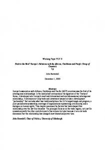
Figure 1—LDIflare areas (cm2) in the three groups of subjects. LDIflare was reduced in both diabetic groups compared with the NC group (DN, P ⬍ 0.0001; DC, P ⫽ 0.01, respectively) and was also significantly reduced in the DN group compared with the DC group (P ⬍ 0.0001).
impaired sensation if they could not feel at more than one of the above-mentioned sites for either modality. Subjects with abnormal response using the Neuropen assessment and/or with impaired VPT (ⱖ15 V, i.e., ⬎95th percentile for this age-group) were classified as neuropathic (48). All subjects in groups NC and DC had intact sensation to Neuropen assessment and normal VPT. For the purpose of this study, cold and warm detection thresholds were used as comparative assessment of C-fiber function and not for classification of neuropathy. These tests were performed using CASE IV software version 4.27.1 (WR Medical Electronics, MN). The cold detection threshold (CDT) and warm detection threshold (WDT) were measured using the 4, 2, and 1 stepping algorithm with null stimuli as described by Dyck et al. (26). CDT and WDT were examined using the standard CASE IV thermode applied on the dorsum of the mid foot. For each test, the computer calculated the just-noticeable difference (JND) from the subject’s responses. The concept of JND is based on the ability to discriminate two levels of stimuli. The CASE IV system uses a set of 25 standardized vibratory and thermal stimulation levels. Each level of stimulation corresponds to 1 JND unit. Therefore, a higher JND value reflects a larger change in temperature (thermal) (49). A value of 26 was given if the JND was higher than the maximum of 25. 2932
Statistical analysis Kruskal-Wallis and Mann-Whitney U tests were used to compare the different variables from the three groups. Median and interquartile ranges were used to express the data. Pearson correlation coefficient (r) was used to examine the correlation between the variables. SPSS version 10.0 software (SPSS, Chicago, IL) was used for the statistical analysis of the data. RESULTS Validation In healthy volunteers, the flare area in response to heating was 5.2 cm2 (3.9 –5.9) as compared with 1.62 cm2 (1.45–1.72) after applying EMLA cream (median [interquartile range]; P ⬍ 0.0001). These results confirm the neurogenic nature of the flare. In contrast, the microvascular hyperemia (LDImax) confined to the area of skin in direct contact with the heater probe and to which the EMLA was applied was unaffected, indicating that this component is non-neurogenic (before: 659.0 arbitrary perfusion units [PU] [602.7–711.2]; after: 629.0 PU [583.5– 676.0]; P ⫽ 0.64). Studies in type 2 diabetes The subjects in the three groups were age matched, and there was no statistically significant difference in the age between the three groups. Similarly, the baseline foot skin temperature was not signifi-
cantly different in the three study groups: NC, 30.0 (29.9 –31.0); DC, 30.6 (29.0 – 32.1); and DN, 31.5 (30.0 –32.0). LDImax was reduced in groups DC and DN compared with group NC (P ⫽ 0.01 and P ⫽ 0.001, respectively), but there was no significant difference between the diabetic groups DC and DN (P ⫽ 0.2). The neurogenic component (LDIflare) was also significantly reduced in groups DC and DN compared with group NC (P ⫽ 0.01 and P ⬍ 0.0001, respectively). In contrast to the LDImax, which was similar in the diabetic groups, the LDIflare in group DN was also reduced when compared with group DC (P ⬍ 0.0001) (Fig. 1). In the diabetic subjects, the LDIflare inversely correlated with the VPT and WDT (r ⫽ ⫺0.64 and ⫺0.57; P ⫽ 0.001 and 0.003, respectively) but was not significantly correlated with CDT (P ⫽ 0.10). In contrast to the LDIflare, LDImax did not significantly correlate with any of the neurological parameters (VPT, P ⫽ 0.07; WDT, P ⫽ 0.56; and CDT, P ⫽ 0.08). Furthermore, there was no correlation between the LDIflare and LDImax (P ⫽ 0.46). With regard to duration of diabetes, BMI, HbA1c, and skin temperature, there was no significant correlation with the LDIflare within the diabetic subjects (r ⫽ ⫺0.2, P ⫽ 0.3; r ⫽ ⫺0.2, P ⫽ 0.4; and r ⫽ ⫺0.3, P ⫽ 0.1, respectively). Of importance, C-fiber function as-
DIABETES CARE, VOLUME 27, NUMBER 12, DECEMBER 2004
Krishnan and Rayman
Table 2—LDIflare, LDImax, and thermal thresholds in the three groups NC
DC
DN
2
LDIflare area (cm ) 5.5 (3.9–5.8) 2.8 (2.5–3.8)* 1.3 (0.9–1.8)† LDImax (PU) 533.0 (504.0–623.0) 409.5 (333.2–523.5)* 380.0 (280.5–484.0)‡ CDT (JND) 7.35 (8.0–12.0) 13.7 (10.5–18.8) 18.9 (16.0–23.7) WDT (JND) 18.8 (16.2–20.0) 19.5 (12.7–22.7) 26.0 (26.0–26.0) Foot temperature (°C) 30.0 (29.9–31.0) 30.6 (29.0–32.1) 31.5 (30.0–32.0) Data are medians (interquartile ranges). *P ⫽ 0.01; †P ⬍ 0.0001; ‡P ⫽ 0.001.
sessed using the new method (LDIflare) demonstrated a significant difference between groups DC and NC (P ⫽ 0.01) (Fig. 1), whereas the other tests of C-fiber function (CDT and WDT [CASE IV]) did not demonstrate a significant difference (Table 2). However, CDT values in the DC group were intermediate between the NC and DN groups. The lack of significant difference between the DC and NC groups for this modality may be due to the small number of subjects in the current study. CONCLUSIONS — In this study, a novel method of evoking and measuring the neurogenic flare was used to assess C-fiber function. In previous studies, the flare was stimulated by a variety of invasive methods, predominantly iontophoresis or injection of acetylcholine or histamine (39,46,50,51). However, interpreting the response using these methods is difficult because the mode of delivery of the drug (current induced or injection) can itself cause axon reflex vasodilatation; in addition, dissemination of the substance through the skin cannot be regulated (52,53). It is also theoretically possible that diffusion of the drug through the glycosylated diabetic skin could be reduced, interfering with the response. Furthermore, these studies use single-point laser Doppler probes, which cannot determine the flare area. Instead, they simply examine the percent change in microvascular blood flow at a predefined distance from the site of delivery of the vasoactive substances. For these reasons, it is not surprising that these methods are associated with a high coefficient of variation (41,44,46,50,54). In contrast, our technique uses a small heater probe, which enables the neurogenic flare to be induced by a single localized and easily reproducible stimulus. Furthermore, the area of the flare and thus the extent of intact innervation are
assessed rather than simply the increase in the blood flow. The coefficient of variation is excellent (6.8%), and the stimulus is physiological in that it is a natural noxious stimulus. Although heating to 44°C is minor trauma, there is wide experience of using probes at this temperature to assess maximum hyperemia with no evidence of adverse events. Moreover, the transcutaneous oxygen tension monitors used in neonates and during anesthesia use electrodes heated to 44°C. We have demonstrated in healthy subjects that the LDIflare is dependent on normal innervation by showing that application of topical local anesthetic significantly diminishes its size. In the diabetic subjects, the strong correlation of LDIflare with VPT and WDT further supports the neurological nature of the flare. In contrast, the LDImax appears independent of innervation because in the healthy subjects it was not significantly affected by the topical anesthesia and in the diabetic patients it was equally impaired in the groups with and without neuropathy. Furthermore, the LDImax was not significantly correlated with any of the neurological parameters. The reduction in LDImax in type 2 diabetic subjects confirms our previous observations and those of others (46,47,55). The pathogenesis of this abnormality is not fully understood, but we have previously demonstrated in type 1 diabetic subjects an inverse relationship with capillary basement membrane thickness and have suggested that this may limit microvascular distensibility (56). It has also been suggested that reduced nitric oxide synthesis or smooth muscle response to nitric oxide may be involved (57). Other investigators have also suggested that the LDImax may be partially mediated by the axon reflex (58). As stated above, this is not supported by our findings.
DIABETES CARE, VOLUME 27, NUMBER 12, DECEMBER 2004
One of the most interesting findings was that the group without clinical neuropathy also had significantly reduced flare responses compared with the healthy control subjects. This demonstrates that C-fiber dysfunction occurs relatively early in type 2 diabetes before it is detected by the currently available methods. In conclusion, this is a simple, noninvasive, and objective method to assess Cfiber function that has significant practical advantages over other methods. We believe the LDIflare will be of value in screening for early neuropathy, and because of its excellent reproducibility, it is ideally suited for assessing therapeutic interventions aimed at preventing or reversing C-fiber dysfunction. Moreover, because C-fiber loss has a putative pathological role in the development of diabetic foot ulcers, its early detection may enable this risk to be assessed more precisely.
References 1. Dyck PJ, Kratz KM, Karnes JL, Litchy WJ, Klein R, Pach JM, Wilson DM, PC OB, Melton LJ, Service FJ: The prevalence by staged severity of various types of diabetic neuropathy, retinopathy, and nephropathy in a population-based cohort: the Rochester Diabetic Neuropathy Study. Neurology 43:817– 824, 1993 [erratum in Neurology 43:2345, 1993] 2. Young MJ, Boulton AJ, Macleod AF, Williams DR, Sonksen PH: A multicentre study of the prevalence of diabetic peripheral neuropathy in the United Kingdom hospital clinic population. Diabetologia 36:150 –154, 1993 3. Tesfaye S, Stevens LK, Stephenson JM, Fuller JH, Plater M, Ionescu Tirgoviste C, Nuber A, Pozza G, Ward JD: Prevalence of diabetic peripheral neuropathy and its relation to glycaemic control and potential risk factors: the EURODIAB IDDM Complications Study. Diabetologia 39:1377– 1384, 1996 4. Galer BS, Gianas A, Jensen MP: Painful diabetic polyneuropathy: epidemiology, pain description, and quality of life. Diabetes Res Clin Pract 47:123–128, 2000 5. McNeely MJ, Boyko EJ, Ahroni JH, Stensel VL, Reiber GE, Smith DG, Pecoraro RF: The independent contributions of diabetic neuropathy and vasculopathy in foot ulceration: how great are the risks? Diabetes Care 18:216 –219, 1995 6. Pecoraro RE, Reiber GE, Burgess EM: Pathways to diabetic limb amputation: basis for prevention. Diabetes Care 13: 513–521, 1990
2933
Test of C-fiber function in type 2 diabetes
7. Dyck PJ, Bushek W, Spring EM, Karnes JL, Litchy WJ, O’Brien PC, Service FJ: Vibratory and cooling detection thresholds compared with other tests in diagnosing and staging diabetic neuropathy. Diabetes Care 10:432– 440, 1987 8. Abbott CA, Carrington AL, Ashe H, Bath S, Every LC, Griffiths J, Hann AW, Hussein A, Jackson N, Johnson KE, Ryder CH, Torkington R, Van Ross ER, Whalley AM, Widdows P, Williamson S, Boulton AJ: The North-West Diabetes Foot Care Study: incidence of, and risk factors for, new diabetic foot ulceration in a community-based patient cohort. Diabet Med 19: 377–384, 2002 9. Paisley AN, Abbott C, van Schie C, Boulton AJM: A comparison of the Neuropen against standard quantitative sensorythreshold measures for assessing peripheral nerve function. Diabet Med 19:400 – 405, 2002 10. Abbott CA, Vileikyte L, Williamson S, Carrington AL, Boulton AJ: Multicenter study of the incidence of and predictive risk factors for diabetic neuropathic foot ulceration. Diabetes Care 21:1071–1075, 1998 11. Kastenbauer T, Sauseng S, Sokol G, Auinger M, Irsigler K: A prospective study of predictors for foot ulceration in type 2 diabetes. J Am Podiatr Med Assoc 91:343– 350, 2001 12. Young MJ, Breddy JL, Veves A, Boulton AJ: The prediction of diabetic neuropathic foot ulceration using vibration perception thresholds: a prospective study. Diabetes Care 17:557–560, 1994 13. Boulton AJ, Kubrusly DB, Bowker JH, Gadia MT, Quintero L, Becker DM, Skyler JS, Sosenko JM: Impaired vibratory perception and diabetic foot ulceration. Diabet Med 3:335–337, 1986 14. Ziegler D, Mayer P, Gries FA: Evaluation of thermal, pain, and vibration sensation thresholds in newly diagnosed type 1 diabetic patients. J Neurol Neurosurg Psychiatry 51:1420 –1424, 1988 15. Ziegler D, Mayer P, Wiefels K, Gries FA: Assessment of small and large fiber function in long-term type 1 (insulin-dependent) diabetic patients with and without painful neuropathy. Pain 34:1–10, 1988 16. Levitt NS, Stansberry KB, Wynchank S, Vinik AI: The natural progression of autonomic neuropathy and autonomic function tests in a cohort of people with IDDM. Diabetes Care 19:751–754, 1996 17. Vinik AI, Park TS, Stansberry KB, Pittenger GL: Diabetic neuropathies. Diabetologia 43:957–973, 2000 18. Celiker R, Basgoze O, Bayraktar M: Early detection of neurological involvement in diabetes mellitus. Electromyogr Clin Neurophysiol 36:29 –35, 1996 19. Vinik AI, Erbas T, Stansberry KB, Pit-
2934
20.
21.
22.
23.
24.
25.
26.
27.
28.
29.
30.
31.
tenger GL: Small fiber neuropathy and neurovascular disturbances in diabetes mellitus. Exp Clin Endocrinol Diabetes 109: 451– 473, 2001 Singleton JR, Smith AG, Bromberg MB: Increased prevalence of impaired glucose tolerance in patients with painful sensory neuropathy. Diabetes Care 24:1448 –1453, 2001 Singleton JR, Smith AG, Bromberg MB: Painful sensory polyneuropathy associated with impaired glucose tolerance. Muscle Nerve 24:1225–1228, 2001 Smith AG, Ramachandran P, Tripp S, Singleton JR: Epidermal nerve innervation in impaired glucose tolerance and diabetesassociated neuropathy. Neurology 57: 1701–1704, 2001 Murakawa Y, Zhang W, Pierson CR, Brismar T, Ostenson CG, Efendic S, Sima AA: Impaired glucose tolerance and insulinopenia in the GK-rat causes peripheral neuropathy. Diabete Metab Res Rev 18:473– 483, 2002 Ali Z, Carroll M, Robertson KP, Fowler CJ: The extent of small fibre sensory neuropathy in diabetics with plantar foot ulceration. J Neurol Neurosurg Psychiatry 52: 94 –98, 1989 Sosenko JM, Kato M, Soto R, Bild DE: Comparison of quantitative sensorythreshold measures for their association with foot ulceration in diabetic patients. Diabetes Care 13:1057–1061, 1990 Dyck PJ, O’Brien PC, Kosanke JL, Gillen DA, Karnes JL: A 4, 2, and 1 stepping algorithm for quick and accurate estimation of cutaneous sensation threshold. Neurology 43:1508 –1512, 1993 Dyck PJ, Zimmerman IR, Johnson DM, Gillen D, Hokanson JL, Karnes JL, Gruener G, O’Brien PC: A standard test of heat-pain responses using CASE IV. J Neurol Sci 136:54 – 63, 1996 Gruener G, Dyck PJ: Quantitative sensory testing: methodology, applications, and future directions. J Clin Neurophysiol 11: 568 –583, 1994 Shy ME, Frohman EM, So YT, Arezzo JC, Cornblath DR, Giuliani MJ, Kincaid JC, Ochoa JL, Parry GJ, Weimer LH: Quantitative sensory testing: Report of the Therapeutics and Technology Assessment Subcommittee of the American Academy of Neurology. Neurology 60: 898 –904, 2003 Maser RE, Nielsen VK, Bass EB, Manjoo Q, Dorman JS, Kelsey SF, Becker DJ, Orchard TJ: Measuring diabetic neuropathy: assessment and comparison of clinical examination and quantitative sensory testing. Diabetes Care 12:270 –275, 1989 Freeman R, Chase KP, Risk MR: Quantitative sensory testing cannot differentiate simulated sensory loss from sensory neuropathy. Neurology 60:465– 470, 2003
32. Gelber DA, Pfeifer MA, Broadstone VL, Munster EW, Peterson M, Arezzo JC, Shamoon H, Zeidler A, Clements R, Greene DA, Porte D, Laudadio C, Bril V: Components of variance for vibratory and thermal threshold testing in normal and diabetic subjects. J Diabetes Complications 9:170 –176, 1995 33. Lindberger M, Schroder HD, Schultzberg M, Kristensson K, Persson A, Ostman J, Link H: Nerve fibre studies in skin biopsies in peripheral neuropathies. I. Immunohistochemical analysis of neuropeptides in diabetes mellitus. J Neurol Sci 93:289 –296, 1989 34. McCarthy BG, Hsieh ST, Stocks A, Hauer P, Macko C, Cornblath DR, Griffin JW, McArthur JC: Cutaneous innervation in sensory neuropathies: evaluation by skin biopsy. Neurology 45:1848 –1855, 1995 35. Holland NR, Stocks A, Hauer P, Cornblath DR, Griffin JW, McArthur JC: Intraepidermal nerve fiber density in patients with painful sensory neuropathy. Neurology 48:708 –711, 1997 36. Hirai A, Yasuda H, Joko M, Maeda T, Kikkawa R: Evaluation of diabetic neuropathy through the quantitation of cutaneous nerves. J Neurol Sci 172:55– 62, 2000 37. Griffin JW, McArthur JC, Polydefkis M: Assessment of cutaneous innervation by skin biopsies. Curr Opin Neurol 14:655– 659, 2001 38. Polydefkis M, Hauer P, Griffin JW, McArthur JC: Skin biopsy as a tool to assess distal small fiber innervation in diabetic neuropathy. Diabetes Technol Ther 3: 23–28, 2001 39. Hutchison KJ, Johnson BW, Williams HT, Brown GD: The histamine flare response in diabetes mellitus. Surg Gynecol Obstet 139:566 –568, 1974 40. Aronin N, Leeman SE, Clements RS Jr: Diminished flare response in neuropathic diabetic patients: comparison of effects of substance P, histamine, and capsaicin. Diabetes 36:1139 –1143, 1987 41. Walmsley D, Wiles PG: Early loss of neurogenic inflammation in the human diabetic foot. Clin Sci (Colch) 80:605– 610, 1991 42. Walmsley D, Wales JK, Wiles PG: Reduced hyperaemia following skin trauma: evidence for an impaired microvascular response to injury in the diabetic foot. Diabetologia 32:736 –739, 1989 43. Schmelz M, Michael K, Weidner C, Schmidt R, Torebjork HE, Handwerker HO: Which nerve fibers mediate the axon reflex flare in human skin? Neuroreport 11:645– 648, 2000 44. Walmsley D, Wiles PG: Assessment of the neurogenic flare response as a measure of nociceptor C fibre function. J Med Eng Technol 14:194 –196, 1990
DIABETES CARE, VOLUME 27, NUMBER 12, DECEMBER 2004
Krishnan and Rayman
45. Caselli A, Rich J, Hanane T, Uccioli L, Veves A: Role of C-nociceptive fibers in the nerve axon reflex-related vasodilation in diabetes. Neurology 60:297–300, 2003 46. Parkhouse N, Le Quesne PM: Impaired neurogenic vascular response in patients with diabetes and neuropathic foot lesions. N Engl J Med 318:1306 –1309, 1988 47. Rayman G, Williams SA, Spencer PD, Smaje LH, Wise PH, Tooke JE, Hassan A: Impaired microvascular hyperaemic response to minor skin trauma in type I diabetes. BMJ (Clin Res Ed) 292:1295–1298, 1986 48. Wiles PG, Pearce SM, Rice PJ, Mitchell JM: Vibration perception threshold: influence of age, height, sex, and smoking, and calculation of accurate centile values. Diabet Med 8:157–161, 1991 49. Dyck PJ, Zimmerman IR, PC OB, Ness A, Caskey PE, Karnes J, Bushek W: Introduction of automated systems to evaluate touch-pressure, vibration, and thermal cutaneous sensation in man. Ann Neurol
4:502–510, 1978 50. Benarroch EE, Low PA: The acetylcholine-induced flare response in evaluation of small fiber dysfunction. Ann Neurol 29: 590 –595, 1991 51. Berg TJ, Levy DM, Reid G, Abraham RR: The effects of vasoactive intestinal polypeptide and substance P on methacholine-induced sweating and vascular flare in diabetic neuropathy. Clin Auton Res 5: 159 –164, 1995 52. Durand S, Fromy B, Bouy P, Saumet JL, Abraham P: Current-induced vasodilation during water iontophoresis (5 min, 0.10 mA) is delayed from current onset and involves aspirin sensitive mechanisms. J Vasc Res 39:59 –71, 2002 53. Droog EJ, Sjoberg F: Nonspecific vasodilatation during transdermal iontophoresis: the effect of voltage over the skin. Microvasc Res 65:172–178, 2003 54. Treede RD: Vasodilator flare due to activation of superficial cutaneous afferents in humans: heat-sensitive versus hista-
DIABETES CARE, VOLUME 27, NUMBER 12, DECEMBER 2004
55.
56.
57.
58.
mine-sensitive fibers. Neurosci Lett 141: 169 –172, 1992 Krishnan ST, Baker NR, Carrington AL, Rayman G: Comparative roles of microvascular and nerve function in foot ulceration in type 2 diabetes. Diabetes Care 27: 1343–1348, 2004 Rayman G, Malik RA, Sharma AK, Day JL: Microvascular response to tissue injury and capillary ultrastructure in the foot skin of type I diabetic patients. Clin Sci (Colch) 89:467– 474, 1995 Veves A, Akbari CM, Primavera J, Donaghue VM, Zacharoulis D, Chrzan JS, DeGirolami U, LoGerfo FW, Freeman R: Endothelial dysfunction and the expression of endothelial nitric oxide synthetase in diabetic neuropathy, vascular disease, and foot ulceration. Diabetes 47:457–463, 1998 Vinik AI, Erbas T, Park TS, Stansberry KB, Scanelli JA, Pittenger GL: Dermal neurovascular dysfunction in type 2 diabetes. Diabetes Care 24:1468 –1475, 2001
2935


![The [Wikipedia] - Semantic Scholar](https://m.moam.info/img/260x300/the-wikipedia-semantic-scholar_599b88541723dd0c4031cf29.jpg)