of Dale & Slack (1987) who refer to many other very careful publications in this field. was added to 6 .... several laboratories (e.g. Jones & Woodland, 1987a,b).
27
Development 105, 27-33 (1989) Printed in Great Britain © The Company of Biologists Limited 1989
The localization of an inductive response
J. B. GURDON Cancer Research Campaign Molecular Embryology Unit, Department of Zoology, University of Cambridge, Downing Street, Cambridge CB2 3EJ, UK
Summary Combinations of tissues from Xenopus blastulae have been used to identify several mechanisms that limit the number of animal cells forming muscle after induction by vegetal cells. The results disagree with a model in which direct physical contact or very close proximity between animal and vegetal cells restricts the number of cells that receive the inductive signal. Rather it seems that a diffusible inducer is released by vegetal cells, and spreads through 4-8 animal cell diameters, equivalent to a distance of 80jon, from the nearest vegetal cells. Several factors seem to cooperate to prevent the further spread of the mesoderm-forming induction. These in-
clude the slow diffusion and/or instability of the inducer, the time of loss of competence of animal cells to respond to induction, and the amount of vegetal tissue that releases inducer for a limited time. The combination of these, and perhaps other, processes seems to ensure that a consistent minority of animal cells are induced to form muscle, thereby leaving other animal cells available to form the nervous system and epidermis.
Introduction
number of cells is reduced, or because cell division and changes of cell position take place during a period of several days before a response is seen. The particular induction analysed here is the mesoderm-forming induction in amphibia. This takes place over a few hours, and can proceed in the absence of cell division and cell movement, making it possible to identify several processes that determine the number and location of cells that respond to induction.
There are many instances in development and adult life where cell differentiation is influenced by products of other cells. In all these cases, it is important that only a limited number of cells in certain positions should respond to these signals. In long-range cell interactions, such as those mediated by hormones, the cells that respond to the hormone are selected by their possession of receptors. For very short-range interactions, such as ones involving neurotransmitters, only cells with membranes located very close to the point of release of transmitters respond; dilution and or instability of the transmitter causes its concentration to decrease strongly from the site of its release. Embryonic induction is intermediate in range, some of the cells that respond being up to 100 fim or 10 cell diameters away from the source of the inducer. It is not known what determines the number and position of cells that respond, though it is clear that more cells have the capacity to respond than normally do so. It is also of crucial importance that not too many cells respond to induction, since those that do not must contribute to different cell-types which would otherwise be deficient. The experiments described here aim to identify mechanisms that localize the response to induction and this requires an experimental system in which inducing and responding cells can be varied in number and position. In most examples of embryonic induction, this cannot be achieved, because the response is lost as the
Key words: Xenopus, induction, muscle, competence, animal cells, vegetal cells.
Materials and methods All experiments were carried out with embryos of Xenopus laevis. Embryo tissues and cells were cultured in modified Barth saline (MBS, Gurdon, 1977). Embryos were staged according to Nieuwkoop & Faber (1956). Glass moulds were constructed from small pieces of 1 mm thick microscope slide attached by Araldite to each other and to a large piece which served as a floor. The top of each chamber was open. Any dead cells which emerged after placing embryo fragments into the moulds were removed at intervals. The moulds with embryo fragments were cultured in MBS until removed for fixation. Nuclepore filters were secured vertically in the middle of a glass mould, so that appropriately sized wells on each side of the filter provided a tight fit for vegetal or animal pieces of a blastula. For some experiments, animal:vegetal conjugates were cultured in solid gelatin (BDH), made up in Ca -free MBS. A 5 % gelatin solution was maintained at 25°C until required. Then 2 (A of a lOmgmF 1 stock solution of cytochalasin B (Sigma) in dimethylsulphoxide (kept at —20°C until required)
28
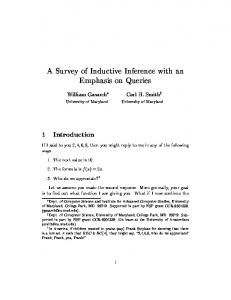
/. B. Gurdon Animal pole
g
Nerve Epidermis
Muscle Muscle
Dorsal
Vegetal pole
Endoderm (gut)
Fig. 1. Diagrams illustrating the localized response to mesoderm-forming induction in Xenopus. (A) A stage-8 blastula cut vertically in half. Arrows represent inductive effect from vegetal cells moving upwards towards the animal pole. The inductive effect is stronger on the dorsal side (left in the diagram) than on the ventral side. (B) Diagram of a transverse section through a stage-8 blastula, indicating the position of cells that will subsequently form the tissues named. The cells filled in with black will form mesoderm, and about half of these will form axial muscle of a tail-bud tadpole. Note that the inductive influence does not spread far enough towards the animal pole to affect cells destined to contribute nerve or epidermis. These diagrams are based on several lineage analyses of early Xenopus embryos, of which the most recent is that of Dale & Slack (1987) who refer to many other very careful publications in this field. was added to 6 ml of the 5 % gelatin solution (making a 3jigm\~l cytochalasin B solution). This was poured into a 35 mm plastic dish containing a thin layer of 2% agarose (FMC, type LGT) in Ca2+-free MBS. Immediately after this, conjugates that had been formed in Ca2+-containing MBS were washed in Ca2+-free MBS and allowed to fall through the liquid gelatin solution, coming to rest on the solid agarose base. The whole dish was then placed in a 4°C refrigerator for 15 min during which time the gelatin solidified, and was then transferred to an incubator for culture, usually at 19°C. For fixation, large syringe needles were used to cut out a solid cube of gelatin containing a conjugate, and the whole cube was immersed in a solution of 50% methanol in MBS. After 8-24 h at room temperature, a 5 ml bottle containing the gelatin-conjugate in 50% MBS was placed at 37°C for 2 min, causing the gelatin to dissolve. Rotation of the bottle dispersed the gelatin, which was removed by careful washing with 50% methanol:MBS, and which was then replaced by 100 % methanol. After a further fixation of a few hours or more at 4°C, the conjugates could now be embedded in wax and sectioned by routine procedures. This particular fixative turned out to be best for the antigen recognized by the Kintner & Brockes (1984) antibody. The fixation by 50% methanol:MBS was devised to avoid the gross distortion that results from immersing 5 % gelatin cubes in 100% methanol. Nuclease protection analysis and other procedures were the same as used before in this laboratory (Gurdon et al. 1985). Results
A localized response takes place in experimental conditions The mesoderm-forming induction that takes place during the first few hours of amphibian development is too well known to need description (Nieuwkoop, 1969; Nakamura et al. 1970; review by Gurdon, 1987). We need to know, for the present discussion, that the cells that respond to this induction by forming muscle are located at and just above the equator of the blastula
(Fig. 1). The cells that are near the animal pole of the embryo are not induced to form muscle or other mesodermal tissues, but form the whole nervous system and much of the epidermis. Therefore, the localization of response to mesodermal induction is of crucial importance in normal development. Combinations of animal and vegetal regions of a blastula after removal of the equatorial one third have been widely used for the analysis of the mesodermforming induction; these constructions have the advantage that neither the animal nor vegetal pieces when cultured on their own will express muscle-specific genes, but the combination of the two always do so very strongly (Gurdon et al. 1985). Muscle cells are recognized in cultured animal:vegetal conjugates, in this and previous work, by the muscle-specific antibody of Kintner & Brockes (1984). Muscle cells are present in one (or sometimes two) coherent groups, and are located in an approximately central position (Fig. 2A). Therefore animakvegetal conjugates provide a good experimental system in which to investigate mechanisms responsible for the location of muscle cells resulting from induction. There is no predisposition among animal cells The simplest explanation for the location of induced muscle cells would be a predisposition for muscle differentiation among the more equatorial cells of the animal hemisphere. This has been tested by placing cells from various regions of the animal hemisphere in conjugation with vegetal tissue. It is observed that about one third of all animal cells express muscle genes, whether they are taken as a piece of tissue from the animal pole or from the dorsal, ventral or lateral parts of the temperate zone. This is also true if cells from different positions are dissociated, mixed and reaggregated before being placed on vegetal tissue for indue-
The localization of an inductive response
29
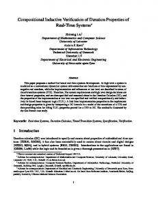
Fig. 2. The localization of muscle cells in animal: vegetal conjugates. An animal one third was placed on a vegetal one third of a stage-8 to -9 blastula, as described by Nieuwkoop (1969) and as illustrated by Gurdon et al. (1985); the conjugate was cultured until control embryos reached stage 28. At this stage, conjugates were fixed, sectioned and reacted with the Kintner & Brockes (1984) muscle-specific antibody (shown white). (A) A conjugate cultured in normal MBS medium, which permits cell division and cell rearrangement (transverse section). B. A conjugate embedded in solid gelatin (5% in Ca2+-free MBS) containing 3^gml~x cytochalasin B, which inhibits cell division and cell migration (transverse section). Note that muscle cells are located near the middle of each conjugate, in a homogeneous group. Though hard to quantify, the total volume of cells positive for muscle antigen seems somewhat reduced in Ca2+-free cytochalasin experiments; it is possible that physical contact helps to somewhat increase the number of cells that respond to induction. tion. The outer strongly pigmented cells can be separated and tested independently of the inner animal cells, and are somewhat less strongly and less frequently induced to form muscle, but this is almost certainly attributable to the impermeability of the outer coat of their surface. Details of these experiments are not given here since they are in agreement with the experience of several laboratories (e.g. Jones & Woodland, 1987a,b). Even though there may be some small variation in the predisposition of animal cells from different regions to become muscle, it is clear that this cannot explain the equatorial location of muscle cells in conjugates and in normal development.
Muscle cells are near inducing cells, not in a midposition between two poles Another hypothesis to account for the equatorial location of induced muscle cells is that they are in a midposition between the animal and vegetal poles of a blastula or conjugate. This may be contrasted with another possibility that induced muscle cells are always derived from the animal cells that are closest to vegetal tissue and hence closest to the source of inducer. These ideas have been distinguished by placing vegetal tissue and several pieces of animal tissue in the configuration shown in Fig. 3. It can be seen that the midposition between the two poles (fragment 4) does not coincide with the region where animal cells are closest to vegetal
cells (fragment 2). Using RNase protection to recognize transcripts of a muscle-specific actin gene, it can be seen in Fig. 3 that muscle genes are not activated in the midposition of the conjugate, but are strongly and uniquely so where animal cells are in contact with vegetal cells. We, therefore, conclude that the central or equatorial position of muscle cells in normal embryos is attributable to their proximity to vegetal cells, and not to their distance from animal and vegetal poles. Inductive response requires proximity not physical contact A simple way of limiting the number of animal cells that respond to induction would exist if physical contact between vegetal and animal cells, or such close proximity as normally separates two cells (~l-2/«n), were required for the inducer to be successfully transmitted. Under these conditions, only a monolayer of animal cells would be induced during the blastula stage, but the combination of cell movement, which is considerable at this stage of Xenopus development, and cell division could result in the formation of a coherent group of muscle cells by the postneurula stage (as explained in Fig. 4A). The need for physical contact has been tested initially by use of Nuclepore filters. Grunz & Tacke (1986) have reported that vegetal tissue can transmit its inductive effect through a Nuclepore filter with 0-1 /mi pores. Our
30
/. B. Gurdon
Glass mould
Cell division and cell movement
I
-I-
I
I
i
i
B Muscle actin
Cytoskeletal actin'
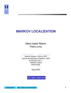
Fig. 3. Muscle cells are induced where animal cells are closest to vegetal cells, and not in a midposition between two poles of a conjugate. The upper part of the figure shows a glass mould into which one vegetal and eight animal pieces of a blastula were fitted. When control embryos reached stage 20, the multiple conjugate, in which all pieces had fused, was removed from the mould and cut into six fragments. Each fragment had RNA extracted, which was analysed by nuclease protection for its content of muscle or cytoskeletal actin mRNA, as described by Gurdon et al. (1985). The midposition of the conjugate is indicated by an arrow (fragment 4), and the only region where animal and vegetal cells are in contact by a + (fragment 2). This experiment was carried out three times independently, in each case with a result like that shown.
Cell division and cell movement inhibited by Cytochalasin and Ca-free medium Fig. 4. Diagrams to illustrate two ways by which a localized subset of animal cells can become positive for muscle differentiation. The small cells at the top of each diagram are animal and the large cells in the lower half are vegetal. (A) This assumes that only those animal cells of a conjugate that are in physical contact with vegetal cells can respond to a muscle-forming induction; a compact group of muscle cells would be formed by subsequent movement and division of the induced cells. (B) This shows the muscle forming induction spreading through or past about four diameters of animal cells in the centre of the conjugate (see Fig. 2B). Conjugates cultured in the presence of cytochalasin (as shown) would acquire a homogeneous group of muscle cells by the restricted spread of inducer; the reason why equatorial cells on the periphery are negative for muscle antigen is not certain, but may be connected with a 'community effect' in which groups of cells enhance each other's differentiation (Gurdon, in preparation) or with some bias of cells on the external surface to become epidermis. The cells filled in which black represent ones that receive an inductive stimulus (left of A and B), and which respond by forming muscle (right of A and B). own results agree with this conclusion to the extent that animal pieces of a blastula can respond to vegetal tissue on the other side of a 0-1 [im Nuclepore filter (Fig. 5A) and that cytoplasmic processes do not extend through these pores within the period of ~4h, after which competence ends. We do, however, observe a major weakening of the response across a filter. Using nuclease resistance as a quantitative assay, we find that about half of the test animal pieces are not detectably induced across a filter (i.e. to less than one tenth of the normal extent); of the other half that are induced, the response is weakened by 3-10 times (Fig. 5B). This very substantial weakening of the transfilter response suggests that this induction involves a diffusible molecule which is unstable or which is ineffective at a lower than normal concentration, or both. The conclusion from transfilter experiments is that the localization of inductive effect does not depend on physical contact, but is more likely to involve a restricted spread of the inducer from its source. It is conceivable that the
The localization of an inductive response inductive effect of vegetal cells can only be transmitted over a few microns to adjacent animal cells, which can then pass on the effect to a few other animal cells, as in homoiogenetic induction (Mangold & Spemann, 1927). The inductive effect can spread over a few cell diameters To show directly that the inductive influence of vegetal tissue can spread to animal cells not in contact with it, conditions have been used which were previously found to inhibit cell division and cell movement in conjugates: 1 5% gelatin in Ca2+-free MBS containing
An +
+ +
RNA - analyses
-
Filter
Veg B
An:An No filter
An:Veg No filter
An:Veg Stages +filter 10 18
M ! •
nh.i
cs Fig. 5. The muscle-forming induction passes through, but is substantially weakened by, a Nuclepore filter 10 jxm thick with 0 1 ;um pores. (A) The result of placing animal and vegetal pieces of a blastula on each side of a filter in the same culture dish. When control embryos had reached stage 20, animal pieces were removed and assayed for actin transcripts by nuclease protection. In the representative experiment illustrated, each of the three animal pieces that were opposite vegetal pieces, but none of the other three animal pieces in the same medium, were positive for muscle actin RNA. (B) Examples of nuclease protection assays on animal pieces of a blastula cultured with animal or vegetal pieces, with or without a 0-1 pm filter, as indicated. Whole control embryos (tracks labelled stages 10,18) contain muscle actin RNA (M) at stage 18, but not at stage 10, and they contain more cytoskeletal actin RNA (CS) at stage 18 than at stage 10. For 30 analyses of each of the three kinds, the average muscle: cytoskeletal RNA ratio was 2-6 for an: veg with no filter, 0-21 for an: veg with a filter, and