www.jkns.or.kr
http://dx.doi.org/10.3340/jkns.2012.52.2.80
Print ISSN 2005-3711 On-line ISSN 1598-7876
Copyright © 2012 The Korean Neurosurgical Society
J Korean Neurosurg Soc 52 : 80-84, 2012
Clinical Article
The Meaning of the Prognostic Factors in Ruptured Middle Cerebral Artery Aneurysm with Intracerebral Hemorrhage Ji Woong Oh, M.D.,1 Ji-Yong Lee, M.D.,2 Myeong Sub Lee, M.D.,3 Hyen-Ho Jung, M.D.,1 Kum Whang, M.D.;1 Brain Research Group Departments of Neurosurgery,1 Neurology,2 Radiology,3 Wonju College of Medicine, Yonsei University, Wonju, Korea Objective : This study analyzed the relationship between prognosis and multiple clinical factors of ruptured middle cerebral artery (MCA) aneurysm with intracerebral hemorrhage (ICH), to aid in predicting the results of surgical treatment. Methods : Enrolled subjects were 41 patients with ruptured MCA aneurysm with ICH who were treated with surgical clipping. Clinical factors such as gender, age, and initial Glasgow coma scale were assessed while radiological factors such as the volume and location of hematoma, the degree of a midline shift, and aneurysm size were considered retrospectively. Prognosis was evaluated postoperatively by Glasgow outcome scale. Results : Age and prognosis were correlated only in the groups with ICH over 31 mL or ICH at the frontal lobe or sylvian fissure. When initial mental status was good, only patients with ICH on the temporal lobe had a better prognosis. If the midline shift was less than 4.5 mm, the probability of better prognosis was 95.5% (21 of 22). If the midline shift was more than 4.5 mm, the probability of poor prognosis was 42.1% (8 of 19). Patients with ICH less than 31 mL had higher survival rates, whereas if the ICH was more than 31 mL, 41.2% (7 of 17) had a poor clinical pathway. Conclusion : Even if the initial clinical condition of the patient was not promising, by carefully examining and taking into account all factors, neurosurgeons can confidently recommend surgical treatment for these patients. Key Words : Ruptured middle cerebral artery aneurysm · Intracerebral hemorrhage · Prognostic outcome.
cal data suggested that patients with ruptured MCA aneurysms with ICH have a non-classical clinical course that requires further study. Therefore, authors analyzed the relationship between multiple clinical factors of ruptured MCA aneurysms with ICH and general prognosis, to help predict the results of surgical treatment.
INTRODUCTION Middle cerebral artery (MCA) aneurysms are 20% of saccular aneurysms. They are occasionally found incidentally or because of a mass effect, but most are found when they rupture8). After Dott2) treated ruptured aneurysms with surgical methods, many treatment modalities have been developed and reported17). When MCA aneurysms rupture, approximately 30-50% lead to a subarachnoid hemorrhage (SAH) combined with an intracerebral hemorrhage (ICH), with a mortality rate of 1041%4,16). The mechanism of ICH developed by aneurysmal rupture is still controversial but the most commonly accepted hypothesis is that the ICH develops when fibrin, hematoma, or the fibrosis of the arachnoid membrane obstructs the subarachnoid space, or an aneurysm attached to the pia ruptures16). These ICHs are a risk factor for poor prognosis and surgical complications in ruptured MCA aneurysm patients. However, our clini-
MATERIALS AND METHODS We retrospectively reviewed 68 patients with MCA aneurysm who underwent neck clipping after aneurysm rupture between January 2006 and December 2010. We excluded 27 patients who did not have accompanying ICH. The remaining 41 patients with accompanying ICH were evaluated. Aneurysm, SAH and ICH were diagnosed with computed tomography (CT) and conventional cerebral angiography. Of the 41 total patients, 27 were evaluated with preoperative angiography (Table 1).
Received : February 15, 2012 • Revised : May 30, 2012 • Accepted : August 19, 2012 Address for reprints : Kum Whang, M.D. Departments of Neurosurgery, Wonju College of Medicine, Yonsei University, 162 Ilsan-dong, Wonju 220-701, Korea Tel : +82-33-741-0593, Fax : +82-33-746-2287, E-mail :
[email protected] • This is an Open Access article distributed under the terms of the Creative Commons Attribution Non-Commercial License (http://creativecommons.org/licenses/by-nc/3.0) which permits unrestricted non-commercial use, distribution, and reproduction in any medium, provided the original work is properly cited. • •
80
The Prognostic Factors in Ruptured MCA with ICH | JW Oh, et al.
Clinical, Radiologic evaluation
2). By classifying the patients based on hematoma amount, its volume less than 31 mL had some correlation where younger age meant better prognosis. However, if the amount of hemato-
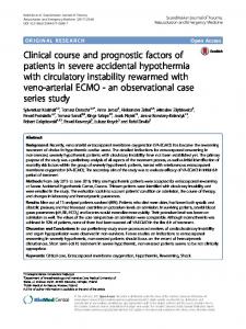
Clinical status was evaluated with gender, age, and Glasgow coma scale (GCS). Clinical outcome was evaluated with the GOS6). Patients with a GOS of 4-5 were considered to be a favorable outcome group. On contrary, patients with a GOS of 1-3 were considered to be an unfavorable outcome group. According to hematoma location, radiologic findings were classified into temporal, sylvian fissure, and frontal groups (Fig. 1). Hematoma volume was measured with abc/2 methods. We also evaluated lesion lateralization, degree of midline shift, and aneurysm size.
Table 1. Demographic statistics of ruptured MCA, aneurysm with ICH Prognostic factors Age Range
36-74 (yrs)
Mean
58.2 (yrs)
Gender Male
11 (26.8%)
Female
30 (73.2%)
Aneurysmal size
Surgical methods and postoperative management All patients were operared with a routine pterional approach. If severe brain swelling was found, duroplasty and decompressive craniectomy were performed. The medial transsylvian approach and lateral transsylvian approach were chosen appropriately in each situation with a goal for less brain retraction and less brain injury. All surgery was performed on the day of admission. Postoperative CT brain contrast perfusion images were obtained for all patients to evaluate perfusion values such as cerebral blood flow, cerebral blood volume, and mean transit time. According to each perfusion value, we performed osomotherapy with mannitol, or treated with a calcium channel blocker such as Nimodipine®. In case of vasospasm events, 3H therapy of hypertension, hypervolemia, hemodilution was performed.
Small
11 (26.8%)
Large
29 (70.7%)
Giant
1 (2.4%)
Lateralization Dominant
23 (56.0%)
Non-dominant
18 (43.9%)
GCS score Mild
19 (46.3%)
Moderate
17 (41.5%)
Severe
5 (12.1%)
Location Frontal
18 (43.9%)
Sylvian fissure Temporal
Statistical analysis
11 (26.8%) 12 (29.3%)
MCA : middle cerebral artery, ICH : intracerebral hemorrhage, GCS : Glasgow
All parameters were evaluated retrospectively. Prognostic fac- coma scale tors were evaluated with a chi-square Table 2. Age and clinical outcome according to location test, independent t-test, receiver operaAge (yrs) tion characteristic (ROC) curve, McNeICH Location Outcome ≤65 mar test, and nonparametric chi-square Frontal Unfavorable 0 test with significance defined as less Favorable 13 than 0.05. All statistical analysis was Sylvian fissure Unfavorable 0 performed with commercial software Favorable 8 (Predictable Analysis Soft Ware, VerTemporal Unfavorable 3 sion 18.0, IBM Inc.) Favorable
RESULTS Age Patients’ mean age was 58.2 years. Patients aged 65 years and younger had a significantly better prognosis than those over 65 years (p0.05
Frontal
Unfavorable Favorable
0 0
1 8
0 6
Sylvian fissure
Unfavorable
1
2
0
Favorable
1
3
7
Unfavorable
2
3
0
Favorable
1 5
0 17
6 19
Temporal Total
p-value
Severe
moderate group (12.1%) and 19 were in the mild group (46.3%). Furthermore, there was a statistical significance between the initial mental status and clinical outcome (p0.05).
Hematoma location ICHs were divided into three groups based on location : 18 had frontal ICH, 11 had sylvian ICH (26.8%), and 12 had temporal ICH (29.3%). Of the three locations, the temporal ICH group had the most favorable clinical outcome (42.6%, 5/12) while in the frontal ICH, survival was 6.7%, and in the sylvian fissure ICH, it was 21.4%. However, no significant conclusions could be drawn.
Lateralization of brain lesion Patients who had ICH in the dominant hemisphere were 23 which outnumbered those who had ICH on the non-dominant hemisphere (18/41). Of 23 patients with ICH in the dominant hemisphere, 5 had unfavorable outcome while 18 others had favorable outcome. In contrast, of patients with ICH in the non-dominant hemisphere, 4 had an unfavorable outcome while 14 had a favorable outcome. However, this was not significant (p>0.05).
Midline shift Of the 41 patients, 24 were had a midline shift of the brain parenchyma, with a midline shift range of 3 mm to 15 mm. The average length of the midline shift was 7.5 mm. Patients with a poor prognosis showed a mean midline shift of 8.36 mm while those with a good prognosis had a mean midline shift of 3.3 mm, for a 5.06 mm difference (p