visit our website at https://www.elsevier.com/. Publisher: Sara ...... With qualitative detection, immunochromatographic test ...... Hindiyeh MY, Keller N, Mandelboim M, et al. High rate of ...... Lee JO, Kim DY, Lim JH, Seo MD, Yi HG, Kim YJ, et al.
The Microbiology of Respiratory System Infections
Page left intentionally blank
The Microbiology of Respiratory System Infections
Edited by
Kateryna Kon Mahendra Rai
AMSTERDAM • BOSTON • HEIDELBERG • LONDON NEW YORK • OXFORD • PARIS • SAN DIEGO SAN FRANCISCO • SINGAPORE • SYDNEY • TOKYO
Academic Press is an imprint of Elsevier
Academic Press is an imprint of Elsevier 125 London Wall, London EC2Y 5AS, United Kingdom 525 B Street, Suite 1800, San Diego, CA 92101-4495, United States 50 Hampshire Street, 5th Floor, Cambridge, MA 02139, United States The Boulevard, Langford Lane, Kidlington, Oxford OX5 1GB, UK Copyright © 2016 Elsevier Inc. All rights reserved. No part of this publication may be reproduced or transmitted in any form or by any means, electronic or mechanical, including photocopying, recording, or any information storage and retrieval system, without permission in writing from the publisher. Details on how to seek permission, further information about the Publisher’s permissions policies and our arrangements with organizations such as the Copyright Clearance Center and the Copyright Licensing Agency, can be found at our website: www.elsevier.com/permissions. This book and the individual contributions contained in it are protected under copyright by the Publisher (other than as may be noted herein). Notices Knowledge and best practice in this field are constantly changing. As new research and experience broaden our understanding, changes in research methods, professional practices, or medical treatment may become necessary. Practitioners and researchers must always rely on their own experience and knowledge in evaluating and using any information, methods, compounds, or experiments described herein. In using such information or methods they should be mindful of their own safety and the safety of others, including parties for whom they have a professional responsibility. To the fullest extent of the law, neither the Publisher nor the authors, contributors, or editors, assume any liability for any injury and/or damage to persons or property as a matter of products liability, negligence or otherwise, or from any use or operation of any methods, products, instructions, or ideas contained in the material herein. Library of Congress Cataloging-in-Publication Data A catalog record for this book is available from the Library of Congress British Library Cataloguing-in-Publication Data A catalogue record for this book is available from the British Library ISBN: 978-0-12-804543-5 For information on all Academic Press publications visit our website at https://www.elsevier.com/
Publisher: Sara Tenney Acquisition Editor: Linda Versteeg-buschman Editorial Project Manager: Halima Williams Production Project Manager: Julia Haynes Designer: Matt Limbert Typeset by Thomson Digital
Contents List of Contributors.............................................................................................................................. xiii Preface..................................................................................................................................................xvii
CHAPTER 1 �Influenza Virus Infections: Clinical Update, Molecular Biology, and Therapeutic Options......................................................................1 G. Franci, L. Palomba, A. Falanga, C. Zannella, V. D’Oriano, L. Rinaldi, S. Galdiero, M. Galdiero 1 Introduction..................................................................................................................1 2 Classification................................................................................................................2 3 Virion Structure and Genomic Architecture................................................................2 4 Viral Replication..........................................................................................................7 5 Clinical Features and Pathogenesis in Humans..........................................................10 6 Genesis of Antigenic Influenza Variants and Pandemics...........................................13 7 Influenza Treatment....................................................................................................20 8 Conclusions................................................................................................................22 References.........................................................................................................................23
CHAPTER 2 �Influenza Viral Infection in the Respiratory System—Potential Ways of Monitoring............................................................................33 S.C.B. Gopinath, T. Lakshmipriya, U. Hashim, M.K. Md Arshad, R.M. Ayub, T. Adam 1 Introduction................................................................................................................33 2 Probes Involved in Influenza Detection.....................................................................34 2.1 Influenza Detection by Antibody......................................................................35 2.2 Influenza Detection by Aptamer.......................................................................36 2.3 Influenza Detection by Glycan.........................................................................36 3 Detection of Influenza Virus......................................................................................36 3.1 Immunochromatographic Test..........................................................................36 3.2 Surface Plasmon Resonance.............................................................................37 3.3 Surface Plasmon Fluorescence Spectroscopy...................................................37 3.4 Waveguide Mode Sensor..................................................................................38 3.5 Gold Nanoparticle Based Colorimetric Assay..................................................38 3.6 Disc Platform—Interferometry.........................................................................39 3.7 Fluorescent Capturing.......................................................................................39 3.8 Enzyme Linked Immunosorbent Assay............................................................40 4 Conclusions................................................................................................................41 References.........................................................................................................................41
v
vi
Contents
CHAPTER 3 �SARS Coronavirus Infections of the Lower Respiratory Tract and their Prevention...........................................................................45 N. Petrovsky 1 Introduction................................................................................................................45 2 Inactivated Whole Virus Vaccines.............................................................................46 3 Recombinant Spike Protein Vaccines........................................................................46 4 SARS-Associated Eosinophilic Lung Immunopathology..........................................47 5 SARS Pathology in the Elderly..................................................................................48 6 Prevention of Vaccine-Exacerbated SARS Lung Immunopathology........................48 7 Conclusions and Future Prospects..............................................................................50 Acknowledgments............................................................................................................50 References........................................................................................................................50
CHAPTER 4 �The Middle East Respiratory Syndrome Coronavirus Respiratory Infection: An Emerging Infection from the Arabian Peninsula..............55 J.A. Al-Tawfiq, Z.A. Memish 1 Introduction................................................................................................................55 2 The Organism.............................................................................................................56 3 MERS-CoV Epidemiology........................................................................................56 4 Clinical Presentations.................................................................................................57 5 Treatment of MERS-CoV..........................................................................................59 6 Preventive and Control of MERS-CoV......................................................................59 7 Summary....................................................................................................................59 References........................................................................................................................60
CHAPTER 5 Respiratory Infections of the Human Bocavirus...................................65 O. Schildgen, V. Schildgen 1 Introduction................................................................................................................65 2 HBoV Biology............................................................................................................65 3 Epidemiology.............................................................................................................69 4 Clinical Features.........................................................................................................69 5 Coinfections and Persistence......................................................................................71 6 Diagnostics.................................................................................................................71 7 Summary and Perspective..........................................................................................72 References........................................................................................................................72
CHAPTER 6 �Circulation of Respiratory Pathogens at Mass Gatherings, with Special Focus on the Hajj Pilgrimage..........................................81
P. Gautret, S. Benkouiten 1 Introduction................................................................................................................81
Contents
vii
2
Respiratory Tract Infections at the Hajj.....................................................................82 2.1 Syndromic Surveillance Data...........................................................................83 2.2 Isolation of Respiratory Pathogens in Ill Hajj Pilgrims...................................83 2.3 �Systematic Screening of Respiratory Pathogens in Hajj Pilgrims Before and After the Pilgrimage�����������������������������������������������������������������������������������84 3 Respiratory Tract Infections at Other Mass Gatherings.............................................86 3.1 Religious Mass Gatherings...............................................................................86 3.2 Sport Mass Gatherings......................................................................................86 3.3 Festivals and Private Mass Gatherings.............................................................87 4 Conclusions................................................................................................................87 References.........................................................................................................................88
CHAPTER 7 �Indoor Air Pollution Due to Mycoflora Causing Acute Lower Respiratory Infections..............................................................95 T. Dubey Introduction................................................................................................................95 1.1 Direct Association of Fungi With Development of Asthma............................95 1.2 Characterization of Fungi.................................................................................96 1.3 Historical Background......................................................................................96 1.4 Diseases Caused by Molds in Humans.............................................................96 Association of Mold With Asthma and Allergies-Major Aspects.............................98 2.1 Pathophysiology................................................................................................98 2.2 Ecological Studies...........................................................................................101 2.3 Methods to Study............................................................................................105 2.4 �Induced Outbreak of Mold Allergies Due to Human Activity—Case Study�������������������������������������������������������������������������������������108 3 Conclusions..............................................................................................................109 References.......................................................................................................................110 1 2
CHAPTER 8 �Is There a Link Between Environmental Allergens and Parasitism?...............................................................................113 I. Postigo, J.A. Guisantes, J. Martínez 1 Introduction..............................................................................................................113 2 Allergens..................................................................................................................115 2.1 Allergens as Cause of Sensitization................................................................115 2.2 Allergens as Type I Allergies’ Diagnoses and Treatment Tools...................115 2.3 The Biological Function and Nature of Allergens.........................................116 2.4 Defining the Concept of Allergens.................................................................117 3 Helminth Parasites and Allergy................................................................................118 4 Concluding Remarks................................................................................................120 References.......................................................................................................................121
viii
Contents
CHAPTER 9 Respiratory Infections in Immunosuppressed Patients.......................125 S.R. Konduri, A.O. Soubani 1 Introduction..............................................................................................................125 2 Pathophysiology Predisposing Patients to Infections...............................................125 3 Bacterial Pneumonia................................................................................................128 4 Fungal Infections......................................................................................................130 5 Cytomegalovirus......................................................................................................135 6 Community Respiratory Viruses..............................................................................137 7 Conclusions..............................................................................................................137 References.......................................................................................................................138
CHAPTER 10 �Metallo-Beta-Lactamase Producer Pseudomonas aeruginosa: An Opportunistic Pathogen in Lungs.................................................143 S.U. Picoli, A.L.S. Gonçalves 1 Introduction..............................................................................................................143 2 P. aeruginosa Resistant to Beta-Lactam Antibiotics...............................................144 3 Detection Tests of MBLs in P. aeruginosa..............................................................146 3.1 Combined Disk (CD) Test..............................................................................147 3.2 Carbapenem Inactivation Method...................................................................147 3.3 Nonphenotypic Tests......................................................................................148 4 Conclusions and Future Perspectives.......................................................................149 References.......................................................................................................................149
CHAPTER 11 �Mycobacterium Tuberculosis: Clinical and Microbiological Aspects...........................................................................................153 R.Y. Ramírez-Rueda 1 Introduction..............................................................................................................153 2 Description of Causal Microorganism.....................................................................154 3 Respiratory Disease Caused by M. tuberculosis......................................................156 3.1 Latent Tuberculosis.........................................................................................156 3.2 Active Tuberculosis........................................................................................157 4 Diagnosis of Pulmonary Tuberculosis.....................................................................157 4.1 Clinical Diagnosis of Pulmonary Tuberculosis..............................................157 4.2 Laboratory Diagnostic of Pulmonary Tuberculosis........................................157 4.3 New Perspectives in TB Diagnostic...............................................................162 5 Conclusion and Future Perspectives........................................................................162 References.......................................................................................................................163
CHAPTER 12 Pulmonary Aspergillosis: Diagnosis and Treatment...........................167 S. Quereshi, P. Paralikar, R. Pandit, M. Razzaghi-Abyaneh, K. Kon, M. Rai 1 Introduction..............................................................................................................167
Contents
ix
2 Classification and Microbiology of Pulmonary Aspergillosis.................................168 2.1 Allergic Bronchopulmonary Aspergillosis.....................................................168 2.2 Chronic Pulmonary Aspergillosis...................................................................169 2.3 Tracheopulmonary Aspergillosis....................................................................170 2.4 Invasive Aspergillosis or Invasive Pulmonary Aspergillosis.........................171 3 Relationship of Pulmonary Aspergillosis with Immunocompromised Patients.......172 4 Morphological and Molecular Identification of Aspergilli......................................172 5 Clinical Diagnosis of Aspergillosis..........................................................................174 5.1 Microscopy......................................................................................................174 5.2 Histopathology................................................................................................174 5.3 Galactomannam Antigen Test.........................................................................175 5.4 Beta-d-Glucan Test.........................................................................................175 5.5 Chest Radiography and Computed Tomography...........................................175 5.6 Lateral Flow Device........................................................................................175 5.7 Volatile Organic Compounds.........................................................................176 6 Treatment of Aspergillosis.......................................................................................176 6.1 Amphotericin B...............................................................................................177 6.2 Voriconazole...................................................................................................177 6.3 Itraconazole.....................................................................................................177 6.4 Posaconazole...................................................................................................177 7 Multidrug Resistance................................................................................................178 8 Conclusions..............................................................................................................179 References.......................................................................................................................179
CHAPTER 13 Laboratory Diagnosis of Pneumocystis jirovecii Pneumonia.............185 O. Matos, F. Esteves 1 Introduction..............................................................................................................185 2 Laboratory Diagnosis of PcP....................................................................................186 2.1 Current Methods for Diagnosis of PcP...........................................................187 2.2 New Alternatives for the Diagnosis of PcP....................................................201 3 Conclusions..............................................................................................................202 References.......................................................................................................................202
CHAPTER 14 �Antimicrobial Approaches Against Bacterial Pathogens Which Cause Lower Respiratory System Infections................................................211 1 2 3 4
A.F. Jozala, D. Grotto, L.C.L. Novaes, V. de Carvalho Santos-Ebinuma, M. Gerenutti, F.S. Del Fiol Introduction..............................................................................................................211 Bacterial Pathogens Which Cause Lower Respiratory System Infections...............212 2.1 WHO’s Data About Antimicrobial Resistance...............................................213 Antimicrobial Therapies...........................................................................................213 3.1 Antimicrobial Approaches Following Clinical Guidelines............................213 Probiotic Treatment..................................................................................................215
x
Contents
5 Natural Medicines....................................................................................................215 6 Conclusions and Future Perspectives.......................................................................220 References.......................................................................................................................221
CHAPTER 15 �Nanotechnological Applications for the Control of Pulmonary Infections.......................................................................223 A.P. Ingle, S. Shende, R. Pandit, P. Paralikar, S. Tikar, K. Kon, M. Rai Introduction..............................................................................................................223 Some Important Pulmonary Infections....................................................................224 2.1 Pneumonia.......................................................................................................224 2.2 Tuberculosis....................................................................................................224 2.3 Pulmonary Aspergillosis.................................................................................225 2.4 Nontuberculous Mycobacterial Pulmonary Infection.....................................225 Existing Treatments for Pulmonary Infections........................................................225 Limitations and Side Effects of the Treatment of Pulmonary Infections.................227 Nanotechnology in Medicine...................................................................................228 5.1 �Applications of Different Nanoparticles for the Treatment of Pulmonary Infections����������������������������������������������������������������������������������228 5.2 Nanotechnology for Pulmonary Drug Delivery.............................................229 6 Conclusions..............................................................................................................231 References.......................................................................................................................231 1 2 3 4 5
CHAPTER 16 �Volatile Oils: Potential Agents for the Treatment of Respiratory Infections........................................................................................237 A. Pasdaran, A. Pasdaran, D. Sheikhi Introduction..............................................................................................................237 Traditional Remedies in Respiratory Infections.......................................................240 Screening of the Antibacterial Effects of Essential Oils..........................................242 3.1 �Laboratory Methods of Evaluation of Antibacterial Activity of Essential Oils����������������������������������������������������������������������������������������������251 4 Screening of the Antiviral Effects of Essential Oils................................................253 5 Role of Inflammation in Respiratory Tract Infections.............................................253 6 Conclusions..............................................................................................................256 Acknowledgements.........................................................................................................257 References.......................................................................................................................257 1 2 3
CHAPTER 17 �Current Therapeutics and Prophylactic Approaches to Treat Pneumonia......................................................................................263 A. Krishnamurthy, E. Palombo 1 Introduction..............................................................................................................263 1.1 �Microbiology and Pathogenesis of Streptococcus Pneumoniae—the Major Cause of Pneumonia���������������������������������������������������������������������������������������263 1.2 Biology of Pneumococcal Pneumonia............................................................264
Contents
xi
2 Childhood Pneumonia..............................................................................................264 3 Adult Pneumonia and Community-Acquired Pneumonia........................................265 4 Vaccination...............................................................................................................265 4.1 HIB Vaccine....................................................................................................266 4.2 Pneumococcal Vaccines..................................................................................266 5 Current Antiinfective Treatments Against Bacterial Pathogens................................... 267 5.1 Current Antiinfective Antimicrobials.............................................................268 6 Current Antiinfective Treatments Against Viral Pathogens....................................269 7 Antibiotic Resistance and its Impact........................................................................270 8 Advances in Antibiotic Treatment for Pneumonia...................................................271 9 �Newer Targets for the next Generation Antimicrobials for Combating Drug Resistance....................................................................................................... 273 9.1 Targeting Bacterial Proteins...........................................................................273 9.2 Combining β-Lactamase Enzyme With β-Lactam Antibacterial Drugs........274 9.3 Immunomodulatory Strategies........................................................................274 10 Conclusions and Future Perspectives.......................................................................274 References.......................................................................................................................274 Subject Index.......................................................................................................................................281
Page left intentionally blank
List of Contributors T. Adam Universiti Malaysia Perlis, Institute of Nano Electronic Engineering (INEE); Universiti Malaysia Perlis (UniMAP), School of Electronic Engineering Technology, Faculty of Engineering Technology, Kangar, Perlis, Malaysia J.A. Al-Tawfiq Speciality Internal Medicine Department, Johns Hopkins Aramco Healthcare, Dhahran, Kingdom of Saudi Arabia; Department of Medicine, Indiana University School of Medicine, Indianapolis, IN, United States M.K. Md Arshad Universiti Malaysia Perlis, Institute of Nano Electronic Engineering (INEE), Kangar, Perlis, Malaysia R.M. Ayub Universiti Malaysia Perlis, Institute of Nano Electronic Engineering (INEE), Kangar, Perlis, Malaysia S. Benkouiten Aix Marseille University, Research Unit on Emerging Infectious and Tropical Diseases (URMITE); University Hospital Institute for Infectious Diseases (Méditerranée Infection), Marseille, France V. D’Oriano University of Naples Federico II, Department of Experimental Medicine, Naples, Italy V. de Carvalho Santos-Ebinuma São Paulo State University, Araraquara, SP, Brazil F.S. Del Fiol University of Sorocaba, Sorocaba, SP, Brazil T. Dubey TBON-LAB, Investment Blvd. Hayward, CA, United States F. Esteves Department of Genetics, Toxicogenomics & Human Health (ToxOmics), NOVA Medical School/Faculdade de Ciências Médicas, Universidade Nova de Lisboa, Portugal A. Falanga University of Naples Federico II, Department of Pharmacy, Naples, Italy G. Franci University of Naples Federico II, Department of Experimental Medicine, Naples, Italy M. Galdiero University of Naples Federico II, Department of Experimental Medicine, Naples, Italy S. Galdiero University of Naples Federico II, Department of Pharmacy, Naples, Italy
xiii
xiv
List of Contributors
P. Gautret Aix Marseille University, Research Unit on Emerging Infectious and Tropical Diseases (URMITE); University Hospital Institute for Infectious Diseases (Méditerranée Infection), Marseille, France M. Gerenutti University of Sorocaba, Sorocaba, SP, Brazil A.L.S. Gonçalves MSc at Universidade Federal do Rio Grande do Sul, Porto Alegre, Rio Grande do Sul, Brazil S.C.B. Gopinath Universiti Malaysia Perlis, Institute of Nano Electronic Engineering (INEE), Kangar; Universiti Malaysia Perlis, School of Bioprocess Engineering, Arau, Perlis, Malaysia D. Grotto University of Sorocaba, Sorocaba, SP, Brazil J.A. Guisantes University of The Basque Country, Department of Immunology, Microbiology and Parasitology, Faculty of Pharmacy and Laboratory of Parasitology and Allergy, Research Center Lascaray, Paseo University, Vitoria, Spain U. Hashim Universiti Malaysia Perlis, Institute of Nano Electronic Engineering (INEE), Kangar, Perlis, Malaysia A.P. Ingle Nanobiotechnology Laboratory, Department of Biotechnology, SGB Amravati University, Amravati, Maharashtra, India A.F. Jozala University of Sorocaba, Sorocaba, SP, Brazil K. Kon Department of Microbiology, Virology and Immunology, Kharkiv National Medical University, Kharkiv, Ukraine S.R. Konduri Wayne State University School of Medicine, Division of Pulmonary, Critical Care and Sleep Medicine, Detroit, Michigan, United States A. Krishnamurthy Swinburne University of Technology, Department of Chemistry and Biotechnology, Faculty of Science, Engineering and Technology, Hawthorn, Victoria, Melbourne, Australia T. Lakshmipriya Universiti Malaysia Perlis, Institute of Nano Electronic Engineering (INEE), Kangar, Perlis, Malaysia J. Martínez University of The Basque Country, Department of Immunology, Microbiology and Parasitology, Faculty of Pharmacy and Laboratory of Parasitology and Allergy, Research Center Lascaray, Paseo University, Vitoria, Spain
List of Contributors
xv
O. Matos Medical Parasitology Unit, Group of Opportunistic Protozoa/HIV and Other Protozoa, Global Health and Tropical Medicine, Instituto de Higiene e Medicina Tropical, Universidade Nova de Lisboa, Portugal Z.A. Memish Ministry of Health; Alfaisal University, College of Medicine, Riyadh, Kingdom of Saudi Arabia L.C.L. Novaes RWTH Aachen University, Aachen, Germany L. Palomba University of Naples Federico II, Department of Experimental Medicine, Naples, Italy E. Palombo Swinburne University of Technology, Department of Chemistry and Biotechnology, Faculty of Science, Engineering and Technology, Hawthorn, Victoria, Melbourne, Australia R. Pandit Nanobiotechnology Laboratory, Department of Biotechnology, SGB Amravati University, Amravati, Maharashtra, India P. Paralikar Nanobiotechnology Laboratory, Department of Biotechnology, SGB Amravati University, Amravati, Maharashtra, India A. Pasdaran Phytochemistry Research Center, Shahid Beheshti University of Medical Sciences, Tehran, Iran A. Pasdaran Guilan University of Medical Sciences, Department of Pharmacognosy, School of Pharmacy, Research and Development Center of Plants and Medicinal Chemistry, Rasht; Shiraz University of Medical Sciences, Medicinal Plants Processing Research Center, Shiraz; Phytochemistry Research Center, Shahid Beheshti University of Medical Sciences, Tehran, Iran N. Petrovsky Vaxine Pty Ltd, Department of Endocrinology, Flinders Medical Centre; Flinders University, Faculty of Medicine, Adelaide, Australia S.U. Picoli Universidade Feevale, Novo Hamburgo, Rio Grande do Sul, Brazil I. Postigo University of The Basque Country, Department of Immunology, Microbiology and Parasitology, Faculty of Pharmacy and Laboratory of Parasitology and Allergy, Research Center Lascaray, Paseo University, Vitoria, Spain S. Quereshi Department of Microbiology and Biotechnology, Indira Priyadarshini College, Chhindwara, Madhya Pradesh, India M. Rai Nanobiotechnology Laboratory, Department of Biotechnology, SGB Amravati University, Amravati, Maharashtra, India
xvi
List of Contributors
R.Y. Ramírez-Rueda Pedagogical and Technological University of Colombia, Faculty of Health Sciences, School of Nursing, Tunja, Colombia M. Razzaghi-Abyaneh Department of Mycology, Pasteur Institute of Iran, Tehran, Iran L. Rinaldi Second University of Naples, Internal Medicine of Clinic Hospital of Marcianise, Department of Medicine, Surgery, Neurology, Geriatric and Metabolic Diseases, ASL Caserta, Italy O. Schildgen Witten/Herdecke University, Department of Pathology, gGmbH clinics of Cologne, Cologne, Germany V. Schildgen Witten/Herdecke University, Department of Pathology, gGmbH clinics of Cologne, Cologne, Germany D. Sheikhi Regulations (GCP/ICH), Pharmaceuticals, Denmark S. Shende Nanobiotechnology Laboratory, Department of Biotechnology, SGB Amravati University, Amravati, Maharashtra, India A.O. Soubani Wayne State University School of Medicine, Division of Pulmonary, Critical Care and Sleep Medicine, Detroit, Michigan, United States S. Tikar Nanobiotechnology Laboratory, Department of Biotechnology, SGB Amravati University, Amravati, Maharashtra, India C. Zannella University of Naples Federico II, Department of Experimental Medicine, Naples, Italy
Preface Respiratory infections include a diverse group of bacterial, viral, and fungal infections of upper and lower respiratory systems. Some of them have been known for a long time, such as tuberculosis and influenza, whereas others have recently emerged, such as coronoviral infections SARS, MERS, and human bocaviruses. Despite the presence of advanced hospital techniques and discovery of new antimicrobial drugs, respiratory infections are still associated with significant mortality and morbidity worldwide. The high healthcare importance of this group of diseases makes it necessary to have a well-structured source of up-to-date scientific information, discussing existent clinical and diagnostic guidelines as well as new and perspective trends in the diagnosis, treatment, and prophylaxis of respiratory system infections. The present book has been divided into three sections according to the types of respiratory pathogens. The first section contain reviews on the most common and epidemiologically important respiratory viruses, such as influenza virus, severe acute respiratory system coronavirus, recently discovered Middle East respiratory syndrome coronavirus and human bocavirus. The second section is devoted to the respiratory infections caused by bacterial and fungal pathogens, including Mycobacterium tuberculosis, multidrug resistant bacteria, such as metallo beta lactamase producing Pseudomonas aeruginosa, and fungal pathogens including Aspergillus spp., Pneumocystis jirovecii, and other fungi. Special attention has been paid to the questions of circulation of respiratory pathogens during mass gatherings, connection between indoor air pollution and respiratory diseases, association of allergic respiratory diseases with the presence of parasites, and to respiratory infections in patients with hematological malignancies. The third section of this book discusses treatment approaches against different types of bacterial infections of lower respiratory tract. This section reviews classical antimicrobial and phytomedical approaches as well as application of nanotechnology against respiratory pathogens. This book would be very useful for graduate and postgraduate students, researchers, university teachers, scientists, medical practitioners, and specialists from pharmaceutical and laboratory diagnostic companies.
xvii
Page left intentionally blank
CHAPTER
INFLUENZA VIRUS INFECTIONS: CLINICAL UPDATE, MOLECULAR BIOLOGY, AND THERAPEUTIC OPTIONS
1
G. Franci*, L. Palomba*, A. Falanga**, C. Zannella*, V. D’Oriano*, L. Rinaldi†, S. Galdiero**, M. Galdiero* *University of Naples Federico II, Department of Experimental Medicine, Naples, Italy; **University of Naples Federico II, Department of Pharmacy, Naples, Italy; †Second University of Naples, Internal Medicine of Clinic Hospital of Marcianise, Department of Medicine, Surgery, Neurology, Geriatric and Metabolic Diseases, ASL Caserta, Italy
1 INTRODUCTION Influenza is an ancient and deadly disease which has sickened and killed millions of people in local epidemics and global pandemics. Nowadays, it is common knowledge that influenza is a highly infectious viral illness, but before the discovery of viruses the etiological factor of influenza was not known and, therefore, we had to relay solely on the clinical picture characterized by a sudden onset of high fever, cough, headache, muscle and joint pain, unwell feeling, sore throat, and runny nose. These symptoms were clearly described by Hippocrates roughly 2400 years ago, but historical data on influenza were of difficult interpretation, since these symptoms can be similar to those of other respiratory diseases, therefore not distinctive enough. The word Influenza originated in the 15th century from the Italian language, meaning “influence” since the disease was ascribed to unfavorable astrological influences. A different origin could be the word “influsso” for describing the sweating characteristic of the illness or meaning “influence of the cold.” It was not until 1703 when J. Hugger’s thesis submitted at the University of Edinburgh and named “De Catarrho epidemio, vel Influenza, prout in India occidentali sese ostendit” that the Englishspoken world directly associated “influenza” with the disease and its symptoms. After that the name influenza and its shorthand “flu” came into more general use.1 The influenza virus was first isolated from pigs in 1930 by Shope and Lewis.2 This seminal discovery was followed by the isolation in ferrets of influenza A virus by Smith, Andrewes, and Laidlaw.3 In 1936, Burnet demonstrated that influenza virus could be grown in chicken embryonated eggs,4 opening the path for the study of the characteristics of the virus. It is estimated that influenza virus infects every year 5–10% of the adult population worldwide and 20–30% of the children. Even though most patients recover from flu symptoms within a short period The Microbiology of Respiratory System Infections. http://dx.doi.org/10.1016/B978-0-12-804543-5.00001-4 Copyright © 2016 Elsevier Inc. All rights reserved.
1
2
CHAPTER 1 INFLUENZA VIRUS INFECTIONS
and without serious sequelae, the estimates indicate from 3–5 million cases of serious illness and over 250,000 deaths per year. Therefore, due to its medical importance, influenza viruses have been the focus of extensive research to decipher the molecular mechanisms that dominate cell invasion and pathogenesis.
2 CLASSIFICATION Influenza virus belongs to the Orthomyxoviridae family, represented by negative-strand RNA viruses whose genome is divided into six to eight individual RNA segments. The orthomyxovirus family name is derived from the Greek words “orthos” which means “correct” while “myxa” stands for “mucus”. The family is subdivided into four genera: Influenzavirus, Isavirus, Thogotovirus, and Quaranjavirus. The first genera contain viruses that cause disease in vertebrates, including birds, humans, and other mammals, while isaviruses are fish-infecting viruses (infectious salmon anemia virus).5 Thogotoviruses have been primarily associated with either hard or soft ticks and they have a wide geographic distribution. Moreover, they can infect several mammals but only a few cases have been reported of human infections and a novel thogotovirus (Bourbon virus) has only recently been associated with a febrile illness and death of a human patient in the United States in 2014.6 Quaranjaviruses predominantly infect arthropods and birds.7 Here we focus our attention on the influenza viruses, including influenza A, B, and C, which are of greater medical importance. The most common and also the most medically important of the influenza viruses are those designated type A, which infect humans and a wide array of other mammals, and predominantly birds. In particular, aquatic birds represent the wildlife reservoirs of the virus and play a role of paramount importance in the creation of human epidemic and pandemic influenza strains. Influenza B is not known to give rise to pandemics, and has a more limited host spectrum, in fact it has only recently been found to cause infections in seals, apart from man.8 Type C influenza viruses infect humans and swine, but only cause mild respiratory illness or no symptoms at all. More importantly, type C influenza viruses are not able to produce epidemics and, therefore, are of limited medical interest.
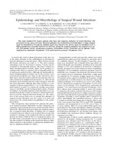
3 VIRION STRUCTURE AND GENOMIC ARCHITECTURE Influenza viruses are pleomorphic, spherical particles (about 100 nm in diameter), although filamentous forms can occur. The virions are relatively unstable in the environment and influenza viruses are inactivated by heat, dryness, extremes of pH, and detergents. Virions are enveloped and their lipid membrane is derived from the host cell. The envelope lodges different glycosylated proteins that project from the surface of the virus. These proteins are the hemagglutinin (HA), the neuraminidase (NA), and the M2 ion channel proteins. The morphology of influenza A virus particles is, therefore, characterized by distinctive spikes, with lengths from 10 to 14 nm, which are readily observable in electron micrographs of virus particles. The approximate ratio between HA and NA is 4:1. The matrix protein (M1) is situated just beneath the envelope, and underlying the M1 layer a helical superstructure, representing the core of the virus particle which is made of the ribonucleoprotein (RNP) complex, is observed. The RNP complex consists of the viral RNA segments, which are coated with the nucleoprotein (NP) and associated with the heterotrimeric polymerase complex (PB1, PB2, and PA) (Fig. 1.1). The chemical composition of virus particles is approximately 1% RNA, 5–8% carbohydrate, 20% lipid, and approximately 70% protein.9
3 Virion structure and genomic architecture
3
FIGURE 1.1 Schematic Representation of the Structure of Influeanza Virus
The genomes of influenza A and B consist of eight separate negative-sense, single-stranded RNA segments known as viral RNAs. The influenza C genome has only seven segments. Each viral RNA segment exists as a structure known as viral ribonucleoprotein (vRNP) complex in which the RNA strand is wrapped by the NP and forms a helical hairpin that is bound on one end by a single heterotrimeric polymerase complex. Within the RNPs, RNA molecules account for the genetic material of the virus, NP monomers cover and protect it, whereas the polymerase complex is responsible for the transcription and replication.10 Noncoding sequences are present at both 5′ and 3′ ends of the viral gene segments; partially complementary sequences are located at the extreme termini which are highly conserved between all segments in all influenza viruses. When base-paired, these ends function as the viral promoter that is required for replication and transcription. Each of the eight segments of the influenza A virus genome encodes for the viral proteins (Table 1.1). The whole influenza genome encodes for 10 major proteins, although alternative protein products have been characterized from several genome segments.11 The complete coding capacity of the influenza virus genome is only partially known and processes like splicing, and the use of alternative initiation codons or overlapping frames are employed by the virus to augment the variety of viral products that are generated during the infectious cycle. In fact, among the RNAs present, 2 of them (segment 7 and 8) are transcribed to messenger RNAs (mRNAs) which are then spliced into two chains encoding a further gene product each. Additionally, segment 2 presents at the 5′ end an alternative open reading frame that encodes for the 87 aa long PB1-F2 polypeptide. The full expression capacity of the influenza genome is further complicated by the abundant synthesis of small viral noncoding RNAs, whose roles in productive infections are still largely unknown.12,13 The three largest RNA segments each encode for one of the viral polymerase subunits, PB2, PB1, and PA.14 The influenza virus RNA-dependent RNA polymerase is a 250-kDa complex of these three genetic products. Electron microscopy studies have shown a compact structure with the three subunits
4
CHAPTER 1 INFLUENZA VIRUS INFECTIONS
Table 1.1 Schematic Representation of the Protein Products of Influenza A Virus Gene Segments Genome Segment
Name
1
PB2
PB2-S1
2
Protein
Function Component of the RNA polymerase (cap recognition) Binds to PB1, inhibits signaling pathways
PB1
Component of the RNA polymerase (enlongation)
N40
Regulates PB1 and PB1-F2 expression
PB1-F2
Virulence factor (proapoptotic)
PA
Component of the RNA polymerase (endonuclease activity)
PA-X
Modulates host response and virulence
PA-N155
Unknown
PA-N182
Unknown
4
HA
Surface glycoprotein, binding to receptor and fusion mediator, major antigen
5
NP
RNA-binding and RNA synthesis. RNP nuclear import
6
NA
Surface glycoprotein, neuraminidase activity
3
3 Virion structure and genomic architecture
5
Table 1.1 Schematic Representation of the Protein Products of Influenza A Virus Gene Segments (cont.) Genome Segment
Name
7
M1
Matrix protein, multiple roles in virion assembly and infection
M2
Membrane protein, ion channal activity
M42
Can replace M2 in M2-null viruses
NS1
Multifunctional protein, INF antagonist activity
NS2/NEP
Mediates RNP nuclear export
NS3
Associated to new host adaptation ability
8
Protein
Function
tightly associated.15 PB1, representing the core of the complex and the most conserved of the polymerase subunits, contains the enzymatic motifs needed for RNA polymerization activity.16 As previously remembered, the second segment also encodes for an accessory protein, PB1-F2, from an alternate open reading frame within the PB1 gene. PB1-F2, which is unique to influenza A, has been found to localize to mitochondria and exhibiting a pro-apoptotic activity. The PB2 subunit recognizes and binds to the host mRNA cap structures generated by the cellular transcription machinery17,18 and the acidic protein PA has an endonucleolytic activity necessary for the viral cap-snatching process.19,20 Segment 4 codes for the HA protein. The mature HA protein is a trimeric type I integral membrane glycoprotein (with a single transmembrane span, N-terminus in the ectodomain and C-terminus in the cytosol) which is found in the lipid envelope of virions and on the surface of infected cells.21 HA is the most abundant proteic component of the viral envelope and is the major target for neutralizing antibodies. HA undergoes several posttranslational modifications including glycosylation,22,23 palmitoylation,24 proteolytic cleavage, disulphide bond formation, and conformational changes. Host cell proteases operate the maturation of the precursor HAO molecule into its HA1 and HA2 subunits (which are linked by a disulphide bridge) (Fig. 1.2, Part 1: HA1 in blue and HA2 in red) and this cleavage mechanism is essential for the fusion activity of HA. Moreover, HA is in charge of the binding of virions to host cell surface receptors (sialic acid).25
6
CHAPTER 1 INFLUENZA VIRUS INFECTIONS
FIGURE 1.2 Influenza Virus HA Mediated Entry Mechanism (1) Influenza HA binds to sialic acids. (2) Low pH conformational change of HA releases the fusion peptide at the N-terminus of HA2 and a conformational change locates the fusion peptide on the distal part of the extended helix and insertion of the fusion peptide into the cell membrane occurs. The transmembrane domain links the HA2 with the viral envelope. (3) At final low pH, a further conformational change drives a refolding mechanism which leads to the formation of a trimeric coiled-coil (the six-helix bundle) that positionates both the transmembrane domains and the fusion peptides in the same fused membrane.
RNA segment 5 of influenza encodes RNA-binding protein NP.26 This is a highly basic protein whose main function is encapsidation of the viral RNA (an NP monomer of 56 kD binds 24 bases of RNA)27 leaving accessibility to the polymerase as a template for transcription.28 NP also plays a crucial role in transporting the viral RNPs into the nucleus.29,30 In fact, RNPs are too large to passively diffuse through the 9 Å nuclear pores.31 In order to shuttle RNPs in and out of nuclei and from the cytoplasm to the cell periphery, NP makes multiple viral and cellular protein associations involving distinct NP recognition sequences (NLSs, nuclear localization signals, and NES, nuclear export signals). During the late stages of infection, NP also associates with the cytoskeleton32 and NP has been found to bind to filamentous-actin.33 Segment 6 of influenza A encodes the NA protein which is the second major viral glycoprotein. This type II integral membrane protein (single transmembrane span, C-terminus in the ectodomain and N-terminus in the cytosol) is a tetramer and possesses critical sialidase (NA) activity that is needed for a productive release of viral particles from the infected cell.34 Segment 7 of influenza A encodes two proteins, the M1 and M2 protein. M1 is expressed from a collinear transcript, while M2 is derived from an alternative spliced mRNA. M1 associates with lipid membranes and plays an essential role in viral budding. It also regulates the movement of RNPs out of the nucleus and inhibits viral RNA synthesis at later stages of viral replication. M2 is a tetrameric type III membrane protein that has ion channel activity. It functions primarily during virus entry where it is responsible for acidifying the core of the particle which triggers dissociation of M1 from the viral RNPs (uncoating).
4 Viral replication
7
The shortest RNA (segment 8 in influenza A) encodes the NS1 protein from a collinear transcript and the NEP/NS2 protein from an alternatively spliced transcript. NS1 is a RNA-binding protein that is expressed at high levels in infected cells and is useful to inhibit host antiviral response, besides interfering with the host mRNA processing. The NEP/NS2 protein mediates the nuclear export of newly synthesized RNPs, corresponding with its expression at later times during viral infections. Recently, several additions have been made to the list of gene products coded by the influenza A genome.35 As just described earlier in the paragraph, the PB1-F2 protein was discovered in 2001 and is encoded by an alternative open translation initiation sites near the 5′ end of the PB1 gene.36 Moreover, a third protein is made by the same mechanism by the PB1 gene, named PB1-N40. PB1-N40 is an N-terminal 39 aa polypeptide which is translated from the fifth AUG codon in frame with the PB1 start.37 In addition, segment 1, coding for PB2, produces a newly discovered viral protein, termed PB2-S1, encoded by a novel spliced mRNA in which the region corresponding to nucleotides 1513– 1894 of the PB2 mRNA is deleted.38 Novel polypeptides are also encoded by genomic segment 3 and are named PA-X, PA-N155, and PA-N182. The first of them is derived from a second open reading frame (X-ORF), accessed via ribosomal frameshifting39 and modulates host response.40 The other two (PA-N155 and PA-N182) do not have a polymerase activity, and have been found to be important for virulence and pathogenesis.41 Expression of specific spliced viral products throughout infection is also applied for two of the smallest segments, M1 and NS1. It was already known for a while that protein products from segment 7 included the matrix (M1) and ion channel (M2) proteins, made from a spliced transcript, but a further protein product with an alternative ectodomain and again by a splicing mechanism, has been recently identified, the M42.42 Finally, segment 8, besides the nonstructural (NS) protein NS1, also encodes a nuclear export protein NS2/NEP, and NS3 by alternative mRNA splicing.43
4 VIRAL REPLICATION Influenza viruses bind to neuraminic acids (sialic acids) on the surface of cells to initiate the entry process25 (Fig. 1.3). Notwithstanding the ubiquitous nature of sialic acids, HAs of influenza viruses infecting different animal species show some preference for particular glycosidic linkages of the receptor. Human viruses preferentially bind to sialic acids linked to galactose by alfa 2,6 glycosidic bonds (SAα2,6Gal). On the other hand, avian viruses show a preference for alfa 2,3 glycosidic bonds (SAα2,3Gal).44,45 The preference of different influenza subtypes for diverse host-species is explained by the fact that SAα2,6Gal is found mostly in human trachea, while SAα2,3Gal is abundant in the gut epithelium of several bird species, but it should be noted that this represents a preference for receptorantireceptor linkage and not an absolute specificity. In fact, avian and human cells contain sialic acids with both linkages, though differently expressed and distributed. In addition, high viral inoculum or adaptive point mutations in the HA gene may circumvent this problem.46 HA is able to perform both action of binding and mediation of fusion of juxtaposing membranes. HA is expressed as a trimeric rod-shaped molecule on the virion surface and is produced as a HA0 precursor which is then cleaved into two subunits HA1 (the globular more external part of the molecule) and HA2 (holding a transmembrane domain) by host cell proteases. The cleavage is needed for the full activity of the molecule. The major structural features of the HA trimer are: a long stem of triple-stranded coiled-coil of α-helices and a globular ectodomain exposed to the environment and derived from the HA1 portion of the monomers.47 The stalk region of HA connects the molecule to the virion envelope by a short hydrophobic
8
CHAPTER 1 INFLUENZA VIRUS INFECTIONS
FIGURE 1.3 Replication Cycle of Influenza Virus (1) Influenza virus enters cells by receptor-mediated endocytosis following the interaction between HA and sialic acids on the cell surface; the virus initially (2) localizes to early endosomes, and then (3) reaches late endosomes where a proton pump generates a gradual acidification of the endosome; HA undergoes sequential conformational changes which finally promotes the fusion of the viral and endosomal membranes. Influenza virus genetic material is released into the cytoplasm and (4) then is transported to the cell nucleus, where (5) replication occurs. (6) Protein prouction and their posttranslational modification happen in the cytoplasm and finally (7) new capsid are assembled and released by membrane budding.
sequence (transmembrane domain),25 while the globular head is responsible for the receptor binding through a receptor binding pocket located on its distal tip. The interaction between sialic acid and HA is considered to be of low affinity; therefore, in order to increase the total intensity of the interaction, several HA molecules cooperate to link more sialic acids molecules leading to a high-avidity binding on the cell surface.48 After binding to the receptor (Fig. 1.2, part 1), influenza viruses are taken up into the cell. Early imaging studies revealed that the virus enters the cell by receptor-mediated endocytosis.49–51 The main mechanism is still recognized to be by clathrin-mediated endocytosis, although a nonclathrin, noncaveolae pathway has been also attributed to influenza entry.52,53 Regardless of whether the influenza viruses are taken up by endocytosis or macropinocytosis, they end up exploiting the transport system via distinct endosomal stages and consequently changes in pH to release their viral ribonucleoprotein complexes (vRNPs) into the cytoplasm. After internalization by either uptake pathways, the virus initially localizes to early endosomes and then reaches late endosomes. A crucial function is devoted to proton pumps, which mobilitate protons from cytoplasm into the endosomal lumen; therefore, allowing for a gradual acidification of the endosome content.54,55 Initial lowering of pH in endosomes radically changes HA structure, which is rendered vulnerable to be digested by proteases. Consequently, HA is cleaved into the two subunits, connected together by disulphide bonds. Once the influenza virus is in the acidic environment of the late endosome, HA undergoes further conformational changes
4 Viral replication
9
that expose the fusion peptide at the N-terminus of the HA2 and locate it toward the endosomal membrane.56,57 This in turn leads to the interaction of HA with the membranes of the endosome. Following the stepwise conformational changes, the fusion peptide is finally inserted into the target endosomal membrane (Fig. 1.2, part 2) and juxtaposition of the viral and endosomal membranes is reached.58–60 The crystal structures of pre- and postfusion HA (Fig. 1.2, part 1 and 3, respectively) have been solved, while only models based on such structures are available to predict the intermediate stages of the HA gradual conformational modifications. Furthermore, it has been demonstrated that several HA trimers (at least nine) are needed to execute membrane fusion by concurrent conformational changes to release the required energy.61 To proceed with fusion between viral and endosomal membranes with the increasing acidification, HA trimers tilt (Fig. 1.2, part 3) at the fusion site and a hemifusion stage is created where the outer leaflets of the interacting membranes are blended together and only the internal leaflets of the two membranes preserve the content from the virus and the endosome to mix together.62,63 Finally, both membranes fuse and a so-called fusion pore is established.64–66 As a consequence of these chain of processes, the influenza virus genetic material, in the form of a RNP complex, can be released through the fusion pore into the cytoplasm.67 Once fusion of viral and endosomal membranes has been achieved, uncoating of the viral capsid is necessary to release the VRNPs into the cytosol. This process appears to require the coordinated action of two viral proteins, namely M1 and M2. Considering influenza virion particle, M1 can be visualized by electron microscopy forming a structured stratum beneath the viral envelope.68,69 M1 serves to link the viral membrane containing the glycoproteins with the RNPs in the virus core through interactions between the middle domain of M1 with the NP on the RNP.70 During the uncoating process, the interactions of M1 with either the viral membrane or the vRNPs need to be released for allowing complete uncoating and subsequent transport of the RNPs into the nucleus. This requires the activity of the viral protein M2 which mediates the influx of H+ ions from the endosome into viral particle. This ion channel activity of M2 is regulated by pH, in fact, lowering the pH, the ion channel activity increases.71 Upon acidification of the endosome, M2 facilitates the proton influx from the endosome into the virion, leading to a pH values drop inside the virus particle.72 This change in pH inside the virus particle causes conformational changes in M1, therefore, the interaction between the RNPs and M1 is greatly weakened or lost pushing the release of the vRNPs into the cytoplasm.73,74 Finally the eight segments are transported as one moiety to the nucleus75 where RNA synthesis of influenza virus occurs. RNP complexes are not able to passively diffuse through nuclear pores (since the small size of the pores); therefore, proteins belonging to the family of α-importins recognize nuclear localization signals (NLS) on the NP protein and play an important role in the transport of RNA complexes.76 In order for the genome to be transcribed, it first must be converted into a positive sense RNA to serve as a template for the production of viral RNAs. Once in the nucleus, genomic ssRNA of negative polarity serves as a template for the synthesis of two different ssRNAs of positive polarity: (1) messenger RNAs (mRNAs), and (2) full-length complementary copies (cRNAs). Messenger RNAs are the incomplete copies of the template and they are capped and polyadenylated. The viral RNA- dependent RNA polymerase (RdRp) is made up of the three viral proteins: PB1, PB2, and PA. During transcription, viral RdRp interacts with the host polymerase II.77 PB2 shows endonuclease activity and binds to the 5′ methylated caps of cellular mRNAs cleaving the cellular mRNAs’ 10–15 nucleotides 3′ to the cap structure (this phenomenon is called “cap-snatching”). This cellular capped RNA fragment is then used by the viral RdRp to prime viral transcription.78–81 It is recognized that the three subunits of the polymerase act cooperatively and the synthesis of RNAs is a concerted action of all subunits. The synthesis of full-length complimentary copy (cRNA) of the vRNA does not need capped primer and the
10
CHAPTER 1 INFLUENZA VIRUS INFECTIONS
cRNA chain is not prematurely terminated and polyadenylated, as is the case of viral mRNA synthesis. The next step of replication of influenza genomic segments is the copying of vRNA on the template of positive strand cRNAs. This process also generates full-length products which assemble with NP and polymerase subunits and form RNP complexes. Such complexes are finally exported into the cytoplasm; the M1 protein and NS2 protein play a vital role in the translocation of these macromolecular structures.30 Packaging of eight different RNA segments in the form of RNPs into virion shells is a poorly understood phenomenon, though many models have been proposed.82–84 Some facts suggest the presence of packaging signals at both ends of the genomic segments; however, the precise sequences or structures responsible for specific packaging are still not well defined. All eight segments probably form a supercomplex before arrival at the plasma membrane binding sites.75 Before being directed into lipid rafts, these two glycoproteins are posttranslationally modified. The modifications take place in the endoplasmic reticulum and in the Golgi apparatus. In the endoplasmic reticulum, these proteins become correctly folded and glycosylated. They are also assembled into oligomers: HA into trimers and NA into tetramers. Capsids get assembled underneath the cell membrane where the inserted viral glycoprotein interacts with the matrix and the RNPs and the envelope virions exit by membrane budding. NA is required to remove sialic acids in order to allow the virus to leave its host cells.
5 CLINICAL FEATURES AND PATHOGENESIS IN HUMANS Influenza A viruses cause a respiratory illness known as “flu” in humans of all age groups all across the world. Generally, the disease is mild, but in immunocompromised individuals, pregnant women, children, and the aged, it may cause severe symptoms and in some cases it can be life threatening.85 Direct person-to-person spread during acute infections represents the major mechanism for maintenance of the virus in human populations. Influenza is generally regarded as a seasonal disease for temperate climate regions.86 In the northern hemisphere, epidemics usually peak between January and April,87,88 while in the southern hemisphere, major outbreaks hit between May and September. On a global perspective, influenza virus infections are detectable throughout the year.88 The period of incubation is 1–3 days and the most effective mechanism of spread among humans is by aerosol production.89,90 Notwithstanding the fact that influenza is generally considered a mild disease, the collective burden can be significant. In fact, the direct costs include hospitalizations, medical fees, drugs, and testing, while indirect costs such as loss of productivity mainly derive from school and workplace absenteeism. Children younger than 2 years of age and the elderly have the highest hospitalization rates.91 The severity of infections depend on the level of preexisting immunity, age of each individual, and virulence of the virus involved, all of these factors can thoroughly vary among individual outbreaks.92–94 Reinfection with a closely related variant can occur,95,96 but symptoms are usually less severe than those resulting from a previous encounter with a similar virus strain. In fact, it was reported that, during the H1N1 pandemic in 2009, older subjects exposed to viruses of previous pandemics (mainly 1918 and 1957) could count on a partial protection against the 2009 strain.97–100 As previously noted, influenza infections can, in some cases, lead to a fatal outcome; however, it is difficult to determine the number of influenza virus–related deaths, because of the absence of a laboratory diagnosis which do not allow the certification of influenza as a primary cause of death. Anyway, fatal cases have been increasing over the last decades, perhaps as a consequence of an ever increasing number of elderly and/or immunocompromised individuals. Mortality can affect all age
5 Clinical features and pathogenesis in humans
11
groups but is principally reported in people older than 65 years (90%).101,102 Major circumstances that lead to a more severe outcome of the infection in the elderly are poor cardiovascular and pulmonary conditions as well as metabolic or neoplastic diseases.103 Human influenza viruses replicate almost exclusively in superficial cells of the respiratory tract, with virus being recoverable from the upper and lower tracts of infected people. The optimal site of growth in the respiratory tract for influenza viruses is, in part, determined by the prevalence of the SAα2,3Gal or SAα2,6Gal receptors. The latter is abundantly expressed on human epithelial cells of the whole respiratory tract, allowing human strains of influenza to bind and infect cells, while SAα2,3Gal is only present on nonciliated cuboidal bronchiolar cells at the junction between the respiratory bronchiole and alveolus, and on cells lining the alveolar wall. This finding offers an explanation not only for the severe pneumonia in humans with avian strains but also for their limited spread in humans.104,105 Virus replication reaches its peak after about 48 h, then the virus titer starts to slowly decline but virus shedding is still present after days 6–8. A good positive correlation has been found between the amount of virus shed and the symptomatology. Interestingly, children still shed virus after 12–13 days following the onset of symptoms. Therefore, children are major contributors in the spread of influenza higher titers and more prolonged shedding highlights the important role of this population.106,107 Influenza A virus induces changes throughout the respiratory tract, but the most clinically important pathology develops in the lower respiratory tract.108–110 In uncomplicated influenza infections, acute inflammation of the larynx, trachea, and bronchi showing mucosal inflammation and edema are present. Infected cells become vacuolated, edematous, and lose cilia before desquamating. Infiltration of neutrophils and mononuclear cells lead to submucosal edema and hyperemia.85 In more severe primary viral pneumonia, an interstitial pneumonitis develops with a predominantly mononuclear leukocyte infiltration. Some influenza types have a higher pathogenicity (HPAI) (discussed below) and the pathologic changes associated with HPAI viruses include a hemophagocytic syndrome, renal tubular necrosis, lymphoid depletion, and diffuse alveolar damage with interstitial fibrosis.111–115 Necrotizing changes may occur with rupture of alveoli and bronchiole walls. At the cellular level, induction of apoptosis represents an additional mechanism of cell destruction.116 Regeneration of the epithelium begins 3–5 days after the onset of clinical illness. Complete healing of the epithelial damage takes up to one month. From a clinical point of view, infection with influenza A viruses results in an array of alternatives ranging from asymptomatic infection to primary viral pneumonia that rapidly progresses to a fatal outcome. The typical uncomplicated influenza syndrome is tracheobronchitis with some involvement of small airways.117 The onset of illness is usually abrupt, with headache, chills, and dry cough, which are rapidly followed by high fever, myalgias, malaise, and anorexia. Fever is the most prominent sign of infection and generally peaks within 24 h with values of 38–40°C.85 Other respiratory tract symptoms include nasal obstruction, rhinorrhea, sneezing, and pharyngeal inflammation. Sometimes, conjunctival inflammation may occur. As the fever declines, the respiratory signs and symptoms may become more prominent. The cough that produces small amounts of mucoid or purulent sputum often arise after a dry and hacking one, and persists for 1–2 additional weeks. As a consequence of the loss of the mucociliary stratum, a higher temporary predisposition to secondary sinusitis and bacterial pneumonia develops, but little permanent damage in the lung remains.118 The clinical manifestations of influenza in children are similar to those in adults, but higher fevers that may be accompanied by febrile convulsions can often be observed. Also otitis media, croup, pneumonia, myositis, and gastrointestinal manifestations are more frequent in children than in adults.85
12
CHAPTER 1 INFLUENZA VIRUS INFECTIONS
Three distinct syndromes of severe pneumonia can follow influenza infection in children or adults, where the etiology may be viral (direct consequence of viral infection of lungs), secondary bacterial infection (impairment of local defense mechanisms), and mixed (combined viral–bacterial pneumonia).119 Primary influenza virus pneumonia may hit predominantly individuals at high risk for the complications of influenza virus infection (ie, the elderly or patients with cardiopulmonary disease),120 therefore, usually occurs in patients with elevated left atrial pressure but has also been described in patients with chronic lung disease and in pregnant women. The typical case of primary viral pneumonia has a sudden start after the onset of influenza clinical illness and quickly progresses to a severe form of pneumonia with rapid respiration rate, tachycardia, cyanosis, high fever, and hypotension. The illness may progress rapidly to hypoxemia and death in 1–4 days. Survivors can develop diffuse interstitial fibrosis.119 Combined pneumonia, due to both viral and bacterial pathogens, is the most common complication in the course of influenza epidemics. Several bacteria can be involved but most often Streptococcus pneumoniae, Staphylococcus aureus, and Haemophilus influenzae are identified.118 From the clinical point of view, this syndrome may be indistinguishable from primary viral pneumonia, except for the fact that the symptoms of pneumonia, generally, appear after the influenza symptoms. The diagnosis depends solely on the demonstration of bacteria in the sputum or in the pleural fluid. Secondary bacterial pneumonia can establish on debilitated respiratory tissues. This is a major complication of influenza that contributes dramatically to the increased mortality. The bacterial pathogens most frequently involved are S. pneumoniae, S. aureus, and H. influenzae.121 The pathogenesis of this disease is due to a correlation between viral and bacterial factors. In fact, influenza viruses favor the colonization of bacteria in the lung by acting on the epithelial cells of the upper respiratory tract, thus removing the physical barrier which opposes the lung bacterial colonization, and reducing the ciliary mucus clearance. In this syndrome, individuals recovering from a typical influenza illness and a period of improvement of 2–3 days after the acute phase develop shaking chills, pleuritic chest pain, and an increase in cough productive of bloody or purulent sputum with the reappearance of fever and the onset of dyspnea. Often, influenza virus is no longer recoverable and the same bacterial pathogens as the earlier event are involved. The myositis and rhabdomyolysis have been reported more frequently in children while viremia is highly unusual in influenza virus infections.122 Instead, serious cardiovascular complications such as myocarditis, heart failure and acute myocardial infarction in patients previously compromised may follow influenza infections. Reye syndrome is a rare complication observed mainly in children and is a rapidly progressive noninflammatory encephalopathy and fatty infiltration of the viscera, especially the liver, which results in severe hepatic dysfunction with elevated serum transaminase and ammonia levels. This syndrome is seen as a consequence of several viral infections, such as varicella, respiratory and gastrointestinal viral infections. A wide spectrum of CNS disease can be observed during influenza A virus infections in humans,123 ranging from irritability, drowsiness, boisterousness, and confusion to serious manifestations such as seizures, psychosis, delirium, and coma. Two specific CNS syndromes are associated with influenza infections: influenzal encephalopathy (may be fatal) and postinfluenzal encephalitis (extremely rare). In the latter, recovery is achieved in most cases.
6 Genesis of antigenic influenza variants and pandemics
13
6 GENESIS OF ANTIGENIC INFLUENZA VARIANTS AND PANDEMICS The reason why influenza still remains a perennial clinical problem worldwide is its unparalleled ability to evade the host’s immune system. The virus accomplishes this evasion by frequently altering the antigenicity of its surface proteins HA and NA. Antigenic differences in their NP and M1 allow influenza viruses to be classified as types A, B, or C influenza, while type A viruses further subtyping is based on the relatedness of their HA and NA glycoproteins. Currently, 16 HA (from H1 to H16) and 9 NA (from N1 to N9) subtypes have been identified in type A viruses.85 Type A influenza viruses have been isolated from various animals, including humans, pigs, horses, sea mammals, and birds. Wild waterfowl, shorebirds, and gulls are the natural reservoirs of influenza A virus and can be infected with viruses harboring combinations of 16 different HA subtypes and nine different NA subtypes. Recently, two novel influenza A virus subtypes (H17N10 and H18N11) have been identified from two different bats, namely the little yellow-shouldered bat (Sturnira lilium) and the flat-faced fruit-eating bat (Artibeus jamaicensis planirostris).124,125 Influenza viruses of these subtypes have not been isolated from any other animal species and it is actually unknown whether these viruses might be able to cross the species barrier. However, putative functional domains present on some viral proteins are conserved; the N-terminal domain of the H17N10 polymerase subunit PA which has preserved its functional and structural features. On the other hand, structural and functional analyses of the H18/N11 subtype indicate that sialic acids are not used for virus attachment and particle release, suggesting bat’s derived influenza viruses employ a different mechanism of attachment and triggering of membrane fusion in order to gain access into the host cells. Taken together, these findings indicate that bats host a potentially important pool of influenza viruses that may likely represent a novel zoonotic reservoir for mammals.126 The epidemiology of human influenza viruses is dictated by their continuous antigenic variation to escape the host immune response. Influenza viruses possess two different and complex mechanisms that allow them to reinfect humans and cause disease, namely antigenic drift (accumulation of mutations over time) and antigenic shift (rearrangement of viral RNA segments in cells infected with two or more different viruses, also known as “reassortment”). For human influenza A viruses, the evolutionary rates differ among the proteins, likely reflecting differences in the selective pressure of the host.127 In fact, the HA and NA proteins evolve faster than the PB2, PB1, PA, NP, and M1 proteins, because residue substitutions in the antigenic domains of the surface glycoproteins HA and NA are likely to produce selective advantages by allowing mutated strains to evade preexisting immunity.128 Antigenic drift, therefore, occurs as a result of point mutations in specific genes leading to minor, gradual, antigenic changes in the HA or NA proteins.129 At the nucleotide level, the reported mutation rates range from ∼5 × 10⁻⁴ to ∼8 × 10⁻³ nucleotide substitutions per site per year.130 Due to the relatively low fidelity of the RdRP, which also lacks a proofreading capacity (5′–3′ exonuclease activity), a common characteristic of all RNA polymerases, influenza constantly accumulates mutations within its genome during replication. It is important to note that these mutations may be silent or may alter the virulence and pathogenicity of the virus. For instance, if a highly pathogenic avian virus acquires the necessary mutations that facilitate its ability to efficiently enter and replicate in humans, then the virus can become a serious threat to humans. The same would apply for a virus of minor pathogenicity but is very efficacious in its human-to-human spread when it eventually accumulates enough mutations to become highly virulent. The selection of antigenic drift variants is a sequential event with the stepwise accumulation of mutations while an antigenic shift can occur all of a sudden, and at irregular and unpredictable intervals. This second mechanism
14
CHAPTER 1 INFLUENZA VIRUS INFECTIONS
of antigenic change is much less frequent since it only occurs when two different viruses coinfect the same individual. The genetic mutational mechanism is an exchange of whole segments between the two viruses. This is only possible because the influenza virus genome consists of eight separated RNA segments, therefore, coinfection of one host cell with two different viruses can result in progeny viruses containing gene segments of both parental viruses. In this way, by reassortment of genomic segments, a new virus is created, resulting in an unpredictable pathogenicity of the new virus, which may possess a surface antigenic pattern completely novel to the population. In fact, considering that this reassortment event usually occurs between viruses coming from different host-species (eg, a human virus and an avian virus), the new virus may immediately present HA and/or NA antigenic determinants from the avian strain against which the human population lacks any significant immunity. Once the pandemic strain is created (generally by antigenic shift), the new virus can further change its virulence determinants as it continues to replicate (generally by antigenic drift). Some influenza virus strains are largely asymptomatic in chickens [and are considered low pathogenic avian influenza (LPAI) viruses], while other strains cause severe disease in chickens that is often fatal within 48 h [and are considered highly pathogenic avian influenza (HPAI) viruses]. Outbreaks of HPAI viruses can cause disaster for the poultry industry.131 Anseriformes (mainly ducks, geese, and swans) and Charadriiformes (mainly gulls, terns, and waders) are considered the natural host reservoirs of LPAI viruses.127 In wild birds, the LPAI viruses predominantly infect epithelial cells of the intestinal tract132,133 and are subsequently excreted in the faeces. However, infection of wild birds with LPAI viruses is typically subclinical and occurs in the absence of obvious lesions.134,135 LPAI viruses can cross the species barrier and infect wild marine mammals as well as domesticated mammals and birds. Swine (Sus scrofa domesticus) can be easily infected by avian influenza viruses. Disease manifestations can range from an acute respiratory tract disease to an inapparent infection.136 Pigs have been traditionally perceived to act as a “mixing vessel” to facilitate the generation of novel reassortant influenza viruses. This is because swine respiratory epithelial cells express both α2,3- and α2,6-linked sialic acids136 and the simultaneous presence of both SAα2,6Gal and SAα2,3Gal would allow swine to be potentially infected by both avian and human viruses. However, more recent data showed that SAα2,3Gal receptors on swine tracheal cells are in reality not that abundant as previously considered,137 but the importance of swine in the reassortment mechanism is to be found in the presence of large numbers of pigs in close proximity to other animal species, especially domesticated water birds,138 increasing the risk of an interspecies transmission event, at least in some world areas. However, it was also reported that the crucial determinant for influenza tropism is the structure of the underlying glycocalyx, not exclusively the terminal SA linkage.139 Avian viruses prefer SA in a cone-like topology that is present in both SAα2,3Gal and SAα2,6Gal with short underlying glycans and allows HA to contact Neu5Ac and galactose sugars in a trisaccharide motif. Human viruses are reported to prefer an umbrella-like glycan topology, which is unique to SAα2,6Gal with long underlying glycans. Therefore, the α2,6 alone is not sufficient for human transmission and the SA topology is of importance to conditionate influenza tropism, not just the SA linkage present.139 A further event that may lead to pandemics is represented by the fact that terrestrial poultry can easily become infected with numerous different LPAI viruses (eg. H5, H6, H7, H9 viruses) which become endemic in the population.140,141 However, the most relevant consequence of poultry infections with LPAI viruses is the high possibility of evolution in these species from LPAI viruses into HPAI viruses by the usual antigenic drift mechanism of point mutations. As earlier described, HA undergoes a posttranslational cleavage into HA1 and HA2 subunits by host proteases, with the generation of a fusogenic domain at the amino terminus of HA2 that induces fusion
6 Genesis of antigenic influenza variants and pandemics
15
between the viral envelope and the endosomal membrane.142 Proteolytic activation is fundamental for viral infectivity and organ dissemination,143 and the HA glycoprotein has a key role in influenza virus pathogenicity.144 HA0 of seasonal human influenza A viruses, as well as LPAI viruses have the consensus cleavage site motif XT/X-R and are selectively processed by a limited number of trypsin-type processing proteases, while in HPAI viruses, endoproteolytic processing of the HA0 occurs through ubiquitous cellular processing proteases, which recognize a multibasic consensus cleavage site motifs, such as, R-X-K/R-R and K-X-K/R-R.145 Influenza viruses of subtypes H5 and H7 may become highly pathogenic after introduction into poultry and cause outbreaks of HPAI. The switch from an LPAI phenotype to the HPAI phenotype of these H5 and H7 influenza A viruses is achieved by the introduction of basic amino acid residues into the HA0 cleavage site by substitution or insertion, resulting in the so-called multibasic cleavage site (MBCS), which facilitates systemic virus replication.146–148 The main processing proteases reported for human influenza viruses in animals and humans that recognize single basic motifs are pancreatic trypsin,143 plasmin from calf and chicken,149 blood clotting factor Xa from chick embryo,150 tryptase Clara from rat lungs,151 mini-plasmin from rat lungs,152 ectopic anionic trypsin from rat lungs,153 porcine mast cell tryptase,154 tryptase TC30 from porcine lungs155 and transmembrane protease serine (TMPRSS) 2, and type II membrane protein human airway trypsin-like protease (HAT).156 In addition to the host cellular proteases, microbial proteases also proteolytically activate influenza virus HA0 in bacterial infection of the airways and may play a role in the spread of the virus.157 The processing proteases for the single basic motif are predominantly distributed in the respiratory and intestinal tract, and most of the epidemic human influenza viruses known to date are pneumotropic in which the virus proliferates in these organs. On the other hand, the multiple basic residues motifs are readily cleaved by ubiquitously present intracellular processing proteases, such as furin and proprotein convertases (PCs)5/6.158–160 In addition, type II membrane serine proteases (TTSP) of mosaic serine protease large form (MSPL) and its splice variant transmembrane protease serine 13 (TMPRSS13) have recently been identified.161 The effect of the experimental introduction of an MBCS into a primary LPAI H6N1 virus, A/Mallard/Sweden/81/2002, has recently been described and such introduction resulted in trypsin-independent replication in vitro and enhanced pathogenesis in a chicken model.162 Influenza A viruses can, therefore, cause seasonal diseases as well pandemic episodes which are mainly characterized by the fact that, due to massive genetic variations, novel strains to which humans have little or no immunity, can be generated. All of the known subtypes of influenza A viruses can infect birds, except subtypes H17N10 and H18N11, which have only been found in bats and only two influenza A virus subtypes (ie, H1N1 and H3N2) are currently in general circulation among humans. There are also some subtypes that infect other animals, such as, H7N7 which can cause flu disease in horses, and H3N8 virus infection that can also be found in dogs. Historical data suggests that there may have been at least 14 pandemics over the past 500 years (1509–2009), which is approximately one pandemic every 36 years. But our understanding of pandemic influenza viruses at the viral level is quite limited since pandemic viruses have only been isolated from 1957 onward, and the 1918 pandemic virus was reconstructed using an “archaeovirological” approach.163 We do not yet know the subtypes or genetic makeup of the pandemics before 1918, therefore we will consider as “pandemic era” the events from 1918 onward. In the last 100 years we have experienced five pandemics which happened in the years: (1) 1918–1919; (2) 1957–1958; (3) 1968; (4) 1977–1978; and (5) 2009–2010. The most important data on these pandemics are summarized in Table 1.2.
16
CHAPTER 1 INFLUENZA VIRUS INFECTIONS
Table 1.2 Details on the Four Major Pandemics of the Last 100 Years Years
1918–19
1957–58
1968–69
2009–10
Name
Spanish flu
Asian flu
Hong Kong flu
Swine flu
Area of emergence
Unclear
Southern China
Southern China
Mexico – Southern USA
Subtype
H1N1
H2N2
H3N2
H1N1
Estimated mortality worldwide
20–50 million
1–4 million
1–2 million
125–570 thousands
Estimated fatality rate
2–3%
0.2%
0.2%
0.03%
Age groups most affected
Young adults
Children
Across all age groups Young people 5–40 years of age
Details
Since no pre-1918 influenza viruses are available, the origin and eventual shift from an animal host remain unknown. The high mortality was the result of bacterial pneumonia
This virus emerged as a linear descendent of the 1918 H1N1 with aquisition of three novel genes from an unknown avian virus (HA, NA, and PB1)
Reassortment event with an avian influenza virus with novel H3 and PB1 gene segments. The other six genes were retained from the 1957 H2N2
HA, polymerase genes, NP and NS derived from a triple-reassortant virus circulating in North American swine, while the NA and M gene derived from the Eurasian avian-like swine H1N1 lineage. The triple-reassortant itself comprised genes derived from avian (PB2 and PA), human H3N2 (PB1) and classical swine (HA, NP, and NS) lineages
The 1918–1919 Pandemic Virus: The Spanish influenza pandemic first struck in 1918 as an unusual respiratory disease that was associated with a disproportionate increase in deaths among young adults. The origin of the Spanish influenza is not clear, but reconstructions of the viral genomes from the tissues of several victims demonstrated that the causative agent was an avian-descended H1N1 virus. Epidemiological features of the pandemic were also unprecedented, including its appearance in up to three waves within the first year and the extremely high mortality registered. The pandemic killed an estimated 50 million people.164 The 1957–1958 Pandemic Virus: The “Asian flu” originated in the Yunan Province of China in the spring of 1957, spreading rapidly to South-East Asia and Japan, and subsequently to Australia, Indonesia and India, and after to Europe, Africa, North and South America, and the Caribbean. In just 6 months, the pandemic had covered the globe. A second wave occurred during the autumn of 1957. In
6 Genesis of antigenic influenza variants and pandemics
17
addition, if the origin of the virus has been connected to the 1918 H1N1 pandemic virus, reassortment events took place to generate a new virus especially for its external antigenic makeup. The gene segments encoding HA and NA were replaced by an avian-like H2 subtype HA and an N2 subtype NA,165 with the other five gene segments retained from the 1918-derived H1N1 lineage. Altogether, it affected about 40–50% of the global population, with 25–30% experiencing clinical disease. The 1968 Pandemic Virus: This pandemic also originated in China, in 1968, spreading to Hong Kong and thereafter to India, Australia, Europe and USA. The 1968 H3N2, named the “Hong Kong”, pandemic, was caused by a reassortment event between a circulating human H2N2 virus and an avian strain. The new configuration was derived by acquiring novel HA (H3 subtype) and PB1 gene segments.166 Compared to the earlier pandemics of the century, the Hong Kong flu was relatively mild (reduction of the duration and severity of the clinical illness), possibly as a result of immunity in the population against the N2 subtype NA which the new pandemic H3N2 virus shared with the circulating H2N2 virus. The 1977–1978 Pandemic Virus: Strictly speaking, there was a fourth pandemic, the Russian Flu, in the 20th century. It was sparked off by an H1N1 strain which appeared in 1977. This pandemic was considered a benign pandemic, or not a “true” pandemic, and primarily involved subjects born after 1950 (so people aged under 25 years), since the older population already presented a protective immunity as a consequence of prior experience with the H1N1 strain. Molecular characterization of this virus showed that both the HA and NA antigens were remarkably similar to those of the 1950s.167 It is believed to not be probable that an influenza virus could have been maintained in nature for 20 years without accumulating mutations, therefore it has been suggested that the 1977 epidemic resulted from the accidental release of a laboratory strain from the 1950s. The 2009–2010 Pandemic Virus: The 2009 H1N1 pandemic virus was derived by reassortment between two preexisting swine influenza viruses, a North American swine H1N2 “triple-reassortant” lineage virus and a Eurasian H1N1 swine lineage virus (Fig. 1.4). It is not currently known whether the novel virus emerged first in humans or swine.168,169 The 2009 pandemic virus is also connected to the 1918 pandemic virus.170 In fact, it derives from multiple reassortment events ultimately ascribed to the “classical” swine H1N1 lineage that circulated enzootically in swine in North America since 1918. The novel H1N1 virus was first detected in a widespread outbreak in Mexico in March–April 2009,171 but may have been circulating in people as early as late 2008.172,173 While severe pneumonia has been described, especially associated with the initial Mexican outbreak,98 most cases in the United States and in other countries have been self-limited, and appeared clinically similar to seasonal influenza.174 Human infection with H1N1 have generally resulted in low mortality, although certain subgroups (including pregnant women, chronic medical conditions sufferers, immunosuppressed people) had significantly higher risk of severe disease. The 2009 H1N1 pandemic derived its NA and M gene segments from the European avian-like H1N1 lineage and its remaining six gene segments (PB2, PB1, PA, HA, NP, and NS) from the North American swine H1N2 “triple” reassortant lineage. The HA, NP, and NS gene segments of this lineage are derived from the classical swine H1N1 (1918 origin) lineage.173 Notwithstanding the evidence that the combination of one of the HA subtypes with one of the NA subtypes allows for a wide genetic diversity which could lead to a pandemic event, within the brief period of modern molecular virology, of the 16 HA subtypes known to exist, pandemics have only been caused by viruses of the H1, H2, and H3 subtypes. All of the other HA subtypes have, up to now, been demonstrated to produce novel influenza strains with a very low or nonexistent capacity for humanto-human transmission. Nevertheless, the possibility of further adaptation of the virus in humans and
18
CHAPTER 1 INFLUENZA VIRUS INFECTIONS
FIGURE 1.4 Antigenic Shift of the Generation of the Pandemic Virus H1N1 of 2009
the possibility to acquire a sustained human-to-human transmission must be taken into consideration assuming that the nature of the next pandemic virus cannot be predicted, but that it will arise from one of the 16 known HA subtypes in avian or mammalian species. In recent years, several novel reassorted influenza viruses (eg, H5N1, H7N9, H9N2, H10N8) have trespassed the host-species barrier and are under surveillance by the scientific community and public health systems. It is still unclear whether these viruses can actually cause pandemics or just isolated episodes. The ecological conditions in many Asian countries are such that new viruses with pandemic potential may arise relatively easily. These conditions include the year-round circulation of influenza viruses, along with the density of human, swine, and domestic poultry populations, often living in close proximity. These conditions may facilitate genetic reassortment or direct transfer of avian viruses to humans. The prominent subtypes of avian influenza A viruses known to infect both birds and humans are described below. Influenza A(H5N1) was first detected in 1996 in geese in China and reported in humans in 1997 during a poultry outbreak in Hong Kong.175 It has since been detected in poultry and wild birds in more than 50 countries in Africa, Asia, Europe, and the Middle East. Since their reemergence in 2003,112 highly pathogenic avian influenza (HPAI) A(H5N1) viruses of the A/goose/Guangdong/1/96 hemagglutinin (HA) lineage have become enzootic in six countries (Bangladesh, China, Egypt, India, Indonesia, and Vietnam).176,177 More than 700 human infections with Asian HPAI H5N1 viruses have been reported since 2003 and Indonesia, Vietnam, and Egypt have reported the highest number of human HPAI Asian H5N1 cases to date. The first report of a human infection with Asian H5N1 in the Americas was in Canada in 2014 and occurred in a traveler who had recently visited China.178 Most human
6 Genesis of antigenic influenza variants and pandemics
19
infections with HPAI Asian H5N1 have occurred after prolonged and close contact with sick or dead poultry or wild infected birds. Human-to-human spread is rare and approximately 60% of the cases are fatal. One human infection in China was caused by an A(H5N6) virus in 2014.179 The influenza A(H7N9) virus is one subgroup among the larger group of H7 viruses, which normally circulate among birds. This particular A(H7N9) virus was not detected in humans until it was found in March 2013 in China. The patients were two men aged 87 and 27 years and a woman aged 35 years. All three patients already presented unfavorable medical conditions and died.180 Two waves were identified, the first from Feb. to May 2013, with 133 cases, and the second from Oct. 2013 to Feb. 2014. By the end of May 2014, a total of 439 cases of H7N9 infection had been reported to the WHO, with a case fatality rate of 30%.181 Illness in humans may include conjunctivitis and/or upper respiratory tract symptoms. Live poultry markets were indicated to likely be the principal source for human infections because most of the infected patients were reported to have had recent contacts with live poultry.182 The novel H7N9 virus was of avian origin with HA derived from the duck H7N3 subtype, the NA gene from a migratory bird H7N9 virus in Korea and the six internal genes from poultry H9N2 viruses.180 It seems that this novel reassortant could be transmitted from poultry to humans more easily than H5N1 and a recent epidemiological study showed that a considerable proportion of people involved in the poultry industry have been exposed to H7, suggesting a high diffusion of mild or asymptomatic infections.183 Influenza A H9 viruses have been identified worldwide in wild birds and poultry and they are all LPAI viruses. Rare, sporadic H9N2 virus infections of humans have been reported to cause generally mild upper respiratory tract illness. The first cases of infection with an H9N2 influenza strain were reported in China and affected two children.184 The symptoms of infection with H9N2 are mild in comparison with those of H5N1 infection.185 At a molecular level, it has been described that the internal genes of H9N2 are similar to those of the H5N1 that caused the infections in 1997,186 therefore, H9N2 has been posed under control of health authorities as a potential pandemic strain. Influenza A (H10N8) viruses have circulated mainly in wild birds since their first detection in quails in Italy in 1965.187 The first H10N8 avian influenza virus was isolated from a duck in Guangdong in the beginning of 2012.188 This novel avian influenza strain appeared for the first time in humans by the end of 2013. So far, three cases have been recorded, all in China, with two fatal outcomes. The virus isolated from the human cases was genetically distinguishable from bird H10N8 viruses, with long genetic distances between their HA and NA genes, and also contained the H9N2-derived internal genes.189 It is quite difficult to assess the real pandemic potential posed by H10N8, in fact, despite the severe infections caused in humans, leading to death in two out of three cases, the reported human cases are too sporadic to allow any sensible epidemiological study. Furthermore, no other cases have been reported since February 2014. Finally, we should note the appearance in Taiwan of influenza A H6N1 in 2013 when this novel virus was identified in a 20 year-old female patient.190 As of September 2014, there has been no documentation of person-to-person transmission of the virus or further cases. Molecular characterization of H6N1 revealed that it is a typical avian influenza virus of low pathogenicity likely derived from reassortment events from different H6N1 lineages circulating in chickens in Taiwan, which might not replicate and propagate well in the upper airway of mammals.191 On the other hand, a single HA mutation of the human isolate might have increased its mammalian receptor binding ability and, hence, its pathogenicity in humans. Based on these data, and considering the cocirculation and potential reassortment with several other influenza subtypes, this H6N1 virus merits consideration.
20
CHAPTER 1 INFLUENZA VIRUS INFECTIONS
7 INFLUENZA TREATMENT The most adopted strategy to fight influenza infections is through vaccination, however, vaccines need to be reformulated each year due to the genetic variability of the virus and they are not always completely protective. Moreover, in case of pandemics, it is hardly impossible to predict the newly emerging strain and, therefore, containment of pandemics by vaccination is restrained by the time-limiting factor (the period going from the understanding of the pandemic risk to the worldwide diffusion of the infection). The available drugs are somewhat limited in their use by the rapidly increasing drug resistances, therefore, additional antiinfluenza therapeutics are urgently needed. Thus, the rest of the chapter will discuss the current and past treatments that have been available for influenza in recent years together with some of the most interesting compounds under development. The first identified inhibitor against influenza virus was amantadine192 (Fig. 1.5a). Amantadine is an amine derivative of adamantane consisting of an adamantane backbone that has an amino group substituted at one of the four methyne positions. Rimantadine is a closely related derivative of adamantane with similar biological properties. Both compounds are M2 ion channel inhibitors and were licensed as first generation antiinfluenza drugs and are still commercially available as Symmetrel (Endo Pharmaceuticals) and Flumadine (Forest Pharmaceuticals), respectively. The M2 transmembrane protein serves to selectively transport protons inside the viral particle for directing the uncoating of the particle and the release of RNPs into the cell cytosol. Both drugs exert an action on M2 ion channels by intercalating into the interior of the channel.193 In fact, they both bind to the N-terminal channel lumen disabiliting its pump activity. Their efficacy is limited to influenza A only, since influenza B M2 channel pore lumen contains polar, and not hydrophobic, amino acids.74 Furthermore, rapid emergence of drug-resistant virus strains (almost 100% for the circulating seasonal H3N2 and 2009 pandemic flu) restricts the use of both drugs
FIGURE 1.5 Compounds to Fight Influenza Virus (a) M2 ion channel inhibitors; (b) neuraminidase inhibitors.
7 Influenza treatment
21
for prevention and treatment in favor of new-generation influenza drugs, the NA inhibitors (Fig. 1.5b). The second generation of antiinfluenza drugs act in the late stage of infection by inhibiting the release of virus particles from infected cells. Influenza virus NA is one of the two major surface glycoprotein antigens embedded in the viral envelope. Its main activity is to cleave off the terminal SA from the host cell receptors to release of progeny viruses from the membranes of infected cells, to which the virus would remain linked via the interaction with HA. Blocking the NA sialidase activity ends up in trapping the newly formed viruses on the cell surface, impairing their ability to infect neighboring cells and, therefore, impeding the spread of the infection in the organism.194,195 NA inhibitors show a broadspectrum of activity since both influenza A and B, as well as different NA subtypes keep the structural characteristics highly conserved.196 NA inhibitors are structural analogs of sialic acid and were first synthesized in the 1970s.197 Two NA inhibitors have been approved by regulatory authorities in the United States and Europe, Zanamivir (inhaled drug, 10 mg/dose; trademark Relenza, GlaxoSmithKline) and Oseltamivir (oral drug, 75 mg/dose; trademark Tamiflu, Roche).198,199 Both drugs require twice-daily administration for treatment. Furthermore, Peramivir and Laninamivir have been approved by regulatory authorities in some parts of Asia.200 Peramivir is a cyclopentane compound that potently and selectively inhibits influenza virus NA,201 and an intravenous formulation has been approved for influenza treatment (single intravenous drip infusion, 300 mg/dose; Rapiacta) in Japan and as Peramiflu in South Korea. The US Food and Drug Administration (FDA) has only recently approved intravenous use of peramivir (Rapivab, manufactured by BioCryst Pharmaceuticals), as a drug available for adult influenza patients who have trouble taking an oral or inhaled antiviral, while in Europe ending of phase III clinical trials are approaching. The other compound, Laninamivir (R-125489), is structurally similar to zanamivir and its octanoyl prodrug, laninamivir octanoate (CS-8958) is used for influenza treatment in Japan where it was approved and is commercially available as Inavir (Daiichi Sankyo Co., Ltd, Tokyo).202,203 In contrast to the currently available drugs, which require multiple dosing, for example, twice-daily for 5 days, it was confirmed that the single inhalation of CS-8958 is sufficient to treat influenza in humans.204 Although the target site for the NA inhibitors is highly conserved, again a number of resistant mutations arise as a consequence of drug-induced selective pressure.205 The mutations include substitutions in both NA and HA that reduce either the affinity of NA for the inhibitor or the affinity of HA for the sialylated glycoproteins. Among the compounds under development, a nucleoside analog is currently in phase III clinical trials by the FDA, favipiravir. Favipiravir (T-705; 6-fluoro-3-hydroxy-2-pyrazinecar-boxamide) is a pyrazine derivative which was identified by Furuta el al. in 2002.206 T-705 has been shown to inhibit influenza A, B, and C viruses in vitro and in an in vivo model.206 T-705 is converted by cellular kinases to its active form, ribofuranosyl triphosphate, and the mechanism of action is through direct inhibition of viral replication and transcription, which represent an exclusive feature among antiinfluenza drugs. A combination of favipiravir with NA inhibitors has demonstrated a synergistic activity; therefore, broadening the therapeutic options for the management of infections by highly pathogenic avian strains and for severely ill patients. Since human cells do not present RdRP domains but are conserved among different families of RNA viruses, a favipivar mechanism targeting RNA viral polymerases makes this molecule an attractive drug candidate for other RNA viruses including agents belonging to the navirus, bunyavirus, and flavivirus families.207 Further compounds in actual development can be grouped for their mechanism of action and can be divided considering the phase of the viral cycle they interrupt. One of the viral targets of considerable interest is the viral RNA-dependent RNA polymerase (RdRP), which as earlier described, is a
22
CHAPTER 1 INFLUENZA VIRUS INFECTIONS
heterotrimer composed of subunits PB1, PB2, and PA. The three polymerase subunits interact with each other, in particular the N-terminus of PB1 interacts with the C-terminus of PA,208 while the Cterminus of PB1 binds the N-terminus of PB2;209 in addition, a weak transient interaction has been proposed for PA and PB2.210 We already described favipiravir, the best characterized antiinfluenza nucleoside analog, but in view of the fact that interactions between the three polymerase subunits are essential for function, and therefore conserved along different viral strains, a synthetic peptide corresponding to residues 1–37 of PB2 was shown to inhibit the PB1/PB2 interaction in vitro, but also a peptide derived from amino acids 731–757 of PB1 can disrupt the interaction between the subunits.211–213 A further antiviral target is the HA and the attachment or fusion mechanisms in the early stages of the viral infections. Several small molecules and peptides have been developed and of note among them are EB peptide and its derivatives, FluPep and arbidol (ARB; 1-metyl-2-phenyl-thiomethyl-3-carbotoxy4-dimetylaminomethyl-5-hydroxy-6-bromoindolehydrochloridemonohydrate). EB is a 20 amino acid peptide derived from signal sequence of fibroblast growth factor 4 showing a broad-spectrum activity against human, swine, and avian influenza A H1N1, H2N2, H3N2, H5N1, H5N9, and H7N3 strains and influenza B viruses. Several data have demonstrated the ability of the peptide to inhibit viral attachment.214,215 FluPep is a mix of predominantly hydrophobic α-helical peptides capable of interaction with HA blocking the viral fusion. These peptides are derived from Tkip peptide, which is a mimetic for the suppressor of cytokine signaling protein, known to be active in modulating inflammatory cytokine responses and known as an effective antiviral drug against Poxviruses.216 A variety of influenza subtypes were inhibited by FluPep in nanomolar concentrations in cell culture.217 Finally, the small molecule arbidol exhibits a broad-spectrum of activity not only against influenza viruses A and B but also against other viruses such as RSV, parainfluenza virus, coxsackie virus, rhinovirus, hepatitis B virus (HBv), and HCV.218,219 This drug has been approved in Russia and in China for treatment and prophylaxis of influenza viruses. Studies with viruses resistant to ARB bearing mutations which map in the HA2 subunit have confirmed that ARB interacts with HA and acts by stabilizing its structure, thus preventing the low pH-induced fusogenic change of HA2.220 Recently, biochemical studies showed that ARB interacts both with cell membrane phospholipids and with aromatic residues of viral glycoproteins on the surface of enveloped viruses.221 This mechanism of action of ARB can prevent the fusogenic change in viral glycoproteins required for membrane fusion and could explain the broad-spectrum of antiviral activity of this compound.
8 CONCLUSIONS Influenza A viruses are zoonotic pathogens that continuously circulate and change in several animal hosts, including birds, pigs, horses, and humans. The emergence of novel virus strains that are capable of causing human epidemics or pandemics needs to be considered a serious possibility; therefore, surveillance and characterization (by high-throughput whole-genome sequencing) of naturally occurring influenza viruses is an imperative task of the world health system to establish drift variants and genetic changes that might be of epidemiological relevance. A great progress has been achieved in recent years in our understanding of the host tropism and virulence of influenza viruses which could lead to additional and enhanced antiviral interventions. Drugs directed against diverse viral or host targets and against distinct steps of influenza virus replication cycle would represent a terrific weapon to challenge infections by several strategies also able to minimize the development of resistant viruses. Therefore, influenza pathogenesis
REFERENCES
23
remains an indispensable field of study to elucidate the molecular requirements to understand both viral and host factors that play a crucial part in disease severity and outcome.
REFERENCES 1. Potter CW. A history of influenza. J Appl Microbiol 2001;91:572–9. 2. Shope RE, Lewis P. Swine influenza : I. Experimental transmission and pathology. J Exp Med 1931;54:349– 59. 3. Smith W, Andrews CH, Laidlaw PP. A virus obtained from influenza patients. Lancet 1933;225:66–8. 4. Burnet FM. Influenza virus on the developing egg I. changes associated with the development of an eggpassage strain of virus. Br J Exper Pathol 1936;17:282–95. 5. Krossoy B, Hordvik I, Nilsen F, Nylund A, Endresen C. The putative polymerase sequence of infectious salmon anemia virus suggests a new genus within the Orthomyxoviridae. J Virol 1999;73:2136–42. 6. Kosoy OI, Lambert AJ, Hawkinson DJ, et al. Novel thogotovirus associated with febrile illness and death, United States 2014. Emerg Infect Dis 2015;21:760–4. 7. Allison AB, Ballard JR, Tesh RB, et al. Cyclic avian mass mortality in the northeastern United States is associated with a novel orthomyxovirus. J Virol 2015;89:1389–403. 8. Osterhaus AD, Rimmelzwaan GF, Martina BE, Bestebroer TM, Fouchier RA. Influenza B virus in seals. Science 2000;288:1051–3. 9. Shaw ML, Palese P. Orthomyxoviridae. In: Knipe DM, Howley PM, editors. Fields Virology. sixth ed. Philadelphia: Lippincott Williams & Wilkins; 2013. p. 1151–85. 10. Resa-Infante P, Jorba N, Coloma R, Ortin J. The influenza virus RNA synthesis machine: advances in its structure and function. RNA Biol 2011;8:207–15. 11. Gerber M, Isel C, Moules V, Marquet R. Selective packaging of the influenza A genome and consequences for genetic reassortment. Trends Microbiol 2014;22:446–55. 12. Perez JT, Varble A, Sachidanandam R, et al. Influenza A virus-generated small RNAs regulate the switch from transcription to replication. PNAS 2010;107:11525–30. 13. Perez JT, Zlatev I, Aggarwal S, et al. A small-RNA enhancer of viral polymerase activity. J Virol 2012;86:13475–85. 14. Rodriguez-Frandsen A, Alfonso R, Nieto A. Influenza virus polymerase: functions on host range, inhibition of cellular response to infection and pathogenicity. Virus Res 2015;209:23–38. 15. Area E, Martin-Benito J, Gastaminza P, et al. 3D structure of the influenza virus polymerase complex: localization of subunit domains. PNAS 2004;101:308–13. 16. Kobayashi M, Toyoda T, Ishihama A. Influenza virus PB1 protein is the minimal and essential subunit of RNA polymerase. Arch Virol 1996;141:525–39. 17. Blaas D, Patzelt E, Kuechler E. Identification of the cap binding protein of influenza virus. Nucleic Acids Res 1982;10:4803–12. 18. Braam J, Ulmanen I, Krug RM. Molecular model of a eucaryotic transcription complex: functions and movements of influenza P proteins during capped RNA-primed transcription. Cell 1983;34:609–18. 19. Dias A, Bouvier D, Crepin T, et al. The cap-snatching endonuclease of influenza virus polymerase resides in the PA subunit. Nature 2009;458:914–8. 20. Yuan P, Bartlam M, Lou Z, et al. Crystal structure of an avian influenza polymerase PA(N) reveals an endonuclease active site. Nature 2009;458:909–13. 21. Skehel JJ, Waterfield MD. Studies on the primary structure of the influenza virus hemagglutinin. PNAS 1975;72:93–7. 22. Deom CM, Schulze IT. Oligosaccharide composition of an influenza virus hemagglutinin with hostdetermined binding properties. J Biol Chem 1985;260:14771–4.
24
CHAPTER 1 INFLUENZA VIRUS INFECTIONS
23. Schulze IT. Effects of glycosylation on the properties and functions of influenza virus hemagglutinin. J Infect Dis 1997;176(Suppl. 1):S24–8. 24. Veit M, Serebryakova MV, Kordyukova LV. Palmitoylation of influenza virus proteins. Biochem Soc Trans 2013;41:50–5. 25. Skehel JJ, Wiley DC. Receptor binding and membrane fusion in virus entry: the influenza hemagglutinin. Annu Rev Biochem 2000;69:531–69. 26. Ritchey MB, Palese P, Kilbourne ED. RNAs of influenza A, B, and C viruses. J Viroly 1976;18:738–44. 27. Ortega J, Martin-Benito J, Zurcher T, Valpuesta JM, Carrascosa JL, Ortin J. Ultrastructural and functional analyses of recombinant influenza virus ribonucleoproteins suggest dimerization of nucleoprotein during virus amplification. J Virol 2000;74:156–63. 28. Baudin F, Bach C, Cusack S, Ruigrok RW. Structure of influenza virus RNP. I. Influenza virus nucleoprotein melts secondary structure in panhandle RNA and exposes the bases to the solvent. EMBO J 1994;13:3158–65. 29. Martin K, Helenius A. Transport of incoming influenza virus nucleocapsids into the nucleus. J Virol 1991;65:232–44. 30. O’Neill RE, Jaskunas R, Blobel G, Palese P, Moroianu J. Nuclear import of influenza virus RNA can be mediated by viral nucleoprotein and transport factors required for protein import. J Biol Chem 1995;270:22701–4. 31. Paine PL, Moore LC, Horowitz SB. Nuclear envelope permeability. Nature 1975;254:109–14. 32. Husain M, Gupta CM. Interactions of viral matrix protein and nucleoprotein with the host cell cytoskeletal actin in influenza viral infection. Curr Sci 1997;73:40–7. 33. Digard P, Elton D, Bishop K, Medcalf E, Weeds A, Pope B. Modulation of nuclear localization of the influenza virus nucleoprotein through interaction with actin filaments. J Virol 1999;73:2222–31. 34. Gong J, Xu W, Zhang J. Structure and functions of influenza virus neuraminidase. Curr Med Chem 2007;14:113–22. 35. Vasin AV, Temkina OA, Egorov VV, Klotchenko SA, Plotnikova MA, Kiselev OI. Molecular mechanisms enhancing the proteome of influenza A viruses: an overview of recently discovered proteins. Virus Res 2014;185:53–63. 36. Chen W, Calvo PA, Malide D, et al. A novel influenza A virus mitochondrial protein that induces cell death. Nature Med 2001;7:1306–12. 37. Wise HM, Foeglein A, Sun J, et al. A complicated message: identification of a novel PB1-related protein translated from influenza A virus segment 2 mRNA. J Virol 2009;83:8021–31. 38. Yamayoshi S, Watanabe M, Goto H, Kawaoka Y. Identification of A novel viral protein expressed from the PB2 segment of influenza A virus. J Virol 2016;90:444–56. 39. Jagger BW, Wise HM, Kash JC, et al. An overlapping protein-coding region in influenza A virus segment 3 modulates the host response. Science 2012;337:199–204. 40. Schrauwen EJ, de Graaf M, Herfst S, Rimmelzwaan GF, Osterhaus AD, Fouchier RA. Determinants of virulence of influenza A virus. Eur J Clin Microbiol Infect Dis 2014;33:479–90. 41. Muramoto Y, Noda T, Kawakami E, Akkina R, Kawaoka Y. Identification of novel influenza A virus proteins translated from PA mRNA. J Virol 2013;87:2455–62. 42. Wise HM, Hutchinson EC, Jagger BW, et al. Identification of a novel splice variant form of the influenza A virus M2 ion channel with an antigenically distinct ectodomain. PLoS Pathog 2012;8 e1002998. 43. Selman M, Dankar SK, Forbes NE, Jia JJ, Brown EG. Adaptive mutation in influenza A virus non-structural gene is linked to host switching and induces a novel protein by alternative splicing. Emerg Microbes Infect 2012;1 e42. 44. Matrosovich MN, Gambaryan AS, Teneberg S, et al. Avian influenza A viruses differ from human viruses by recognition of sialyloligosaccharides and gangliosides and by a higher conservation of the HA receptorbinding site. Virology 1997;233:224–34.
REFERENCES
25
45. Wilks S, de Graaf M, Smith DJ, Burke DF. A review of influenza haemagglutinin receptor binding as it relates to pandemic properties. Vaccine 2012;30:4369–76. 46. Stevens J, Blixt O, Glaser L, et al. Glycan microarray analysis of the hemagglutinins from modern and pandemic influenza viruses reveals different receptor specificities. J Mol Biol 2006;355:1143–55. 47. Wilson IA, Skehel JJ, Wiley DC. Structure of the haemagglutinin membrane glycoprotein of influenza virus at 3 A resolution. Nature 1981;289:366–73. 48. Sauter NK, Bednarski MD, Wurzburg BA, et al. Hemagglutinins from two influenza virus variants bind to sialic acid derivatives with millimolar dissociation constants: a 500-MHz proton nuclear magnetic resonance study. Biochemistry 1989;28:8388–96. 49. Patterson S, Oxford JS, Dourmashkin RR. Studies on the mechanism of influenza virus entry into cells. J Gen Virol 1979;43:223–9. 50. Matlin KS, Reggio H, Helenius A, Simons K. Infectious entry pathway of influenza virus in a canine kidney cell line. J Cell Biol 1981;91:601–13. 51. Yoshimura A, Kuroda K, Kawasaki K, Yamashina S, Maeda T, Ohnishi S. Infectious cell entry mechanism of influenza virus. J Virol 1982;43:284–93. 52. Lakadamyali M, Rust MJ, Zhuang X. Endocytosis of influenza viruses. Microbes and infection/Institut Pasteur 2004;6:929–36. 53. Grove J, Marsh M. The cell biology of receptor-mediated virus entry. J Cell Biol 2011;195:1071–82. 54. Galloway CJ, Dean GE, Marsh M, Rudnick G, Mellman I. Acidification of macrophage and fibroblast endocytic vesicles in vitro. PNAS 1983;80:3334–8. 55. Perez L, Carrasco L. Involvement of the vacuolar H(+)-ATPase in animal virus entry. J Gen Virol 1994;75(Pt 10):2595–606. 56. Carr CM, Kim PS. A spring-loaded mechanism for the conformational change of influenza hemagglutinin. Cell 1993;73:823–32. 57. Bullough PA, Hughson FM, Skehel JJ, Wiley DC. Structure of influenza haemagglutinin at the pH of membrane fusion. Nature 1994;371:37–43. 58. Tsurudome M, Gluck R, Graf R, Falchetto R, Schaller U, Brunner J. Lipid interactions of the hemagglutinin HA2 NH2-terminal segment during influenza virus-induced membrane fusion. J Biol Chem 1992;267:20225–32. 59. Weber T, Paesold G, Galli C, Mischler R, Semenza G, Brunner J. Evidence for H(+)-induced insertion of influenza hemagglutinin HA2 N-terminal segment into viral membrane. J Biol Chem 1994;269:18353–8. 60. Durrer P, Galli C, Hoenke S, et al. H + -induced membrane insertion of influenza virus hemagglutinin involves the HA2 amino-terminal fusion peptide but not the coiled coil region. J Biol Chem 1996;271:13417–21. 61. Markovic I, Leikina E, Zhukovsky M, Zimmerberg J, Chernomordik LV. Synchronized activation and refolding of influenza hemagglutinin in multimeric fusion machines. J Cell Biol 2001;155:833–44. 62. Tatulian SA, Hinterdorfer P, Baber G, Tamm LK. Influenza hemagglutinin assumes a tilted conformation during membrane fusion as determined by attenuated total reflection FTIR spectroscopy. EMBO J 1995;14:5514–23. 63. Chernomordik LV, Frolov VA, Leikina E, Bronk P, Zimmerberg J. The pathway of membrane fusion catalyzed by influenza hemagglutinin: restriction of lipids, hemifusion, and lipidic fusion pore formation. J Cell Biol 1998;140:1369–82. 64. Spruce AE, Iwata A, White JM, Almers W. Patch clamp studies of single cell-fusion events mediated by a viral fusion protein. Nature 1989;(342):555–8. 65. Melikyan GB, Niles WD, Cohen FS. Influenza virus hemagglutinin-induced cell-planar bilayer fusion: quantitative dissection of fusion pore kinetics into stages. J Gen Virol 1993;102:1151–70. 66. Melikyan GB, Niles WD, Peeples ME, Cohen FS. Influenza hemagglutinin-mediated fusion pores connecting cells to planar membranes: flickering to final expansion. J Gen Virol 1993;102:1131–49.
26
CHAPTER 1 INFLUENZA VIRUS INFECTIONS
67. Stegmann T. Membrane fusion mechanisms: the influenza hemagglutinin paradigm and its implications for intracellular fusion. Traffic 2000;1:598–604. 68. Ruigrok RW, Barge A, Durrer P, Brunner J, Ma K, Whittaker GR. Membrane interaction of influenza virus M1 protein. Virology 2000;267:289–98. 69. Fontana J, Steven AC. At low pH, influenza virus matrix protein M1 undergoes a conformational change prior to dissociating from the membrane. J Virol 2013;87:5621–8. 70. Noton SL, Medcalf E, Fisher D, Mullin AE, Elton D, Digard P. Identification of the domains of the influenza A virus M1 matrix protein required for NP binding, oligomerization and incorporation into virions. J Gen Virol 2007;88:2280–90. 71. Pinto LH, Holsinger LJ, Lamb RA. Influenza virus M2 protein has ion channel activity. Cell 1992;69:517–28. 72. Wharton SA, Belshe RB, Skehel JJ, Hay AJ. Role of virion M2 protein in influenza virus uncoating: specific reduction in the rate of membrane fusion between virus and liposomes by amantadine. J Gen Virol 1994;75(Pt 4):945–8. 73. Zhirnov OP. Solubilization of matrix protein M1/M from virions occurs at different pH for orthomyxo- and paramyxoviruses. Virology 1990;176:274–9. 74. Pinto LH, Lamb RA. The M2 proton channels of influenza A and B viruses. J Biol Chem 2006;281:8997–9000. 75. Chou YY, Heaton NS, Gao Q, Palese P, Singer RH, Lionnet T. Colocalization of different influenza viral RNA segments in the cytoplasm before viral budding as shown by single-molecule sensitivity FISH analysis. PLOS Pathog 2013;9 e1003358. 76. Cros JF, Palese P. Trafficking of viral genomic RNA into and out of the nucleus: influenza Thogoto and Borna disease viruses. Virus Res 2003;95:3–12. 77. Engelhardt OG, Smith M, Fodor E. Association of the influenza A virus RNA-dependent RNA polymerase with cellular RNA polymerase II. J Virol 2005;79:5812–8. 78. Plotch SJ, Bouloy M, Krug RM. Transfer of 5′-terminal cap of globin mRNA to influenza viral complementary RNA during transcription in vitro. PNAS 1979;76:1618–22. 79. Bouloy M, Morgan MA, Shatkin AJ, Krug RM. Cap and internal nucleotides of reovirus mRNA primers are incorporated into influenza viral complementary RNA during transcription in vitro. J Virol 1979;32:895–904. 80. Bouloy M, Plotch SJ, Krug RM. Both the 7-methyl and the 2’-O-methyl groups in the cap of mRNA strongly influence its ability to act as primer for influenza virus RNA transcription. PNAS 1980;77:3952–6. 81. Krug RM. Priming of influenza viral RNA transcription by capped heterologous RNAs. Curr Top Microbiol Immunol 1981;93:125–49. 82. Smith GL, Hay AJ. Replication of the influenza virus genome. Virology 1982;118:96–108. 83. Enami M, Sharma G, Benham C, Palese P. An influenza virus containing nine different RNA segments. Virology 1991;185:291–8. 84. Bancroft CT, Parslow TG. Evidence for segment-nonspecific packaging of the influenza a virus genome. J Virol 2002;76:7133–9. 85. Wright PF, Neumann G, Kawaoka Y. Orthomyxoviruses. In: Knipe DM, Howley PM, editors. Fields virology. 6th ed. Philadelphia: Lippincott Williams & Wilkins; 2013. p. 1186–243. 86. Tamerius J. Influenza is generally regarded as a seasonal disease for temperate climate regions. Environ Health Perspect 2011;119:439–55. 87. Azziz Baumgartner E, Dao CN, Nasreen S, et al. Seasonality, timing, and climate drivers of influenza activity worldwide. J Infect Dis 2012;206:838–46. 88. Noble GR. Epidemiological and clinical aspects of influenza. In: Beare AS, editor. Basic and applied influenza research. Boca Raton: CRC Press; 1982. p. 11–50. 89. Hall CB. The spread of influenza and other respiratory viruses: complexities and conjectures. Clin Infect Dis 2007;45:353–9.
REFERENCES
27
90. Patrozou E, Mermel LA. Does influenza transmission occur from asymptomatic infection or prior to symptom onset? Public Health Rep 2009;124:193–6. 91. Glezen WP. Serious morbidity and mortality associated with influenza epidemics. Epidemiol Rev 1982; 4:25–44. 92. Wilkinson TM, Li CK, Chui CS, et al. Pre-existing influenza-specific CD4+ T cells correlate with disease protection against influenza challenge in humans. Nature Med 2012;18:274–80. 93. Couch RB, Kasel JA. Immunity to influenza in man. Annu Rev Microbiol 1983;37:529–49. 94. Hayward AC, Fragaszy EB, Bermingham A, et al. Comparative community burden and severity of seasonal and pandemic influenza: results of the Flu Watch cohort study. Lancet Respir Med 2014;2:445–54. 95. Sonoguchi T, Sakoh M, Kunita N, Satsuta K, Noriki H, Fukumi H. Reinfection with influenza A (H2N2, H3N2, and H1N1) viruses in soldiers and students in Japan. J Infect Dis 1986;153:33–40. 96. de Jong JC, Claas EC, Osterhaus AD, Webster RG, Lim WL. A pandemic warning? Nature 1997;389:554. 97. Centers for Disease, C., and Prevention. Serum cross-reactive antibody response to a novel influenza A (H1N1) virus after vaccination with seasonal influenza vaccine. MMWR Morb Mortal Wkly Rep 2009;58:521–4. 98. Chowell G, Bertozzi SM, Colchero MA, et al. Severe respiratory disease concurrent with the circulation of H1N1 influenza. N Engl J Med 2009;361:674–9. 99. Hancock K, Veguilla V, Lu X, et al. Cross-reactive antibody responses to the 2009 pandemic H1N1 influenza virus. N Engl J Med 2009;361:1945–52. 100. Itoh Y, Shinya K, Kiso M, et al. In vitro and in vivo characterization of new swine-origin H1N1 influenza viruses. Nature 2009;460:1021–5. 101. Thompson WW, Shay DK, Weintraub E, et al. Mortality associated with influenza and respiratory syncytial virus in the United States. JAMA 2003;289:179–86. 102. Elliot AJ, Fleming DM. Influenza and respiratory syncytial virus in the elderly. Expert Rev Vaccines 2008;7:249–58. 103. Neuzil KM, Reed GW, Mitchel EF, Simonsen L, Griffin MR. Impact of influenza on acute cardiopulmonary hospitalizations in pregnant women. Am J Epidemiol 1998;148:1094–102. 104. Nicholls JM, Chan MC, Chan WY, et al. Tropism of avian influenza A (H5N1) in the upper and lower respiratory tract. Nature Med 2007;13:147–9. 105. Shinya K, Ebina M, Yamada S, Ono M, Kasai N, Kawaoka Y. Avian flu: influenza virus receptors in the human airway. Nature 2006;440:435–6. 106. Frank AL, Taber LH, Wells CR, Wells JM, Glezen WP, Paredes A. Patterns of shedding of myxoviruses and paramyxoviruses in children. J Infect Dis 1981;144:433–41. 107. Ip DK, Lau LL, Chan KH, et al. The dynamic relationship between clinical symptomatology and viral shedding in naturally acquired seasonal and pandemic influenza virus infections. Clin Infect Dis 2015. 108. Zambon MC. The pathogenesis of influenza in humans. Rev Med Virol 2001;11:227–41. 109. Hers JF, Mulder J. Broad aspects of the pathology and pathogenesis of human influenza. Am Rev Respir Dis 1961;83(2):84–97 Pt 2. 110. Guarner J, Paddock CD, Shieh WJ, et al. Histopathologic and immunohistochemical features of fatal influenza virus infection in children during the 2003–2004 season. Clin Infect Dis 2006;43:132–40. 111. Gill JR, Sheng ZM, Ely SF, et al. Pulmonary pathologic findings of fatal 2009 pandemic influenza A/H1N1 viral infections. Arch Pathol Lab Med 2010;134:235–43. 112. Peiris JS, Yu WC, Leung CW, et al. Re-emergence of fatal human influenza A subtype H5N1 disease. Lancet 2004;363:617–9. 113. Subbarao K, Klimov A, Katz J, et al. Characterization of an avian influenza A (H5N1) virus isolated from a child with a fatal respiratory illness. Science 1998;279:393–6. 114. To KF, Chan PK, Chan KF, et al. Pathology of fatal human infection associated with avian influenza A H5N1 virus. J Med Virol 2001;63:242–6.
28
CHAPTER 1 INFLUENZA VIRUS INFECTIONS
115. Ungchusak K, Auewarakul P, Dowell SF, et al. Probable person-to-person transmission of avian influenza A (H5N1). N Engl J Med 2005;352:333–40. 116. Hinshaw VS, Olsen CW, Dybdahl-Sissoko N, Evans D. Apoptosis: a mechanism of cell killing by influenza A and B viruses. J Virol 1994;68:3667–73. 117. Kuiken T, Taubenberger JK. Pathology of human influenza revisited. Vaccine 2008;26(Suppl 4):D59–66. 118. Rynda-Apple A, Robinson KM, Alcorn JF. Influenza and bacterial superinfection: illuminating the immunologic mechanisms of disease. Infect Immun 2015;83:3764–70. 119. Rothberg MB, Haessler SD, Brown RB. Complications of viral influenza. Am J Med 2008;121:258–64. 120. Walsh EE, Cox C, Falsey AR. Clinical features of influenza A virus infection in older hospitalized persons. J Am Geriatr Soc 2002;50:1498–503. 121. Smith AM, McCullers JA. Secondary bacterial infections in influenza virus infection pathogenesis. Curr Top Microbiol Immunol 2014;385:327–56. 122. Agyeman P, Duppenthaler A, Heininger U, Aebi C. Influenza-associated myositis in children. Infection 2004;32:199–203. 123. Studahl M. Influenza virus and CNS manifestations. J Clin Virol 2003;28:225–32. 124. Tong S, Zhu X, Li Y, et al. New world bats harbor diverse influenza A viruses. PLOS Pathog 2013;9 e1003657. 125. Tong S, Li Y, Rivailler P, et al. A distinct lineage of influenza A virus from bats. PNAS 2012;109:4269–74. 126. Mehle A. Unusual influenza A viruses in bats. Viruses 2014;6:3438–49. 127. Webster RG, Bean WJ, Gorman OT, Chambers TM, Kawaoka Y. Evolution and ecology of influenza A viruses. Microbiol Rev 1992;56:152–79. 128. Murphy BR, Clements ML. The systemic and mucosal immune response of humans to influenza A virus. Curr Top Microbiol Immunol 1989;146:107–16. 129. Wilson IA, Cox NJ. Structural basis of immune recognition of influenza virus hemagglutinin. Annu Rev Immunol 1990;8:737–71. 130. Chen R, Holmes EC. Avian influenza virus exhibits rapid evolutionary dynamics. Mol Biol Evol 2006;23:2336–41. 131. Short KR, Richard M, Verhagen JH, et al. One health, multiple challenges: the inter-species transmission of influenza A virus. One Health 2015;1:1–13. 132. Daoust PY, Kibenge FS, Fouchier RA, van de Bildt MW, van Riel D, Kuiken T. Replication of low pathogenic avian influenza virus in naturally infected Mallard ducks (Anas platyrhynchos) causes no morphologic lesions. J Wildl Dis 2011;47:401–9. 133. Hofle U, Van de Bildt MW, Leijten LM, et al. Tissue tropism and pathology of natural influenza virus infection in black-headed gulls (Chroicocephalus ridibundus). Avian Pathol 2012;41:547–53. 134. Franca MS, Brown JD. Influenza pathobiology and pathogenesis in avian species. Curr Top Microbiol Immunol 2014;385:221–42. 135. Pantin-Jackwood MJ, Swayne DE. Pathogenesis and pathobiology of avian influenza virus infection in birds. Rev Sci Tech 2009;28:113–36. 136. Janke BH, Influenza. A virus infections in swine: pathogenesis and diagnosis. Vet Pathol 2014;51:410–26. 137. Trebbien R, Larsen LE, Viuff BM. Distribution of sialic acid receptors and influenza A virus of avian and swine origin in experimentally infected pigs. Virol J 2011;8:434. 138. Gray GC, Trampel DW, Roth JA. Pandemic influenza planning: shouldn’t swine and poultry workers be included? Vaccine 2007;25:4376–81. 139. Chandrasekaran A, Srinivasan A, Raman R, et al. Glycan topology determines human adaptation of avian H5N1 virus hemagglutinin. Nat Biotechnol 2008;26:107–13. 140. Huang K, Zhu H, Fan X, et al. Establishment and lineage replacement of H6 influenza viruses in domestic ducks in southern China. J Virol 2012;86:6075–83.
REFERENCES
29
141. Chu YC, Cheung CL, Hung Leung CY, et al. Continuing evolution of H9N2 influenza viruses endemic in poultry in southern China. Influenza Other Respir Viruses 2011;5(Suppl. 1):68–71. 142. White J, Kartenbeck J, Helenius A. Membrane fusion activity of influenza virus. EMBO J 1982;1:217–22. 143. Klenk HD, Rott R, Orlich M, Blodorn J. Activation of influenza A viruses by trypsin treatment. Virology 1975;68:426–39. 144. Webster RG, Rott R. Influenza virus A pathogenicity: the pivotal role of hemagglutinin. Cell 1987;50:665–6. 145. Horimoto T, Kawaoka Y. Influenza: lessons from past pandemics, warnings from current incidents. Nat Rev Microbiol 2005;3:591–600. 146. Banks J, Speidel ES, Moore E, et al. Changes in the haemagglutinin and the neuraminidase genes prior to the emergence of highly pathogenic H7N1 avian influenza viruses in Italy. Arch Virol 2001;146:963–73. 147. Bosch FX, Garten W, Klenk HD, Rott R. Proteolytic cleavage of influenza virus hemagglutinins: primary structure of the connecting peptide between HA1 and HA2 determines proteolytic cleavability and pathogenicity of Avian influenza viruses. Virology 1981;113:725–35. 148. Horimoto T, Kawaoka Y. Molecular changes in virulent mutants arising from avirulent avian influenza viruses during replication in 14-day-old embryonated eggs. Virology 1995;206:755–9. 149. Lazarowitz SG, Goldberg AR, Choppin PW. Proteolytic cleavage by plasmin of the HA polypeptide of influenza virus: host cell activation of serum plasminogen. Virology 1973;56:172–80. 150. Gotoh B, Ogasawara T, Toyoda T, Inocencio NM, Hamaguchi M, Nagai Y. An endoprotease homologous to the blood clotting factor X as a determinant of viral tropism in chick embryo. EMBO J 1990; 9:4189–95. 151. Kido H, Yokogoshi Y, Sakai K, et al. Isolation and characterization of a novel trypsin-like protease found in rat bronchiolar epithelial Clara cells. A possible activator of the viral fusion glycoprotein. J Biol Chem. 1992;267:13573–9. 152. Murakami M, Towatari T, Ohuchi M, et al. Mini-plasmin found in the epithelial cells of bronchioles triggers infection by broad-spectrum influenza A viruses and Sendai virus. Eur J Biochem 2001;268:2847–55. 153. Towatari T, Ide M, Ohba K, et al. Identification of ectopic anionic trypsin I in rat lungs potentiating pneumotropic virus infectivity and increased enzyme level after virus infection. Eur J Biochem 2002;269:2613–21. 154. Chen Y, Shiota M, Ohuchi M, et al. Mast cell tryptase from pig lungs triggers infection by pneumotropic Sendai and influenza A viruses. Purification and characterization. Eur J Biochem 2000;267:3189–97. 155. Sato M, Yoshida S, Iida K, Tomozawa T, Kido H, Yamashita M. A novel influenza A virus activating enzyme from porcine lung: purification and characterization. Biol Chem 2003;384:219–27. 156. Bottcher E, Matrosovich T, Beyerle M, Klenk HD, Garten W, Matrosovich M. Proteolytic activation of influenza viruses by serine proteases TMPRSS2 and HAT from human airway epithelium. J Virol 2006;80:9896–8. 157. Tashiro M, Ciborowski P, Klenk HD, Pulverer G, Rott R. Role of Staphylococcus protease in the development of influenza pneumonia. Nature 1987;325:536–7. 158. Klenk HD, Garten W. Host cell proteases controlling virus pathogenicity. TIM 1994;2:39–43. 159. Horimoto T, Nakayama K, Smeekens SP, Kawaoka Y. Proprotein-processing endoproteases PC6 and furin both activate hemagglutinin of virulent avian influenza viruses. J Virol 1994;68:6074–8. 160. Remacle AG, Shiryaev SA, Oh ES, et al. Substrate cleavage analysis of furin and related proprotein convertases. A comparative study. J Biol Chem 2008;283:20897–906. 161. Kido H, Okumura Y, Takahashi E, et al. Host envelope glycoprotein processing proteases are indispensable for entry into human cells by seasonal and highly pathogenic avian influenza viruses. J Mol Genet Med 2008;3:167–75. 162. Munster VJ, Schrauwen EJ, de Wit E, et al. Insertion of a multibasic cleavage motif into the hemagglutinin of a low-pathogenic avian influenza H6N1 virus induces a highly pathogenic phenotype. J Virol 2010;84:7953–60.
30
CHAPTER 1 INFLUENZA VIRUS INFECTIONS
163. Taubenberger JK, Hultin JV, Morens DM. Discovery and characterization of the 1918 pandemic influenza virus in historical context. Antivir Ther 2007;12:581–91. 164. Taubenberger JK, Morens DM. Influenza: the once and future pandemic. Public Health Rep 2010;125(Suppl 3):16–26. 165. Scholtissek C, von Hoyningen V, Rott R. Genetic relatedness between the new 1977 epidemic strains (H1N1) of influenza and human influenza strains isolated between 1947 and 1957 (H1N1). Virology 1978;89:613–7. 166. Kawaoka Y, Krauss S, Webster RG. Avian-to-human transmission of the PB1 gene of influenza A viruses in the 1957 and 1968 pandemics. J Virol 1989;63:4603–8. 167. Nakajima K, Desselberger U, Palese P. Recent human influenza A (H1N1) viruses are closely related genetically to strains isolated in 1950. Nature 1978;274:334–9. 168. Garten RJ, Davis CT, Russell CA, et al. Antigenic and genetic characteristics of swine-origin 2009 A(H1N1) influenza viruses circulating in humans. Science 2009;325:197–201. 169. Dunham EJ, Dugan VG, Kaser EK, et al. Different evolutionary trajectories of European avian-like and classical swine H1N1 influenza A viruses. J Virol 2009;83:5485–94. 170. Morens DM, Taubenberger JK, Fauci AS. The persistent legacy of the 1918 influenza virus. N Engl J Med 2009;361:225–9. 171. Perez-Padilla R, de la Rosa-Zamboni D, Ponce de Leon S, et al. Pneumonia and respiratory failure from swine-origin influenza A (H1N1) in Mexico. N Engl J Med 2009;361:680–9. 172. Fraser C, Donnelly CA, Cauchemez S, et al. Pandemic potential of a strain of influenza A (H1N1): early findings. Science 2009;324:1557–61. 173. Smith GJ, Vijaykrishna D, Bahl J, et al. Origins and evolutionary genomics of the 2009 swine-origin H1N1 influenza A epidemic. Nature 2009;459:1122–5. 174. Novel Swine-Origin Influenza AVIT, Dawood FS, Jain S, et al. Emergence of a novel swine-origin influenza A (H1N1) virus in humans. N Engl J Med 2009;360:2605–15. 175. Centers for Disease C and Prevention. Isolation of avian influenza A(H5N1) viruses from humans--Hong Kong, May-December 1997. MMWR Morb Mortal Wkly Rep 1997;46:1204–7. 176. Tran TH, Nguyen TL, Nguyen TD, et al. Avian influenza A (H5N1) in 10 patients in Vietnam. N Engl J Med 2004;350:1179–88. 177. Dudley JP. Age-specific infection and death rates for human A(H5N1) avian influenza in Egypt. Euro Surveill 2009;14. 178. Maurer-Stroh S, Li Y, Bastien N, et al. Potential human adaptation mutation of influenza A(H5N1) virus, Canada. Emerg Infect Dis 2014;20:1580–2. 179. Yang ZF, Mok CK, Peiris JS, Zhong NS. Human Infection with a Novel Avian Influenza A(H5N6) Virus. N Engl J Med 2015;373:487–9. 180. Gao R, Cao B, Hu Y, et al. Human infection with a novel avian-origin influenza A (H7N9) virus. N Engl J Med 2013;368:1888–97. 181. WHO. Human infections with avian influenza A(H7N9) virus—summary of surveillance and investigation findings. Geneva, Switzerland: WHO; 2014. 182. Liu J, Xiao H, Wu Y, et al. H7N9: a low pathogenic avian influenza A virus infecting humans. Curr Opin Virol 2014;5:91–7. 183. Wang D, Yang L, Gao R, et al. Genetic tuning of the novel avian influenza A(H7N9) virus during interspecies transmission, China, 2013. Euro Surveill 2014;19(25):pii: 20836. 184. Peiris M, Yuen KY, Leung CW, et al. Human infection with influenza H9N2. Lancet 1999;354:916–7. 185. Butt KM, Smith GJ, Chen H, et al. Human infection with an avian H9N2 influenza A virus in Hong Kong in 2003. J Clin Microbiol 2005;43:5760–7. 186. Guan Y, Shortridge KF, Krauss S, Webster RG. Molecular characterization of H9N2 influenza viruses: were they the donors of the “internal” genes of H5N1 viruses in Hong Kong? PNAS. 1999;96:9363–7.
REFERENCES
31
187. De Marco MA, Campitelli L, Foni E, et al. Influenza surveillance in birds in Italian wetlands (1992–1998): is there a host restricted circulation of influenza viruses in sympatric ducks and coots? Vet Microbiol 2004;98:197–208. 188. Jiao P, Cao L, Yuan R, et al. Complete genome sequence of an H10N8 avian influenza virus isolated from a live bird market in Southern China. J Virol 2012;86:7716. 189. Chen H, Yuan H, Gao R, et al. Clinical and epidemiological characteristics of a fatal case of avian influenza A H10N8 virus infection: a descriptive study. Lancet 2014;383:714–21. 190. Wei SH, Yang JR, Wu HS, et al. Human infection with avian influenza A H6N1 virus: an epidemiological analysis. Lancet Respir Med 2013;1:771–8. 191. Shi W, Shi Y, Wu Y, Liu D, Gao GF. Origin and molecular characterization of the human-infecting H6N1 influenza virus in Taiwan. Protein Cell 2013;4:846–53. 192. Davies WL, Grunert RR, Haff RF, et al. Antiviral activity of 1-adamantanamine (amantadine). Science 1964;144:862–3. 193. Leonov H, Astrahan P, Krugliak M, Arkin IT. How do aminoadamantanes block the influenza M2 channel, and how does resistance develop? J Am Chem Soc 2011;133:9903–11. 194. Matrosovich MN, Matrosovich TY, Gray T, Roberts NA, Klenk HD. Neuraminidase is important for the initiation of influenza virus infection in human airway epithelium. J Virol 2004;78:12665–7. 195. Suzuki T, Takahashi T, Guo CT, et al. Sialidase activity of influenza A virus in an endocytic pathway enhances viral replication. J Virol. 2005;79:11705–15. 196. Yen HL, Hoffmann E, Taylor G, et al. Importance of neuraminidase active-site residues to the neuraminidase inhibitor resistance of influenza viruses. J Virol 2006;80:8787–95. 197. Schulman JL, Palese P. Susceptibility of different strains of influenza A virus to the inhibitory effects of 2-deoxy-2,3-dehydro-n-trifluoracetylneuraminic acid (FANA). Virology 1975;63:98–104. 198. von Itzstein M, Wu WY, Kok GB, et al. Rational design of potent sialidase-based inhibitors of influenza virus replication. Nature 1993;363:418–23. 199. von Itzstein M, Thomson R. Anti-influenza drugs: the development of sialidase inhibitors. Handb Exp Pharmacol 2009;189:111–54. 200. Burnham AJ, Baranovich T, Govorkova EA. Neuraminidase inhibitors for influenza B virus infection: efficacy and resistance. Antiviral Res 2013;100:520–34. 201. Sidwell RW, Smee DF. Peramivir (BCX-1812, RWJ-270201): potential new therapy for influenza. Expert Opin Investig Drugs 2002;11:859–69. 202. Koyama K, Takahashi M, Oitate M, et al. CS-8958, a prodrug of the novel neuraminidase inhibitor R-125489, demonstrates a favorable long-retention profile in the mouse respiratory tract. Antimicrob Agents Chemother 2009;53:4845–51. 203. Kiso M, Kubo S, Ozawa M, et al. Efficacy of the new neuraminidase inhibitor CS-8958 against H5N1 influenza viruses. PLOS Pathog 2010;6 e1000786. 204. Yamashita M. Laninamivir and its prodrug, CS-8958: long-acting neuraminidase inhibitors for the treatment of influenza. Antiviral Chem Chemother 2010;21:71–84. 205. Samson M, Pizzorno A, Abed Y, Boivin G. Influenza virus resistance to neuraminidase inhibitors. Antiviral Res 2013;98:174–85. 206. Furuta Y, Takahashi K, Fukuda Y, et al. In vitro and in vivo activities of anti-influenza virus compound T-705. Antimicrob Agents Chemother 2002;46:977–81. 207. Furuta Y, Gowen BB, Takahashi K, Shiraki K, Smee DF, Barnard DL. Favipiravir (T-705), a novel viral RNA polymerase inhibitor. Antiviral Res 2013;100:446–54. 208. Ohtsu Y, Honda Y, Sakata Y, Kato H, Toyoda T. Fine mapping of the subunit binding sites of influenza virus RNA polymerase. Microbiol Immunol 2002;46:167–75. 209. Poole EL, Medcalf L, Elton D, Digard P. Evidence that the C-terminal PB2-binding region of the influenza A virus PB1 protein is a discrete alpha-helical domain. FEBS Lett 2007;581:5300–6.
32
CHAPTER 1 INFLUENZA VIRUS INFECTIONS
210. Hemerka JN, Wang D, Weng Y, et al. Detection and characterization of influenza A virus PA-PB2 interaction through a bimolecular fluorescence complementation assay. J Virol 2009;83:3944–55. 211. Ghanem A, Mayer D, Chase G, et al. Peptide-mediated interference with influenza A virus polymerase. J Virol 2007;81:7801–4. 212. Wunderlich K, Mayer D, Ranadheera C, et al. Identification of a PA-binding peptide with inhibitory activity against influenza A and B virus replication. PloS One 2009;4 e7517. 213. Li C, Ba Q, Wu A, Zhang H, Deng T, Jiang T. A peptide derived from the C-terminus of PB1 inhibits influenza virus replication by interfering with viral polymerase assembly. FEBS J 2013;280:1139–49. 214. Jones JC, Turpin EA, Bultmann H, Brandt CR, Schultz-Cherry S. Inhibition of influenza virus infection by a novel antiviral peptide that targets viral attachment to cells. J Virol 2006;80:11960–7. 215. Jones JC, Settles EW, Brandt CR, Schultz-Cherry S. Identification of the minimal active sequence of an antiinfluenza virus peptide. Antimicrob Agents Chemother 2011;55:1810–3. 216. Ahmed CM, Dabelic R, Waiboci LW, Jager LD, Heron LL, Johnson HM. SOCS-1 mimetics protect mice against lethal poxvirus infection: identification of a novel endogenous antiviral system. J Virol 2009;83:1402–15. 217. Nicol MQ, Ligertwood Y, Bacon MN, Dutia BM, Nash AA. A novel family of peptides with potent activity against influenza A viruses. J Gen Virol 2012;93:980–6. 218. Boriskin YS, Leneva IA, Pecheur EI, Polyak SJ. Arbidol: a broad-spectrum antiviral compound that blocks viral fusion. Curr Med Chem 2008;15:997–1005. 219. Pecheur EI, Lavillette D, Alcaras F, et al. Biochemical mechanism of hepatitis C virus inhibition by the broad-spectrum antiviral arbidol. Biochemistry 2007;46:6050–9. 220. Leneva IA, Russell RJ, Boriskin YS, Hay AJ. Characteristics of arbidol-resistant mutants of influenza virus: implications for the mechanism of anti-influenza action of arbidol. Antiviral Res 2009;81:132–40. 221. Teissier E, Zandomeneghi G, Loquet A, et al. Mechanism of inhibition of enveloped virus membrane fusion by the antiviral drug arbidol. PloS One 2011;6:e15874.
CHAPTER
INFLUENZA VIRAL INFECTION IN THE RESPIRATORY SYSTEM—POTENTIAL WAYS OF MONITORING
2
S.C.B. Gopinath*,**, T. Lakshmipriya*, U. Hashim*, M.K. Md Arshad*, R.M. Ayub*, T. Adam*,† *Universiti Malaysia Perlis, Institute of Nano Electronic Engineering (INEE), Kangar, Perlis, Malaysia; **Universiti Malaysia Perlis, School of Bioprocess Engineering, Arau, Perlis, Malaysia; †Universiti Malaysia Perlis, School of Electronic Engineering Technology, Faculty of Engineering Technology, Kangar, Perlis, Malaysia
1 INTRODUCTION Respiratory diseases caused by different viruses usually affect the lung, followed by the trachea and upper part of the respiratory track. Influenza is one of the seasonal RNA viruses that leads to respiratory disease and which spreads easily via airborne droplets. Influenza has three types: Influenza A, B, and C. Among these, A and B are common in humans and C is not common. Influenza virus with ∼100 nm in diameter is formed by different proteins and RNA1,2 (Fig. 2.1). Influenza A and B have eight RNA strands and C has seven. Influenza has been classified by its two major proteins [hemagglutinin (HA) and neuraminidase (NA)] on its surface, which are mainly involved in the multiplication and infection. Until now, 17 HAs (H1–H17) and nine NAs (N1–N9) have been found in different influenza strains. Influenza virus is named according to the number of HA and NA, such as H1N1, H2N2, H3N2, H5N1 and so on.3–5 Structural studies on these surface proteins elucidate further understanding and help precise classification (Fig. 2.2a,b). New strains of influenza emerge due to seasonal variations, genetic reassortments, the involvement of intermediate hosts, and other environmental influences6,7 (Fig. 2.3). Currently available vaccines cannot prevent the appearance of new strains and, therefore, it is vital to develop novel detection systems to diagnose influenza viruses, which paves the way to generate vaccinations to prevent new influenza viruses. Currently, in most cases the antibody-based probes that are predominantly used for influenza virus detection5,8 cannot differentiate among influenza subtypes. Other probes such as aptamer and glycan have been developed to take a step to distinguish influenza subtypes.3,9–16 These probes have allowed several sensing strategies to be generated from laboratory to industrial scale, and which connects the gap between the different scales.16 On the other hand, in nanotechnology top-down and bottom-up approaches have been used to develop the current plaforms.17–23 Currently, the primary aim of sensors is to bring the bench-top sensors to the front-line of medical practitioners. In this chapter, we will discuss the different strategies involved in the detection of influenza disease with novel sensing methods.
The Microbiology of Respiratory System Infections. http://dx.doi.org/10.1016/B978-0-12-804543-5.00002-6 Copyright © 2016 Elsevier Inc. All rights reserved.
33
34
CHAPTER 2 INFLUENZA VIRAL INFECTION
FIGURE 2.1 Image of an Intact Influenza Virus
FIGURE 2.2 Crystal Structures of Influenza Surface Proteins
It has a size of about 100 nm in diameter. The major surface proteins are hemagglutinin (HA) and neuraminidase (NA).
3D structures of Hemagglutinin (HA: PDB Accession: 2VIU) and Neuraminidase (NA: PDB Accession: 3SAL) are displayed.
FIGURE 2.3 Emergence of Influenza Viruses Different influenza pandemics are indicated. Influenza hosts are displayed.
2 PROBES INVOLVED IN INFLUENZA DETECTION As stated earlier, the choice of probe (antibody, aptamer, and glycan) is the important factor in the development of sensors for the diagnosis of influenza (Fig. 2.4a–c). These probes have been generated for influenza viruses having higher affinities to the surface proteins (Hemagglutinin-HA; neuraminidaseNA), and they facilitate the whole virus detection. These probes can also be generated for proteins
2 Probes involved in influenza detection
35
FIGURE 2.4 Probes used for influenza detection (a) Antibody as probe, (b) aptamer as probe, and (c) glycan as probe. Sialic acids are shown for glycan-based probes α-2,6 and α-2,3.
residing inside the virus, such as structural proteins, and in this case the virus has to lyse before being diagnosed. Both antibody and aptamer bind, based on affinities to the target, whereas glycan has both affinity and functional interaction to the target/host cell. Usually, in most viruses glycan mediates the infection, which helps to attach to the host cells. During infection or interaction, the HA of influenza will interact with the glycan present on the host cell.24 After multiplication of viral particles, NA cleaves these glycans (sialic acids) to infect neighboring cells.
2.1 INFLUENZA DETECTION BY ANTIBODY Antibodies can be generated by injecting the antigen of interest into the animal or a recombinant antibody can be prepared using the DNA sequence of the antigen. Several sensing systems have been demonstrated for influenza using suitable antiinfluenza antibodies. The binding of the antibody and the target (antigen) from the influenza virus generates a signal that can be used to identify the influenza viruses. As stated above, to detect a whole influenza virus, the antigen should be either hemagglutinin or neuraminidase (Fig. 2.2). Gopinath et al.4,5 used HA antibody to detect the influenza viruses and discriminated between HA of human and bird influenza viruses. Again in the case of anti-HA antibody, this can be either anti-HA1 or anti-HA2 antibodies. Researchers have used enzyme linked immunosorbent assay (ELISA) to detect influenza viruses with the HA1 antibodies. In most cases, using antibodies means that we can only differentiate types of influenza viruses. Because of its larger size, in several cases we cannot differentiate subtypes. To substitute antibodies, researchers are currently looking at using aptamers as a probe to detect and differentiate influenza viruses.
36
CHAPTER 2 INFLUENZA VIRAL INFECTION
2.2 INFLUENZA DETECTION BY APTAMER Aptamer is an artificial chemical antibody that is generated from the randomized nucleic acid library by three simple steps: binding, separation, and amplification. The selected aptamer has a high binding affinity with the target molecule.25–30 In most cases, aptamer was found to be better than antibodies. Additionally, aptamers have more advantages, such as being easy to prepare, having no variation with different preparations, higher sensitive, they are easy to modify, and they are nonimmunogenic. In the past, several generated aptamers against influenza viruses have been reported to have high affinity. Moreover, since aptamer binds with only a few bases on the target molecule, it has a higher chance to differentiate subtypes of influenza viruses. Among the different target/antigens from influenza viruses, HA is a suitable target for designing the detection strategy because of its high abundance (Fig. 2.4b). HA accounts for 80% of the influenza surface proteins; next to HA, NA molecules are higher in number on influenza surface. Gopinath et al.10,11 have selected the aptamers against both influenza A (H3N2) and B (Johannesburg) viruses, using either HA or whole virus. The same team also generated the aptamer against H1N1; the selected aptamer has a high binding affinity with the HA protein.14 Lakshmipriya et al.31 have selected the aptamers that can be used against intact influenza B virus (B/Tokio) and HA protein of influenza B virus (B/Jilin).
2.3 INFLUENZA DETECTION BY GLYCAN The initiation of viral infection is highly dependent on the presence of receptor molecules on the host cells and it is mediated predominantly by either α-2,6 or α-2,3 linked sialic acid (glycan) chains in human and bird (avian) influenza, respectively32 (Fig. 2.4c). However, following the emergence of different strains in the past several decades, the specificities of these glycans have become varied. Some exceptional cases have been reported, irrespective of glycan specificities, due to special viral adaptations. Furthermore, the avian influenza virus could also infect humans when a close physical touch between human and bird occurred. Different amino acids from the HA protein have been found to be specifically involved in the host interaction. Different sensing strategies have previously been formulated based on glycan to diagnose and discriminate influenza viruses.33,34 The main advantage of sialic acid based sensing is to discriminate avian and human influenza viruses.
3 DETECTION OF INFLUENZA VIRUS 3.1 IMMUNOCHROMATOGRAPHIC TEST Using the above probes, several sensing methods have been reported and have achieved different levels by both qualitative and quantitative detection. With qualitative detection, immunochromatographic test (ICT) has been demonstrated to be reliable. ICT operates based on the lateral flow of solutions, with different set-ups of pad. This system immobilized with gold nanoparticle (GNP)-conjugated antiinfluenza antibody. Upon the interaction of the GNP-conjugated antibody and influenza virus, there will be an accumulation of this complex, which causes the appearance of red colored lines that indicate an influenza positive sample. At the same time, it displays a control red-line and no color formation with a negative line (influenza A or B), depending on the samples to be analyzed (Fig. 2.5). However, this system does not predominantly demonstrate for subtyping (within influenza A or B) purposes.
3 Detection of Influenza virus
37
FIGURE 2.5 Immunochromatography Test This is a lateral flow system operating based on different pads. It uses antiinfluenza antibodies conjugated with gold nanoparticles.
Furthermore, physicians have claimed that ICT can only detect influenza when the patient has attained a high fever. This means that it could not detect an influenza virus in the earlier stages and it needs the higher viral count that happens with the on-set of high fever. Even though ICT has been successful in several instances, for quantitative analyses it is necessary to look for established sensors. The following section will discuss sensors that can detect the target from influenza virus in a quantitative manner.
3.2 SURFACE PLASMON RESONANCE Surface Plasmon Resonance (SPR) is the favorite sensor system to analyze biomolecular interactions on the sensing surface in the lower femtomolar range. In an SPR system, light hits a prism and the substrate is then reflected. The angle of the reflected light will be changed depending on the molecules binding on the substrate. Changes in the reflection are considered as the real binding of the ligand and analyte. Using the SPR based principle, various kinds of sensor have been proposed to detect the target against its appropriate partner, such as antibody or aptamer. Among SPR based sensors, BIACORE is the most well established sensing system and it is used to detect biomolecules with higher sensitivity. In the BIACORE system, various sensing surfaces are readily available. Basically, the entire gold surface in this system was modified with different linkers, which are suited for different biomolecular interactive analyses. Using the BIACORE system, Gopinath and colleagues have analyzed the interactions of influenza and aptamers in several different studies with human influenza A viruses.10,11,14,33 With these aptamers, they could reach a picomolar level sensitivity during analysis of influenza belonging to H3N2 and influenza B. With a further step, they attained a femtomolar level sensitivity during analysis of aptamer and H1N1 (swine flu) virus. Similarly, Suenaga et al. have made different studies with avian influenza viruses on SPR.34–37
3.3 SURFACE PLASMON FLUORESCENCE SPECTROSCOPY Surface plasmon resonance fluorescence spectroscopy (SPFS) works on a similar principle to SPR, the only difference is that SPFS uses fluorescence to detect the analyte. Lakshmipriya et al.31 have detected
38
CHAPTER 2 INFLUENZA VIRAL INFECTION
intact influenza B virus and HA protein of influenza B using SPFS by their appropriate aptamer or antibody. They compared the interaction between antibody and aptamer against influenza viruses and showed an improved detection level. Furthermore, they found that aptamer behaves better than antibody and that it could discriminate influenza types and subtypes. Comparative studies between SPFS and radio isotope labeling revealed the better performance of SPFS. The authors attested to the higher success rate of the operation of SPR, SPFS, and other similar systems, such as waveguide mode sensors, which are is based on Kretschmann configuration.
3.4 WAVEGUIDE MODE SENSOR A waveguide mode sensor is used to detect analyte molecules in a solution and has been used to diagnose influenza virus. Even though the principles of SPR and a waveguide mode sensor are similar, in the waveguide mode system the light that hits the prism passes through the surface as a guiding mode and not as a surface mode.38,39 Depending on the molecular binding on the substrate environment, the reflected light will be changed, and changes in the reflection are considered as the real binding. Gopinath et al.10,11 have demonstrated the use of the binding events for the detection of an influenza virus against antiinfluenza antibody. In their study, they used an antibody raised in the laboratory against H3N2 influenza and had better discriminating ability than commercial antiinfluenza antibody. This anti-H3N2 antibody is able to discriminate types and subtypes of influenza viruses among H3N2, H1N1, and influenza B. To improve the sensitivity of their waveguide mode sensor, they conjugated GNP on the surface of influenza viruses. They were able to attain a sensitivity level of 8 × 10³ PFU/ mL. They also demonstrated waveguide mode detection of influenza in the absence of GNP and in the presence of dye materials.40
3.5 GOLD NANOPARTICLE BASED COLORIMETRIC ASSAY GNP-based colorimetric assay has been formulated to detect different kinds of molecule with the naked eye. This assay is more suitable for controlled assembly and disassembly of aptamers or antibodies on the GNP in the presence (or absence) of target molecules. Gold has naturally no charge, but GNP will get a surface charge, which may be either positive or negative charge depending on the reagents that are used. Prepared GNP is usually in a dispersed condition (red colored solution) and when we add charged ions they induce aggregation (blue/purple-colored solution).19 Usually monovalent or divalent ions can be used to induce the aggregation. Sodium chloride (NaCl) has predominantly been used to induce the aggregation. When aptamer or antibody is used with dispersed GNP, it keeps the same status (dispersed red solution), even in the presence of NaCl, and the aptamer/antibody will bind to GNP by electrostatic interaction or chemical reaction. However, in the presence of appropriate target molecules to the aptamer/antibody, GNP will be aggregated (blue solution) in the presence of NaCl. Under this condition, a complex of aptamer/antibody target will be formed and this will induce the aggregation. Lee et al.41 have developed a colorimetric assay for influenza detection, they used glycan (sialic acid) as the probe and reached a sensitivity of up to 512 HA titer (for influenza B/Victoria and B/Yamagata). In this case, the assay is slightly modified, but this is not like controlled assembly and disassembly. They formulated that in the presence of influenza virus, the complex will be formed with sialic acidconjugated GNP and this will induce virus mediated aggregation. Gopinath et al.19 have revealed the nonfouling effect of HA protein on the GNP and they compared their results with other molecules.
3 Detection of Influenza virus
39
3.6 DISC PLATFORM—INTERFEROMETRY As mentioned above, sensing platforms have been reported to be appropriate systems for detection of influenza virus and other molecules. However, most of the sensing designs are suitable for analyzing a limited number of samples. A sensing platform/system suitable for high-throughput screening with the facility to analyze higher number of samples is highly appreciated. The disc platform is suitable for high-throughput sampling. The disc platform is basically designed based on a commercial compact disc (CD) or digital versatile disc (DVD). Varma et al.42 have shown CD-based detections of biomolecules with antigen–antibody interactions. Later, Gopinath et al.43–45 have shown the progression with the DVD platform and analyzed different samples, including influenza viruses. These two platforms have spiral tracks on the surface that can accommodate several thousands of samples. The cost of the sensing surface is cheaper and easier for surface functionalization. In most cases, gold was used as the top substrate layer and it can be easily linked to biomolecules through thiol groups. Gopinath et al.44 have used five layer structures, with a top gold layer, for biomolecular interactive analyses. For the interactive analysis of influenza virus-A against its antibody, they attained a dissociation constant to the lower nanomolar level. This level is comparable to the detection level of BIACORE with the same molecules.
3.7 FLUORESCENT CAPTURING In most cases, these platforms are defined as label-free, except for SPFS. However, other fluorescent labelling techniques are also used for the diagnosis of influenza. For example, fluorescent labelling has the advantage of higher sensitivity, although it has been argued that it is difficult to label the molecule due to the unavailability of the site for labelling or because the labelling site may be hidden. Several different labelling materials, such as rhodamine, cyanine, Alexa Flour, and fluorescein, are routinely used for different bio-labelling strategies (Fig. 2.6a,b). In the SPFS method for influenza virus detection, Lakshmipriya et al.31 used cyanine as the fluorescent material to label the aptamer. Nomura et al.8 used Alexa Flour 700 for the detection of influenza virus. They used a new kind of SPR system without involving the use of a prism (prism-free). A CCD camera system has been used to capture the
FIGURE 2.6 Fluorescent-Based Detection Strategies (a) Aptamer-conjugated fluorescent; (b) antibody-conjugated fluorescent; (i–iii) different ways of detection.
40
CHAPTER 2 INFLUENZA VIRAL INFECTION
fluorescent signaling upon interaction of influenza virus and antiinfluenza H3N2 antibody. With this system, the authors have shown a clear discrimination between influenza subtypes using antibody.
3.8 ENZYME LINKED IMMUNOSORBENT ASSAY ELISA is the gold standard method to detect a wide range of target molecules assisted with appropriate partner molecules. ELISA is used not only for the detection but also for the basic screening of many important diseases, such as HIV, influenza, and so on. Gopinath et al.14 have demonstrated ELISA based detection of influenza virus using anti-H3N2 antibody and discriminated against other influenza viruses. Due to its high sensitivity and selectivity, ELISA helps to identify the target molecules, even in the human crude samples (serum, urine, and saliva). Until now, antibodies have commonly been used as a probe to detect biomolecules in ELISA. In general, the binding of the target (antigen) and the probe (antibody) on the ELISA plate was detected by the enzyme (eg, horseradish peroxidase (HRP), alkaline phosphatase) conjugated with a secondary antibody and detected by chromogenic substrates. Biotin-streptavidin conjugation has also been used to improve the ELISA methods. In this case, biotinylated secondary antibody was detected by streptavidin-conjugated enzyme. Various kinds of pattern are used in the ELISA method to improve the detection methods, such as direct, indirect, sandwich, and competitive ELISA. In most of the cases, antibody was used as the probe due its strong binding, stability, and selectivity. The sandwich ELISA is usually conducted by the monoclonal or polyclonal antibody of the specific target (Fig. 2.7). In some cases, only the Fc region of the antibody has been used as the capture molecule on the ELISA plate. After aptamer generation, some researchers have used aptamer as the probe instead of antibody. This method is called aptamer linked immunosorbent assay (ALISA).46 Since aptamer has a higher sensitivity than the antibody, it is possible to increase the
FIGURE 2.7 Enzyme Linked Immunosorbent Assay (ELISA) There are basically three types of ELISAs (direct, indirect, and sandwich). Here a sandwich ELISA is shown as an example.
REFERENCES
41
limit of detection when aptamer is used as the probe. In addition, there is a possibility to do a sandwich assay with two different aptamers for the same target. Since both aptamer and antibody are suitable as detection molecules, there is a higher possibility of increasing the limit of detection with the sandwich patterns using aptamer and antibody.
4 CONCLUSIONS Currently, the treatment for influenza is focused against the neuraminidase protein, which is one of the major proteins on the surface of the influenza virus, by a so called drug “Tamiflu.” Current vaccines cannot prevent all types of influenza viruses because current vaccination is only suitable for particular influenza viruses, while every year different subtypes of influenza viruses are emerging. Therefore, it is necessary to generate a new vaccine, and a good starting point for this purpose is to diagnose new influenza strains and generate an efficient detection system that is suitable for early diagnosis. Moreover, the use of currently available antibodies does not allow us to create a detection system which can distinguish influenza subtypes. Development of a complement detection system which uses different molecules, such as antibody with aptamer, aptamer with glycan or glycan with antibody, will increase the possibility of developing novel detection systems. Further optimization of studies performed with viruses/molecules other than influenza viruses can be coupled for influenza detection strategies.47–52
REFERENCES 1. Noda T, Sagara H, Yen A, Takada A, Kida H, Cheng RH, Kawaoka Y. Architecture of ribonucleo-protein complexes in influenza a virus particles. Nature 2006;439:490–2. 2. Shima T, Fujimaki M, Yoshida A, Gopinath SCB, Kuwahara M, Ohki Y, Awazu K. Detection of influenza viruses attached to an optical disk. J Biomat Nanobiotechnol 2013;4:145–50. 3. Gopinath SCB. Anti-viral aptamers. Arch Virol 2007;152:2137–57. 4. Gopinath SCB, Awazu K, Fujimaki M, Shimizu K, Shima T. Observations of immuno-gold conjugates on influenza viruses using waveguide-mode sensor. PLoS One 2013;8:e69121. 5. Gopinath SCB, Awazu K, Fujimaki M, Shimizu K. Evaluation of Anti-A/Udorn/307/1972 antibody specificity to Influenza viruses using a waveguide mode sensor. PLoS One 2013;8:e81396. 6. Webster RG, Bean WJ, Gorman OT, Chambers TM, Kawaoka Y. Evolution and ecology of influenza A viruses. Microbiol Rev 1992;56:152–79. 7. Neumann G, Noda T, Kawaoka Y. Emergence and pandemic potential of swine- origin H1N1 influenza virus. Nature 2009;459:931–9. 8. Nomura K, Gopinath SCB, Lakshmipriya T, Fukuda N, Wang X, Fujimaki M. An angular fluidic channel for prism-free surface-plasmon-assisted fluorescence capturing. Nat Commun 2013;4:2855. 9. Jeon SH, Kayhan B, Ben-Yedidia T, Arnon R. A DNA aptamer prevents influenza infection by blocking the receptor binding region of the viral hemagglutinin. J Biol Chem 2004;279:48410–9. 10. Gopinath SCB, Misono TS, Kawasaki K, Mizuno M, Imai T, Odagiri T, Kumar PKR. An RNA aptamer that distinguishes between closely related human influenza viruses and inhibits hemagglutinin-mediated membrane fusion. J Gen Virol 2006;87:479–87. 11. Gopinath SCB, Sakamaki Y, Kawasaki K, Kumar PKR. An efficient RNA aptamer against human influenza B virus hemagglutinin. J Biochem 2006;139:837–46. 12. Gopinath SCB, Awazu K, Fujimaki M. Waveguide-mode sensors as aptasensors. Sensors 2012;12:2136–51.
42
CHAPTER 2 INFLUENZA VIRAL INFECTION
13. Gopinath SCB, Tang TH, Yeng C, Citartan M, Tominaga J, Lakshmipriya T. Sensing strategies for influenza surveillance. Biosens Bioelectron 2014;61:357–69. 14. Gopinath SCB, Kumar PKR. Aptamers that bind to the hemagglutinin of recent pandemic H1N1 and inhibits efficiently the agglutination. Acta Biomaterialia 2013;9:8932–41. 15. Hidari KIPJ, Suzuki T. Glycan receptor for influenza virus. Open Antimicrob Agents J 2010;2:26–33. 16. Fathil MF, Md Arshad MK, Gopinath SC, Hashim U, Adzhri R, Ayub RM, Ruslinda AR, Nuzaihan MNM, Azman AH, Zaki M, Tang TH. Diagnostics on acute myocardial infarction: cardiac troponin biomarkers. Biosens Bioelectron 2015;70:209–20. 17. Yuan Y, Gopinath SCB, Kumar PKR. Regeneration of commercial Biacore chips to analyze biomolecular interactions. Opt Engg 2011;50 034402-1-6. 18. Perumal V, Hashim U, Gopinath SCB, Haarindraprasad R, Ravichandran M, Balakrishnan SR, Poopalan P. A new nano-worm structure from gold-nanoparticle mediated random curving of zinc oxide nanorod. Biosens Bioelectron 2016;78:14–22. 19. Gopinath SCB, Lakshmipriya T, Awazu K. Colorimetric detection of controlled assembly and disassembly of aptamers on unmodified gold nanoparticles. Biosen Bioelectron 2014;51:115–23. 20. Haarindraprasad R, Hashim U, Gopinath SCB, Kashif M, Veeradasan P, Balakrishnan SR, Foo KL. Low temperature annealed zinc oxide nanostructured thin film-based transducer: characterization for sensing applications. PloS One 2015;10:e0132755. 21. Perumal V, Hashim U, Gopinath SCB, Haarindraprasad R, Foo KL, Balakrishnan SR, Poopalan P. ‘Spotted nanoflowers’-gold-seeded zinc oxide nanohybrid for selective bio-capture. Sci Rep 2015;5:12231. 22. Balakrishnan SR, Hashim U, Gopinath SCB, Poobalan P, Hariram R, Omar I, Dhahi TS, Haarindraprasad R, Veeradasan P. Polysilicon nanogap lab-on-a-chip: a point-of-care immunosensing human chorionic gonadotropin in clinical human urine. Plos One 2015;10:e0137891. 23. Nadzirah Sh, Azizah N, Hashim U, Gopinath SCB, Kashif M. Titanium dioxide nanoparticle-based interdigitated electrodes: a novel current to voltage DNA biosensor recognizes E. coli O157:H7. Plos One 2015;10:e0139766. 24. Skehel JJ, Wiley DC. Receptor binding and membrane fusion in virus entry: the influenza hemagglutinin. Ann Rev Biochem 2000;69:531–69. 25. Ellington AD, Szostak JW. In vitro selection of RNA molecules that bind specific ligands. Nature 1990;346:818–22. 26. Kumarevel T, Gopinath SCB, Nishikawa S, Mizuno H, Kumar PKR. Identification of important chemical groups of the hut mRNA for HutP interactions that regulates the hut operon in Bacillus subtilis. Nucleic Acids Res 2004;32:3904–12. 27. Gopinath SCB, Balasundaresan D, Akitomi J, Mizuno H. An RNA aptamer that discriminates bovine factor IX from human factor IX. J Biochem 2006;140:667–76. 28. Gopinath SCB, Awazu K, Fujimaki M, Sugimoto K, Ohki Y, Komatsubara T, Tominaga J, Gupta KC, Kumar PKR. Influence of nonometric holes on the sensitivity of waveguide plasmon: a label-free nano-sensor to analyze RNA-aptamer ligand interactions. Anal Chem 2008;80:6602–9. 29. Gopinath SCB, Hayashi K, Kumar PKR. Aptamer that binds to the gD protein of herpes simplex virus-1 and efficiently inhibits viral entry. J Virol 2012;86:6732–44. 30. Gopinath SCB. Aptamers. In: Meyers RA, editor. Encyclopedia of analytical chemistry. Chichester: John Wiley; 2011. p. 93–120. 31. Lakshmipriya T, Fujimaki M, Gopinath SCB, Awazu K. Generation of anti-influenza Aptamers using systematic evolution of ligands by exponential enrichment for sensing applications. Langmuir 2013;29:15107–15. 32. Suzuki Y. Sialobiology of influenza: molecular mechanism of host range variation of influenza viruses. Biol Pharm Bulletin 2005;28:399–408.
REFERENCES
43
33. Gopinath SCB, Awazu K, Fujimaki M. Detection of influenza viruses by a waveguide-mode sensor. Anal Method 2010;2:1880–4. 34. Suenaga E, Kumar PKR. An aptamer that binds efficiently to the hemagglutinins of highly pathogenic avian influenza viruses (H5N1 and H7N7) and inhibits hemagglutinin–glycan interactions. Acta Biomaterialia 2014;10:1314–23. 35. Suenaga E, Mizuno H, Kumar PKR. Monitoring influenza hemagglutinin and glycan interactions using surface plasmon resonance. Biosens Bioelectron 2012;32:195–201. 36. Gopinath SCB, Awazu K, Fujimaki M, Shimizu K. Neu5Acα2,6Gal and Neu5Acα2,3Gal receptor specificities on influenza viruses determined by a waveguide-mode sensor. Acta Biomaterialia 2013;9:5080–7. 37. Suenaga E, Mizuno H, Kumar PKR. Influenza virus surveillance using surface plasmon resonance. Virulence 2012;3:464–70. 38. Gopinath SCB, Misono T, Kumar PKR. Prospects of ligand-induced aptamers. Crit Rev Anal Chem 2008;38:34–47. 39. Fujimaki M, Nomura K, Sato K, Kato T, Gopinath SCB, Wang X, Awazu K, Ohki Y. Detection of coloured nanomaterials using evanescent field-based waveguide sensors. Opt Exp 2010;18:15732–40. 40. Gopinath SCB, Awazu K, Fujimaki M, Sugimoto K, Ohki Y, Komatsubara T, Tominaga J, Kumar PKR. Monitoring surface-assisted biomolecular assembly by means of evanescent-field-coupled waveguide-mode nanobiosensors. Anal Bioanal Chem 2009;394:481–8. 41. Lee C, Gaston MA, Weiss AA, Zhang P. Colorimetric viral detection based on sialic acid stabilized gold nanoparticles. Biosens Bioelectron 2013;42:236–41. 42. Varma MM, Nolte DD, Inerowicz HD, Regnier FE. Spinning-disk self-referencing interferometry of antigen– antibody recognition. Opt Lett 2004;29:950–2. 43. Gopinath SCB, Awazu K, Kumar PKR, Tominaga J. Monitoring biomolecular interactions on a digital versatile disc: a BioDVD platform technology. ACS Nano 2008;2:1885–95. 44. Gopinath SCB, Awazu K, Fons P, Tominaga J, Kumar PKR. A sensitive multilayered structure suitable for biosensing on the BioDVD platform. Anal Chem 2009;81:4963–70. 45. Gopinath SCB, Kumar PKR, Tominaga J. A BioDVD media with multilayered structure is suitable for analyzing biomolecular interactions. J Nanosci Nanotechnol 2011;11:5682–8. 46. Toh SY, Citartan M, Gopinath SCB, Tang TH. Aptamer as a modular replacement of antibodies in enzymelinked immunosorbent assay. Biosens Bioelectron 2015;64:392–403. 47. Anbu P, Gopinath SCB, Hilda A, Mathivanan N, Annadurai G. Secretion of keratinolytic enzymes and keratinolysis by Scopulariopsis brevicaulis and Trichophyton mentagrophytes: regression analysis. Canadian J Microbiol 2006;52:1060–9. 48. Gopinath SCB, Shikamoto Y, Mizuno H, Kumar PKR. Snake venom-derived factor IX binding protein specifically blocks the Gla domain-mediated-membrane binding of human factors IX and X. Biocheml J 2007;405:351–7. 49. Gopinath SCB, Anbu P, Lakshmipriya T, Hilda A. Strategies to characterize fungal lipases. BioMed Res Int 2013;2013 ID 154549. 50. Poongodi GL, Suresh N, Gopinath SCB, Chang T, Inoue S, Inoue Y. Dynamic change of NCAM polysialylation on human neuroblastoma (IMR-32) and rat pheochromocytoma (PC-12) cells during growth and differentiation. J Biol Chem 2002;277:28200–11. 51. Gopinath SCB. Anti-coagulant aptamers. Thromb Res 2008;122:838–47. 52. Gopinath SCB, Hilda A, Lakshmipriya T, Annadurai G, Anbu P. Purification of lipase from Geotrichum candidum: conditions optimized for enzyme production using Box-Behnken design. World Microbiol Biotechnol 2003;19:681–9.
Page left intentionally blank
CHAPTER
SARS CORONAVIRUS INFECTIONS OF THE LOWER RESPIRATORY TRACT AND THEIR PREVENTION
3 N. Petrovsky
Vaxine Pty Ltd, Department of Endocrinology, Flinders Medical Centre, Adelaide, Australia; Flinders University, Faculty of Medicine, Adelaide, Australia
1 INTRODUCTION The severe acute respiratory syndrome (SARS) coronavirus is a positive-stranded RNA virus, 29.7 kb in length with approximately 14 open reading frames.1 It was identified as a human pathogen for the first time in 2003 as part of an intensive investigation into the cause of a series of fatal pneumonia cases that started in Hong Kong but then rapidly spread to over 30 different countries.2,3 The outbreak was eventually brought under control by quarantine measures but not before more than 8000 people worldwide were infected and there were over 800 confirmed deaths. This translated into an overall case fatality rate of ∼10% but with mortality rates approaching 50% in the elderly.4 Those most likely to get serious complications or die from SARS virus infection were individuals over 65 years of age or who had a chronic illness, such as, diabetes or hepatitis. SARS virus is spread by aerosol in the form of respiratory droplets or through close personal contact, being absorbed through mucous membranes. It has a typical incubation period between initial infection and development of symptoms of 3–7 days. Symptoms include a high fever, dry cough, shortness of breath, headache, muscle aches, sore throat, fatigue, and diarrhea. Lung histology in fatal cases revealed a marked diffuse alveolar damage, inflammatory cell infiltrate, bronchial epithelial denuda tion, loss of cilia, and squamous metaplasia.5 Deaths resulted from respiratory, heart and/or liver failure. Infected individuals recovering from coronavirus infections may become susceptible to reinfection due to rapidly waning immunity.6,7 In fact, those with waning immunity may be at risk of even more severe disease upon coronavirus reinfection.8 SARS homologous reinfection studies showed that although immune animals cleared lung virus much faster than naive animals, the incidence and severity of lung inflammation was not reduced.9 This suggests that illness severity is not just dictated by SARS viral load but is also influenced by host factors. Given the possibility of future human outbreaks, development of a safe and effective coronavirus vaccine platform would be beneficial. In particular, lessons learned from development of SARS vaccines may also be applicable to other coronavirus infections, such as, the recently emerged Middle East Respiratory Syndrome (MERS) coronavirus.10
The Microbiology of Respiratory System Infections. http://dx.doi.org/10.1016/B978-0-12-804543-5.00003-8 Copyright © 2016 Elsevier Inc. All rights reserved.
45
46
CHAPTER 3 SARS CORONAVIRUS INFECTIONS
2 INACTIVATED WHOLE VIRUS VACCINES Initial attempts to produce SARS vaccine candidates were based on traditional methods where SARS virus was grown in cell culture under biosafety level (BSL)-3 conditions and then inactivated with formaldehyde, beta-propriolactone, ultraviolet irradiation, or a combination of these. These initial inactivated whole virus vaccines provided modest protection in animal models, inducing low titers of neutralizing antibody and earlier lung virus clearance but did not completely prevent infection.11 One such inactivated vaccine was administered to a small number of human subjects in a phase 1 clinical trial and was shown to be able to induce neutralizing antibodies.12 Although no immediate safety issues were identified, the subjects were not exposed to SARS virus and hence any risk of vaccine exacerbation of lung immune-pathology by this vaccine remains unknown. Of note, animals immunized with similar inactivated vaccines developed a severe lung eosinophilic immunopathology when challenged with SARS virus.13–16 This lung pathology was exacerbated further when SARS vaccines were formulated with alum adjuvant in an attempt to increase their immunogenicity.9 Despite inactivated vaccines combined with alum adjuvant being able to reduce virus replication in young animals, they failed to reduce virus replication in older animals in which their use was associated with severe lung eosinophilic pathology postchallenge.13 This finding is particularly concerning, as the elderly are a major target population for SARS vaccines, being most at risk of complications and mortality. If the animal data translates to humans, this suggests that inactivated vaccines might not only fail to protect the elderly but may even increase their risk of serious illness if exposed to SARS virus. The problem of lung eosinophilic pathology was also seen in mice immunized with Venezuelan equine encephalitis virus replicon particles expressing the SARS nucleocapsid protein.17 Similarly, mice immunized with vaccinia virus encoding the SARS nucleocapsid protein developed a severe pneumonia characterized by increased Th2 and Th1 cytokines, reduced IL-10 and TGF-beta, thickening of the alveolar epithelium and lung infiltration by eosinophils, lymphocytes, and neutrophils.15 Hence, given these problems of poor immunogenicity, difficulty of BSL-3 manufacture, poor efficacy in the elderly, and risk of exacerbated disease due to eosinophilic lung immunopathology, inactivated whole virus formulations are poor candidates for safe and effective SARS vaccines.
3 RECOMBINANT SPIKE PROTEIN VACCINES A major advance in vaccine development was the identification that the SARS virus spike (S) protein mediates cell entry via its ability to bind angiotensin-converting enzyme 2 and CD209L, thereby triggering virus endocytosis into target cells.18,19 A human monoclonal antibody binding the S protein N-terminal domain was shown to be able to block infection, thereby identifying S protein as a major target of SARS virus neutralizing antibodies.20 Consistent with this, monkeys could be protected against SARS infection by intranasal immunization with a S protein-encoding live parainfluenza vector.21 S protein was also shown to be the target of CD4 and CD8 T cell responses suggesting these may also be important to SARS protection.22 A recombinant S protein vaccine was manufactured using an insect cell expression system but was found to be considerably less immunogenic that inactivated whole virus vaccine, requiring ∼100 times more antigen to achieve the same level of immunogenicity.23 Attempts to improve the immunogenicity of S protein vaccine
4 SARS-associated eosinophilic lung immunopathology
47
by formulation with alum adjuvant again resulted in severe lung eosinophilic immunopathology in response to SARS virus infection, marking this as another potentially unsafe approach.13,16 This confirmed that the problem of lung eosinophilic immunopathology was not just confined to inactivated or nucleocapsid protein vaccines but was a more general problem of vaccines made from any SARS virus antigen.
4 SARS-ASSOCIATED EOSINOPHILIC LUNG IMMUNOPATHOLOGY What is the mechanistic basis of lung eosinophilic immunopathology? This question is not just of academic interest as SARS-associated lung immunopathology bears a striking resemblance to the lung eosinophilic pathology previously seen with an alum-adjuvanted formalin-inactivated respiratory syncytial virus (RSV) vaccine. In clinical trials this vaccine caused increased mortality when immunized children became infected with RSV.24 Hence, any SARS vaccine that induces lung eosinophilic immunopathology could be equally unsafe in human subjects. Lung eosinophilic immunopathology did not arise in naive mice pretreated with anti-S protein antibody before challenge suggesting it is not due to an antibody-dependent enhancement mechanism.17 Lung immunopathology might instead be due to an aberrant host immune response to SARS virus, exacerbated by vaccine priming. As the ability of inactivated or recombinant vaccines to induce eosinophilic immunopathology is dramatically exacerbated by formulation with alum adjuvant, a known Th2-polarising adjuvant, this implies that the problem might be vaccines that prime an excessive Th-2-response and/or that fail to prime for a sufficient Th1 response. SARS virus itself mediates broad ranging immune modulatory effects that might be expected to lead to strong Th2 immune bias. S protein binds lung surfactant protein D, a collectin found in the lung that activates macrophages but not dendritic cells (DC).25 Autopsy lung samples of patients dying after SARS infection revealed down regulation of type 1 interferon and CXCL10, a chemokine involved in T cell recruitment and inhibition of the STAT1 pathway.26 Conversely, SARS virus enhances production of interleukin-6 (IL-6) and IL-8, inflammatory cytokines that inhibit the ability of DC to prime T cells.27 GU-rich ssRNAs from SARS virus activate TLR7 and TLR8 in mice and thereby induce high levels of proinflammatory cytokines including TNF-α, IL-6, and IL-12 leading to lethal acute lung injury.28 Thus SARS virus has the capacity to exacerbate lung inflammation while at the same time inhibiting antiviral interferon responses. SARS virus uses multiple mechanisms to inhibit the host type 1 interferon response with open reading frame (ORF) 3b, ORF 6, and nucleocapsid proteins all playing a major role; nucleocapsid protein inhibits the synthesis of interferon while ORF 3b and ORF 6 proteins inhibit both interferon synthesis and signaling.29 In addition, ORF 6 protein inhibits nuclear translocation of STAT1. The importance of STAT1 to protection is demonstrated by STAT1 knockout (STAT1−/−) mice being unable to clear SARS virus infection, resulting in increased virus lethality.30 Furthermore, STAT1−/− mice infected with SARS virus had evidence of T cell and macrophage dysregulation with increased alternatively activated macrophages and a Th2-biased immune response. As STAT6 is essential for development of alternatively activated macrophage, the importance of alternatively activated macrophages to SARS pathol ogy was able to be demonstrated using STAT1/STAT6 double-knockout mice, which exhibited reduced lung disease in response to SARS virus challenge.31 On the host side, CD8 T cells are responsible for virus clearance and adoptive transfer of immune splenocytes or SARS-specific T cells reduced lung
48
CHAPTER 3 SARS CORONAVIRUS INFECTIONS
virus titers and enhanced survival.32 The importance of Th1-cellular immunity to viral control may help explain the many mechanisms that SARS virus has developed to subvert Th1-cellular responses. By suppressing Th1 responses, the SARS virus may thereby impart an unbalanced Th2 bias to the antiviral lung immune response, which could amplify and exaggerate any preexisting Th2 bias already imparted by immunization with a Th-2 biased SARS vaccine. This thereby provides a plausible explanation for the vaccine-exacerbated lung eosinophilic pathology observed in recipients of alum-adjuvanted or unadjuvanted inactivated whole virus and recombinant S protein vaccines. This is supported by data from a ferret SARS virus reinfection model where reinfected animals or ferrets previously immunized with an alum-adjuvanted inactivated vaccine candidate failed to mount an effective type 1 interferon response.33 In mice, SARS-associated lung pathology was shown to be prevented by pretreatment of alveolar macrophages with poly(I:C), a TLR3 agonist, which may reflect its ability to prime for a strong type 1 interferon response.34 Hence the aberrant host immune response causing SARS-associated lung immunopathology is complex, with roles played by alternatively activated macrophages, neutrophils, NFkB activation, lack of a type 1 interferon response, and excess IL-Iβ, IL-6, and TNF production due to inflammasome activation by the SARS envelope protein-encoded ion channel.31,35–39
5 SARS PATHOLOGY IN THE ELDERLY Elderly human subjects infected with SARS virus experienced an extremely high mortality rate, approaching 50%.4 SARS-infected aged macaques similarly developed more severe pathology than young adult animals, even though viral replication levels were similar. This suggests that increased SARS mortality in the elderly may reflect as much an aberrant host response as toxic effects of the virus. In keeping with this, aged macaques showed greater NFkB activation in response to SARS infection and this was associated with increased inflammatory gene expression but lower expression of type I interferon.40 Treatment of aged animals with type 1 interferon reduced expression of inflammatory genes, such as IL-8, and reduced lung pathology, despite not changing lung virus levels.40 It has been shown that aged mice exhibit increased lung prostaglandin D2 in response to lung infection that correlates with impairment of DC migration and lower T cell responses.41 More severe clinical disease in aged animals could be attenuated by blocking prostaglandin D2 with small-molecule antagonists.41 Thus, multiple factors likely contribute to the increased mortality in elderly subjects suffering from SARS infection, including intrinsic DC defects, increased production of DC-inhibitory factors, such as prostaglandin D2, predisposition to an increased inflammatory response, and reduced Th1 immune responses. An ongoing challenge is to develop a SARS virus vaccine strategy that is able to overcome all of these obstacles and is thereby safe and effective in the elderly.
6 PREVENTION OF VACCINE-EXACERBATED SARS LUNG IMMUNOPATHOLOGY There is a critical need to identify suitable SARS vaccines that do not run the risk of exacerbating coronavirus-associated lung immunopathology. Advax™ is a polysaccharide adjuvant based on microcrystalline particles of β-d-[2-1]poly(fructo-furanosyl)α-d-glucose (delta inulin).42–44 It has been shown to enhance vaccine immunity against a wide variety of pathogens, including influenza,45,46 Japanese encephalitis,47 West Nile virus,48 and hepatitis B,49 enhancing both humoral and
6 PREVENTION OF VACCINE-EXACERBATED SARS
49
cellular immunity, and inducing a balanced Th1-Th2 response.46,49 It has also been shown to be safe and nonreactogenic in human vaccine trials.50,51 When combined with either recombinant S protein or inactivated SARS, Advax™ adjuvant alone or combined with a TLR9 agonist enhanced neutralizing antibody and cellular immune responses, reduced lung viral load and completely protected against SARS lethality.52 Notably, animals immunized with Advax™ adjuvant formulations had minimal or no lung eosinophilic pathology postchallenge in stark contrast to the severe immunopathology observed in animals immunized with antigen alone or combined with alum adjuvant (Fig. 3.1) thereby highlighting the critical importance of adjuvant selection for SARS vaccine development. Protection against lung immunopathology correlated with a strong memory Th1 response in the Advax™-immunized animals.52
FIGURE 3.1 Lung Eosinophilic Immunopathology in SARS Vaccine Recipients Mice were immunized with various SARS antigen formulations then challenged with SARS virus. Lungs were harvested day 6 postchallenge for staining with H&E plus a specific eosinophil stain (brown).52 After SARS challenge, mice that received only vehicle control had marked lung inflammation but minimal eosinophils (a) By contrast, mice immunized with either alum-adjuvanted S protein (b) or unadjuvanted inactivated whole virus (c) exhibited florid lung eosinophil infiltration. However, minimal eosinophils were seen in mice immunized with the same inactivated whole virus vaccine formulated with Advax-2 adjuvant (d). Pictures courtesy of Bi-Hung Peng, University of Texas Medical Branch, Galveston, Texas.
50
CHAPTER 3 SARS CORONAVIRUS INFECTIONS
TLR agonists including lipopolysaccharide, poly(U) or poly(I:C) when formulated with inactivated SARS virus, have similarly been shown to help prevent lung immunopathology.53 What all of these successful strategies have in common is that they prime for a Th1-immune response, suggesting that this is the key to avoiding lung eosinophilic immunopathology.
7 CONCLUSIONS AND FUTURE PROSPECTS Protection against SARS virus involves coordinated action of memory CD4 and CD8 T cells and neutralizing antibody. Vaccines that fail to induce Th1 immunity against the SARS virus run the risk of exacerbating lung disease.54 Hence, the key to any successful SARS vaccine will be the inclusion of an adjuvant able to induce robust cellular immunity together with long-lived neutralizing antibodies. Important questions remain on how best to protect elderly subjects, the most vulnerable population for lethal human coronaviruses. Another important issue is how best to provide protection against heterologous SARS virus strains. Unfortunately, with the passage of time since the SARS epidemic, funding for SARS research has dried up and some of these questions may never be answered. While the SARS epidemic was halted by quarantine measures, there is an ongoing need for a safe and effective vaccine platform that could be used in the event of future human coronavirus threats. Notably, the MERS coronavirus has emerged to become a major human threat. Unlike SARS, MERS has not been halted by quarantine procedures, creating an urgent need for a safe and effective MERS vaccine.55 It is not yet known whether MERS vaccines will suffer from the same problems as SARS vaccines, such as, low immunogenicity and lung eosinophilic immunopathology, but this seems likely. Hence, similar strategies as described above will be required to make MERS vaccines safe and effective. MERS will almost certainly not be the last new coronavirus to cause human disease, identifying the need for ongoing research into the unique host-pathogen interactions contributing to coronavirus pathology and infection outcomes, and to develop an effective coronavirus vaccine platform.
ACKNOWLEDGMENTS NP is supported by Federal funds from the National Institute of Allergy and Infectious Diseases, National Institute of Health, Department of Health and Human Services under contracts HHSN272200800039C and U01AI061142. The content is solely the responsibility of the author and the funders played no role in the writing of this m anuscript.
REFERENCES 1. Marra MA, Jones SJ, Astell CR, Holt RA, Brooks-Wilson A, Butterfield YS, et al. The genome sequence of the sars-associated coronavirus. Science 2003;300:1399–404. 2. Drosten C, Gunther S, Preiser W, van der Werf S, Brodt HR, Becker S, et al. Identification of a novel coronavirus in patients with severe acute respiratory syndrome. N Engl J Med 2003;348:1967–76. 3. Rota PA, Oberste MS, Monroe SS, Nix WA, Campagnoli R, Icenogle JP, et al. Characterization of a novel coronavirus associated with severe acute respiratory syndrome. Science 2003;300:1394–9.
REFERENCES
51
4. Graham RL, Donaldson EF, Baric RS. A decade after sars: strategies for controlling emerging coronaviruses. Nat Rev Microbiol 2013;11:836–48. 5. Nicholls J, Dong XP, Jiang G, Peiris M. Sars: clinical virology and pathogenesis. Respirology 2003;8(Suppl) :S6–8. 6. Callow KA, Parry HF, Sergeant M, Tyrrell DA. The time course of the immune response to experimental coronavirus infection of man. Epidemiol Infect 1990;105:435–46. 7. Tang F, Quan Y, Xin ZT, Wrammert J, Ma MJ, Lv H, et al. Lack of peripheral memory B cell responses in recovered patients with severe acute respiratory syndrome: a six-year follow-up study. J Immunol 2011;186:7264–8. 8. Wang SF, Tseng SP, Yen CH, Yang JY, Tsao CH, Shen CW, et al. Antibody-dependent sars coronavirus infection is mediated by antibodies against spike proteins. Biochem Biophys Res Commun 2014;451:208–14. 9. Clay C, Donart N, Fomukong N, Knight JB, Lei W, Price L, et al. Primary severe acute respiratory syndrome coronavirus infection limits replication but not lung inflammation upon homologous rechallenge. J Virol 2012;86:4234–44. 10. Assiri A, McGeer A, Perl TM, Price CS, Al Rabeeah AA, Cummings DA, et al. Hospital outbreak of middle east respiratory syndrome coronavirus. N Engl J Med 2013;369:407–16. 11. Darnell ME, Plant EP, Watanabe H, Byrum R, St Claire M, Ward JM, et al. Severe acute respiratory syndrome coronavirus infection in vaccinated ferrets. J Infect Dis 2007;196:1329–38. 12. Lin JT, Zhang JS, Su N, Xu JG, Wang N, Chen JT, et al. Safety and immunogenicity from a phase I trial of inactivated severe acute respiratory syndrome coronavirus vaccine. Antivir Ther 2007;12:1107–13. 13. Bolles M, Deming D, Long K, Agnihothram S, Whitmore A, Ferris M, et al. A double-inactivated severe acute respiratory syndrome coronavirus vaccine provides incomplete protection in mice and induces increased eosinophilic proinflammatory pulmonary response upon challenge. J Virol 2011;85:12201–15. 14. See RH, Zakhartchouk AN, Petric M, Lawrence DJ, Mok CP, Hogan RJ, et al. Comparative evaluation of two severe acute respiratory syndrome (SARS) vaccine candidates in mice challenged with SARS coronavirus. J Gen Virol 2006;87:641–50. 15. Yasui F, Kai C, Kitabatake M, Inoue S, Yoneda M, Yokochi S, et al. Prior immunization with severe acute respiratory syndrome (SARS)-associated coronavirus (Sars-Cov) nucleocapsid protein causes severe pneumonia in mice infected with SARS-Cov. J Immunol 2008;181:6337–48. 16. Tseng CT, Sbrana E, Iwata-Yoshikawa N, Newman PC, Garron T, Atmar RL, et al. Immunization with Sars Coronavirus vaccines leads to pulmonary immunopathology on challenge with the Sars Virus. PLoS ONE 2012;7:e35421. 17. Deming D, Sheahan T, Heise M, Yount B, Davis N, Sims A, et al. Vaccine efficacy in senescent mice challenged with recombinant Sars-Cov bearing epidemic and zoonotic spike variants. PLoS Med 2006;3:e525. 18. Li W, Moore MJ, Vasilieva N, Sui J, Wong SK, Berne MA, et al. Angiotensin-converting enzyme 2 is a functional receptor for the Sars Coronavirus. Nature 2003;426:450–4. 19. Jeffers SA, Tusell SM, Gillim-Ross L, Hemmila EM, Achenbach JE, Babcock GJ, et al. Cd209l (L-Sign) is a receptor for severe acute respiratory syndrome coronavirus. Proc Natl Acad Sci USA 2004;101:15748–53. 20. Berger A, Drosten C, Doerr HW, Sturmer M, Preiser W. Severe acute respiratory syndrome (Sars)—paradigm of an emerging viral infection. J Clin Virol 2004;29:13–22. 21. Bukreyev A, Lamirande EW, Buchholz UJ, Vogel LN, Elkins WR, St Claire M, et al. Mucosal immunisation of African Green Monkeys (Cercopithecus aethiops) with an attenuated parainfluenza virus expressing the Sars Coronavirus spike protein for the prevention of Sars. Lancet 2004;363:2122–7. 22. Huang J, Cao Y, Du J, Bu X, Ma R, Wu C. Priming with Sars Cov S DNA and boosting with Sars Cov S epitopes specific for Cd4+ and Cd8+ T cells promote cellular immune responses. Vaccine 2007;25:6981–91. 23. Zhou Z, Post P, Chubet R, Holtz K, McPherson C, Petric M, et al. A recombinant baculovirus-expressed S Glycoprotein vaccine elicits high titers of Sars-associated coronavirus (Sars-Cov) neutralizing antibodies in mice. Vaccine 2006;24:3624–31.
52
CHAPTER 3 SARS CORONAVIRUS INFECTIONS
24. Openshaw PJ, Culley FJ, Olszewska W. Immunopathogenesis of vaccine-enhanced Rsv disease. Vaccine 2001;20(Suppl. 1):S27–31. 25. Leth-Larsen R, Zhong F, Chow VT, Holmskov U, Lu J. The Sars coronavirus spike glycoprotein is selectively recognized by lung surfactant protein D and activates macrophages. Immunobiology 2007;212:201–11. 26. Kong SL, Chui P, Lim B, Salto-Tellez M. Elucidating the molecular physiopathology of acute respiratory distress syndrome in severe acute respiratory syndrome patients. Virus Res 2009;145:260–9. 27. Yoshikawa T, Hill T, Li K, Peters CJ, Tseng CT. Severe acute respiratory syndrome (Sars) Coronavirusinduced lung epithelial cytokines exacerbate Sars pathogenesis by modulating intrinsic functions of monocyte-derived macrophages and dendritic cells. J Virol 2009;83:3039–48. 28. Li Y, Chen M, Cao H, Zhu Y, Zheng J, Zhou H. Extraordinary Gu-rich single-strand Rna identified from Sars coronavirus contributes an excessive innate immune response. Microbes Infect 2013;15:88–95. 29. Kopecky-Bromberg SA, Martinez-Sobrido L, Frieman M, Baric RA, Palese P. Severe acute respiratory syndrome coronavirus open reading frame (Orf) 3b, Orf 6, and nucleocapsid proteins function as interferon antagonists. J Virol 2007;81:548–57. 30. Zornetzer GA, Frieman MB, Rosenzweig E, Korth MJ, Page C, Baric RS, et al. Transcriptomic analysis reveals a mechanism for a prefibrotic phenotype in Stat1 knockout mice during severe acute respiratory syndrome coronavirus infection. J Virol 2010;84:11297–309. 31. Page C, Goicochea L, Matthews K, Zhang Y, Klover P, Holtzman MJ, et al. Induction of alternatively activated macrophages enhances pathogenesis during severe acute respiratory syndrome coronavirus infection. J Virol 2012;86:13334–49. 32. Zhao J, Zhao J, Perlman S. T cell responses are required for protection from clinical disease and for virus clearance in severe acute respiratory syndrome coronavirus-infected mice. J Virol 2010;84:9318–25. 33. Cameron MJ, Kelvin AA, Leon AJ, Cameron CM, Ran L, Xu L, et al. Lack of innate interferon responses during Sars coronavirus infection in a vaccination and reinfection Ferret model. PLoS ONE 2012;7:e45842. 34. Zhao J, Wohlford-Lenane C, Zhao J, Fleming E, Lane TE, McCray Jr PB, et al. Intranasal treatment with poly(I*C) protects aged mice from lethal respiratory virus infections. J Virol 2012;86:11416–24. 35. Fett C, DeDiego ML, Regla-Nava JA, Enjuanes L, Perlman S. Complete protection against severe acute respiratory syndrome coronavirus-mediated lethal respiratory disease in aged mice by immunization with a mouse-adapted virus lacking E protein. J Virol 2013;87:6551–9. 36. Nieto-Torres JL, DeDiego ML, Verdia-Baguena C, Jimenez-Guardeno JM, Regla-Nava JA, FernandezDelgado R, et al. Severe acute respiratory syndrome coronavirus envelope protein ion channel activity promotes virus fitness and pathogenesis. PLoS Pathog 2014;10:e1004077. 37. Smits SL, van den Brand JM, de Lang A, Leijten LM, van Ijcken WF, van Amerongen G, et al. Distinct severe acute respiratory syndrome coronavirus-induced acute lung injury pathways in two different nonhuman primate species. J Virol 2011;85:4234–45. 38. Rockx B, Baas T, Zornetzer GA, Haagmans B, Sheahan T, Frieman M, et al. Early upregulation of acute respiratory distress syndrome-associated cytokines promotes lethal disease in an aged-mouse model of severe acute respiratory syndrome coronavirus infection. J Virol 2009;83:7062–74. 39. DeDiego ML, Nieto-Torres JL, Regla-Nava JA, Jimenez-Guardeno JM, Fernandez-Delgado R, Fett C, et al. Inhibition of Nf-Kappab-mediated inflammation in severe acute respiratory syndrome coronavirus-infected mice increases survival. J Virol 2014;88:913–24. 40. Smits SL, de Lang A, van den Brand JM, Leijten LM, van IWF, Eijkemans MJ, et al. Exacerbated innate host response to Sars-Cov in aged non-human primates. PLoS Pathog 2010;6:e1000756. 41. Zhao J, Zhao J, Legge K, Perlman S. Age-related increases in Pgd(2) expression impair respiratory Dc migration, resulting in diminished T cell responses upon respiratory virus infection in mice. J Clin Invest 2011;121:4921–30. 42. Petrovsky N, Aguilar JC. Vaccine adjuvants: current state and future trends. Immunol Cell Biol 2004;82: 488–96.
REFERENCES
53
43. Petrovsky N, Cooper PD. Carbohydrate-based immune adjuvants. Expert Rev Vaccines 2011;10:523–37. 44. Cooper PD, Petrovsky N. Delta inulin: a novel, immunologically active, stable packing structure comprising beta-d-[2 -> 1] poly(fructo-furanosyl) alpha-d-glucose polymers. Glycobiology 2011;21:595–606. 45. Layton RC, Petrovsky N, Gigliotti AP, Pollock Z, Knight J, Donart N, et al. Delta inulin polysaccharide adjuvant enhances the ability of split-virion H5n1 vaccine to protect against lethal challenge in ferrets. Vaccine 2011;29:6242–51. 46. Honda-Okubo Y, Saade F, Petrovsky N. Advax, a polysaccharide adjuvant derived from delta inulin, provides improved influenza vaccine protection through broad-based enhancement of adaptive immune responses. Vaccine 2012;30:5373–81. 47. Larena M, Prow NA, Hall RA, Petrovsky N, Lobigs M. Je-Advax vaccine protection against Japanese encephalitis virus mediated by memory B cells in the absence of Cd8+ T cells and pre-exposure neutralizing antibody. J Virol 2013;87:4395–402. 48. Petrovsky N, Larena M, Siddharthan V, Prow NA, Hall RA, Lobigs M, et al. An inactivated cell culture Japanese encephalitis vaccine (Je-Advax) formulated with delta inulin adjuvant provides robust heterologous protection against West Nile encephalitis via cross-protective memory B cells and neutralizing antibody. J Virol 2013;87:10324–33. 49. Saade F, Honda-Okubo Y, Trec S, Petrovsky N. A novel hepatitis B vaccine containing a, a polysaccharide adjuvant derived from delta inulin, induces robust humoral and cellular immunity with minimal reactogenicity in preclinical testing. Vaccine 2013;31:1999–2007. 50. Gordon DL, Sajkov D, Woodman RJ, Honda-Okubo Y, Cox MM, Heinzel S, et al. Randomized clinical trial of immunogenicity and safety of a recombinant H1n1/2009 pandemic influenza vaccine containing advax polysaccharide adjuvant. Vaccine 2012;30:5407–16. 51. Gordon D, Kelley P, Heinzel S, Cooper P, Petrovsky N. Immunogenicity and safety of advax, a novel polysaccharide adjuvant based on delta inulin, when formulated with hepatitis B surface antigen: a randomized controlled phase 1 study. Vaccine 2014;32:6469–77. 52. Honda-Okubo Y, Barnard D, Ong CH, Peng BH, Tseng CT, Petrovsky N. Severe acute respiratory syndromeassociated coronavirus vaccines formulated with delta inulin adjuvants provide enhanced protection while ameliorating lung eosinophilic immunopathology. J Virol 2015;89:2995–3007. 53. Iwata-Yoshikawa N, Uda A, Suzuki T, Tsunetsugu-Yokota Y, Sato Y, Morikawa S, et al. Effects of toll-like receptor stimulation on eosinophilic infiltration in lungs of Balb/C mice immunized with Uv-inactivated severe acute respiratory syndrome-related coronavirus vaccine. J Virol 2014;88:8597–614. 54. Channappanavar R, Fett C, Zhao J, Meyerholz DK, Perlman S. Virus-specific memory Cd8 T cells provide substantial protection from lethal severe acute respiratory syndrome coronavirus infection. J Virol 2014;88:11034–44. 55. Ma C, Wang L, Tao X, Zhang N, Yang Y, Tseng CT, et al. Searching for an ideal vaccine candidate among different mers coronavirus receptor-binding fragments—the importance of immunofocusing in subunit vaccine design. Vaccine 2014;32:6170–6.
Page left intentionally blank
CHAPTER
THE MIDDLE EAST RESPIRATORY SYNDROME CORONAVIRUS RESPIRATORY INFECTION: AN EMERGING INFECTION FROM THE ARABIAN PENINSULA
4
J.A. Al-Tawfiq*,**, Z.A. Memish†,‡ * Speciality Internal Medicine Department, Johns Hopkins Aramco Healthcare, Dhahran, Kingdom of Saudi Arabia; **Department of Medicine, Indiana University School of Medicine, Indianapolis, IN, United States; †Ministry of Health, Riyadh, Kingdom of Saudi Arabia; ‡ Alfaisal University, College of Medicine, Riyadh, Kingdom of Saudi Arabia
1 INTRODUCTION Coronaviruses (CoV) are a group of viruses known to cause mild to severe diseases in humans. Known human coronaviruses causing disease belong to the genera alpha-coronavirus and beta-coronavirus. These viruses usually cause mild upper respiratory tract disease in humans. The Middle East Respiratory Syndrome Coronavirus (MERS-CoV) belongs to the beta-coronaviruses and was first identified in the Kingdom of Saudi Arabia in 2012.1 The virus was isolated from the sputum of a 60-year-old man who presented with community acquired pneumonia and subsequently developed a fatal disease associated with acute renal failure and respiratory failure.1 Since Apr. 2012 to date, the virus has caused a total of 1611 cases including 575 deaths that were reported by the World Health Organization in 26 countries.2 The majority of these cases occurred in the Arabian Peninsula and the other cases were linked to this geographic area, usually through travel. The disease has a wide range of clinical presentation and epidemiology.3–7 The clinical spectrum ranges from mild disease to a rapidly fatal disease. The presence of asymptomatic cases was also described. Three main factors contribute to the transmission of MERS-CoV, these are the virus, the host, and the environment. Cases occurred as sporadic patients, limited intrafamilial transmission, and clusters of healthcare associated transmissions. The sporadic cases may result from camel to human transmission with subsequent cases being secondary cases among human contacts. The virus seems to have a peculiar tendency to cause healthcare-associated transmissions as exemplified by multiple hospital outbreaks, as will be discussed later. The emergence of MERS-CoV caused great attention to the emergent respiratory pathogens and the potential for global spread of the disease with the current spread of globalization. Understanding the pathogen, the mode of The Microbiology of Respiratory System Infections. http://dx.doi.org/10.1016/B978-0-12-804543-5.00004-X Copyright © 2016 Elsevier Inc. All rights reserved.
55
56
CHAPTER 4 THE MIDDLE EAST RESPIRATORY SYNDROME
transmission, and the spectrum of the diseases allows the development of preventive measures and the application of effective infection control practices. The prospect for the development of a novel therapy or the use of previous therapy for the treatment of MERS-CoV would further enhance our abilities to combat the disease. Here, we review the epidemiology of the disease, clinical presentations, and the outcome.
2 THE ORGANISM Coronaviruses are parts of the Nidovirales order. The name stems from the presence of crown-like spikes on their surfaces. Coronaviruses were first identified as human pathogens in the mid-1960s. Coronaviruses are enveloped RNA viruses and there are four virus clusters within the Coronavirinae subfamily: alpha, beta, gamma, and delta coronaviruses. Pathogenic human coronaviruses are classified into the genera alpha-coronavirus (HCoV-229E and HCoV-NL63) and beta-coronavirus (HCoVOC43, HCoV-HKU1, and SARS-CoV).1 MERS-CoV emerged as a significant pathogen after the initial identification in 2012 from a patient with rapidly fatal community acquired pneumonia and is the first human coronavirus in lineage C of the beta-coronavirus genus.1,8 The MERS-CoV virus is known to have multiple clades circulating in humans. In one study, four different phylogenetic MERS-CoV clades were circulating in Saudi Arabia in Sep. 2012 to May 2013.9 Only one clade persisted at the end of the observation period.9 The length of each clade was different: Al-Hasa clade from Apr. 21, 2013 to Jun. 22, 2013 (62 days), Riyadh_3 clade from Feb. 5, 2013 to Jul. 2, 2013 (147 days), Buraidah_1 clade from May 3, 2013 to Aug. 5, 2013 (84 days), and Hafr-Al-Batin_1 clade from Jun. 4, 2013 to Oct. 1, 2013 (119 days).9 Most of the cases in the 2014 Jeddah outbreak belong to a single clade indicating human-to-human transmission.10 The imported case into South Korea showed that the MERSCoV is a recombinant of groups 3 and 5 elements and that the recombination event occurred in the second half of 2014.11
3 MERS-COV EPIDEMIOLOGY Since Apr. 2012 to Oct. 2015, a total of 1611 cases including 575 deaths have been reported by the World Health Organization in 26 countries.2 Most of these cases were reported from Saudi Arabia (Table 4.1). Multiple healthcare associated infections occurred within Saudi Arabia and contributed to the significant increase in the number of the cases. The most studied outbreaks occurred in Al-Hasa,7 Jeddah,12–16 and Riyadh.12–16 The Al-Hasa outbreak occurred in Apr. 2013 and involved 23 confirmed cases and 11 probable cases of MERS-CoV in 4 hospitals.7 In Mar.–Apr. 2014, a large number of cases were reported in Saudi Arabia and the United Arab Emirates.12–16 During the 2014 Jeddah outbreak, a total of 14 hospitals were involved and they had a total of 128 cases.10,13 The largest outbreak outside the Arabian Peninsula occurred in the Republic of Korea and was initiated by an index patient after returning from a trip to multiple countries in the Middle East (Bahrain/Saudi Arabia/UAE/Qatar).16 In about 2 weeks, the outbreak involved 5 health care facilities and there were 63 cases.17 Subsequently, the outbreak in the Republic of Korea involved 72 health care facilities and 6 health care facilities had nosocomial transmission.18 The total number of cases as of Jun. 26, 2015 were 182 cases with 31 deaths.19,20
4 Clinical presentations
57
Table 4.1 Number of Cases and Deaths of MERS-CoV Among Most Frequent Countries Country
Number of Cases
Number of Deaths (% Case Fatality Rate)
Saudi Arabia
1255
539 (43)
South Korea
185
36 (19.5)
United Arab Emirates
81
11 (13.6)
Jordan
35
14 (40)
Qatar
13
5 (38.5)
All countries
1611
275 (35.7)
4 CLINICAL PRESENTATIONS The clinical presentation of MERS-CoV varies from asymptomatic or mildly symptomatic cases to severe and often fatal disease. A large number of the patients had underlying medical comorbidities.3–7 These comorbidities include: diabetes mellitus (44%), cardiac disease (21%), renal failure (26%), hemodialysis (6.2%), and hypertension (24%) (Table 4.2).6,7,21–25 According to the Saudi Ministry of Health, 38% of the cases were primary, 45% were healthcareassociated infection, and 14% were household infections.26 These numbers summarize three epidemiological pattern of the disease: sporadic cases occurring in the communities, probably from an animal contact, and human to human transmission as a result of healthcare-associated infection and intrafamilial transmission of MERS-CoV.3,5,7,27–29 Most of the affected patients were adults with a mean age of 56 years (range: 14–94) years4,21 and a number of pediatric cases were described.30–32 A study of 1898 combined nasal and throat swabs yielded no MERS-CoV by PCR in children 2 million participants).63,64 A measles outbreak (34 cases) occurred at a church gathering in the US, 2005 (500 participants) among unvaccinated participants and another was recorded during the 2010 Taizé festival in France (3500 participants), affecting 27 individuals mainly unvaccinated subjects including secondary and tertiary cases mainly in Germany.65,66 In 2006, an outbreak of mumps with 214 identified patients occurred after an Easter festival in Austria.67 A syndromic survey conducted during the 2013 Kumb Mela pilgrimage in India (estimated 100 million participants), evidenced a peak in upper respiratory tract infection symptoms just after the bathing day.68 Of 412,703 patients who attended to outpatient departments of the hospitals, respiratory infections accounted for 70% of illnesses.69
3.2 SPORT MASS GATHERINGS We found some examples of outbreak at sport events. One involved 36 individuals participating in the Winter Olympiad in Salt Lake City (2002) (1.5 million participants) who tested positive for influenza among 188 individuals presenting with influenza like illness.70 Special Olympic World Games are
4 Conclusions
87
reserved for athletes with disabilities; after the Minneapolis gathering in 1991 (20000 participants) a small outbreak of measles (25 cases) was recorded involving not only athletes but also volunteers and a few spectators, most of whom had not been previously vaccinated.71 Another small measles outbreak (7 cases) occurred subsequent to an international youth sport event in Pennsylvania in 2007 (265,000 participants), and again the majority had previously received no or one dose of vaccine.72
3.3 FESTIVALS AND PRIVATE MASS GATHERINGS Person-to-person transmission of influenza, measles, and mumps viruses has been recorded in the context of large scale open air festivals. Outbreaks of respiratory tract infections due to influenza A(H1N1) pdm09 (totalizing 71 cases) were recorded during the year 2009 at various music festivals in Belgium, Hungary, and Serbia (100,000–4,000,000 participants each).73–75 Outbreaks of measles occurred at various MGs (totalizing 281 cases), including a large wedding in Spain (2010),76 a music festival in Germany in 2011 (50,000 participants),77 an international dog show in Slovenia in 2014 (11,000 participants) resulting in a chain of transmission in Italia,78,79 and at the Disney theme parks in California in 2014–2015 (24 million annual attendance) resulting in a multistate outbreak.80,81 Finally, an outbreak of mumps (77 cases) was recorded at an annual village festival in Spain in 2006 (>4500 participants).82
4 CONCLUSIONS The Hajj cough is very common, probably resulting of crowded conditions during ritual performance. It affects all individuals whatever their comorbidities, age, and adherence to preventive measures against respiratory infections including use of face mask, hand hygiene, social distancing, and vaccination.22 Vaccination against influenza may be considered, although contradictive results have been obtained.83 A metaanalysis of observational studies demonstrated a significant effect of vaccination on laboratoryproven influenza although the overall effect of the vaccine on disease is yet to be seen and controlled studies exploring the efficacy of influenza vaccine among attendees of MGs are not available.84 Studies are necessary to evaluate knowledge, attitudes, and practices of Hajj pilgrims regarding vaccination. Randomized controlled trials are needed to assess the efficacy of vaccines and improve the vaccination coverage in this vulnerable population.83 Nonpharmaceutical methods, such as face mask use, hand washing or use of hand gel, cough etiquette, social distancing, can be theoretically effective to mitigate the interhuman transmission of respiratory viruses 85 and are consequently recommended to Hajj pilgrims by national public health agencies 86 However, given the limited and inconclusive evidence of their effectiveness at the Hajj, prospective cohort studies are required to confirm whether or not such interventions are effective in interrupting or reducing the spread of respiratory pathogens at the Hajj.87 A trial is being conducted to provide evidence on the efficacy of face masks in preventing viral respiratory tract infections among pilgrims. However, the adherence to face mask use by Hajj pilgrims remains a challenge.87 By contrast, and despite their poor knowledge of the usefulness of hand hygiene in preventing respiratory tract infections, hand washing compliance of pilgrims is quite good.87 Pending further rigorous studies on their effectiveness, we recommend effective hand hygiene practices, notably the use of alcoholbased hand gel, since it requires less time than classical hand washing, and act more rapidly. Social distancing measures are not realistic given the high density to which pilgrims are exposed at holy sites.87
88
CHAPTER 6 CIRCULATION OF RESPIRATORY PATHOGENS
ransmission of respiratory pathogens at the Hajj Muslim pilgrimage in the Kingdom of Saudi Arabia T is highly frequent, resulting in high acquisition rates of virus and bacteria carriage. The Hajj may, therefore, contribute to the globalization of common respiratory pathogens resulting from the crosscontamination of participants harboring pathogens that easily spread among pilgrims. Numerous viral and bacterial clinical infections of the upper and lower respiratory tract do occur during the pilgrimage leading to a high number of in- and outpatients in Saudi medical structures. Over the past 10 years, the emergence of several viruses including influenza AH1N1, severe acute respiratory syndrome (SARS)CoV, and MERS-CoV have been a great concern for the international medical community.5 However, no major outbreaks have occurred at the Hajj, up to now. We must remember that aside from the highly publicized exotic and emerging pathogens, we must be alert to the circulation of common pathogens, which silently cause much more casualties than the exotic newcomers which occupy the forefront of the stage and get all the headlines.88 Although outbreaks are less frequently reported in or after MGs outside the Hajj and Umrah pilgrimages, they do sometimes occur. Most common were vaccine preventable diseases, mainly measles and influenza, while outbreaks of mumps have rarely been recorded. Almost all measles outbreaks at MGs have occurred among unvaccinated or incompletely vaccinated individuals.84 However, the effectiveness of vaccination in this context remains to be evaluated. It is also noticeable that many outbreaks occurring at MGs result in international spread of communicable diseases. Despite extensive surveillance, reports of outbreaks of respiratory tract infections at large sport events including the Olympics, the FIFA, and EURO football cups are scant. This is likely because the crowd density is much lower and because collective housing is not common at these events. Large music festivals, by contrast, share some characteristics with the Hajj regarding crowd density and housing conditions in tent camps. The younger age of the participants at music festivals likely account for the distinct pattern of pathogens responsible for outbreaks in this context, compared to that of the Hajj.89 Other large religious MGs, like the Kumb Mela, have been poorly studied and the data concerning the health status of the pilgrims and potential health risks are scarce.90 Hopefully, public health research projects, developed in the context of international scientific collaboration, will elucidate the dynamics of communicable diseases transmission during the MGs and the consequence for their international spread. Large multinational cohort surveys are needed to better assess the risk of respiratory pathogens acquisition at MGs. The role of host factor including vaccination status, underlying chronic diseases age, and so on, and the role of environmental factors on the transmission of viruses and bacteria also warrant investigation. Interventional studies addressing the effectiveness of preventive measures are needed. The results of these studies will allow the implementation of evidencebased recommendations on prevention strategy including notably vaccination, hand hygiene, and face mask use. Investigations are also needed to evaluate the relationship link between communication and behavior in order to better adapt preventive message according to needs of specific communities.
REFERENCES 1. Memish ZA, Stephens GM, Steffen R, Ahmed QA. Emergence of medicine for mass gatherings: lessons from the Hajj. Lancet Infect Dis. 2012;12:56–65. 2. World Health Organization. Communicable disease alert and response for mass gatherings. Technical workshop. Geneva, Switzerland, April 29–30, 2008. www.who.int/csr/resources/publications/WHO_HSE_ EPR_2008_8c.pdf.
REFERENCES
89
3. Kok J, Blyth CC, Dwyer DE. Mass gatherings and the implications for the spread of infectious diseases. Future Microbiol. 2012;7:551–3. 4. Abubakar I, Gautret P, Brunette GW, Blumberg L, Johnson D, Poumerol G, et al. Global perspectives for prevention of infectious diseases associated with mass gatherings. Lancet Infect Dis. 2012;12:66–74. 5. Memish ZA, Zumla A, Alhakeem RF, Assiri A, Turkestani A, Al Harby KD, et al. Hajj: infectious disease surveillance and control. Lancet 2014;383:2073–82. 6. Alnabulsi H, Drury J. Social identification moderates the effect of crowd density on safety at the Hajj. Proc Natl Acad Sci USA 2014;111:9091–6. 7. Barasheed O, Almasri N, Badahdah AM, Heron L, Taylor J, McPhee K, et al. Pilot randomised controlled trial to test effectiveness of facemasks in preventing influenza-like illness transmission among Australian Hajj Pilgrims in 2011. Infect Disord Drug Targets 2014;14:110–6. 8. Shafi S, Booy R, Haworth E, Rashid H, Memish ZA. Hajj: health lessons for mass gatherings. J Infect Public Health 2008;1:27–32. 9. Paterson FW. Letter from abu dhabi: welfare of the Hajj. Br Med J 1980;280:1261–2. 10. Al-Zahrani AG, Choudhry AJ, Al-Mazroa MA, Turkistani AH, Nouman GS, Memish ZA. Pattern of diseases among visitors to Mina health centers during the Hajj season, 1429 H (2008 G). J Infect Public Health 2012;5:22–34. 11. Al-Ghamdi SM, Akbar HO, Qari YA, Fathaldin OA, Al-Rashed RS. Pattern of admission to hospitals during muslim pilgrimage (Hajj). Saudi Med J 2003;24:1073–6. 12. Yousuf M, Al-Saudi DA, Sheikh RA, Lone MS. Pattern of medical problems among Haj pilgrims admitted to King Abdul Aziz hospital, Madinah Al-Munawarah. Ann Saudi Med. 1995;15:619–21. 13. Al-Harbi MA. Pattern of surgical and medical diseases among pilgrims attending Al-Noor Hospital, Makkah. J Famy Commun Med 2000;7:21–4. 14. Madani TA, Ghabrah TM, Al-Hedaithy MA, Alhazmi MA, Alazraqi TA, Albarrak AM, et al. Causes of hospitalization of pilgrims in the Hajj season of the Islamic year 1423 (2003). Ann Saudi Med 2006;26:346–51. 15. Madani TA, Ghabrah TM, Albarrak AM, Alhazmi MA, Alazraqi TA, Althaqafi AO, et al. Causes of admission to intensive care units in the Hajj period of the Islamic year 1424 (2004). Ann Saudi Med 2007;27:101–5. 16. Baharoon S, Al-Jahdali H, Al Hashmi J, Memish ZA, Ahmed QA. Severe sepsis and septic shock at the Hajj: etiologies and outcomes. Travel Med Infect Dis 2009;7:247–52. 17. Mandourah Y, Al-Radi A, Ocheltree AH, Ocheltree SR, Fowler RA. Clinical and temporal patterns of severe pneumonia causing critical illness during Hajj. BMC Infect Dis 2012;12:117. 18. Meysamie A1, Ardakani HZ, Razavi SM, Doroodi T. Comparison of mortality and morbidity rates among Iranian pilgrims in Hajj 2004 and 2005. Saudi Med J 2006;27:1049–53. 19. Deris ZZ, Hasan H, Sulaiman SA, Wahab MS, Naing NN, Othman NH. The prevalence of acute respiratory symptoms and role of protective measures among Malaysian Hajj pilgrims. J Travel Med 2010;17:82–8. 20. Al-Jasser FS, Kabbash IA, Al-Mazroa MA, Memish ZA. Patterns of diseases and preventive measures among domestic hajjis from Central, Saudi Arabia. Saudi Med J 2012;33:879–86. 21. Razavi SM, Sabouri-Kashani A, Ziaee-Ardakani H, Tabatabaei A, Karbakhsh M, Sadeghipour H, et al. Trend of diseases among Iranian pilgrims during five consecutive years based on a Syndromic Surveillance System in Hajj. Med J Islam Repub Iran 2014;27:179–85. 22. Gautret P, Benkouiten S, Griffiths K, Sridhar S. The inevitable Hajj cough: surveillance data in French pilgrims, 2012–2014. Travel Med Infect Dis 2015;13(6):485–9. 23. Al-Herabi AZ. Road map of an ear, nose, and throat clinic during the 2008 Hajj in Makkah, Saudi Arabia. Saudi Med J 2009;30:1584–9. 24. Al-Herabi AZ. Impact of pH1N1 influenza A infections on the otolaryngology, head and neck Clinic during Hajj, 2009. Saudi Med J 2011;32:933–8. 25. Imani R, Karimi A, Habibian R. Acute respiratory viral infections among Tamattu’ Hajj pilgrims in Iran. Life Sci J 2013;10:449–53.
90
CHAPTER 6 CIRCULATION OF RESPIRATORY PATHOGENS
26. Rashid H, Shafi S, Haworth E, El Bashir H, Ali KA, Memish ZA, et al. Value of rapid testing for influenza among Hajj pilgrims. Travel Med Infect Dis 2007;5:310–313. 27. Rashid H, Shafi S, Booy R, El Bashir H, Ali K, Zambon M, et al. Influenza and respiratory syncytial virus infections in British Hajj pilgrims. Emerg Health Threats J 2008;1:e2. 28. Rashid H, Shafi S, Haworth E, El Bashir H, Memish ZA, Sudhanva M, et al. Viral respiratory infections at the Hajj: comparison between UK and Saudi pilgrims. Clin Microbiol Infect 2008;14:569–74. 29. Alborzi A, Aelami MH, Ziyaeyan M, Jamalidoust M, Moeini M, Pourabbas B, et al. Viral etiology of acute respiratory infections among Iranian Hajj pilgrims, 2006. J Travel Med 2009;16:239–42. 30. Moattari A1, Emami A, Moghadami M, Honarvar B. Influenza viral infections among the Iranian Hajj pilgrims returning to Shiraz, Fars province. Iran. Influenza Other Respir Viruses 2012;6:e77–9. 31. Barasheed O, Rashid H, Alfelali M, Tashani M, Azeem M, Bokhary H, et al. Viral respiratory infections among Hajj pilgrims in 2013. Virol Sin 2014;29:364–71. 32. Memish ZA, Almasri M, Turkestani A, Al-Shangiti AM, Yezli S. Etiology of severe communityacquired pneumonia during the 2013 Hajj-part of the MERS-CoV surveillance program. Int J Infect Dis 2014;25:186–90. 33. Aberle JH, Popow-Kraupp T, Kreidl P, Laferl H, Heinz FX, Aberle SW. Influenza A and B Viruses but Not MERS-CoV in Hajj Pilgrims, Austria, 2014. Emerg Infect Dis 2015;21:726–7. 34. German M, Olsha R, Kristjanson E, Marchand-Austin A, Peci A, Winter AL, Gubbay JB. Acute respiratory infections in travelers returning from MERS-CoV-affected areas. Emerg Infect Dis 2015;21(9):1654–6. 35. Gautret P, Charrel R, Benkouiten S, Belhouchat K, Nougairede A, Drali T, et al. Lack of MERS coronavirus but prevalence of influenza virus in French pilgrims after 2013 Hajj. Emerg Infect Dis 2014;20:728–30. 36. Thomas HL, Zhao H, Green HK, Boddington NL, Carvalho CF, Osman HK, et al. Enhanced MERS coronavirus surveillance of travelers from the Middle East to England. Emerg Infect Dis 2014;20:1562–4. 37. Shahkarami M, Yen C, Glaser C, Xia D, Watt J, Wadford DA. Laboratory testing for Middle East respiratory syndrome coronavirus, California, USA, 2013–2014. Emerg Infect Dis 2015;21:1664–6. 38. Sridhar S, Brouqui P, Parola P, Gautret P. Imported cases of Middle East respiratory syndrome: an update. Travel Med Infect Dis 2015;13:106–9. 39. El-Sheikh SM1, El-Assouli SM, Mohammed KA, Albar M. Bacteria and viruses that cause respiratory tract infections during the pilgrimage (Haj) season in Makkah, Saudi Arabia. Trop Med Int Health 1998;3:205–9. 40. Alzeer A, Mashlah A, Fakim N, Al-Sugair N, Al-Hedaithy M, Al-Majed S, et al. Tuberculosis is the commonest cause of pneumonia requiring hospitalization during Hajj (pilgrimage to Makkah). J Infect 1998;36:303–6. 41. Asghar AH, Ashshi AM, Azhar EI, Bukhari SZ, Zafar TA, Momenah AM. Profile of bacterial pneumonia during Hajj. Indian J Med Res 2011;133:510–3. 42. Abdulrahman NK, Chaudhry AJ, Al Mazroa M. Etiology of upper respiratory tract infection among international pilgrims arriving for Hajj 2010 G. Saudi Epidemiol Bull 2011;19:14–5. 43. El Bashir H, Haworth E, Zambon M, Shafi S, Zuckerman J, Booy R. Influenza among UK pilgrims to Hajj, 2003. Emerg Infect Dis 2004;10:1882–3. 44. Kandeel A1, Deming M, Elkreem EA, El-Refay S, Afifi S, Abukela M, et al. Pandemic (H1N1) 2009 and Hajj Pilgrims who received Predeparture Vaccination, Egypt. Emerg Infect Dis 2011;17:1266–8. 45. Memish ZA, Assiri AM, Hussain R, Alomar I, Stephens G. Detection of respiratory viruses among pilgrims in Saudi Arabia during the time of a declared influenza A(H1N1) pandemic. J Travel Med 2012;19:15–21. 46. Gautret P, Charrel R, Belhouchat K, Drali T, Benkouiten S, Nougairede A, et al. Lack of nasal carriage of novel corona virus (HCoV-EMC) in French Hajj pilgrims returning from the Hajj 2012, despite a high rate of respiratory symptoms. Clin Microbiol Infect 2013;19:e315–7. 47. Ziyaeyan M, Alborzi A, Jamalidoust M, Moeini M, Pouladfar GR, Pourabbas B, et al. Pandemic 2009 influenza A (H1N1) infection among 2009 Hajj Pilgrims from Southern Iran: a real-time RT-PCR-based study. Influenza Other Respir Viruses 2012;6:e80–4.
REFERENCES
91
48. Benkouiten S, Charrel R, Belhouchat K, Drali T, Salez N, Nougairede A, et al. Circulation of respiratory viruses among pilgrims during the 2012 Hajj pilgrimage. Clin Infect Dis 2013;57:992–1000. 49. Ashshi A, Azhar E, Ayman Johargy A, Asghar A, Momenah A, Turkestani A, et al. Demographic distribution and transmission potential of influenza A and 2009 pandemic influenza A H1N1 in pilgrims. J Infect Dev Ctries 2014;8:1169–75. 50. Benkouiten S, Charrel R, Belhouchat K, Drali T, Nougairede A, Salez N, et al. Respiratory viruses and bacteria among pilgrims during the 2013 Hajj. Emerg Infect Dis 2014;20:1821–7. 51. Memish ZA, Assiri A, Almasri M, Alhakeem RF, Turkestani A, Al Rabeeah AA, et al. Prevalence of MERSCoV nasal carriage and compliance with the Saudi health recommendations among pilgrims attending the 2013 Hajj. J Infect Dis 2014;210:1067–72. 52. Annan A, Owusu M, Marfo KS, Larbi R, Sarpong FN, Adu-Sarkodie Y, et al. High prevalence of common respiratory viruses and no evidence of Middle East Respiratory Syndrome Coronavirus in Hajj pilgrims returning to Ghana, 2013. Trop Med Int Health 2015;20:807–12. 53. Memish ZA, Assiri A, Turkestani A, Yezli S, Al Masri M, Charrel R, et al. Mass gathering and globalization of respiratory pathogens during the 2013 Hajj. Clin Microbiol Infect 2015;21:e571–9. 54. Al-Tawfiq JA, Zumla A, Memish ZA. Respiratory tract infections during the annual Hajj: potential risks and mitigation strategies. Curr Opin Pulm Med 2013;19:192–7. 55. Memish ZA, Assiri A, Almasri M, Alhakeem RF, Turkestani A, Al Rabeeah AA, Akkad N, Yezli S, Klugman KP, O’Brien KL, van der Linden M, Gessner BD. Impact of the Hajj on pneumococcal transmission. Clin Microbiol Infect 2015;21:77e8–77e11. 56. Benkouiten S, Gautret P, Belhouchat K, Drali T, Salez N, Memish ZA, et al. Acquisition of Streptococcus pneumoniae carriage in pilgrims during the 2012 Hajj. Clin Infect Dis 2014;58:e106–9. 57. Verhoeven PO, Gautret P, Haddar CH, Benkouiten S, Gagnaire J, Belhouchat K, et al. Molecular dynamics of Staphylococcus aureus nasal carriage in Hajj pilgrims. Clin Microbiol Infect 2015;21:650e5–8. 58. Memish ZA, Balkhy HH, Almuneef MA, Al-Haj-Hussein BT, Bukhari AI, Osoba AO. Carriage of Staphylococcus aureus among Hajj pilgrims. Saudi Med J 2006;27:1367–72. 59. Wilder-Smith A, Foo W, Earnest A, Paton NI. High risk of Mycobacterium tuberculosis infection during the Hajj pilgrimage. Trop Med Int Health 2005;10:336–9. 60. Wilder-Smith A, Earnest A, Ravindran S, Paton NI. High incidence of pertussis among Hajj pilgrims. Clin Infect Dis 2003;37:1270–2. 61. Hassan S, Imtiaz R, Ikram N, Baig MA, Safdar R, Salman M, et al. Public health surveillance at a mass gathering: urs of Baba Farid, Pakpattan district, Punjab, Pakistan, December 2010. East Mediterr Health J 2013;19(Suppl. 2):S24–8. 62. Blyth CC, Foo H, van Hal SJ, Hurt AC, Barr IG, McPhie K, et al. Influenza outbreaks during World Youth Day 2008 mass gathering. Emerg Infect Dis 2010;16:809–15. 63. Zepeda HM, Perea-Araujo L, Zarate-Segura PB, Vázquez-Pérez JA, Miliar-García A, Garibay-Orijel C, et al. Identification of influenza A pandemic (H1N1) 2009 variants during the first 2009 influenza outbreak in Mexico City. J Clin Virol 2010;48:36–9. 64. Zepeda-Lopez HM, Perea-Araujo L, Miliar-García A, Dominguez-López A, Xoconostle-Cázarez B, Lara-Padilla E, et al. Inside the outbreak of the 2009 influenza A (H1N1)v virus in Mexico. PLoS One 2010;5:e13256. 65. Parker AA, Staggs W, Dayan GH, Ortega-Sánchez IR, Rota PA, Lowe L, et al. Implications of a 2005 measles outbreak in Indiana for sustained elimination of measles in the United States. N Engl J Med 2006;355:447–55. 66. Pfaff G, Lohr D, Santibanez S, Mankertz A, van Treeck U, Schonberger K, Hautmann W. Spotlight on measles 2010: measles outbreak among travellers returning from a mass gatering, Germany, September to October 2010. Euro Surveill 2010;15(50.) pii: 19750.
92
CHAPTER 6 CIRCULATION OF RESPIRATORY PATHOGENS
67. Schmid D, Holzmann H, Alfery C, Wallenko H, Popow-Kraupp TH, Allerberger F. Mumps outbreak in young adults following a festival in Austria, 2006. Euro Surveill 2008;13 pii: 8042. 68. Singh BP, Mudera CP. Mass gathering health care. Popul Health Manag 2014;17:316–7. 69. Cariappa MP, Singh BP, Mahen A, Bansal AS. Kumbh Mela 2013: healthcare for the millions. Med J Armed Forces India 2015;71:278–81. 70. Gundlapalli AV, Rubin MA, Samore MH, Lopansri B, Lahey T, McGuire HL, et al. Influenza, Winter Olympiad, 2002. Emerg Infect Dis 2006;12:144–6. 71. Ehresmann KR, Hedberg CW, Grimm MB, Norton CA, MacDonald KL, Osterholm MT. An outbreak of measles at an international sporting event with airborne transmission in a domed stadium. J Infect Dis 1995;171:679–83. 72. Centers for Disease Control, Prevention (CDC). Multistate measles outbreak associated with an international youth sporting event—Pennsylvania, Michigan, and Texas, August–September 2007. MMWR Morb Mortal Wkly Rep 2008;57:169–73. 73. Gutiérrez I, Litzroth A, Hammadi S, Van Oyen H, Gérard C, Robesyn E, et al. Community transmission of influenza A (H1N1)v virus at a rock festival in Belgium, 2–5 July 2009. Euro Surveill 2009;14 pii = 19294. 74. Botelho-Nevers E, Gautret P, Benarous L, Charrel R, Felkai P, Parola P. Travel-related influenza A/H1N1 infection at a rock festival in Hungary: one virus may hide another one. J Travel Med 2010;17:197–8. 75. Ristic M, Seguljev Z, Nedeljkovic J, Ilic S, Injac D, Dekic J. Importation and spread of pandemic influenza virus A(H1N1) in autonomous province of Vojvodina in preepidemic period. Med Pregl 2010;63:502–5. 76. Lopez HB, Laguna SJ, Marin R, I, Gallardo G, V, Perez ME, Mayoral Cortes JM. Spotlight on measles 2010 An ongoing outbreak of measles in an unvaccinated population in Granada,Spain, October to November 2010 an ongoing outbreak of measles in an unvaccinated population in Granada, Spain, October to November 2010. Euro Surveill 2010; 15, pii: 19746. 77. Santibanez S, Prosenc K, Lohr D, Pfaff G, Jordan Markocic O, Mankertz A. Measles virus spread initiated at international mass gatherings in Europe, 2011. Euro Surveill 2014;19 pii: 20891. 78. Grgicˇ-Vitek M, Frelih T, Ucakar V, Fafangel M, Jordan Markocic O, Prosenc K, et al. An outbreak of measles associated with an international dog show in Slovenia, November 2014. Euro Surveill 2015;20:pii:21012. 79. Filia A, Riccardo F, Del Manso M, D’Agaro P, Magurano F, Bella A. Regional contact points for measles surveillance. Measles outbreak linked to an international dog show in Slovenia - primary cases and chains of transmission identified in Italy, November to December 2014. Euro Surveill 2015;20:pii: 21050. 80. McCarthy M. Measles outbreak linked to Disney theme parks reaches five states and Mexico. BMJ 2015;350:h436. 81. Zipprich J, Winter K, Hacker J, Xia D, Watt J, Harriman K. Centers for Disease Control and Prevention (CDC). Measles outbreak—California, December 2014–February 2015. MMWR Morb Mortal Wkly Rep 2015;64:153–4. 82. Gerstel L, Lenglet A, Garcia CM. Mumps outbreak in young adults following a village festival in the Navarra region, Spain, August 2006. Euro Surveill 2006;11 pii 3078. 83. Alqahtani AS, Rashid H, Heywood AE. Vaccinations against respiratory tract infections at Hajj. Clin Microbiol Infect 2015;21:115–27. 84. Alqahtani AS, Alfelali M, Arbon P, Booy R, Rashid H. Burden of vaccine preventable diseases at large events. Vaccine 2015;33(48):6552–63. 85. Jefferson T, Del Mar CB, Dooley L, Ferroni E, Al-Ansary LA, Bawazeer GA, et al. Physical interventions to interrupt or reduce the spread of respiratory viruses. Cochrane Database Syst Rev 2011;7 CD006207. 86. Alqarni H, Memish ZA, Assiri AM. Health conditions for travellers to Saudi Arabia for the pilgrimage to Mecca (Hajj)—2015. J Epidemiol Glob Health 2015;6(1):7–9. 87. Benkouiten S, Brouqui P, Gautret P. Non-pharmaceutical interventions for the prevention of respiratory tract infections during Hajj pilgrimage. Travel Med Infect Dis 2014;12:429–42.
REFERENCES
93
88. Charrel RN, Hajj. Umrah, and other mass gatherings: which pathogens do you expect? Beware of the tree that hides the forest! Travel Med Infect Dis 2014;12:418–9. 89. Botelho-Nevers E, Gautret P. Outbreaks associated to large open air festivals, including music festivals, 1980 to 2012. Euro Surveill 2013;18:20426. 90. Sridhar S, Gautret P, Brouqui P. A comprehensive review of the Kumbh Mela: identifying risks for spread of infectious diseases. Clin Microbiol Infect 2015;21:128–33.
Page left intentionally blank
CHAPTER
INDOOR AIR POLLUTION DUE TO MYCOFLORA CAUSING ACUTE LOWER RESPIRATORY INFECTIONS
7 T. Dubey
TBON-LAB, Investment Blvd. Hayward, CA, United States
1 INTRODUCTION Allergy and asthma are common health issues among humans and they may not get attention until they turn into acute respiratory infections. Mostly respiratory diseases are developed due to continuous exposure to an unhealthy or polluted environment, either indoor or outdoor locations. Prolonged breathing time in polluted air is mostly possible inside a building, which can be a home or work place (with few exceptions such as agriculture fields and construction zones where field workers are exposed for more than 4 h duration). Normally the preliminary symptoms of allergy are ignored unless they develop into chronic respiratory infections. Asthma is a common disorder that afflicted the health of 24.6 million persons, including children, in the United States during 2009.1 According to physicians the disease becomes severe when sensitivity toward mold becomes very high.
1.1 DIRECT ASSOCIATION OF FUNGI WITH DEVELOPMENT OF ASTHMA Primary reasons suspected for such infections are bacteria and virus, while mold as pioneer invader is still a questionable factor directly associated with such infections. There is a big discussion going on to prove that fungal spores accumulating in breathing air can lead to severe respiratory diseases. Research published by Knutsen et al.2 has explained the details of fungal species responsible for asthma. They found direct association between increased fungal exposure and loss of asthma control. Jaakkolla et al.3 found that increased sensitivity for Aspergillus and Cladosporium species increased the risk for adult-onset asthma. Harley et al.4 found that children exposed to ascospores and basidiospores during first 3 years of childhood are at risk of asthma. The reason for increased sensitivity for fungi can be a combination of genetic and environmental factors. Respiratory diseases can be due to outdoor mold spore exposure or due to indoor mold spores accumulated due to a damp environment. Recent reviews from the United States,5 Europe,6 and World Health Organization7 have also confirmed a damp indoor environment as a major factor in the development of respiratory diseases. Fungal distribution inside buildings may vary depending on the presence of favorable temperature, moisture, and nutritional sources. The presence of mold spores are best represented in house dust collected The Microbiology of Respiratory System Infections. http://dx.doi.org/10.1016/B978-0-12-804543-5.00007-5 Copyright © 2016 Elsevier Inc. All rights reserved.
95
96
CHAPTER 7 INDOOR AIR POLLUTION
by HEPA filter. Dust formation takes place due to accumulation of airborne organic and inorganic particulate matter originating from multiple indoor and outdoor sources. House dust consists of a fibrous mixture of hairs, skin cells, dust mites, textile fibers with a particle size range of 10−3 to 1 mm (ibid.). Besides many other components, the fungi are known to be major microbiota of house dusts. Scotts8 reported from his research on fungi from house dust that outdoor mycoflora act as major supplier of indoor phylloplane spores when wind speed and precipitation favor the release of spores. A large magnitude of fungal spores in house dust can be either amplified within the dust itself or imported from surrounding indoor amplification site or source. The indoor sources include household items varying from small pieces of rotten food from refrigerator, indoor house plant, soil from shoe closets, decaying wood surface, wooden picture frames, or a house pet.9–11 Table 7.1 shows different environment conditions from residential and industrial/occupational areas responsible for promoting the fungal growth to cause HP and related allergies to exposed persons.
1.2 CHARACTERIZATION OF FUNGI Fungi are defined as eukaryotic, filamentous, and mostly spore bearing organisms. They grow on plants and animals as saprophytic or parasitic micro- or macroorganisms. Fungal spores vary in morphology and are ubiquitous. Approximately one million fungal species are present in the environment. Many fungal species are associated with human diseases such as asthma, allergies, respiratory diseases, and skin infections. Respiratory allergy can vary from 6% among general population to 30% among atopic individuals. Toxic metabolites produced by fungal cells can cause allergic manifestations such as asthma, rhinitis, allergic bronchopulmonary mycoses, hypersensitive pneumonitis (HP), and allergic bronchopulmonary aspergilosis (ABPA).
1.3 HISTORICAL BACKGROUND Airborne fungal spores were first associated with allergy attacks among patients by Storm Van Leeuwen.12 He suspected that fungi in the home and mattresses of a patient in Holland who suffered from asthma could be causative agents. Similar studies in Germany and the United States have indicated that airborne fungi could cause asthma and hay fever in many patients. An incident of asthma in a patient who was exposed to fungal spores from the bark of maple logs was reported by Towey et al.13 Another case of asthma attack was noted by Cobe14 in a greenhouse worker who inhaled the spores of Cladosporium from a tomato plant. Gambale et al.15 studied the airborne fungi of Sao Paulo state of Brazil and reported Cladosporium, Dreschlera, Fusarium, Alternaria, Penicillium, Aspergillus, and hyphal fragments as most common fungal genera.
1.4 DISEASES CAUSED BY MOLDS IN HUMANS More information is required to track the pathway of individual mold spore type from various stages: 1. Inhaled by a person; 2. Production of specific toxin or allergen in specific area of respiratory tract; 3. Major symptoms generated due to specific allergen; 4. Cause the asthma; and finally, 5. Its proper treatment by using appropriate antigen.
1 Introduction
97
Table 7.1 Various Sources of Allergens Produced by Fungal Species to Cause Hypersensitive Pneumonitis Source Domestic/Residential Exposure
Disease
Allergen
Excessive moisture: Leakage area, kitchen sink, ceiling, heating ventilation air conditioning filters (HVAC), wall board, shower, basement sewage conditions, Sauna
Humidifier air conditioner (from contaminated water), Cephalosporium-hypersensitive pneumonitis (HP), Sauna taker’s lung, hot tub lung
Penicillium expansum, Aureobasidium pullulans, Thermophilic actinomycetes, Acremonium (=Cephalosporium), Cladosporium sp.
Spoiled food: Refrigerated food-moldy cheese, rotting fruits, and vegetables
Cheese washer’s lung
Penicillium casei
Decoration: House plants, live Christmas trees, wooden frames, fish aquariums
Asthma, allergies, Housewife’s lung
Alternaria, Aspergillus, Cladosporium, Aurebasidium pullulans
Pets: Dogs, cats, and fishes
General asthma, Fisherman’s lung Paecilomyces sp., Saprolegnia sp.
Clothes and shoes in closet, sports items/balls
Aspergillosis, Sportsman’s allergies
Aspergillus sp. Smut/Myxomycetes
Outside vegetation: trees, mushrooms, fruits and vegetables, soil
Lycoperdonosis
Lycoperdon sp./Puffballs, Aspergillus sp., Penicillium sp.
Travel: Items inside car, parks/garden soil: bird droppings, heavy dust
Japanese Summer house-HP, Valley fever
Trichosporon sp., Coccidioides imitis
Occupational/Industrial Exposure
Disease
Allergen
Agricultural: Compost, field equipments, vehicles, storage of grain, hay and silage, and sugarcane workers
Farmers lung, compost lung, Begassosis
Aspergillus fumigatus, thermophilic fungi, actinomycetes, and other soil microorganisms
Mushroom workers
Mushroom worker’s lung
Micropolyspora faeni is a bacteria associated with mushrooms
Construction workers lung-dust-burrowing animals
Valley fever disease
Valley fever fungus and other soil fungi
Moldy barley and moldy grapes
Malt and wine worker’s lung
Aspergillus fumigatus, Aspergillus clavatus, Botrytis cinera
Tobacco workers:moldy tobacco
Tobacco worker’s disease
Aspergillus sp.
Wood workers: Cedar, mahogany and oak dust and pine and spruce pulp
Wood worker’s lung
Alternaria species and wood dust fungi
This can be difficult task because more than one fungal species are inhaled by the human body, which may be producing similar toxins but may not have similar antigen for treatment. Due to this state of uncertainty, we need to look for other options such as prevention of exposure of sensitive persons to toxic mold. This can be possible if we know more about the etiology of predominating mold genera inside and outside a building. Air monitoring of quality and quantity of
98
CHAPTER 7 INDOOR AIR POLLUTION
specific mold genera in surrounding atmosphere is a major tool to watch and prevent the accumulation of spores inside a building.
2 ASSOCIATION OF MOLD WITH ASTHMA AND ALLERGIES-MAJOR ASPECTS This chapter will discuss various aspects of predominating mold species associated with indoor air quality and their role in causing any respiratory problems among humans. Major aspects to explore the association of mold species with asthma and allergies and the mechanism of their development inside human body include the following: 1. Pathophysiology: mechanism of development of disease by producing fungal metabolites; 2. Ecological studies: various factors causing mold growth inside the building; 3. Methods to study the mold and related problems in indoor and outdoor environments; and, 4. Induced outbreak of mold allergies due to human activity (eg, bringing a live Christmas tree from the outside of a nursery to the inside of a residential or commercial building).
2.1 PATHOPHYSIOLOGY 2.1.1 Toxic metabolites The major toxic metabolites produced by different groups of fungi responsible for these allergies include: 1. B-Glucan and Dectin receptor: glucans are major components of fungi and may form 60% of dry weight of fungi; 2. Fungal protease: can damage epithelial tissues to enter mucosal layer; 3. Chitinase: can cause increased IgE levels and increase in asthma; 4. HLA-class II antigen: which is found in patients with ABPA; and, 5. IL4RA and IL 13 polymorphisms: genes containing single nucleotide polymorphism (SNPs), which are associated with severe asthma. Mycotoxins and volatile organic compounds are produced as a major toxic chemical by fungal cells to cause mycotoxicoses but less is known about their exposure limits to cause the disease. Information available on mycotoxins is insufficient from medical textbooks and can be unrecognized by medical professionals, unless they affect large numbers of people. In 1999, the World Health Organization16 published a bulletin to explain the chemical nature of various mycotoxins and the etiology of disease due to production of specific mycotoxin by a mold species. This bulletin also explains the identification of mycotoxin producing fungi, their epidemiological, clinical, and histological nature and their role (when available) in outbreaks of mycotoxicoses resulting from exposure to different fungi. The various groups of mycotoxins as explained by WHO bulletin are given as follows: 1. Aflatoxins: These are produced by different Aspergillus species growing on nuts, cereal, and rice if exposed to water or high humidity. These toxins can be harmful to human health. The two major Aspergillus species that produce aflatoxins are: a. Aspergillus flavus: which produces only B aflatoxins; and,
2 Association of mold with asthma and allergies-major aspects
99
b. A. parasiticus: which produces both B and G aflatoxins. Aflatoxins [M.sub.1] and [M.sub.2] are oxidative metabolic products of aflatoxins [B.sub.1] and [B.sub.2] produced by animals following ingestion, and so appear in milk (both animal and human), urine, and feces. Aflatoxicol is a reductive metabolite of aflatoxin [B.sub.1]. Aflatoxins are acutely toxic, immunosuppressive, mutagenic, teratogenic, and carcinogenic compounds. The main target organ for toxicity and carcinogenicity is the liver. The evaluation of epidemiological and laboratory results carried out in 1987 by the International Agency for Research on Cancer (IARC) provided evidence that naturally occurring mixtures of aflatoxins can be carcinogenic in humans. Therefore, they are classified as Group 1 carcinogens, except for aflatoxin [M.sub.1], which is possibly carcinogenic to humans (Group 2B). 2. Ochratoxins: These are present as secondary metabolites of Aspergillus and Penicillium strains, found on cereals, coffee, bread, and meat and food from animal origin. The most frequent is ochratoxin A, which is also the most toxic. It has been shown to be nephrotoxic, immunosuppressive, carcinogenic, and teratogenic in all experimental animals tested so far. 3. Trichothecenes: Currently, 148 Trichothecenes have been isolated but only a few have been found to contaminate food and feed. They are produced mostly by members of the Fusarium genus, although other genera [eg, Trichoderma, Trichothecium, Myrothecium, and Stachybotrys (black mold)] are also known to produce these compounds. Deoxynivalenol (DON), also known as vomitoxin, nivalenol (NIV), diacetoxyscirpenol (DAS) are most frequently observed contaminants. On the other hand T-2 toxin is rare. In experimental animals, trichothecenes are 40 times more toxic when inhaled than when given orally. In several cases, trichothecene mycotoxicosis was caused by a single ingestion of bread containing toxic flour or rice. Trichothecenes are also very common in air samples collected during the drying and milling process on farms, in the ventilation systems of private houses and office buildings. Trichothecens may accumulate in residential buildings if the walls have high humidity due to seepage. A higher concentration of mold spores can increase the concentration of Trichothecens and can change a healthy green building into a sick moldy building. This can be proven when the buildings and ventilation systems are thoroughly cleaned and symptoms of airborne toxicosis disappear. 4. Zearalenone: Earlier known as F-2, this is produced mainly by Fusarium graminearum and related species, principally in wheat and maize. Zearalenone and its derivatives produce estrogenic effects in various animal species and cause infertility, vulval oedema, vaginal prolapse, mammary hypertrophy in females, and atrophy of testes and enlargement of mammary glands in males. 5. Fumonisins: These are mycotoxins produced by Fusarium moniliforme and related species when they grow in maize. Only Fumonisins B.sub.1and B.sub.2 are of toxicological significance. Disaster due to this mycotoxin Fumonisin [B.sub.1] was best observed during the outbreak of acute food-borne disease in 27 villages of India. The individuals affected were from the poorest social strata, who had consumed maize and sorghum harvested and left in the fields during unseasonable rains. The main features of the disease were transient abdominal pain, borborygmus, and diarrhea, which began half an hour to 1 h following consumption of unleavened bread prepared from moldy sorghum or moldy maize.
100
CHAPTER 7 INDOOR AIR POLLUTION
2.1.2 TTC concept The threshold of toxicologic concern (TTC) is based on a known structure-activity relationship. Frawley17 and Munro18 emphasized methods of evaluation of safety and characterization of risk to determine the daily chemical intake level that is acceptable for the human body without causing adverse health effects. This was assumed for a 70 year life span (Hardin et al.19). The TTC concept was further extended to other inhaled substances by converting a TTC expressed in microgram/person/day to an airborne concentration (ng/M3). The resulting concentration of no toxicologic concern (CONTC) (30 ng/M3) is considered as a generic airborne concentration that is harmless to human. It was also suggested that mycotoxin levels of dust and fungal spores from agricultural fields have potential to produce mycotoxins greater than CONTC levels. On the other hand, common exposure to mycotoxins inside buildings are below CONTC and harmless. Unacceptable relationship of airborne mold exposure and clinical manifestations of allergic rhinitis. Bush et al.20 evaluated various data to establish a clear relationship between mold and respiratory diseases and gave following reasons for the unclear relationship between the mold as causal organism and human respiratory diseases. 1. Presence of IgE antibodies to molds as part of polysensitization among atopic patients (those with allergic asthma, allergic rhinitis, and atopic dermatitis). 2. Lower airway diseases (asthma) have well known allergic responses to inhaled mold antigens. 3. There is no evidence to prove that exposure to outdoor airborne molds can cause allergic rhinitis. In addition, the studies on the contribution of indoor molds to upper airway allergy are even less compelling. 4. Exposure to airborne molds is not sufficient to prove them to be a factor in atopic dermatitis, urticaria, angioedema, or anaphylaxis. It was also suggested that patients with suspected mold allergy should be evaluated by means of a standard clinical method for evaluation of potential allergies (skin or blood testing) for IgE antibodies in response to appropriate mold antigens.
2.1.3 Role of prick test
According to Menezes et al.,21 the skin test is a deliberate and controlled exposure to a suspected allergen conducted mainly to confirm clinical atopic sensitivity. The skin test has recently become a widespread diagnostic aid. A basic premise of skin test is that reaginic antibody will fix to the skin mast cell similar to that occurring in other target organs. It involves an interaction between the allergen and IgE fixed to mast cells in the skin with liberation of chemical mediators, which results in local erythema and wheal formation. In a clinical review by the European Respiratory Society, Kousha et al.22 emphasized the fact that air born fungi are spread through atmospheric air and if we know the ecological distribution of anemophilous fungi in a city then specific treatment of allergic manifestations induced by inhaled allergens can be easier. Their use in an individual’s allergy is widespread. The authors used the prick test in 50 patients suffering from asthma and rhinitis and 10 healthy persons with no resp. allergy as control. Fungal allergens extracts were prepared from 10 of the most predominant mold species found in air by using sodium bicarbonate. All ten of the predominant fungi could provoke skin test reactivity in individuals with resp. allergy in a specific geographic location. Aspergillus, Alternaria, and Dreschlera were positive in all 50 patients. Against this, all of the healthy people used as control tests were negative.
2 Association of mold with asthma and allergies-major aspects
101
2.1.4 Allergic bronchopulmonary aspergillosis Allergic bronchopulmonary aspergillosis (ABPA) is a hypersensitivity reaction to Aspergillus fumigatus antigens. This is generally seen in patients with atopy, asthma, or cystic fibrosis. Kousha et al. have also reported that invasive pulmonary aspergillosis occurs primarily in severely immunodeficient patients and that the number of such patients have increased. The effect of inhalation of A. fumigatus spores to different types of sensitive patients include: 1. Normal host-no sequel; 2. Patients with cavity lung disease: aspergilloma; 3. Chronic lung disease or mild immune-compromised host: chronic necrotizing aspergillosis; 4. Immunocompromised host: invasive pulmonary aspergillosis; and, 5. Patients with asthma, cystic fibrosis, and atopy can lead to allergic reaction.
2.1.5 Sensitization to ABPA Sensitization to Aspergillus antigens is an important phenomenon in asthmatics, especially those with atopy but this also increases the risk of more severe airflow obstruction and more prescriptions for oral corticosteroids. These observations also suggest that it is crucial to screen asthmatic patients for sensitization to Aspergillus antigens and to monitor these patients more closely and exclude the presence of ABPA. ABPA is usually suspected on clinical grounds. The diagnosis is confirmed by radiological and serological testing. Other symptoms include elevated IgE total serum and sputum cultures reveal Aspergillus spp. Elevated Serum IgE could be used as a marker for flare-ups and response to therapy.23 The pathogenesis of ABPA is not completely understood. There is no clear correlation between Aspergillus load in the environment and the development of ABPA.24 More than one immune responses may be involved,25 such as Aspergillus-specific IgE-mediated type I hypersensitivity reactions5, specific IgG-mediated type III hypersensitivity reactions,6 and abnormal T-lymphocyte responses.7
2.2 ECOLOGICAL STUDIES Direct association between mold species and human disease caused due to inhalation of mold spores is a big challenge among medical mycologists. One of the major reasons behind this fact is lack of sufficient data to create a standard for mold concentration inside the buildings. Various efforts are being made to furnish more reliable data from different habitats to prove that fungal species can be one of the strongest initiators in causing the upper and lower respiratory diseases of the human body. Current research data suggest the following: • Various mold spores present in outdoor air can act as source of indoor mold exposure; • The occurrence of various mold species in different geographical regions play a major role in outbreak of allergy season of the area; • Different mold genera are associated with specific substrates inside a building and serve as source of allergies to sensitive persons; • High moisture level supports mold growth and plays a significant role in converting a healthy green building into a moldy and sick building; and, • Mold spore concentration collected from indoor environment must be compared with same from outdoor environment and remediation should be planned accordingly.
102
CHAPTER 7 INDOOR AIR POLLUTION
In the following section we describe the major ecological studies on mold spores associated with indoor air quality.
2.2.1 Studies by James Scott
James Scott in his thesis8 studied the mycoflora from indoor household dust. According to his observations, the development of allergies to fungi follow the biological process similar to other allergens such as pollen, dust, dandruff and so on. The airborne spores of fungal species such as Alternaria, Aspergillus, and Cladosporium are found throughout the world and can cause allergic rhinitis and allergic asthma. Fungal spores are especially important in the tropics where climatic conditions are favorable for growth and sporulation. Higher concentrations of spores can cause an increased incidence of allergic diseases. He isolated Aspergillus and Penicillium as predominant fungal genera collected as vacuum dust. These species were reported as major sources of asthma and allergies to the exposed persons. He also used molecular PCR techniques to identify different species of Penicillium collected from house dusts. These fungal species can be present on different surfaces, including carpet, and were collected as house dust where only small-sized spores with longer viability can survive to accumulate and cause respiratory diseases. Scott also studied the characterization of fungal diversity of house dust and found major predominating fungal genera as Aspergillus, Alternaria, Chaetomium, Fusarium, Myrothesium, Paecilomyces, Penicillium, Stachybotrys, and Tolypocladium. He also emphasized analysis of household dust as a major storehouse for dominant indoor fungal taxa such as Penicillium and its role in causing the human respiratory diseases. Aspergillus and Penicillium types are accumulated in house dust because they are small in size (2–5 um) and can easily penetrate the dust bags of vacuum cleaner to regenerate and accumulate in carpet dust. There were noted correlations between positively associated dust borne fungal species and their ecological similarities, as described by Pope and coworkers.26 They reviewed asthma trends and observed that magnitude of allergen exposure increased the potential for allergic sensitization, and suggested an increase in asthma morbidity and mortality as reflected by hospital admission and statistics. Many of these molds are passive and allochthonous arising from different outdoor habitats. These mold genera also form specific assemblage of species from dust microform of many ecologically homogenous groups,8 as shown: 1. Phylloplane (leaf surface) molds: Allernaria alternata, Cladosporium cladosporioides, Epicoccum, Aspergillus versicolor; 2. Xerophilic molds: Eurotium herbarum, Wallemia saba. They are present due to activities within indoor environment, such as food and clothes;27,28 3. Soil borne fungi, present due to indoor movement through footwear, transferred by pets or by potted ornamental plants29: Trichoderma viride, Penicillium citrienigrun30,31; 4. Assemblage of fungal species as contaminants of water damaged building material: A. versicolor, Aspergillus ustus, Chaetomium globosum, Penicillium aurentiogriseum, Penicillium brevicompactum, Penicillium chrysogenum, and Stachybotrys chartarum. Accumulation of small-spored fungi also found a disproportionate high levels of Aspergillus and Penicillium in household dust which could be due to morphology, long viability, thin wall, and globose shape.32,33
2 Association of mold with asthma and allergies-major aspects
103
2.2.2 Studies by Dubey
Dubey and Amal34 noted that many household items from daily use can provide the substratum for mold growth and accumulation of spores. These materials are used in residential and occupational buildings and can increase the spore concentrations and cause allergies to sensitive persons. They focused on the colonization of indoor household items by Aspergillus species and used viable bulk culture method. An affinity of Aspergillus species with 14 different household substrates was noted, which included fiber, wall material, wall paper, wooden material, leather, textiles, and so on. CFU counts of different species of Aspergillus were recorded from 374 samples collected from different buildings of Northern California during 2003. A. versicolor was most predominant species followed by Aspergillus niger, A. fumigatus, and A. flavus. Wall material, wall paper, and fiber/textile were preferred by 80% of species. More allergic mold from wall material also suggested a need for thorough inspection of wall materials while looking for the source of mold growth. A summary of various items serving as possible mold spore source from residential and occupational environments is presented in Table 7.1.8,9,11,34-38 Dubey39 collected data on ecology and distribution of mold spores from 18 different locations of Northern California. This was a preliminary survey on distribution of mold genera from outdoor environments of 18 different cities of Northern California (Table 7.2). The basic idea behind this survey was the assumption that mapping of outdoor mold concentrations can help to find the source of indoor mold accumulation in specific season of a location. Collection of data from different locations can be used to create a mold forecast system to warn the high alert for allergy season. Their survey also indicated that local environmental factors such as vegetation, rainfall, and wind– velocity support the predominance of specific mold species in the area. High spore counts of predominating fungal species from certain regions could be due to the favorable temperature, moisture, shape and size of spores, and rate of multiplication. Majority of locations showed basidiospores as most dominating spore type followed by Cladosporium, Aspergillus/Penicillium type, and unidentified ascospores.
2.2.3 Studies by Baxter Baxter’s team of investigators had evaluated several thousand buildings from Southern California during 1994–2001, and classified as clean and sick buildings.40 He also gave an illustrated account of standard procedures to study the air-mycoflora of San Diego in Southern California. His studies included the surveys of several residential and commercial buildings for moisture levels, water intrusion, and mold concentrations. These data were used to classify the buildings as healthy and sick buildings (Table 7.3). Currently the American Industrial Hygiene Association and the American Conference of Governmental Industrial Hygienists41 (ACGIH) recommend that indoor sampling data should be compared with the local outdoor environment and/or a control environment. Both of these organizations also acknowledge the absence of standards or acceptable limits for the concentration of fungi due to the absence of data sufficient to establish them. Baxter compared short-term mold spore data collected from well-characterized residential and commercial buildings that have been classified based on predetermined visual inspection criteria. These results were compared with outdoor sampling results that have also been collected on a short-term basis at entry and exit points to buildings, along with ground level outdoor data collected during routine indoor air quality investigations.
104
CHAPTER 7 INDOOR AIR POLLUTION
Table 7.2 Distribution of Predominating Mold Species From 18 Different Cities of Northern California Ascospores
Aspergillus/ Penicillium Type
Total Basidiospores Cladosporium Counts
Spores/M3
Spores/M3
Spores/M3
Spores/M3
Spores/M3
800
1,750
1,000
5,150
1,750
900
5,800
700
1,300
4,050
1,000
1,650
3,750
Date
City
May 14
Alameda
Aug. 14
Brisbane
Jun. 14
Concord
Aug. 15
Concord
500
May 15
Crescent City
4,300
Apr. 15
Danville
2,100
2,250
5,700
Apr. 14
Dublin
1,250
1,000
3,400
Jul. 15
Dublin
1,200
1,300
3,250
Aug. 15
Eureka
2,500
1,250
5,500
Jul. 15
Eureka
1,500
1,750
3,700
May 15
Eureka
500
6,200
Feb. 14
Fremont
2,150
2,200
1,200
6,850
Mar. 15
Fremont
6,000
10,000
1,250
21,000
Jul. 15
Hayward
750
1,750
5,350
May 15
Hayward
1,100
1,200
1,700
4,700
Aug. 14
Hayward
7,640
4,770
2,640
20,500
Mar. 15
Las Gatos
3,200
7,400
Apr. 15
Livermore
1,000
1,500
4,400
Aug. 10
Livermore
1,000
1,250
4,500
May 15
Napa
1,200
6,400
9,350
Aug. 14
Oakland
750
1,250
1,100
4,150
Jun. 15
Oakland
2,000
1,700
7,500
Apr. 15
Oakland
3,000
1,500
6,300
May 14
Petaluma
1,250
3,000
1,750
7,700
Jun. 14
Pleasanton
1,350
3,350
850
6,600
Jul. 15
Redwood City
2,000
1,500
6,400
Aug. 15
San Francisco
1,650
1,300
3,650
Jun. 15
San Francisco
3,250
680
550
4,650
Jun. 15
San Francisco
700
1,250
1,750
4,850
Feb. 14
Sunnyvale
500
1,000
1,350
3,500
Total Observations
30
12
16
23
29
% Occurrence
40
53.3
76.6
96.6
% Dominance
0.08
16.6
52.1
37.9
1,650 1,250 2,000
8,750
4,750
2,000
1,000 2,200 1,150
2,250
300
16,550
2 Association of mold with asthma and allergies-major aspects
105
Table 7.3 Baxter’s Classification of Buildings Based on Moisture Levels Classification
No. of Buildings
No. of Samples
Total Number of Mold Species
Residential buildings (clean)
19
55
16
Residential buildings (water stained)
30
108
17
Residential buildings (mold growth)
77
230
18
Commercial buildings (clean)
37
107
18
Commercial buildings (mold growth)
27
76
16
Baxter et al. [39].
Based on complete visual inspection and comprehensive building history, different buildings were classified under the following groups: 1. “Clean” commercial office and nonindustrial workplace environments; 2. “Moldy” commercial office and nonindustrial workplace environments showing mold growth; 3. “Clean” residential single-family dwellings and apartments; 4. Residential single-family dwellings and apartments showing water staining only; 5. “Moldy” residential single-family dwellings and apartments, showing mold growth.
2.2.3.1 Criteria for building classification To classify a building as “clean,” it has to satisfy all of the following conditions: 1. No history of flooding was observed; 2. No evidence of moisture intrusion; 3. No history of sewer backups; 4. No visible mold growth; 5. No interior surface moisture measurements exceeding 15% (equivalent wood scales); and, 6. Causes for air quality complaint were other than water intrusion or fungal growth were suspected. The criteria for “Water stained” buildings were classified as if they met the following interior conditions (Table 7.3) a. Water staining was observed in single to multiple locations beyond the allowances for the clean building category; and, b. No visual evidence of mold growth was observed. Criteria for “Mold Growth” classification: interior visible mold growth on areas totaling greater than one square foot (0.09 m2) was required.
2.3 METHODS TO STUDY The study of mold concentration from air requires the following steps: 1. Collection of air samples; 2. Preparation and microscopic analysis of spore trap samples;
106
CHAPTER 7 INDOOR AIR POLLUTION
3. Identification and counting of spores; 4. Calculation of LOD (limit of detection); and, 5. Interpretation of spore count data.
2.3.1 Collection of sample Spore trap as slit impaction sampling is currently the most commonly used, and fastest method for the collection of both nonviable and culturable airborne mold spores. Because of the size selection criteria associated with this type of collection device, particles larger than 2.7 µm in aerodynamic diameter are collected with at least 50% collection efficiency. This spore trap method is capable of identification and counting of fungal spores to classify them as genus or morphologically similar groups present in the air regardless of its viability. The method is helpful to decide whether fungal concentration inside a building are “normal” or “atypical” if remediation procedure for mold contaminated environment was satisfactory. Equipment: The most commonly used devices are the Zefon Air-O-Cell (Zefon International, Ocala, Fla.), Allergenco sampler (San Antonio, Texas), and Burkard personal sampler (Burkard Manufacturing Co., Rickmansworth, Hertfordshire, UK).42 Regional outdoor data has been reported through the National Allergy Bureau Aeroallergen Network of the American Academy of Allergy, Asthma, and Immunology.43 Baxter’s data were collected from the rooftops of multistory buildings and over long time intervals (ie, 5–7 days) by using the Burkard 7-day sampler. Indoor Data: In contrast, the majority of the data collected for the evaluation of buildings (indoors and outdoors) were based on short-term impaction samplers These devices are frequently used to collect 5–10-min samples at entry and exit points or fresh air intakes and supply locations of heating and air-conditioning systems.
2.3.2 Preparation and microscopic analysis of spore trap samples This requires aseptical techniques to remove the impaction slide and prepare for microscopic identification and counting through an efficient microscope with known field diameter.
2.3.3 Identification and counting of spores The area of deposition of an Air-O-Cell sample measures approximately 14.5 by 1.1 mm and visually resembles penicillin. This device is an impaction sampler and is not similar to filter sampling devices, the particle deposition density varies significantly along the width of the area from the center to each edge. Accurate counting requires mold spores to be counted in a series of slices, or traverses, perpendicular to the long axis of the rectangular particle deposition area, the diameter of the microscope field of view is measured and used to establish the width of the pathway covered by performing each traverse. The percentage of the sample analyzed is then calculated by multiplying the width of each traverse times the number of traverses, divided by the actual length of the deposition trace. When using the Nikon Labophot-2 microscope at a magnification of 400×, and counting 8–10 traverses, this corresponds to analyzing 25% of the entire trace. • A minimum of four traverse widths separated each analyzed traverse. Analysis stopping rules similar to NIOSH Method 7400 can be employed,44 if spore concentrations are relatively high.
2 Association of mold with asthma and allergies-major aspects
107
• Only spores found within the microscopic field of view or crossing the border of the microscope field of view by more than 50% of the spore area can be counted. • A 100% analysis of a trace is highly recommended by researchers to evaluate the mold concentrations in hospitals, children’s daycare centers, or in senior livings areas where risk of mold exposure is very high.45 Spore Identification Procedures: Mold spore identification is based on comparison with known slide reference standards and reference atlases.46–48 Percentage of unknown or unclassified spores can vary from 5–15% of the total counts of a trace.
2.3.4 Calculation of the limit of detection Spore concentrations are calculated according to the following equation: Cp = L × P (1) DN QT /1000
where Cp = concentration of particles per cubic meter of air (cts/m3), P = number of particles counted L = length of entire deposition trace (mm), D = microscope field of view and traverse width (mm), N = number of sample traverses counted, Q = sample flow rate in liters per minute (L/min), T = time (min) Measurements for the limit of detection (LOD) can vary depending on the sample air volume and the density of particles on the slide. The typical mold spore detection limit ranged from 40 spores/m3 to over 3000 spores/m3 depending on air sample volume and actual total spore density. In Baxter’s experiment, high detection limits occurred when the 100-spore count stopping point was reached in only a few traverses. Thus, LOD values for a single fungal group and building can vary by fivefold or more. The best approach was to use the constant value of zero Measurements of values below 55 to 70 years of age) with acute myelogenous leukemia: a study of the Eastern Cooperative Oncology Group (E1490). Blood 1995;86:457–62. Mattiuzzi GN, Kantarjian H, O’Brien S, Kontoyiannis DP, Giles F, Zhou X, et al. Intravenous itraconazole for prophylaxis of systemic fungal infections in patients with acute myelogenous leukemia and high-risk myelodysplastic syndrome undergoing induction chemotherapy. Cancer 2004;100:568–73. Costa SF, Alexander BD. Non-Aspergillus fungal pneumonia in transplant recipients. Clin Chest Med 2005;26:675–90. Marom EM, Holmes AM, Bruzzi JF, Truong MT, O’Sullivan PJ, Kontoyiannis DP. Imaging of pulmonary fusariosis in patients with hematologic malignancies. AJR Am J Roentgenol 2008;190:1605–9. Husain S, Munoz P, Forrest G, Alexander BD, Somani J, Brennan K, et al. Infections due to Scedosporium apiospermum and Scedosporium prolificans in transplant recipients: clinical characteristics and impact of antifungal agent therapy on outcome. Clin Infect Dis 2005;40:89–99. Maertens J, Demuynck H, Verbeken EK, Zachée P, Verhoef GE, Vandenberghe P, et al. Mucormycosis in allogeneic bone marrow transplant recipients: report of five cases and review of the role of iron overload in the pathogenesis. Bone Marrow Transplant 1999;24:307–12. Saag MS, Graybill RJ, Larsen RA, Pappas PG, Perfect JR, Powderly WG, et al. Practice guidelines for the management of cryptococcal disease. Infectious Diseases Society of America. Clin Infect Dis 2000;30:710–8. Goodrich JM, Reed EC, Mori M, Fisher LD, Skerrett S, Dandliker PS, et al. Clinical features and analysis of risk factors for invasive candidal infection after marrow transplantation. J Infect Dis 1991;164:731–40. Kume H, Yamazaki T, Abe M, Tanuma H, Okudaira M, Okayasu I. Increase in aspergillosis and severe mycotic infection in patients with leukemia and MDS: comparison of the data from the Annual of the Pathological Autopsy Cases in Japan in 1989. 1993 and 1997. Pathol Int 2003;53:744–50. Hughes WT, Feldman S, Aur RJ, Verzosa MS, Hustu HO, Simone JV. Intensity of immunosuppressive therapy and the incidence of Pneumocystis carinii pneumonitis. Cancer 1975;36:2004–9. Varthalitis I, Aoun M, Daneau D, Meunier F. Pneumocystis carinii pneumonia in patients with cancer. An increasing incidence. Cancer 1993;71:481–5. Arend SM, Kroon FP, van’t Wout JW. Pneumocystis carinii pneumonia in patients without AIDS, 1980 through 1993. An analysis of 78 cases. Arch Intern Med 1995;155:2436–41.
142
CHAPTER 9 Respiratory infections in immunosuppressed patients
79. Pagano L, Fianchi L, Mele L, Girmenia C, Offidani M, Ricci P, et al. Pneumocystis carinii pneumonia in patients with malignant haematological diseases: 10 years’ experience of infection in GIMEMA centres. Br J Haematol 2002;117:379–86. 80. Naimey GL, Wuerker RB. Comparison of histologic stains in the diagnosis of Pneumocystis carinii. Acta Cytol 1995;39:1124–7. 81. Kovacs JA, Ng VL, Masur H, Leoung G, Hadley WK, Evans G, et al. Diagnosis of Pneumocystis carinii pneumonia: improved detection in sputum with use of monoclonal antibodies. N Engl J Med 1988;318:589–93. 82. Bollee G, Sarfati C, Thiery G, Bergeron A, de Miranda S, Menotti J, et al. Clinical picture of Pneumocystis jiroveci pneumonia in cancer patients. Chest 2007;132:1305–10. 83. Glenn TJ, Blair JE, Adams RH. Coccidioidomycosis in hematopoietic stem cell transplant recipients. Med Mycol 2005;43:705–10. 84. Torres HA, Aguilera E, Safdar A, Rohatgi N, Raad II, Sepulveda C, et al. Fatal cytomegalovirus pneumonia in patients with haematological malignancies: an autopsy-based case-control study. Clin Microbiol Infect 2008;14:1160–6. 85. Ljungman P, Aschan J, Lewensohn-Fuchs I, Carlens S, Larsson K, Lönnqvist B, et al. Results of different strategies for reducing cytomegalovirus-associated mortality in allogeneic stem cell transplant recipients. Transplantation 1998;66:1330–4. 86. Franquet T, Lee KS, Muller NL. Thin-section CT findings in 32 immunocompromised patients with cytomegalovirus pneumonia who do not have AIDS. AJR Am J Roentgenol 2003;181:1059–63. 87. Tamm M, Traenkle P, Grilli B, Solèr M, Bolliger CT, Dalquen P, et al. Pulmonary cytomegalovirus infection in immunocompromised patients. Chest 2001;119:838–43. 88. Humar A, Lipton J, Welsh S, Moussa G, Messner H, Mazzulli T. A randomised trial comparing cytomegalovirus antigenemia assay vs screening bronchoscopy for the early detection and prevention of disease in allogeneic bone marrow and peripheral blood stem cell transplant recipients. Bone Marrow Transplant 2001;28:485–90. 89. Zaia JA. Prevention and treatment of cytomegalovirus pneumonia in transplant recipients. Clin Infect Dis 1993;17:S392–9. 90. Chemaly RF, Ghosh S, Bodey GP, Rohatgi N, Safdar A, Keating MJ, et al. Respiratory viral infections in adults with hematologic malignancies and human stem cell transplantation recipients: a retrospective study at a major cancer center. Medicine (Baltimore) 2006;85:278–87. 91. Whimbey E, Englund JA, Couch RB. Community respiratory virus infections in immunocompromised patients with cancer. Am J Med 1997;102:10–8.
CHAPTER
METALLO-BETA-LACTAMASE PRODUCER PSEUDOMONAS AERUGINOSA: AN OPPORTUNISTIC PATHOGEN IN LUNGS
10
S.U. Picoli*, A.L.S. Gonçalves** *Universidade Feevale, Novo Hamburgo, Rio Grande do Sul, Brazil; **MSc at Universidade Federal do Rio Grande do Sul, Porto Alegre, Rio Grande do Sul, Brazil
1 INTRODUCTION Pseudomonas aeruginosa is an opportunistic human pathogen that is frequently isolated in hospital infections, especially in nosocomial pneumonia.1 Species of this genus are characterized by their inability to ferment sugars, although most of the strains oxidatively degrade them. However, they are nutritionally versatile bacteria that are able to use a wide variety of substrates, whose optimum growth temperature varies between 30 and 37°C, although they multiply in lower temperatures.2 Furthermore, P. aeruginosa has phenotypic and biochemical characteristics that allow its easy laboratorial identification; for instance, the production of water-soluble pigments named pyocyanin (white to blue-green fluorescence under ultraviolet light) and pyoverdine, in addition to producing a characteristic grape-like odor.3 Given that it is considered to be an opportunistic pathogen, it is generally not a high risk for healthy individuals, although it has been reported as a recurrent community-acquired pneumonia in a young patient who had no risk factors.4 Adversely, due to the wide distribution of these bacteria in the environment, it may be problematic, especially in hospitals, since it remains on artificial respiratory systems used in Intensive Care Unit (ICU) patients, as well as in aqueous solutions.2 Besides these features, P. aeruginosa is capable of producing several virulence factors, which are strongly related to this bacteria potential to cause disease.5 This ability is mediated by the presence of genes that express different virulence factors, including exoS (exoenzyme S), exoU (exoenzyme U), toxA (exotoxin A), LasB (elastase LasB), nan1 (neuraminidase), plcN (nonhemolytic phospholipase C), plcH (hemolytic phospholipase C), pilB (type IV fimbrial biogenesis protein pilB), algD (alginato), among others. The latter gene is responsible for the production of a mucopolysaccharide capsule that gives greater adherence of the bacteria to the respiratory epithelial cells, forming biofilms, which hamper the action of antimicrobial agents and immune system. Regarding the pathogenic potential of P. aeruginosa, another important characteristic is the simultaneous presence of several of this described virulence genes and
The Microbiology of Respiratory System Infections. http://dx.doi.org/10.1016/B978-0-12-804543-5.00010-5 Copyright © 2016 Elsevier Inc. All rights reserved.
143
144
CHAPTER 10 METALLO-BETA-LACTAMASE PRODUCER P. AERUGINOSA
enzymes, such as, the metallo-beta-lactamases (MBL). This association has a significant correlation between the virulence factors and antibiotic resistance patterns in P. aeruginosa multidrug resistant (MDR-PA) isolates.6 Indeed, plasmids are one of the most important virulence mechanisms in multidrug resistant bacteria. They transport antibiotic resistance determinants contained in transposons (mobile genetic elements), which results in constant modifications to DNA.7 Nevertheless, patients hospitalized in ICUs with severe underlying diseases, previous antimicrobial treatment, undergoing invasive procedures, as well as extended periods of hospitalization, are of particular risk of acquiring nosocomial infections by P. aeruginosa. These conditions contribute to the association of the bacteria with high rates of morbidity and mortality in critically ill patients in ICUs, particularly in those with mechanical ventilator associated pneumonia.8 In addition, exposure to several antimicrobial agents in the hospital environment may create conditions for resistance selection among the host microbiota or for pathogen transmission. Consequently, transfer of patients between admission units in the hospital may provide the introduction of new and often highly resistant clones into the ICU.9
2 P. AERUGINOSA RESISTANT TO BETA-LACTAM ANTIBIOTICS Usually, both intrinsic and acquired wide resistance to numerous antibiotics used in medical practice contribute to P. aeruginosa’s pathogenicity, since bacteria from this genus may demonstrate high rates of resistance to most of the antimicrobial agents normally used in hospital environment. Therapeutic options include aminoglycosides, fluoroquinolones, broad-spectrum penicillins, monobactams, ceftazidime, fourth generation cephalosporins and carbapenems. One of the recommendations for antibiotic therapy in treatment of infections caused by P. aeruginosa is the use of association including a betalactam and another antibiotic, usually an aminoglycoside or quinolone, being the beta-lactams the basis of the chosen association.10 However, P. aeruginosa resistance to beta-lactam antibiotics may occur due to enzymatic mechanisms, for instance, the beta-lactamases production via the activation of outer membrane impermeability systems, which are related to porins11 and also via antimicrobial agents expulsion of the bacteria cell, called the efflux pump.10,11 Eventually, more than one mechanism may be present, resulting in a wide resistant pattern to all beta-lactams, being the beta-lactamases production the most studied mechanism over the last few decades. MBL is an enzyme classified in the 3a group of Bush–Jacoby, in which all the biggest families of MBLs codified by plasmids are included, such as, the IMP (IMiPenemase) and the VIM (Verona Integron-encoded Metallo-beta-lactamase). They have high hydrolytic potential on penicillins, cephalosporins, carbapenems, but not on monobactams. Due to zinc metal (Zn2+) present on the enzyme active site, it can be inhibit by metal chelating agents, such as, EDTA (ethylenediaminetetraacetic acid), but not by beta-lactamases inhibitors clinical available, like clavulanic acid or tazobactam.12 The emergence of MBL producing microorganisms has great clinical importance, as they show resistance to many antibiotics used, with sensitivity only to colistin and polymyxin B, limiting treatment options. Therefore, an appropriate screening system should be established, especially for all P. aeruginosa imipenem-resistant, enabling early detection of carbapenemase strains, and hence, preventing the spread of these multidrug resistant strains.13
2 P. AERUGINOSA RESISTANT TO BETA-LACTAM ANTIBIOTICS
145
MBLs are categorized in types according to its amino acid’s sequence. More than 10 MBL variants were described and most of them have been already found in P. aeruginosa: IMP (IMiPenemase), VIM [Verona Integron-encoded Metallo-beta-lactamase, SPM (São Paulo Metallo-beta-lactamase)], GIM (Germany IMipenemase) NDM (New Delhi Metallo-beta-lactamase), FIM (Florence IMipenemase).14–18 The distribution of these enzymes is worldwide and they have been described in different countries of America, Europe, Asia, Africa and Oceania, with mean of variation between 10% and 50%.19 However, its constant local monitoring is recommended, since epidemiology of these carbapenemase not only varies among different countries but also within the same country. Thus, this monitoring becomes more relevant knowing that metallo-beta-lactamase producing P. aeruginosa (MBL-PA) isolates are associated to infection’s rapid progression and evolution to death. In addition, MBL-PA are more resistant than non-MBL-PA strains and high clonal dissemination of MBL-PA strains suggest the cross transmission as an important mechanism of spreading.20 Considering that resistance rates to polymyxin B tend to increase after generalized use of this drug, strict infection control measures should be urgently adopted, otherwise we may be facing untreatable P. aeruginosa nosocomial infections.21 Not all patients on mechanical ventilation develop lung tissue infection, since it depends on other factors such as host defenses and the P. aeruginosa virulence colonizing the lower respiratory tract. Nevertheless, the ventilator associated pneumonia (VAP) is an important respiratory infection seen in ICU patients and it occurs in 7–20% of those who were mechanically ventilated for periods greater than or equal to 48 h.22 VAP is initiated by an inflammation of the lung parenchyma caused by aspirated microorganisms after mechanical ventilation and results in increased hospitalization time, higher treatment cost and higher mortality.23 Additionally, when VAP is caused by P. aeruginosa the infection becomes invasive with rapid progression. It is characterized by acute leukocytosis and fever, which requires increased ventilator support besides raising significantly the mortality rates.24,25 Production of MBL might be associated to higher mortality due to inadequacy of antimicrobial therapy in infections caused by MBL-PA, suggesting that institutions with high prevalence of MBL should review their therapeutic approaches.24 Commonly, nosocomial P. aeruginosa isolates obtained from respiratory material as bronchoalveolar lavage or endotracheal aspirate show antimicrobial resistance mediated mainly by MBLs. Additionally, biofilm formation on endotracheal tube, with subsequent embolization to distal respiratory tract, might be important in the pathogenesis of VAP.26,27 Microbiological criteria for VAP are quantitative and they vary according to the type of clinical material under study, where in findings of ≥106 colony forming units (CFU)/mL in endotracheal aspirate and ≥104 CFU/mL in bronchoalveolar lavage are considered.25 Also, according to the American Toracic Society, all patients with suspected VAP should have blood cultures collected and a positive result can indicate the presence of pneumonia or extrapulmonary infection.28 However, hospital-acquired pneumonia index seen in nonventilated patients is also high (>70%) and the etiological agents are similar to those found in patients with VAP, including P. aeruginosa multidrug resistant (MDR-PA).8,29 Higher survival rate is expected in these patients when polymyxin is used by inhalation in addition to antimicrobial therapy,30 noting that a study demonstrated that only this antibiotic showed overall coverage equal to or superior than 90% in patients with VAP.31 The result of multivariate analysis, conducted by Zavascki et al.,32 showed that recent use of a beta-lactam antibiotic (OR 3.21; 95% CI 1.74–5.93) or a quinolone (OR 3:50; 95% CI 1:46–8:37) was a significant risk factor for infection caused by MBL-PA, even though patient to patient transmission
146
CHAPTER 10 METALLO-BETA-LACTAMASE PRODUCER P. AERUGINOSA
has a major role in the dissemination of isolates. Lung was shown to be the most frequent nosocomial infection site in the patients who were evaluated (50.3%). Regarding the chronic infections that affect lungs, cystic fibrosis (CF) is presented as the most common among Caucasians. This disease is characterized by airway obstruction due to the production of thick and viscous secretions, which increases the individual’s susceptibility to pulmonary infections, being P. aeruginosa one of the main bacteria involved in these infections.33,34 Conversely, it is not entirely clear why infections in CF are chronic, noninvasive and highly resistant to eradication. Production of biofilms has been regarded as the main mechanism, but a study showed that P. aeruginosa might grow in bacterial aggregates, which are greatly resistant to host defenses and to antibiotics independently of the bacterial biofilm production.35 Moreover, MBLs genes (IMP and SPM) were found concomitantly to biofilm production in P. aeruginosa CF isolates, suggesting that overlapping antibiotic resistance mechanisms represents a therapeutic challenge even greater than the one faced in isolates that only produce biofilms.36 However, the majority of CF patients acquire chronic P. aeruginosa infections in early life, which is responsible for much of the morbidity and mortality in individuals with the disease. Furthermore, hypermutable strains of P. aeruginosa were isolated from an elevated percentage of CF patients. P. aeruginosa follows a characteristic evolution pattern defined by selection of hypermutable strains and increase of antibiotic resistance as a consequence of long term infection in CF-affected lung. Besides affecting CF patients, P. aeruginosa is also recognized as a relevant pathogen in chronic obstructive pulmonary disease (COPD). These individual often have acute exacerbations, which cause great impact on these patients’ quality of life. These exacerbations are the main cause of mortality among patients affected by this disease.37
3 DETECTION TESTS OF MBLS IN P. AERUGINOSA P. aeruginosa isolates with phenotype of nonsusceptibility to antipseudomonal broad-spectrum antibiotics such as carbapenems (imipenem and meropenem) may be carriers of some MBL gene. In this context, it is essential that the microbiology laboratory promptly detect the metallo-enzyme, enabling the appropriateness of antimicrobial therapy and contributing to making infection control measures. After observing the carbapenem-resistance phenotype, either by agar diffusion test or by automation system, it is important to confirm the MBL presence. Different methods can be used, ranging from phenotypic tests that are easy to perform and inexpensive, to even the most sensitive and specific ones like the molecular tests (Polimerase Chain Reaction-PCR, DNA probes, cloning, sequencing)38 and MALDI-TOF MS (Matrix-Assisted Laser Desorption-Ionization Time-Of-Flight Mass Spectrometry).39 Unfortunately, the majority of these last resources are not available in most of the clinical laboratories. The following tests may be produced in-house in clinical laboratories, and choice of any of them for routine investigation of MBL should consider the local MBLs epidemiology. Nevertheless, these tests are prone to error due to the great diversity of the enzymes types present in this group (MBLs in P. aeruginosa: VIM, IMP, SPM, NDM, GIM, and FIM). Other factors that may contribute to disagreeing results in MBL detection are: trustworthiness of the inputs used, the concentration of enzyme inhibitor (EDTA) on the carbapenem disk and also,
3 DETECTION TESTS OF MBLS IN P. AERUGINOSA
147
operational errors inherent to test preparation.40 One of the major problems faced on phenotypic MBL screening is the lack of standardization of a single test.
3.1 COMBINED DISK (CD) TEST Variants of this test (Table 10.1) have in common the inoculum preparation equivalent to 0.5 McFarland of the microorganism under test. The inoculum is seeded on Mueller Hinton agar surface (MHA); two disks of the same carbapenem are placed on the agar and enzyme inhibitor (EDTA) is added to one of them (Fig. 10.1). After suitable incubation, typically for 24 h at 35°C, the inhibition diameters difference between the carbapenem disk with EDTA and the carbapenem alone are measured, which leads to a positive or negative result for MBL.
3.2 CARBAPENEM INACTIVATION METHOD The Carbapenem Inactivation Method (CIM) is a new phenotypic test of low cost, which aims to detect the activity of different carbapenemases in Gram-negative bacilli, including MBLs of IMP and VIM types in P. aeruginosa. The test consists in suspending a dense inoculum of the bacteria (a full 10 µL inoculation loop) in 400 µL of water. A meropenem (MEM) 10 µg disk is added in this suspension and it is incubated for at
Table 10.1 Phenotypic Tests for Metallo-Beta-Lactamase (MBL) Detection in Pseudomonas aeruginosa Isolates Resistant to Carbapenems Song et al.41
Sheikh et al.40
Qu et al.42
Pitout et al.43
2 carbapanem disks
IPMa 10 µg
DORb 10 µg
IPMa 10 µg
MEMc 10 µg
EDTAe
10 µL
10 µL 0.5 Md
10 µL 0.5 Md
930 µg
aqueous solution
(30 mg/mL)
(750 µg)
(750 µg)
MBLf
IPMa/EDTAe - IPMa
DORb/EDTAe - DORb IPMa/EDTAe - IPMa
MEMc/EDTAe - MEMc
presence
≥5 mm
≥7 mm
≥6 mm
≥7 mm
Sensibility
100%
100%
100%
100%
Specificity
100%
64%
100%
97%
f
g
Types of MBLs
IMP -6
IMP
IMP -1
IMPg
detected
IMPg-26
VIMh
IMPg-9
VIMh
VIMh-2 a
IPM: Imipenem. DOR: Doripenem. c MEM: Meropenem. d M: Molar. e EDTA: Ethylenediaminetetraacetic acid. f MBL: Metallo-beta-lactamase. g IMP: Imipenemase. h VIM: Verona Integron-encoded Metallo-beta-lactamase. b
g
g
VIMh-2
148
CHAPTER 10 METALLO-BETA-LACTAMASE PRODUCER P. AERUGINOSA
FIGURE 10.1 Phenotypic Detection of Metallo-Beta-Lactamase in Pseudomonas aeruginosa by the Combined Disk Test (CD Test) Above/Upper: two meropenem disks (MEM 10 µg), plus 930 µg EDTA43 on the right disk; MEM/EDTA— MEM = 10.2 mm (MBL positive if ≥7 mm). Bellow/Bottom: two imipenem disks (IPM 10 µg), plus 750 µg EDTA42 on the right disk; IPM/EDTA— IPM = 8.4 mm (MBL positive if ≥6 mm).
least 2 h at 35°C. The disk is removed from the suspension and it is placed on Mueller Hinton agar plate that was previously inoculated with Escherichia coli strain (ATCC 25922) adjusted to 0.5 McFarland. The plate is again incubated at 35°C for at least 6 h, although overnight incubation would be ideal. For interpretation, it is considered carbapenemase producing bacteria in the absence of any inhibition zone around the MEM disk because bacteria present in the suspension inactivates this antibiotic. CIM results had high concordance (98.8%) when compared to the results of molecular tests (PCR) to detect specific genes involved in the resistance. Similarly, positive and negative predictive values were also high, determined at 96.3 and 99.4%, respectively.44 Alternatively, some quick tests for the detection of carbapenemase production may be performed. Among them is the Carba NP (homemade) that is based on the detection of beta-lactam ring hydrolysis of imipenem.45 This assay has been extensively validated for carbapenemase detection, both in Enterobacteriaceae and in Pseudomonas spp.46–48 Reading is performed within 2 h, showing high sensitivity (94.4 %) and specificity (100%).47 In the commercial version of this test, named Rapidec Carba NP, the class B carbapenemase producing samples, the MBL, show a positive colorimetric response in less than 10 min.
3.3 NONPHENOTYPIC TESTS MALDI-TOF MS is one of the most promising and attractive carbapenemase detection techniques in clinical isolates. It has been introduced in the routines of different clinical laboratories because results can be obtained in shorter time when compared to other methods. Despite requiring a high initial investment, this technique allows early identification of a resistance mechanism, making it extremely
REFERENCES
149
useful for the initiation of antimicrobial therapy and it assists in controlling the transmission of this bacteria. MALDI-TOF MS detects the activity of different carbapenemases with high sensitivity (95–99%), including the MBLs, via carbapenem hydrolysis. Briefly, the technique consists in preparing a very dense inoculum (equivalent to 3 McFarland) of the bacteria in a buffer and centrifuge. The pellet is suspended in a buffer containing the carbapenem molecule and it is incubated at 35°C for 4 h. Afterwards, it is centrifuged, a proper matrix (dihydroxybenzoic acid in 50% ethanol) is mixed to the supernatant, and measurement is performed by MALDI-TOF MS. Subsequently, spectra containing peaks representing the carbapenem molecule, its salts or its degradation products are analyzed.49
4 CONCLUSIONS AND FUTURE PERSPECTIVES Respiratory infections associated with opportunistic bacteria such as metallo-beta-lactamase producing P. aeruginosa (MBL-PA) progresses rapidly, resulting in high morbi-mortality rates. The overall increase of this resistance has been reported in several countries and it is in evidence among the scientific community. Although several studies have been performed in an effort to establish a standard test for quick research of this powerful resistance mechanism in P. aeruginosa, it has not yet been possible to elect a single reliable test. This condition can be attributed to the diversity of circulating MBLs types (VIM, IMP, SPM, GIM, NDM, and FIM), reinforcing the need for local molecular epidemiological studies of these enzymes. It is presumed from prior knowledge of this data that it will be possible to establish a quick test, preferably with low cost, reliable, and feasible to be used in clinical laboratories covered by specific geographical location. However, it is evident that strict infection control measures, including restrictive antibiotic use policies in the hospital environment, isolation of patients with MBL-PA, especially ICU patients, should be applied whenever necessary, aiming to avoid the increase of nosocomial infections associated with MBL-PA.
REFERENCES 1. Linch JP. Hospital-acquired infections: realities of risks and resistance. Chest 2001;119:373S–84S. 2. Kiska DL, Gilligan PH. Pseudomonas. In: Murray PR, Baron EJ, Pfaller MA, Tenover FC, Yolken RH, editors. Manual of Clinical Microbiology. 7th ed. Washington: American Society for Microbiology; 1999. p. 517–25. 3. Koneman EW, Allen SD, Janda WM, Schereckenberger PC, Winn WCJ. Bacilos Gram negativos nãofermentadores. Diagnóstico microbiológico: texto e atlas colorido. Rio de Janeiro, Brasil: Medsi; 2001. p. 263–319. 4. Fujii A, Seki M, Higashiguchi M, Tachibana I, Kumanogoh A, Tomono K. Community-acquired, hospitalacquired, and healthcare-associated pneumonia caused by Pseudomonas aeruginosa. Respir Med Case Rep 2014;12:30–3. 5. Pitt TL, Barth AL. Pseudomonas aeruginosa and other medically important Pseudomonads. In: Emmerson AM, Hawkey PM, Gillespie SH, editors. Principles and Practice of Clinical Bacteriology. United Kingdom: Wiley; 1997. p. 493–517. 6. Sonbol FI, Khalil MAEF, Mohamed AB, Ali SS. Correlation between antibiotic resistance and virulence of Pseudomonas aeruginosa clinical isolates. Turk J Med Sci 2015;45:568–77.
150
CHAPTER 10 METALLO-BETA-LACTAMASE PRODUCER P. AERUGINOSA
7. Woodford N, Turton JF, Livermore DM. Multiresistant Gram-negative bacteria: the role of high-risk clones in the dissemination of antibiotic resistance. FEMS Microbiol Rev 2011;35:736–55. 8. American Toracic Society Documents. Guidelines for the management of adults with hospital-acquired, ventilator-associated, and healthcare-associated pneumonia. Am J Respir Crit Care Med 2005;171:388–416. 9. Streit JM, Jones RN, Sader HS, Fritsche TR. Assessment of pathogen occurrences and resistance profiles among infected patients in the intensive care unit: report from the SENTRY Antimicrobial Surveillance Program (North America, 2001). Int J Antimicrob Agents 2004;24:111–8. 10. Nordmann P. Mechanism of resistance to betalactam antibiotics in Pseudomonas aeruginosa. Ann Fr Anesth Reanim 2003;22(6):527–30. 11. Livermore DM. Of Pseudomonas, porins, pumps and carbapenems. J Antimicrob Chemother 2001;47:247–50. 12. Bush K, Jacoby GA. Updated functional classification of beta-lactamases. Antimicrob Agents Chemother 2010;54(3):969–76. 13. EL-Mosallamy WAES, Osman AS, Tabl HAEM, Tabbakh ASM. Phenotypic and genotypic methods for detection of metallo-beta-lactamase (MBL) producing Pseudomonas aeruginosa. Egypt J Med Microbiol 2015;24(3):27–35. 14. Martins AF, Zavascki AP, Gaspareto PB, Barth AL. Dissemination of Pseudomonas aeruginosa producing SPM-1-like and IMP-1-like metallo-beta-lactamases in hospitals from southern Brazil. Infection 2007;35:457–60. 15. Lee K, Lim JB, Yum JH, Yong D, Chong Y, Kim JM, et al. blaVIM-2 cassette-containing novel integrons in metallo-beta-lactamase-producing Pseudomonas aeruginosa and Pseudomonas putida isolates disseminated in a Korean hospital. Antimicrob Agents Chemother 2002;46:1053–8. 16. Castanheira M, Toleman MA, Jones RN, Schmidt FJ, Walsh TR. Molecular characterization of a betalactamase gene blaGIM-1 encoding a new subclass of metallo-beta-lactamase. Antimicrob Agents Chemother 2004;48(12):4654–61. 17. Jovcic B, Lepsanovic Z, Suljagic V, Rackov G, Begovic J, Topisirovic L, et al. Emergence of NDM-1 metallobeta-lactamase in Pseudomonas aeruginosa clinical isolates from Serbia. Antimicrob Agents Chemother 2011;55:3929–31. 18. Pollini S, Maradei S, Pecile P, Olivo G, Luzzaro F, Docquier JD, et al. FIM-1, a new acquired metallobeta-lactamase from a Pseudomonas aeruginosa clinical isolate from Italy. Antimicrob Agents Chemother 2013;57:410–6. 19. Hong DJ, Bae IK, Jang I, Jeong SH, Kang H, Lee K. Epidemiology and characteristics of metallo-betalactamase-producing Pseudomonas aeruginosa. Infect Chemother 2015;47(2):81–97. 20. Lucena A, Dalla Costa LM, Nogueira KS, Matos AP, Gales AC, Paganini MC, et al. Nosocomial infections with metallo-beta-lactamase-producing Pseudomonas aeruginosa: molecular epidemiology, risk factors, clinical features and outcomes. J Hosp Infect 2014;87(4):234–40. 21. Zavascki AP, Goldani LZ, Gaspareto PB, Gonçalves ALS, Martins AF, Barth AL. High prevalence of metallobeta-lactamase-mediated resistance challenging antimicrobial therapy against Pseudomonas aeruginosa in a Brazilian teaching hospital. Epidemiol Infect 2007;135:343–5. 22. Xiao H, Ye X, Liu Q, Li L. Antibiotic susceptibility and genotype patterns of Pseudomonas aeruginosa from mechanical ventilation-associated pneumonia in intensive care units. Biomed Rep 2013;1(4):589–93. 23. Alp E, Güven M, Yıldız O, Aygen B, Voss A, Doganay M. Incidence, risk factors and mortality of nosocomial pneumonia in Intensive Care Units: a prospective study. Ann Clin Microbiol Antimicrob 2004;3:17. 24. Zavascki AP, Barth AL, Fernandes JF, Moro ALD, Gonçalves ALS, Goldani LZ. Reappraisal of Pseudomonas aeruginosa hospital-acquired pneumonia mortality in the era of metallo-beta-lactamase-mediated multidrug resistance: a prospective observational study. Crit Care 2006;10:R114. 25. Fricks-Lima J, Hendrickson CM, Allgaier M, Zhuo H, Wiener-Kronish JP, Lynch SV, et al. Differences in biofilm formation and antimicrobial resistance of Pseudomonas aeruginosa isolated from airways of mechanically ventilated patients and cystic fibrosis patients. Int J Antimicrob Agents 2011;37(4):309–15.
REFERENCES
151
26. Inglis TJ, Millar MR, Jones JG, Robinson DA. Tracheal tube biofilm as a source of bacterial colonization of the lung. J Clin Microbiol 1989;27:2014–8. 27. Adair CG, Gorman SP, Feron BM, Byers LM, Jones DS, Goldsmith CE, et al. Implications of endotracheal tube biofilm for ventilator-associated pneumonia. Intens Care Med 1999;25:1072–6. 28. Luna CM, Videla A, Mattera J, Vay C, Famiglietti A, Vujacich P, et al. Blood cultures have limited value in predicting severity of illness and as a diagnostic tool in ventilator-associated pneumonia. Chest 1999;116:1075–84. 29. Van Eldere J. Multicentre surveillance of Pseudomonas aeruginosa susceptibility patterns in nosocomial infections. J Antimicrob Chemother 2003;51:347–52. 30. Hamer DH. Treatment of nosocomial pneumonia and tracheobronchitis caused by multidrug-resistant Pseudomonas aeruginosa with aerosolized colistin. Am J Respir Crit Care Med 2000;162:328–3230. 31. Bassetti M, Taramasso L, Giacobbe DR, Pelosi P. Management of ventilator-associated pneumonia: epidemiology, diagnosis and antimicrobial therapy. Expert Rev Anti Infect Ther 2012;10(5):585–96. 32. Zavascki AP, Barth LA, Gaspareto PB, Gonçalves ALS, Moro ALD, Fernandes JF, et al. Risk factors for nosocomial infections due to Pseudomonas aeruginosa producing metallo-beta-lactamase in two tertiary-care teaching hospitals. J Antimicrob Chemother 2006;58(4):882–5. 33. Mogayzel PT, Naureckas Jr ET, Robinson KA, Mueller G, Hadjiliadis D, Hoag JB, et al. Cystic fibrosis pulmonary guidelines chronic medications for maintenance of lung health. Am J Respir Crit Care Med 2013;187(7):680–9. 34. Hauser AR, Jain M, Bar-Meir M, McColley SA. Clinical significance of microbial infection and adaptation in cystic fibrosis. Clin Microbiol Rev 2011;24(1):29–70. 35. Staudinger BJ, Muller JF, Halldórsson S, Boles B, Angermeyer A, Nguyen D, et al. Conditions associated with the cystic fibrosis defect promote chronic Pseudomonas aeruginosa infection. Am J Respir Crit Care Med 2014;189(7):812–24. 36. Perez LRR, Antunes ALS, Freitas ALP, Barth AL. When the resistance gets clingy: Pseudomonas aeruginosa harboring metallo-beta-lactamase gene shows high ability to produce biofilm. Eur J Clin Microbiol Infect Dis 2012;31:711–4. 37. Martínez-Solano L, Macia MD, Fajardo A, Oliver A, Martinez JL. Chronic Pseudomonas aeruginosa infection in chronic obstructive pulmonary disease. Clin Infect Dis 2008;47(12):1526–33. 38. Wirth FW, Picoli SU, Cantarelli VV, Gonçalves ALS, Brust FR, Santos LMO, et al. Metallo-betalactamase-producing Pseudomonas aeruginosa in two hospitals from southern Brazil. Braz J Infect Dis 2009;13(3):170–2. 39. Chong PM, McCorrister SJ, Unger MS, Boyd DA, Mulvey MR, Westmacott GR. MALDI-TOF MS detection of carbapenemase activity in clinical isolates of Enterobacteriaceae spp., Pseudomonas aeruginosa and Acinetobacter baumannii compared against the Carba-NP assay. J Microbiol Methods 2015;111:21–3. 40. Sheikh AF, Rostami S, Jolodar A, Tabatabaiefar MA, Khorvash F, Saki A, et al. Detection of metallobeta-lactamases among carbapenem-resistant Pseudomonas aeruginosa. Jundishapur J Microbiol 2014;7(11):e12289. 41. Song W, Hong SG, Yong D, Jeong SH, Kim HS, Kim H-S, et al. Combined use of the modified Hodge test and carbapenemase inhibition test for detection of carbapenemase producing Enterobacteriaceae and metallo-beta-lactamase producing Pseudomonas spp. Ann Lab Med 2015;35(2):212–9. 42. Qu T, Zhang J, Wang J, Tao J, Yu Y, Chen Y, et al. Evaluation of phenotypic tests for detection of metallobeta-lactamase producing Pseudomonas aeruginosa strains in China. J Clin Microbiol 2009;47(4):1136–42. 43. Pitout JDD, Gregson DB, Poirel L, McClure J-A, Le P, Church DL. Detection of Pseudomonas aeruginosa producing metallo-beta-lactamases in a large centralized laboratory. J Clin Microbiol 2005;43(7):3129–35. 44. Van der Zwaluw K, de Haan A, Pluister GN, Bootsma HJ, de Neeling AJ, Schouls LM. The carbapenem inactivation method (CIM), a simple and low-cost alternative for the Carba NP test to assess phenotypic carbapenemase activity in gram negative rods. PLoS ONE 2015;10(3):e0123690.
152
CHAPTER 10 METALLO-BETA-LACTAMASE PRODUCER P. AERUGINOSA
45. Poirel L, Nordmann P. Rapidec Carba NP test for rapid detection of carbapenemase producers. J Clin Microbiol 2015;53(9):3003–8. 46. Nordmann P, Poirel L, Dortet L. Rapid detection of carbapenamase-producing Enterobacteriaceae. Emerg Infect Dis 2012;18:1503–7. 47. Dordet L, Poirel L, Nordmann P. Rapid detection of carbapenemase producing Pseudomonas spp. J Clin Microbiol 2012;50(11):3773–6. 48. Tai AS, Sidjabat HE, Kidd TJ, Whiley DM, Paterson DL, Bell SC. Evaluation of phenotypic screening tests for carbapenemase production in Pseudomonas aeruginosa from patients with cystic fibrosis. J Microbiol Methods 2015;111C:105–7. 49. Hrabák J, Chudácˇková E, Walková R. Matrix-assisted laser desorption ionization–time of flight (MALDITOF) mass spectrometry for detection of antibiotic resistance mechanisms: from research to routine diagnosis. Clin Microbiol Rev 2013;26(1):103–14.
CHAPTER
MYCOBACTERIUM TUBERCULOSIS: CLINICAL AND MICROBIOLOGICAL ASPECTS
11 R.Y. Ramírez-Rueda
Pedagogical and Technological University of Colombia, Faculty of Health Sciences, School of Nursing, Tunja, Colombia
1 INTRODUCTION Tuberculosis (TB) is a chronic infectious disease that usually affects the lungs. It is mainly caused by Mycobacterium tuberculosis (Mt). This infection is transmitted directly without contact, through coughing by micro-droplets of saliva (flügge) that transport the bacterium, which are found inside the airways of people with active tuberculosis. The bacterial wall of Mt contains arabinogalactan, esterified with fatty acids of heavy molecular weight termed mycolic acids, which turns it into in an acid-fast bacillus (AFB), a particular feature of Mycobacterium genus. Virulence factors of Mt are responsible for its survival inside of the human body and also for many of the deleterious effects on the host. Such effects are especially enhanced in immunosuppressed hosts. However, M. tuberculosis infection is usually asymptomatic in healthy people because their immune system acts to develop a barrier (tuberculous granuloma) around the bacterium. Ordinarily, TB appears as a pulmonary syndrome called pulmonary TB. After beating the immune system, Mt replicates slowly but continuously in the lungs until the occurrence of the first symptoms, which marks the beginning of active tuberculosis.1 The oldest evidence of a TB case dates back to 9000 BC. This proof of coevolution with the bacterium was found through studies of paleopathology in the remains of an approximately 1-year-old child found near the Mediterranean Sea.2 Numerous studies have been reported demonstrating the existence of TB through human evolution and its cosmopolitan distribution. Another archeological evidence for the presence of the disease is the demonstration of spinal tuberculosis, or Pott’s disease, in ancient Egyptian mummies over 5500 years ago.3 According to the World Health Organization, TB currently affects more than 9 million people worldwide and causes around 1.5 million deaths per year.4 Morbidity and mortality in TB are associated with coinfection of primary syndromes which affects the immune system, and acquired immune deficiency syndrome (AIDS), is the principal associated cause of TB. AIDS infected people carry a greater risk of developing severe diseases because it accelerates the course of HIV disease by activating viral replication and increased depletion rate of CD4+ T cells. Progress in scaling up interventions to deal with the AIDS/TB coepidemic continues. However, the increase of morbidity is greater than the The Microbiology of Respiratory System Infections. http://dx.doi.org/10.1016/B978-0-12-804543-5.00011-7 Copyright © 2016 Elsevier Inc. All rights reserved.
153
154
CHAPTER 11 CLINICAL AND MICROBIOLOGICAL ASPECTS
efforts made at this level.5 These efforts, and those made in the treatment of TB, consist of improving and introducing new diagnostic techniques, drugs, vaccine research, and surveillance, not only in TB but in the diseases associated with this disease.
2 DESCRIPTION OF CAUSAL MICROORGANISM M. tuberculosis is an AFB, a characteristic conferred to the bacterium by the structure of its bacterial wall, which in addition to the peptidoglycan (common component of bacterial cell wall), has arabinogalactan. The distal ends of the arabinogalactan are esterified with fatty acids of heavy molecular weight, termed mycolic acids, which have a size and a unique structure for mycobacteria. The disease caused by the bacilli is determined by the immune status of the host and factors of virulence within the bacteria. Upon entering the body, Mt is able to survive the attack of macrophages, blocking the acidification of the phagosome (through the production of ammonia), which is essential in the formation of the phagolysosome and the activation of the bactericidal factors released during the merger. An alternative bacterial mechanism to avoid its destruction within the phagolysosome consists of blocking the oxidative phosphorylation process and suppressing the production of reactive Oxygen and Nitrogen species by means of a sulfated glycolipid from the bacterial wall called sulfolipid 1.6 There is no doubt that much of the virulence of the Mt is in its envelope (wall and cell membrane), so that apart from the lipids (present in large number and variety) there are also important proteins such as the Exported repetitive protein (Erp). The absence of Erp in mutant strains decreases the replication of the bacteria within macrophages.7 The mycocerosic acid synthase (mas) is essential for the synthesis of mycocerosic acids and their derivatives. When there is deficiency of one of these, such as phtioceroldimycocerasic acid, strains with attenuated growth in macrophages in vitro and murine models are observed.8 Lipids such as phtioceroldimycocerosate (PDIM) have been also nominated as virulent factors, showing growth reduction in vivo and in murine models when Mt strains defective in PDIM or in fad26/28 (fatty-acid-Coa synthase) were inoculated.9 In addition to the production of PDIM, its transport and localization in the cell membrane of Mt by transporting proteins such as mmpl7 are essential for the virulence. For this reason, in Mt mutant strains for the gene encoding this protein, similar effects were observed as those described above. The importance of Mt growth inside phagocytes is highlighted by the enzyme fibronectin binding protein mycolytransferase and its essential part in the synthesis of mycolic acid, as attenuated growth phenotypes were observed in human monocytes and murine macrophages when inoculated with gene defective strains coding for this enzyme.10 Other enzymes, such as methoxymycolic acid synthase 4 (mmaA4) and mycolic acid cyclopropane synthase (pcaA), are important in the cell permeability and their absence produces phenotypes with decreased growth in vivo and a reduction of mortality in mice.11,12 Mt also has virulent factors such as lipoarabinomannane (LAM) which is able to inmunomodulate the host response decreasing the production of interferon-γ, scavenging the Oxygen free radicals and blocking the production of protein-kinase C.13 Moreover, extrapulmonary dissemination could be partially due to the presence of proteins such as heparin-binding hemaglutin (HbhA) which decreases the bacterial phagocytosis in the lung.14 The bacterial metabolism plays an important role in Mt virulence and adaptation to different substrates within the host. This microorganism has several enzymes as isozitratelyase (Icl),15 lipase/esterase LipF, polyketide synthase (FasD33),16 a great number of phospholipases C (Rv2351c, Rv2350c,
2 Description of causal microorganism
155
Rv2349c, Rv1755c, plcA, plcB, plcC, plcD),17 pantothenatesynthetases and aspartate-1-decarboxylase (Rv3602c, Rv3601c, pan C, panD)18; which help metabolize several lipids required for its development and replication, unlike mutant strains without them. The biosynthesis of aminoacids and purines is essential for the development and expression of virulence in Mt, just like lipid synthesis. The bacterium uses enzymes such as isopropylmalate isomerase (LeuD), anthranilate phosphoribosiltransferase (TrpD), pyrroline-5-carboxlate reductase (ProC) and 1-phosphoribosylaminoimidazole-succinocarboxamide synthase (PurC) for the biosynthesis of amino acids and purines.19–21 Magnesium and Iron are essential for the life of various pathogens (including Mt), this is the reason why some transporters and acceptors are required of these metals. In Mt the presence of Mg2+ transport P-type ATPase, ABC transporters such as Iron ABC transporter (coded by mbtB) and Iron-dependent regulatory proteins (IrtAB and ideR) are important for the synthesis of siderophores, the micobactina and the carboximicobactina are the most important to Mt. These are essential in the biosynthesis of cytochromes and hemoproteins, acting also as cofactors of proteins involved in the synthesis of amino acids, pyrimidines and bacterial DNA.22 These siderophores are developed by the bacterium in response to the low concentration of Iron inside the macrophages of mammals (approximately 1000 times less than the concentration required for normal bacterium growth) and, in their absence, Mt has an attenuated growth in macrophages and lungs of murine models.23 Adaptation to different Oxygen concentrations inside the host, in different stages of the disease, is also a significant feature in bacterial virulence. Late stages of TB such as granulomas, cause microaerophilic microenvironments with the concomitant production of Oxygen free radicals (oxidative stress). Enzymes such as alkyl hydroperoxidereductase and catalase-peroxidase enzyme, which catabolize hydrogen peroxide and other organic peroxides, are important in the elimination of Oxygen free radicals. In the absence of such enzymes, Mt and Mycobacterium bovis show a growth decrease in animal models.24 With the same purpose, Mt also exhibits enzymes superoxide dismutase-type which detoxify the bacterium of radicals of Oxygen; also proposing that such enzymes could inhibit the redox signaling necessary for the initiation of the immune response generated by the macrophage.25 Bacterial adaptability to express certain genes in response to external stimuli is also considered as a virulence factor. It determines bacterial survival when subjected to microenvironments such as inside the human body. Sigma type transcriptional factors found in Mt are A, C, D, E, F, G, H, and L; which are responsible in allowing the expression of a diverse repertoire of virulence factors. These serve, among other things, in the transcription of housekeeping genes to activate the latency, withstand increased temperatures, and act as a buffer in the treatment with detergents such as sodium dodecyl sulfate (SDS) or the action of oxidizing agents such as diamides.26–28 Another way bacteria respond to environmental changes is the expression of regulatory factors such as the two-component systems, the regulators of response, and kinases sensors. In Mt, 11 twocomponent systems have been described until now, without which the bacterium usually loses or minimizes its ability to replicate intracellularly in vitro and in animal models. One of the most important two-component system in Mt is the PhoP-Phor system, which is related to the regulation of lipid metabolism and cellular respiration processes.29 Other outstanding two-component systems in Mt are SenX3-RegX3 (which regulates 100 genes involved in the maintenance of the cell envelope, some regulatory functions, and energy metabolism),30 DosR/S/T (responsible of the latent stage)31, and MprA/AprB (which responds to the damage in the cell envelop caused by alkaline pH, detergents, and antibiotics).32
156
CHAPTER 11 CLINICAL AND MICROBIOLOGICAL ASPECTS
To control the expression of large groups of genes, Mt uses other transcriptional regulators such as hspR, which act as transcriptional repressors of the hsp70 heat-shock genes, whose repression starts at 37°C and is suppressed at 45°C. The absence of these transcriptional repressors is reflected in a colony forming units (CFU) reduction in animal model organs.33 Sporulation is an important phenomenon in the survival of bacteria in unfavorable environments for their growth. Mt is unable of sporulation, but it remains latent; for this reason ortholog genes of the WhiB family described originally in S. coelicolor have been studied; as well as genes coding for the WhiD protein, which interrupts the sporulation and septation in this bacterium.34 Studies conducted in a homologous gene to WhiB2 (WhmD) in M. smegmatis have demonstrated that this gene is required for cell division35; whereas in Mt suppression of WhiB2 does not affect the growth of the bacterium in murine models and Guinea pigs; however, it affects its survival. The half-life of Mt was found to be increased in the mutant strains for the gene WhiB3 (350 days), compared with mice infected with wild strains (225 days). Recently, it has been shown that the function of whiB2 in Mt is the control of cell division (Rv3260c gene) as well as in M. smegmatis.36
3 RESPIRATORY DISEASE CAUSED BY M. TUBERCULOSIS Exposure to M. tuberculosis often results in the development of latent tuberculosis (LTB) with a risk of 5–10% for progression to active tuberculosis (ATB). Most TB cases occur in the first 2 years after the establishment of the bacteria in the lungs.37
3.1 LATENT TUBERCULOSIS Mt primary infection begins when the bacilli are able to reach the alveoli, where they are phagocytized by alveolar macrophages that transport them across the lymphatic system to regional lymph nodes (hilar, mediastinal and sometimes supraclavicular or retroperitoneal), producing a nonspecific inflammatory response which is usually asymptomatic. This immune response results in LTB, classically defined as an immunological sensitization to bacteria in the absence of transmission and manifestations of active disease (fever, chills, night sweats, weight lost, cough, hemoptysis, or opacities in the chest X-ray) due to the absence of bacterial replication. A granuloma is formed at the site of inoculation of Mt, which becomes necrotic and finally calcifies. In most cases (in immunocompetent hosts) the bacilli are destroyed, and then the only evidence of infection is a positive tuberculin hypersensitivity skin test; but sometimes bacilli can survive, leading to the LTB.38 The immunological reaction to Mt is evaluated using the tuberculin skin test (TST), also called Mantoux test or through Interferon gamma release assays (IGRAs). A positive result for any of the methods mentioned above is indicative of LTB, whereas microbiological test such as smear microscopy and culture for Mt should be negative, as well as the individual’s ability to transmit the infection.39 The homogeneity in the definition of LTB is currently being discussed because it does not consider how long the focus has been present, which could vary depending on the virulence of the Mt strain and the individual host susceptibility. A fact that reinforces the heterogeneity theory in LTB is the evidence of replication of Mt inside the granuloma of patients suffering LTB; supported by decreased risk of progression to ATB when treatment is performed with isoniazid, an antibiotic that inhibits bacterial cell wall synthesis (a process that occurs only when bacterial replication is active).40
4 Diagnosis of pulmonary tuberculosis
157
3.2 ACTIVE TUBERCULOSIS About 2–10 weeks after the inoculation of Mt, an injury appears demonstrable by X-ray in cases of ATB, which is caused by the necrosis of the primary focus after the accumulation of antigens in that focus. The accumulation of these antigens in sufficient quantities causes cell-mediated hypersensitivity, resulting in necrosis with calcification radiologically visible that could be detected through a tuberculin test or purified protein derivative (PPD). The combination of a peripheral lung injury and a calcified parahilar node is known as Ghon complex. TB disease or ATB develops when this immune response fails and the bacterium is not contained. TB disease or ATB is caused either by reactivation of the primary focus or by progression of a primary infection. The development of ATB occurs (on an average) within 2 years since the Mt primary infection has occurred. ATB is also produced by reinfection, which means a new infection that goes beyond the holding capacity of the immune system. In these cases the host’s immune reaction results in a pathological lesion typically localized, and it often presents extensive tissue destruction and cavitation.41 The risk of ATB is high in immunocompromised situations, such as extremes of age or people with debilitating diseases such as cancer or diabetes.42 When the infection is not contained at that level, bacilli may reach to the bloodstream and spread. Most of the pulmonary lesions or those disseminated lesions scar, turning into foci of potential future reactivation for TB. If such dissemination occurs, it may result in meningeal or miliary tuberculosis, which is particularly fatal in immunocompromised individuals.
4 DIAGNOSIS OF PULMONARY TUBERCULOSIS Pulmonary TB presents a broad spectrum of signs and symptoms in patients, which forces clinicians to establish a differential diagnosis with other lung infectious diseases such as bacterial or viral pneumonia, pneumocystosis, histoplasmosis, acquired bronchiectasis, or lung abscess; or with noninfectious conditions such as asthma, congestive heart failure, nonacquired bronchiectasis, lung cancer, or chronic obstructive pulmonary disease. Accurate diagnosis of TB must combine clinical and laboratory elements in order to confirm the clinical suspicion.
4.1 CLINICAL DIAGNOSIS OF PULMONARY TUBERCULOSIS Pulmonary tuberculosis has nonspecific clinical manifestations. Usually, the affected individual has clinical manifestations over several weeks with cough, expectoration (sometimes hemoptoic), chest pain and nonspecific symptoms (fever or low grade fever, sweating, asthenia, anorexia, and weight loss). The results after the chest examination may be unremarkable, but localized rales or adventitious sounds with asymmetry in breathing sounds can be auscultated. It is essential to suspect the presence of disease in all patients with respiratory symptoms lasting more than 2–3 weeks and in all patients with hemoptysis, independent of the duration of respiratory symptoms. In addition to the clinical diagnosis, the epidemiological suspicion can be added when patients have an underlying disease causing immunosuppression (AIDS, cancer, etc.), are under 5 or over 60 years old, are smokers, alcoholics or drug addicts; and patients who had TB in the past or maintain an intense and prolonged contact with people suffering ATB.43
4.2 LABORATORY DIAGNOSTIC OF PULMONARY TUBERCULOSIS Clinical suspicion of TB requires confirmation by laboratory techniques, which can range from simple microscopic observations to determinations requiring molecular biology and other cutting edge techniques.
158
CHAPTER 11 CLINICAL AND MICROBIOLOGICAL ASPECTS
4.2.1 Microbiological tests Microbiological testing has been and remains the mainstay of the diagnosis of TB. The parameters that contribute to the diagnosis of pulmonary TB can range from simple observation to the confirmation of bacillus species by molecular biological methods. These tests are explained in the following sections. Sputum smear The sputum smear test consists of the microscopic examination of AFB in an extended lung secretion from patients with presumptive symptoms of TB. The smear is stained with Ziehl Neelsen stain (ZN), in which carbolfuchsin is retained by the mycobacterium (due to the presence of mycolic acids and waxes in its cell wall) after discoloration process with acid alcohol. The mycobacterium is observed as red bacilli on a blue background provided by the methylene blue, which acts as contrast dye. The ZN stain sputum allows the detection of all members of Mycobacterium genus, but the presence of AFB is not a sufficient parameter for confirm the presence of Mt in the samples evaluated by this technique.44 Other limitations of this testing include the requirement of 5.000 to 10.000 AFB/mL in the sample (optimally 100.000) in order to be detected. This, along with the fact that approximately a 30–50% of TB patients are smear sputum negative, decreases the sensitivity of smear microscopy, and makes it impossible to consider a negative result as a unique parameter to rule out the disease. The diagnostic sensitivity of smear microscopy is estimated between 22–43% in a single sample and 50–70% with two to three samples examined in different days. Therefore, the recommendation in the diagnosis of pulmonary TB is to examine three sputum samples through ZN staining on three different days, preferably in the morning samples.45 The specificity of the smear sputum microscopy depends on several factors, including the quality of coloration and the expertise of the examiner in order to distinguish AFB from acid-fast elements such as food particles, precipitated dye, environmental AFB, normal microbiota, atypical mycobacteria, or Nocardia species; fibers (cotton, wool, etc.), pollen granules; and even defects on the glass slide where the smear was made. It is also important to consider that false negatives could occur due to deficiencies in the dying of the sample, reading of the test, sputum collection (saliva collection or nasopharyngeal secretions), and the selection of the portion of the sample used for the test.45–47 Fluorescent dyes The fluorescent stains for Mt as auramine O, rhodamine, or a combination of these, represent an alternative to the ZN staining. With these stains the AFB appear fluorescent. With auramine yellow to orange, with rhodamine red, and with auramine-rhodamine are reddish yellow on a dark background when using potassium permanganate as contrast dye.48 An advantage of using fluorochromes in the observation of AFB is that these allow its detection in a shorter time, compared to regular ZN staining. Thus, the bacilli can be observed with low power lenses, and then confirming its morphology with the oil immersion technique. Another feature of the technique is that it allows subsequent staining of smears with ZN in order to study the morphology of the bacilli and its acid-fast properties. The sensitivity of fluorescent stains in the diagnosis of pulmonary TB is greater than in ZN, such sensitivity increases in samples with low quantity of bacilli. The specificity of these two techniques is similar, and some studies suggest that the difference in sensitivity and specificity using different fluorescent compounds is not significant.49 It is important to mention that this technique, as well as the ZN stain, could lead to false positive results due to the presence of artifacts in the preparation50; or false negatives due
4 Diagnosis of pulmonary tuberculosis
159
to poor sampling preparation or poor staining technique, particularly considering the fact that fluorescence is stable for only 3 days even when the glass slide is protected from light.51 Culture This technique is considered to be the gold standard in the diagnosis of TB because it confirms the positive results of the microscopic test. The culture has much a greater sensitivity than the sputum smear staining, being able to detect between 10 and 100 AFB/mL of sputum; allowing the isolation of the mycobacterium for identification and determination of antibiotic susceptibility. The biggest disadvantage of cultures (using traditional methods) is the long waiting time for the results, which ranges from 4 to 8 weeks on solid media as Ogawa Kudoh (OK) or Lowestein Jensen (LJ); a fact that is determined by the metabolic characteristics of Mt.44 Some techniques have been developed to minimize this incubation period that involve the use of liquid media incorporated into automated systems that provide results in a significantly shorter time. Cultures in solid medium Solid consistency of these kind of media can be provided by compounds such as egg (Ogawa Kudoh, Lowenstein Jensen and Coletsos) or agar (7H10 and 7H11 of Middlebrook). The eggbased media are enriched with the addition of an inhibitor called malachite green. These media allow the recovery of most of the mycobacterial species due to their components, inhibitory power of the contaminant flora, and their buffering capability that allows the neutralization of multiple toxic products present in the clinical samples. Furthermore their half-life is long (6 months at a temperature between 2 and 8°C), and the phenotypic characteristics of the isolated strains represent a good degree of fidelity. Among the disadvantages are the slowness with which Mt develops in the media and the difficulties of preparation (coagulation method), leading to variations between batches.52 The agar-based media allows a rapid detection of colony growth of Mt (10–12 days), and its recovery for susceptibility testing. The Middlebrook 7H11 media containing 0.1% of casein hydrolisate favors the recovery of isoniazid-resistant Mt strains. The disadvantages with respect to the egg-based media, are the short half-life (1 month at a temperature between 2 and 8°C) and a higher rate of contamination.53–54 An alternative technique developed for the identification and susceptibility testing of Mt is the mycobacteriophage system. Phage-based assays for Mt detection have high specificity (ranging 0.83–1.00), but modest and variable sensitivity (ranging 0.21–0.88). This method is more useful for detecting rifampicin resistance directly from sputum smear-positive samples or indirectly from culture. The test has a manual format and the results are read visually. Results are available within 2 days after of inoculation of mycobacteriophages, and use basic microbiological equipment available in most laboratories.55 Cultures in liquid media This test uses cultures of pulmonary secretions in liquid media into automated systems for finding Mt, which is the most effective recovery of this bacterium and also the fastest method. Consequently, this test is always recommended for the primary isolation of clinical samples. Liquid media systems are the basis for identification and susceptibility testing in automated incubation and reading systems. The disadvantage of these media is the inability to distinguish colonies and therefore possible contamination, as well as its high cost. However, its performance and adaptation to the new technologies of identification and systematization make them ideal for fast and effective detection of Mt.56 These media can be classified into manual reading media and automated reading
160
CHAPTER 11 CLINICAL AND MICROBIOLOGICAL ASPECTS
media. The manual reading culture media contains mycobacterial growth indicators without apparent presence of bacterium. Such indicators may be tetrazolium salts present in culture media as MB Redox medium or fluorescent compounds such as Ruthenium pentahydrate component of Mycobacterial Growth Indicator Tube (MGIT).57 On the other hand, semiautomated systems such as BACTEC 460 TB use a radiometric marker (palmitic acid labeled with 14C) as an indicator for the detection of mycobacteria, and can be used for the presumptive identification of Mt and susceptibility testing. Unfortunately, due to a lack of integration with an automated reading system, it may have problems of cross-contamination, and there may be a possible production of aerosols during handling and reading. The evolution of automated system makes it possible to produce systems that combine incubation and reading, such as the BACTEC MGIT 960 which uses MGIT media detecting O2 consumption by fluorometric sensors with the ability to perform susceptibility testing for first-line antimycobacterial drugs (including pyrazinamide).58 There are alternatives such as ESP Culture System I, whose detection system uses pressure sensors detecting the O2 consumption of the mycobacteria. The basic medium of this system is a Middlebrook 7H9 media with enriching supplements and inhibition of contaminant agents.59 This system is able to perform susceptibility testing for isoniazid, rifampicin, and Ethambutol.60 There are also automated methods based on colorimetric methods such as MB/BacT ALERT 3D, which uses production of CO2 as indicator of bacterial growth, and Middlebrook 7H9 media with growth and inhibition factors for samples that come from human compartments that are normally nonsterile. Once inoculated, cultures remain in the “incubator-reader” system without additional handling until the equipment notifies the results. Following a positive result, susceptibility testing can be performed for first-line antimycobacterial drugs (excluding pyrazinamide).61
4.2.2 Molecular tests Several methods for detecting DNA of Mt directly from samples or positive cultures have been developed. These tests take a great value today due to the increasingly frequent occurrence of multi-drug resistance Mt strains. Some of the most used tests for MDR detection are the molecular line probe assays, which use multiplex polymerase chain reaction (PCR) amplification and reverse hybridization to identify Mt complex and gene mutations associated with rifampicin and isoniazid resistance. Line probe assays are highly sensitive and specific for detection of rifampicin resistance (≥97% and ≥99%) and isoniazid resistance (≥90% and ≥99%) on culture isolates and smear-positive sputum.62 Amplification and detection of Mt DNA is one of the fastest and most sensitive ways to detect tuberculosis, and may also allow the detection of genetic mutations associated with drug resistance. Some of these methods are complicated and require specialized training, and can only be performed on material that has been subjected to processing and DNA extraction. Between the automatic systems of detection stands the GeneXpert system (Cepheid), which is a closed, self-contained, fully-integrated and automated platform that allows a relatively untrained operator to perform sample processing, DNA amplification, and detection of a large variety of microorganisms that cause infectious diseases in less than 2 h.63 In the TB field, GeneXpert systems has developed a semi-nested RT-PCR assay for Mt that simultaneously detects rifampicin resistance called Xpert MTB/RIF, this assay has shown excellent performance in a multicentre study, where in a unique direct MTB/RIF testing detected 92.2% of cases of pulmonary tuberculosis, including 72.5% of those with smear-negative disease, which was equivalent to that reported for solid culture.64 A striking molecular method is a loop-mediated isothermal amplification assay, which is a simple DNA amplification method that does not require a thermocycler or detection system and reportedly allows visual detection of amplification.65
4 Diagnosis of pulmonary tuberculosis
161
4.2.3 Indirect tests These are assays based on detection of organism’s effects on the host. These can be classified into antibody detection test (humoral response) and tests for cellular response detection, either by direct cell production and its effect in vivo or by the production of some molecules released by leukocytes sensitized with a TB antigen. Serodiagnostic tests Unlike many infectious diseases whose diagnosis can be supported strongly in the application of serodiagnostic tests, it has not been possible to develop a test for TB with high sensitivity and specificity. This is most probably because the primary immune response to Mt is cell-mediated, instead of being a humoral response. Evidence suggests that the humoral response to the bacilli is heterogeneous.66 Currently, the International Standards for TB Care discourage about the use of serological tests in routine practice and no international guidelines recommend their use; however, many commercial serological tests for TB diagnosis are offered for sale in many countries around the world. WHO is emphatic in their policy statements to recommend strongly that these commercial tests not be used for the diagnosis of pulmonary and extrapulmonary TB. Results from studies performed in several countries of the most widely used tests showed sensitivity at 76% and 59%, and specificity at 92% and 91% in smear-positive and smear-negative patients, respectively. In conclusion, currently available commercial serodiagnostic tests provide inconsistent and imprecise results.67 Independent studies, such as one made in India where 1.259 subjects were included in the study, using three different enzyme immunoassays, also conclude that the commercial serological test evaluated showed poor sensitivity and specificity, and suggests no utility for the detection of pulmonary tuberculosis.68 Immunological tests The TST is a traditional method used as a screening method for the diagnosis of TB, which is also called PPD or Mantoux test. Tuberculin is a method based on a delayed-type hypersensitivity reaction, developed in the skin, after intradermal inoculation with PPD. The Mantoux test is typically used to determine immunity to TB in humans and positive reactions develop not only in individuals previously exposed to Mt but also in those previously immunized with the Bacillus of Calmette and Guérin (BCG) vaccine. After 24–72 h later the tuberculin (PPD-RT23) is inoculated in the forearm, in a dose of 0.1 mL; an induration that is larger in diameter than a certain size (ranging from 5 to 15 mm) is considered a positive result and indicates infection with Mt.69 Despite its importance, the Mantoux test is falsely negative in 10–47% of patients with active disease.70 The PPD test has lower sensitivity in populations of immunocompromised patients, recently infected individuals, and very young children. Specificity is low because the PPD contains various antigens widely shared among different species of mycobacteria.71 In the presence of coinfection with HIV, a far higher proportion of patients with active disease will have a false-negative test result. The degree of immunosuppression determines the rate of false negatives, being 30% for patients with a CD4 T-lymphocyte cell count of more than 500/mL, compared with almost a 100% of patients with a CD4 T-lymphocyte cell count of less than 200/mL.72 The Mantoux test will be replaced in the future by specific antigens only found in Mt. Recombinant dimer ESAT-6 (rdESAT-6) has been successfully tested for this purpose. Improvements of this new skin test continue to be developed using other antigens such as CFP-10.73
162
CHAPTER 11 CLINICAL AND MICROBIOLOGICAL ASPECTS
To improve the performance of Mantoux test, new tests such as T-cell-based IGRA have been designed. IGRAs are in vitro blood tests based on interferon-gamma release after stimulation by antigens such as Early Secreted Antigenic Target-6 (ESAT-6) and Culture Filtrate Protein-10 (CFP-10). These antigens are not shared with any of BCG vaccine strains or certain species of nontuberculosis mycobacteria. Indeterminate results are likely in immunocompromised individuals with low CD4+ cell counts.74 There are currently two commercial IGRAs: the QuantiFERON-TB Gold In-Tube (QFT) and the two-stage T-SPOT. TB assay. There is strong evidence that IGRAs, especially QFT, have excellent specificity unaffected by BCG vaccination and appear to correlate well with markers of TB exposure. Regarding the sensitivity, T-SPOT.TB appears to be more sensitive than QFT. This could be partly because the cut-off for T-SPOT.TB is designed to maximize sensitivity, while the cutoff for QFT is designed to maximize specificity. Resuming, the estimated sensitivity of IGRAs in patients with active TB is between 75% and 95%, decreasing in immunocompromised individuals (60–80%), and estimated specificity in healthy persons with no TB disease or exposure is between 95% and 100%.75
4.3 NEW PERSPECTIVES IN TB DIAGNOSTIC In recent years, new alternatives for TB diagnostic have been developed as a solution to the problems that occur with current techniques. Tests such as breathalyser screening test, which use an instrument for detection of volatile organic compounds of Mt, is performed and a readout is obtained in under 10 min. The instrument is fully portable and runs off rechargeable AA batteries.76 The detection of Mt molecules in urine is a test that is based on detection of lipoarabinomannan (LAM) excreted in the urine of TB patients. Urinary antigen detection may be of particular value in diagnosing TB in HIV-coinfected patients. It may prove valuable for rapid and simple diagnosis of TB, particularly in developing countries at peripheral levels.77 An increase of the sensitivity of sputum samples is being searched for through the use of techniques of concentrating sputum, such as sputum filtration. In this technique, sputum is liquefied and passed through a filter, which is then stained or cultured by standard techniques. Filtration considerably concentrates mycobacteria, increasing sensitivity. Another advantage of using concentrating sputum is the reduction in time spent on sputum examination.78 TB diagnosis using immunodominant secreted antigens of Mt is being developed. In this method, skin patches delivering MPT64, ESAT6, or CFP-10 (specific proteins of Mt) are placed on the patient’s skin and in those with active infectious TB a localized immune response consisting of erythema and/or vesiculation appears 3–4 days after application to the skin.79
5 CONCLUSION AND FUTURE PERSPECTIVES Infectious diseases have accompanied people since the beginning of the time. Likewise, because humans were affected by disease, they have always looked for cures. In the case of TB the story is the same and, as in most infectious diseases, despite the significant progress made to fight against it the disease succeeds to the present day. However, at present, the health situation is much better than it was. This is caused in part by the actions implemented against infectious diseases. The development of antibiotics against Mt, implementation in diagnostic methods, prophylaxis through vaccination, and improving the conditions of poverty, have been masterpieces in reducing TB.
REFERENCES
163
The development of molecular biology and the other genomic technologies has aided the diagnosis of TB. For example, it has allowed the fast detection of TB. In comparison with traditional techniques, current methods provide much more information in a shorter time. A more effective TB vaccine is expected to be introduced over the next few decades. In addition, we also expect the introduction of antibiotics that can be used against MDR-Mt, and fast and accurate diagnostic techniques that are more affordable for developing countries. The ultimate goal is the eradication of TB. This should be parallel to the reduction in cases of AIDS, which is one of the main risk factors for acquiring TB. Fundamental and applied research on TB and the maintenance and proper implementation of public health policies should be the cornerstone on which must be founded the fight against one of the most important and ancient infectious diseases in the history of humanity.
REFERENCES 1. Hopewell PC, Kato-Maeda M, Ernst JD. 35 - Tuberculosis. In: Murray JA, Nadel AS, editors. Murray and Nadel’s textbook of respiratory medicine. 6th ed. Philadelphia: W.B. Saunders; 2016 p. 593.e20–628.e20. 2. Holloway KL, Henneberg RJ, de Barros Lopes M, Henneberg M. Evolution of human tuberculosis: a systematic review and meta-analysis of paleopathological evidence. HOMO 2011;62(6):402–58. 3. Daniel TM. The history of tuberculosis. Resp Med 2006;100(11):1862–70. 4. World Health Organization. Global tuberculosis report 2014; 2014. Available from: http://apps.who.int/iris/ bitstream/10665/137094/1/9789241564809_eng.pdf?ua=1. 5. Pawlowski A, Jansson M, Sköld M, Rottenberg ME, Källenius G. Tuberculosis and HIV co-infection. PLoS Pathog 2012;8(2):e1002464. 6. Zhang L, Goren M, Holzer T, Andersen B. Effect of Mycobacterium tuberculosis-derived sulfolipid I on human phagocytic cells. Infect Immun 1988;56:2876–83. 7. Berthet F, Lagranderie M, Gounon P, Laurent-Winter C, Ensergueix D, Chavarot P, et al. Attenuation of virulence by disruption of the Mycobacterium tuberculosis erp gene. Science 1998;282:759–62. 8. Sirakova T, Dubey V, Cynamon M, Kolattukudy P. Attenuation of Mycobacterium tuberculosis by disruption of a mas-like gene or a chalcone synthaselike gene, which causes deficiency in dimycocerosyl phthiocerol synthesis. J Bacteriol 2003;185:2999–3008. 9. Camacho L, Ensergueix D, Perez E, Gicquel B, Guilhot C. Identification of a virulence gene cluster of Mycobacterium tuberculosis by signature-tagged transposon mutagenesis. Mol Microbiol 1999;34: 257–67. 10. Armitige LY, Jagannath C, Wanger AR, Norris SJ. Disruption of the genes encoding antigen 85A and antigen 85B of Mycobacterium tuberculosis H37Rv: effect on growth in culture and in macrophages. Infect Immun 2000;68:767–78. 11. Dubnau EJ, Chan C, Raynaud VP, Mohan MA, Laneelle K, et al. Oxygenated mycolic acids are necessary for virulence of Mycobacterium tuberculosis in mice. Mol Microbiol 2000;36:630–7. 12. Glickman MS, Cox J, Jacobs W. A novel mycolic acid cyclopropane synthetase is required for cording, persistence, and virulence of Mycobacterium tuberculosis. Mol Cell 2000;5:717–27. 13. Chan J, Ran X, Hunter S, Brennan P, Bloom B. Lipoarabinomannan, a possible virulence factor involved in persistence of Mycobacterium tuberculosis within macrophages. Infect Immun 1991;59:1755–61. 14. Pethe K, Alonso F, Biet G, Delogu MJ, Brennan C, Locht C, et al. The heparin-binding haemagglutinin of M. tuberculosis is required for extrapulmonary dissemination. Nature 2001;412:190–4. 15. Dubnau EP, Fontan R, Manganelli S, Soares-Appel S, Smith I. Mycobacterium tuberculosis genes induced during infection of human macrophages. Infect Immun 2002;70:2787–95.
164
CHAPTER 11 CLINICAL AND MICROBIOLOGICAL ASPECTS
16. Rindi L, Fattorini L, Bonanni D, Iona E, Freer G, Tan D, et al. Involvement of the fadD33 gene in the growth of Mycobacterium tuberculosis in the liver of BALB/c mice. Microbiology 2002;148:3873–80. 17. Raynaud C, Guilhot C, Rauzier J, Bordat Y, Pelicic V, Manganelli R, et al. Phospholipases C are involved in the virulence of Mycobacterium tuberculosis. Mol Microbiol 2002;45:203–17. 18. Sambandamurthy VK, Wang X, Chen B, Russell R, Derrick S, Collins F, et al. A pantothenate auxotroph of Mycobacterium tuberculosis is highly attenuated and protects mice against tuberculosis. Nat Med 2002;8:1171–4. 19. Hondalus MK, Bardarov S, Russell R, Chan J, Jacobs WJ, Bloom B. Attenuation of and protection induced by a leucine auxotroph of Mycobacterium tuberculosis. Infect Immun 2000;68:2888–98. 20. Smith DA, Parish T, Stoker NG, Bancroft GB. Characterization of auxotrophic mutants of Mycobacterium tuberculosis and their potential as vaccine candidates. Infect Immun 2001;69:1142–50. 21. Jackson MS, Phalen W, Lagranderie M, Ensergueix D, Chavarot P, Marchal G, et al. Persistence and protective efficacy of a Mycobacterium tuberculosis auxotroph vaccine. Infect Immun 1999;67:2867–73. 22. Quadri L. Iron uptake in Mycobacteria. In: Daffé M, Reyrat J, editors. The mycobacterial cell envelope. Washington: ASM Press; 2008. p. 167–84. 23. Litwin CM, B CS. Role of iron in regulation of virulence genes. Clin Microbiol Rev 1993;6:137–49. 24. Wilson TM, De Lisle GW, Collins DM. Effect of inhA and katG on isoniazid resistance and virulence of Mycobacterium bovis. Mol Microbiol 1995;15:1009–15. 25. Edwards KM, Cynamon MH, Voladri RK, Hager CC, DeStefano MS, Tham KT, et al. Iron-cofactored superoxide dismutase inhibits host responses to Mycobacterium tuberculosis. Am J Respir Crit Care Med 2001;164:2213–9. 26. Steyn AJ, Collins DM, Hondalus MK, Jacobs Jr WR, Kawakami RP, Bloom BR. Mycobacterium tuberculosis WhiB3 interacts with RpoV to affect host survival but is dispensable for in vivo growth. Proc Natl Acad Sci USA 2002;99(5):3147–52. 27. Beaucher J, Rodriguez S, Jacques PE, Smith I, Brzezinski R, Gaudreau L. Novel Mycobacterium tuberculosis anti-sigma factor antagonists control sigma F activity by distinct mechanisms. Mol Microbiol 2002;45: 1527–40. 28. Manganelli R, Dubnau E, Tyagi S, Kramer FM, Smith I. Differential expression of 10 sigma factor genes in Mycobacterium tuberculosis. Mol Microbiol 1999;31:715–24. 29. Gonzalo-Asensio J, Mostowy S, Harders-Westerveen J, Huygen K, Hernández-Pando R, Thole J, et al. PhoP: a missing piece in the intricate puzzle of Mycobacterium tuberculosis virulence. PLoS One 2008;3:e3496 p. Available from: http://dx.doi.org/10.1371/journal.pone.0003496. 30. Parish T, Smith DA, Roberts G, Betts J, N.G S. The senX3-regX3 two-component regulatory system of Mycobacterium tuberculosis is required for virulence. Microbiology 2003;149:1423–35. 31. Boon C, Dick T. Mycobacterium bovis BCG response regulator essential for hypoxic dormancy. J Bacteriol 2002;184:6760–7. 32. Zahrt TC, Deretic V. Mycobacterium tuberculosis signal transduction system required for persistent infections. Proc Natl Acad Sci USA 2001;98:12706–11. 33. Gomez M, Smith I. Determinants of mycobacterial gene expression. In: Hatfull GF, Jacobs Je WR, editors. Molecular genetics of mycobacteria. Washington, DC: American Society for Microbiology; 2000. p. 111–29. 34. Molle V, Palframan WJ, Findlay KC, Buttner M. WhiD and WhiB, homologous proteins required for different stages of sporulation in Streptomyces coelicolor A3(2). J Bacteriol 2000;182:1286–95. 35. Gomez JE, Bishai WR. whmD is an essential mycobacterial gene required for proper septation and cell division. Proc Natl Acad Sci USA 2000;97(15):8554–9. 36. Fu LM, Shinnick TM. Genome-wide exploration of the drug action of capreomycin on Mycobacterium tuberculosis using Affymetrix oligonucleotide GeneChips. J Infection 2007;54(3):277–84. 37. Centers for Disease Control and Prevention (CDC). Basic TB facts, risk factors 2012. Available from: http:// www.cdc.gov/tb/topic/basics/risk.htm.
REFERENCES
165
38. Hartman-Adams H, Clark K, Juckett G. Update on latent tuberculosis infection. Am Fam Physician 2014;89(11):889–96. 39. Salgame P, Geadas C, Collins L, Jones-López E, Ellner JJ. Latent tuberculosis infection—revisiting and revising concepts. Tuberculosis 2015;95(4):373–84. 40. Mack U, Migliori GB, Sester M, Rieder HL, Ehlers S, Goletti D, et al. LTBI: latent tuberculosis infection or lasting immune responses to M. tuberculosis? A TBNET consensus statement. Eur Respir J 2009;33(5): 956–73. 41. Russell DG, Cardona P-J, Kim M-J, Allain S, Altare F. Foamy macrophages and the progression of the human tuberculosis granuloma. Nat Immunol 2009;10(9):943–8. 42. Lönnroth K, Jaramillo E, Williams BG, Dye C, Raviglione M. Drivers of tuberculosis epidemics: the role of risk factors and social determinants. Soc Sci Med 2009;68(12):2240–6. 43. Richeldi L. An update on the diagnosis of tuberculosis infection. Am J Respir Crit Care Med 2006;174(7): 736–42. 44. Pfyffer GE. Mycobacterium: general characteristics, laboratory detection and staining procedures. In: Murray PRBE, Jorgensen JH, Landry ML, Pfaller MA, editors. Manual of clinical microbiology. 9th ed. Washington DC: ASM Press; 2007. p. 543–72. 45. American Thoracic Society, Centers for Disease Control and Prevention, Infectious Diseases Society of America. . Diagnostic standards and classification of tuberculosis in adults and children. Am J Respir Crit Care Med 2000;161(4):1376–95. 46. Takahashi S. Handbook of direct smear examination of sputum for tubercle bacillus. Tokyo, Japan: South-East Asian Medical Information Centre; 1975. 47. Smithwick R. Laboratory manual for acid-fast microscopy. Atlanta, GA, USA: Department of Health, Education and Welfare; 1976. 48. Bogen E. Detection of tubercle bacilli by fluorescence microscopy. Am J Respir Crit Care Med 1941;44(3): 267–71. 49. Steingart KR, Henry M, Ng V, Hopewell PC, Ramsay A, Cunningham J, et al. Fluorescence versus conventional sputum smear microscopy for tuberculosis: a systematic review. Lancet Infec Dis 2006;6(9):570–81. 50. Boyd J, Marr J. Decreasing reliability of acid-fast smear techniques for detection of tuberculosis. Ann Intern Med 1975;82:489–92. 51. Toman K. What are the advantages and disadvantages of fluorescence microscopy? In: Frieden T, editor. Toman’s tuberculosis: case detection, treatment, and monitoring—questions and answer. 2nd ed. Geneva: World Health Organization; 2004. p. 31–4. 52. Mitchison DA, Allen BW, Carrol L, Dickinson JM, Aber VR. A selective oleic acid albumin agar medium for tubercle bacilli. J Med Microbiol 1972;5(2):165–75. 53. Cohn ML, Waggoner RF, McClatchy JK. The 7H11 medium for the cultivation of mycobacteria. Am Rev Resp Dis 1968;98(2):295–6. 54. Joloba ML, Johnson JL, Feng P-JI, Bozeman L, Goldberg SV, Morgan K, et al. What is the most reliable solid culture medium for tuberculosis treatment trials? Tuberculosis 2014;94(3):311–6. 55. Kalantri S, Pai M, Pascopella L, Riley L, Reingold A. Bacteriophage- based tests for the detection of Mycobacterium tuberculosis in clinical specimens: a systematic review and meta-analysis. BMC Infect Dis 2005;5(1):59. 56. Perkins MD. New diagnostic tools for tuberculosis [The Eddie O’Brien Lecture]. Int J Tuberc Lung Dis 2000;4(12):S182–8. 57. Palomino JC, Martin A, Von Groll A, Portaels F. Rapid culture-based methods for drug-resistance detection in Mycobacterium tuberculosis. J Microb Meth 2008;75(2):161–6. 58. Pinheiro MD, Ribeiro MM. Comparison of the Bactec 460TB system and the Bactec MGIT 960 system in recovery of mycobacteria from clinical specimens. Clin Microbiol Infect 2000;6(3):171–3. 59. Woods GL, Fish G, Plaunt M, Murphy T. Clinical evaluation of difco ESP culture system II for growth and detection of mycobacteria. J Clin Microbiol 1997;35(1):121–4.
166
CHAPTER 11 CLINICAL AND MICROBIOLOGICAL ASPECTS
60. Richter E, Rüsch-Gerdes S, Hillemann D. Drug-susceptibility testing in TB: current status and future prospects. Expert Rev Respir Med 2009;3(5):497–510. 61. Ängeby KAK, Werngren J, Toro JC, Hedström G, Petrini B, Hoffner SE. Evaluation of the BacT/ALERT 3D system for recovery and drug susceptibility testing of Mycobacterium tuberculosis. Clin Microbiol Infec 2003;9(11):1148–52. 62. Ling DI, Zwerling AA, Pai M. GenoType MTBDR assays for the diagnosis of multidrug-resistant tuberculosis: a meta-analysis. Eur Respir J 2008;32(5):1165–74. 63. Niemz A, Ferguson TM, Boyle DS. Point-of-care nucleic acid testing for infectious diseases. Trends Biotechnol 2011;29(5):240–50. 64. Boehme CC, Nabeta P, Hillemann D, Nicol MP, Shenai S, Krapp F, et al. Rapid molecular detection of tuberculosis and rifampin resistance. New Engl J Med 2010;363(11):1005–15. 65. Bi A, Nakajima C, Fukushima Y, Tamaru A, Sugawara I, Kimura A, et al. A rapid loop-mediated isothermal amplification assay targeting hspX for the detection of Mycobacterium tuberculosis complex. Jpn J Infect Dis 2012;65(3):247–51. 66. Lyashchenko K, Colangeli R, Houde M, Al Jahdali H, Menzies D, Gennaro M. Heterogeneous antibody responses in tuberculosis. Infect Immun 1998;66(8):3936–40. 67. World Health Organization. Commercial serodiagnostic tests for diagnosis of active tuberculosis Geneva: World Health Organization; 2011. Available from: http://www.who.int/tdr/publications/tdr-researchpublications/diagnostics-evaluation-2/en/. 68. Singh S, Singh J, Kumar S, Gopinath K, Balooni V, Singh N, et al. Poor performance of serological tests in the diagnosis of pulmonary tuberculosis: evidence from a contact tracing field study. PLoS ONE 2012;7(7):e40213. 69. Vukmanovic-Stejic M, Reed JR, Lacy KE, Rustin MHA, Akbar AN. Mantoux Test as a model for a secondary immune response in humans. Immunol Lett 2006;107(2):93–101. 70. Al Zahrani K, Al Jahdali H, Menzies D. Does size matter? Utility of size of tuberculin reactions for the diagnosis of mycobacterial disease. Am J Respir Crit Care Med 2000;162:1419–22. 71. Andersen P, Munk M, Pollock J, Doherty T. Specific immune-based diagnosis of tuberculosis. Lancet 2000;356(9235):1099–104. 72. Markowitz N, Hansen NI, Wilcosky TC, Hopewell PC, Glassroth J, Kvale PA, et al. Tuberculin and anergy testing in HIV-seropositive and HIV-seronegative persons. Ann Intern Med 1993;119(3):185–93. 73. Arend SM, Franken WPJ, Aggerbeck H, Prins C, van Dissel JT, Thierry-Carstensen B, et al. Doubleblind randomized Phase I study comparing rdESAT-6 to tuberculin as skin test reagent in the diagnosis of tuberculosis infection. Tuberculosis 2008;88(3):249–61. 74. Dinnes J, Deeks J, Kunst H, Gibson A, Cummins E. A systematic review of rapid diagnostic tests for the detection of tuberculosis infection. Health Technol Asses 2007;11(3):1–196. 75. Pai M, Denkinger CM, Kik S, Rangaka MX, Zwerling A, Oxlade O, et al. Gamma interferon release assays for detection of Mycobacterium tuberculosis infection. Clin Microbiol Rev 2014;27(1):3–20. 76. Amy S-T, Michael E, Stephen C. Validating a breath collection and analysis system for the new tuberculosis breath test. J Breath Res 2013;7(3):037108. 77. Hamasur B, Bruchfeld J, Haile M, Pawlowski A, Bjorvatn B, Källenius G, et al. Rapid diagnosis of tuberculosis by detection of mycobacterial lipoarabinomannan in urine. J Microbiol Meth 2001;45(1):41–52. 78. Ramos E, Schumacher SG, Siedner M, Herrera B, Quino W, Alvarado J, et al. Optimizing tuberculosis testing for basic laboratories. J Trop Med Hyg 2010;83(4):896–901. 79. Bekmurzayeva A, Sypabekova M, Kanayeva D. Tuberculosis diagnosis using immunodominant, secreted antigens of Mycobacterium tuberculosis. Tuberculosis 2013;93(4):381–8.
CHAPTER
PULMONARY ASPERGILLOSIS: DIAGNOSIS AND TREATMENT
12
S. Quereshi*, P. Paralikar**, R. Pandit**, M. Razzaghi-Abyaneh†, K. Kon‡, M. Rai** *Department of Microbiology and Biotechnology, Indira Priyadarshini College, Chhindwara, Madhya Pradesh, India; **Nanobiotechnology Laboratory, Department of Biotechnology, SGB Amravati University, Amravati, Maharashtra, India; †Department of Mycology, Pasteur Institute of Iran, Tehran, Iran; ‡Department of Microbiology, Virology and Immunology, Kharkiv National Medical University, Kharkiv, Ukraine
1 INTRODUCTION Aspergillus is a cosmopolitan fungus that usually occurs on organic debris, compost, food, stored grain, compost heaps, air vents, and airborne dust or in other decaying vegetation. It is aerobic and grows mostly on a high carbon source such as monosaccharides and polysaccharides. It belongs to Deutromycetes, which is devoid of a sexual reproductive growth phase. The species of Aspergillus which are associated with human illness are Aspergillus fumigatus and Aspergillus niger and less frequently, Aspergillus flavus and Aspergillus clavatus. The term “aspergillosis” indicates, an infection of the airways, which involves the respiratory tract. The cutaneous infection is caused by Aspergillus species.1 Other sites of Aspergillus infection present in human include auditory canal, skin, nails, eyes, sinuses, meninges, and bones. The central nervous system, cardiovascular system, and other tissues may be infected. The majority of Aspergillus species can tolerate temperature up to 49°C. Nearly about 200 species of Aspergillus are known, but it is estimated that around 40 Aspergillus species are responsible to cause infections in humans. Aspergillus reproduces by means of conidia. They are approximately 2–4 µm in diameter. As conidia are airborne, they can be easily inhaled and cause infections in the lungs.2 Gomori methanamine silver stain can be used to observe sputum or mucus hyphae.3,4 Aspergillosis of the head and neck region affects the nasal and paranasal sinuses. Aspergillus is the most common fungus responsible for the paranasal sinus involvement. Generally A. fumigatus is the most predominant causative agent followed by A. flavus. It is a type of invasive aspergillosis, that affects the patients with chronic nasal nodule and mucoid impaction of the sinuses. The immunological pathogenesis is not fully understood, but it is assumed that continuous inhalation of Aspergillus spores results in the colonization in the sputum plugs and leads to minute tissue damage. Spores of Aspergillus secret certain proteolytic enzymes, as a result bronchial wall invasion takes place and antigen absorption increases. Antigen release results in the production of IgE, IgG, and IgA. Hypersensitivity reaction occurs and results in tissue inflammation. Mucus plugs consist of Charcot-Leyden crystals, eosinophils, and hyphae of A. fumigatus.1,4,5 The most frequently affected sinus is the maxillary sinus. It is categorized into invasive and noninvasive types on the basis of the fungal invasion of the bones and the mucosal layer.6 Chronic invasive The Microbiology of Respiratory System Infections. http://dx.doi.org/10.1016/B978-0-12-804543-5.00012-9 Copyright © 2016 Elsevier Inc. All rights reserved.
167
168
CHAPTER 12 Pulmonary aspergillosis: diagnosis and treatment
sinonasal aspergillosis is mostly noticed in immunocompetent patients, residing in dry air climate countries such as India, Saudi Arabia, and Sudan. In Sudan area, invasive sinonasal aspergillosis occurs due to A. flavus. Further progress in this type of aspergillosis for few months leads to granulomatous reactions. Invasive aspergillosis is further subdivided into three types: namely acute, fulminant, chronic invasive, and granulomatous invasive. Noninvasive aspergillosis is classified into two types allergic rhinosinusitis and fungal ball rhinosinusitis. The common symptoms of noninvasive aspergillosis are the invasion of sinus mucosa and bone atrophy.1–4 Aspergilloma is defined as the condition in which accumulation of Aspergillus hyphae, cellular debris, and fibrin are present within a pulmonary cavity. Aspergilloma is a fungus ball mycetoma that develops in a preexisting cavity in the lung parenchyma. Underlying causes of this cavity may include treating tuberculosis or other necrotizing infections, sarcoidosis, CF and emphysematous bullae. The ball of fungus may translocate within the cavity, but does not invade the cavity wall. However, as a complication, hemoptysis (the coughing of blood) may occur. Patients with a previous history of such lung diseases as tuberculosis, sarcoidosis, cystic fibrosis, or other are most susceptible to an aspergilloma. Usually, there are no specific symptoms in aspergilloma, but in many patients the haemoptysis may be infrequent and in small quantity, but can be severe and require urgent medical intervention.7
2 CLASSIFICATION AND MICROBIOLOGY OF PULMONARY ASPERGILLOSIS Pulmonary aspergillosis is a disease of the lungs, which is characterized by the colonization of Aspergillus spores on the invasive infection.4 Aspergillus species, such as A. fumigatus, A. niger, A. terreus, and A. flavus have been known to exhibit many life-threatening diseases in humans, especially in the immunocompromised patients. Generally, these are present in the intrapulmonary region, nasal cavity, auditory canal and cornea. These species generate allergies, chronic and saprophytic conditions and result in different forms of aspergillosis. A. fumigatus is commonly found in the patients suffering from invasive aspergillosis. Other predominantly occurring types of Aspergillus species are A. flavus, A. niger and A. terreus.8–11 Generally, Pulmonary aspergillosis can be subdivided into ABPA, chronic pulmonary aspergillosis (CPA), invasive pulmonary aspergillosis (IPA), and simple pulmonary aspergilloma (SPA).12,13
2.1 ALLERGIC BRONCHOPULMONARY ASPERGILLOSIS ABPA is a lung mycosis that has been recorded in people who are allergic to the Aspergillus. In a case of ABPA, the allergic reaction takes place between the immune system of the host and Aspergillus, which rises into lung inflammation.14 Common problems associated with ABPA are bronchospasm and mucus build up, which eventually lead to the breathing problem, cough, and airway obstruction.15 Bronchiectasis is the disease characterized by damage in the airways, which affects lungs badly and has been observed in the few patients of ABPA. ABPA is a disorder which is caused by A. fumigatus.16 Aspergillus sensitization is the first progression step that results in the advancement of ABPA.17 From clinical findings, it was found that A. flavus, A. niger and A. fumigatus are responsible for ABPA.18 Aspergillus sensitization is defined as an immediate hypersensitivity reaction against antigens of A. fumigatus.19,20 Another disease called allergic bronchopulmonary mycosis ABPM is a disease which is similar to ABPA like syndrome, but it is affected by fungi other than A. fumigatus. Allergic brochopulomonary
2 Classification and microbiology of pulmonary aspergillosis
169
fungosis occurs, when A. fumigatus is present in association with other fungi, such as, Helminthosporium species, Stemphylium lanuginosum, Fusarium vasinfectum, Dreschslera hawaiiensis, Candida species, Curvularia species, Schizophyllum commune, and species of Aspergillus such as A. nidulans, A. niger, A. flavus and A. oryzae.21 In 1952, ABPA was first reported in England by Hinson et al.22 and it has now emerged all over the world. Bronchial colonization of A. fumigatus results in a hypersensitive lung disease ABPA. This is a disease which generally affects patients of asthma and cystic fibrosis. It is estimated that 1–2% of asthma patients, 7–14% steroid-dependent patients, and 2–15% of cystic fibrosis patients are supposed to be the sufferer of ABPA.15 This disease is characterized by asthma, eosinophilia and pulmonary infiltrates. The actual susceptibility of ABPA in asthma patient is not exactly known. It is reported that high concentration of spore exposure increases the chances of ABPA. It is estimated that defects in adaptive and innate immunity may result in the persistence of A. fumigatus.19 Some researchers have stated that exposure to the spores of A. fumigatus may be responsible for ABPA in asthmatic patients. The environment is not considered as the reason for the development of ABPA. Although all asthma patients are exposed to the same environment, very few of them are prone to the ABPA. The outcome of inhalation of A. fumigatus conidia may give rise to hyphal growth. Many types of proteins are released by A. fumigatus particularly pro-inflammatory cytokines by the airway epithelium. Certain proteases released by Aspergillus are directly toxic to the pulmonary epithelial cell and causes cell detachment and cell death.23 Depending on radiological images, ABPA has been classified into three groups, which are: ABPA Seropositive (ABPA-S), ABPA-Central Bronchiectasis (ABPA-CB) and ABPA Other Radiological Findings (ABPA-CB-ORF).19 According to clinical data, the following five different stages of ABPA have been observed:7,14 Stage 1: This is also known as acute stage. In this stage, the level of IgE and IgG specific to A. flavus is increased. Pulmonary infiltrate was observed in the middle and lower lobe of the lungs along with increase in the level of IgE. Stage 2: This is called remission. In this stage, stage I condition persists for more than 6 months. Ig G level in the serum may be normal or slightly higher than the normal range. If stage 2 persists for longer duration, it may result in the development of stage 3. Stage 3: This is known as exacerbation. In this stage, relapse of stage 2 is observed. The in-filtrates of Ig E are observed in the middle and lower lobes of lungs. At this level patient required proper medications for ABPA. Stage 4: This is known as corticosteroid-dependent asthma. At this level, patients are supposed to suffer from constant cough and wheezing like symptoms. Ig E level is increased in the blood even on the oral corticosteroid consumption. Symptoms become more critical if corticosteroids consumption is stopped. Stage 5: This is the last stage, which is known as end stage. In this stage, the patients whose diagnosis was missed during first stage and those patients who had taken treatment for steroids for the treatment of asthma may become prone to bronchiectasis and fibrosis.
2.2 CHRONIC PULMONARY ASPERGILLOSIS CPA is a gradual and progressive inflammatory pulmonary syndrome. CPA is also named as semiinvasive aspergillosis or subacute invasive aspergillosis. The names chronic necrotizing pulmonary aspergillosis (CNPA) and semi-invasive aspergillosis were given by Gefter et al.24 According to recent
170
CHAPTER 12 Pulmonary aspergillosis: diagnosis and treatment
guidelines from the Infectious Diseases Society of America (IDSA), there are three major subtypes of chronic types of pulmonary aspergillosis: (CNPA), chronic cavitary pulmonary aspergillosis (CCPA) and aspergilloma.25 CCPA is characterized by the occurrence of a large number of cavities with the presence or absence of fungal ball along with Aspergillus antibodies and increased in the number of inflammatory markers. Aspergilloma is defined as the condition, in which accumulation of Aspergillus hyphae, cellular debris, and fibrin are present within a pulmonary cavity.7 CNPA progresses gradually in a few months or even in a year, which results in lung destruction, such as, progressive cavitation, fibrosis. Common symptoms associated with CNPA are weight loss, productive cough, chronic sputum, hemosputum, or hemoptysis.26 From the previous case reports, it was found that A. fumigatus is supposed to be the most common causative agent present in the CPA patients. CNPA is observed in middle-aged people and elderly patients.25 Patients with chronic lung disease, such as, pulmonary tuberculosis, cystic fibrosis, chronic obstructive lung disease, pneumoconiosis, lung infarction and sarcoidosis are susceptible to CNPA.27 Immunocompromised patients, such as, suffering from diabetes mellitus, chronic liver disease, malnutrition, and alcoholism are prone to CNPA.7 The pulmonary diseases that are responsible for cavitation and which may result in the CPA are emphysematous bullae, chronic obstructive pulmonary disease, bronchiectasis, histoplasmosi, and rheumatoid nodules.28 CNPA is histologically characterized by necrosis of lung tissue followed by acute or chronic inflammation of the cavity wall. Criteria for the detection of CNPA are divided into three parts: clinical detection, radiological detection and laboratory detection. Clinical symptoms of CNPA are weight loss, productive cough, and hemoptysis. Radiological criteria include the symptoms like cavitary pulmonary lesions, paracavitary infiltrates, new cavity formation or expansion of cavity size. Laboratory criteria include increased levels of inflammatory markers. Laboratory criteria are supposed to be helpful for the early detection and therapy of aspergillosis.7 CNPA differential diagnosis is linked with dreadful lung diseases such as infectious actinomycosis, necrotizing pneumonias tuberculosis, and lung abscesses. Other noninvasive diseases associated with lung cavities are Wegener’s granulomatosis, sarcoidosis, and lymphomatoid granulomatosis. Lung diseases with primary tumors in the lungs are also related with differential diagnosis, including lymphoma and Kaposi’s sarcoma, Langerhans’ cell histiocytosis, and autoimmune diseases such as ankylosing spondylitis, rheumatoid arthritis, systemic lupus erythematosus, and primary amyloidosis.28
2.3 TRACHEOPULMONARY ASPERGILLOSIS Tracheopulmonary aspergillosis is an unusual form of Aspergillus infection, that is confined entirely or predominantly to the bronchitis. Tracheopulmonary aspergillosis is characterized by thick mucus in bronchitis. Patients who are susceptible to tracheopulmonary aspergillosis are lung transplant recipients, patients with AIDS and generally patients with cancer.7 According to bronchoscopic appearance, tracheopulmonary aspergillosis is classified as pseudomembranous, ulcerative and obstructive. Pseudomembranous tracheopulmonary aspergillosis is the excessive invasion of tracheobronchial tree along with mucosa compromising Aspergillus species. It is the most dreadful as compared to other types. In case of obstructive tracheopulmonary aspergillosis, thick mucus and inflammation of tracheobronchial tree is observed. Ulcerative type is generally observed in the recipients of lung transplantation.7 Presence of Aspergillus colonization in the cavities or the airways in the bronchitis leads tracheopulmonary aspergilloma. It is widely spread in the immunocompromised patients, such as, in case of AIDS and
2 Classification and microbiology of pulmonary aspergillosis
171
those who have had lung transplantation. The most common symptoms of this aspergillosis are fever, cough, chest pain and hemoptysis.29
2.4 INVASIVE ASPERGILLOSIS OR INVASIVE PULMONARY ASPERGILLOSIS Invasive aspergillosis or invasive pulmonary aspergillosis (IA or IPA) is an infection of the pulmonary, parenchyma, which is affected by the growing hyphae of Aspergillus. Furthermore, the term invasive aspergillosis can be modified as angioinvasive aspergillosis, if there is vascular invasion by the hyphae of aspergillosis. A. fumigatus is the most common aetiological agent of IPA. A. fumigatus is an omnipresent fungus and its conidia are airborne, so, its exposure is constant and universal to almost all human beings.30 IPA is one of the most widespread forms of invasive disease.16 A defect in the host defense mechanism is the main reason of invasive aspergillosis. The phagocytic function is greatly affected; hence, it is more in case of patients with a haematological patients like acute leukemia and in anaemic patients.7 It is a mycotic infection of immunosuppressed patients who have undertaken chemotherapy and in those hosts who have taken the doses of steroids for malignant or haemopoietic diseases. IPA mainly causes infection in the lungs, which is important reason for the mortality of patients associated hematological malignancy and in solid organ and stem cell transplant recipients,30 allogeneic bone marrow transplantation, and in HIV infected patients with its last stage, chronic granulomatosis.20,31 Patients, who have undergone bone marrow transplantation are susceptible to IPA and in those cases 90% of the patients die.32 Depending on the situation of the patients, it was found that 24% of patients with acute leukemia and 7% of patients with lymphoid malignancies are susceptible to IPA.33 Traditionally, it is reported that the patients who had undergone hematopoietic stem cell transplantation IPA was observed after 10–14 days.34 In the last few years, it has been reported that, IPA is increasing in the patients with chronic obstructive pulmonary disease (COPD).35,36 IPA has been observed in a patient with neutropenia, having neutrophil less than 500/µL or less.36 Various immunological problems are reported for immunocompromised patients who are associated with diabetes mellitus and with alcohol abuse or who may be patients with renal failure, such as neutrophil proliferation, maturation and its life span is affected very badly and these all are the characteristic features of patients with invasive aspergillosis.37 Various clinical and pathological forms of IPA occur, which include acute bronchopneumonia, angioinvasive aspergillosis, acute tracheobronchitis, and pleural aspergillosis.7 Clinical staging of IPA comprises of pleuritic chest pain, dry cough, fever, and dyspnea. Severe and pleuritic chest pain are the symbols of angioinvasive aspergillosis.29 The most frequently observed radiological imaging symbols of IPA include: diffuse pulmonary nodular infiltrates, cavitary invasions, and pleural effusions along with pleural wedge-shaped densities in the pulmonary nodules. One of the main signs of IPA is the low attenuation around the pulmonary nodule which gradually gives rise to cavities. Hitopathologically, a large number of fungal hyphae invasion and discrete nodular destruction are observed along the blood vessels.36 Diagnostically, as per Japanese guidelines, IPA is categorized into three types: “proven infection”, “clinically documented infection or probable infection” and “possible infection”. Proven infection is positive, when presence of Aspergillus hyphae infection is found mycologically and histopathologically. The detection of Aspergillus infection can be done using the sputum and bronchoalveolar lavage specimen. Probable or clinically documented infection is diagnosed when galactomannon antigen and β-d glucan is present in serum or gene is diagnosed by using polymerase chain reaction and such diagnosis
172
CHAPTER 12 Pulmonary aspergillosis: diagnosis and treatment
is positive. Clinically documented infection is known as probable infection by the European Organization for Research and Treatment of Cancer (EORTC). Possible infection is positive when serum, genetic, and imaging finding is positive.7
3 RELATIONSHIP OF PULMONARY ASPERGILLOSIS WITH IMMUNOCOMPROMISED PATIENTS Immunocompromised patients are prone to pulmonary aspergillosis. IPA is the most dreadful type of pulmonary aspergillosis, which results in high mortality and morbidity in immunocompromised patients.38 IPA is observed most frequently in immunocompromised patients with neutropenia, organ transplantation, hematopoieitc stem cell transplant, hematological malignancy, chemotherapy and corticosteroid therapy, chronic granulomatous disease (CGD), advanced stage of AIDS, acute leukemias and in anaemic patient, diabetes mellitus and with alcohol abuse with renal failure.37 From the previous report, it was found that the mortality risk was 50% in patients with neutropenia and 90% mortality risk in patients with hematopoietic stem cell transplants.37 When the neutrophil count is less than 500 cells/ mm3, it results in neutopenia as the number of neutrophil is decreased the immunity is decreased, and it results in the susceptibility to IPA. As far as the risk of IPA in neutropenia is concerned, it is estimated to be 1% until the first 3 weeks and then it is 4% per day. Organ transplantation, especially the lungs, hematopoietic stem transplant is also risk issue for IPA.36 When allogeneic and autologous hematopoietic transplants are compared, it was found that allogeneic transplants have a higher risk for IPA, as compared to autologous transplants. For allogeneic transplants, the risk factor is 2.5–15%, whereas, for autologous transplants it is 0.5–4%.18 From the previous study, it was found that the risk factor is higher in the case of allogenic transplants and the percent of the risk factor in such patients increase with time. It is 5% at 2 months, near about 9% at 6 months, whereas 10% in a year and after 3 years of transplantation risk percentage rises to 11%.18 Reports have revealed that the patients with severe COPD are prone to IPA. Long-lasting use of sterocorticoids, changes in the structural architecture of the lungs, antibiotic doses and repeated hospitalization along with other associated diseases with COPD, such as diabetes mellitus, alcoholism. All these reasons are responsible for the increase of the susceptibility of COPD patients.37 Patients suffering from immunocompromised disease such as HIV, lung transplant recipient and those with nutropenia are prone to tracheobronchial pulmonary aspergillosis.3 ABPA is the most common type of pulmonary aspergillosis and is observed in patients suffering from cystic fibrosis and in asthma. About 1–2 % of asthma patients, 7–14% in steroid dependent patients, and 2–15 % cystic fibrosis patients are supposed to be susceptible to ABPA.17
4 MORPHOLOGICAL AND MOLECULAR IDENTIFICATION OF ASPERGILLI Even though many novel methods are available for identification of different species of Aspergillus, morphological features are essential tools in the identification of Aspergilli.39 Morphological characteristics are subdivided into two sections: macro-morphological and micro-morphological characteristics. The micro-morphological features, which are used to distinguish Aspergillus species involve, the shape of the conidia head, vesicle shape and diameter, stipe length, width, texture and color, conidial size,
4 Morphological and molecular identification of Aspergilli
173
shape, texture and color, seriation, size. Macromorphological characteristics depend on various features such as colony color, texture, production of soluble pigments by the fungi in the media, formation of sclerotia, cleisthethocia, and reverse color of the plate. Depending on the seriation genus Aspergillus is either uniseriate or biseriate. The morphological characteristics of Aspergillus vary with the use of growth media.40 Culture and microscopy are preliminary factors which are useful in the morphological studies. For the identification of the fungus color of the colonies is very significant. A section of the colony color can be used in the identification of Aspergillus species. Yellowish-green, green, and deep-green color colony sections were observed for sections of Flavi, Fumigati, and Nidulantes, respectively. A. niger showed a black or brownish shade.39,41 Morphological characteristics of Aspergillus in culture are very important for the identification and classification up to the genus level (Fig. 12.1). Microscopic characteristics for the identification is based on conidial heads, stipes, color and length vesicles shape and seriation, metula covering, conidia size, shape, and roughness, also colony features including diameter after 7 days, color of conidia, mycelia, exudates and reverse, colony texture, and shape. Morphological characters of the isolate are compared with morphological characters of the pure Aspergillus culture. After morphological classification of the isolates, pure cultures of Aspergillus isolates can be maintained on potato dextrose agar. After proper growth and sporulation cultures of Aspergillus can be examined using lactophenol cotton-blue mount for the sporulation after 10, 20, and 30 days. All the morphological and microscopic identification of Aspergillus species can be confirmed by comparing with the characters given in the Atlas of Clinical Fungi.42
FIGURE 12.1 Structure of Aspergillus
174
CHAPTER 12 Pulmonary aspergillosis: diagnosis and treatment
The molecular diagnosis technique is excellent because it facilitates easy, early, and correct identification of aspergillosis which can help in the antifungal treatment. Single nucleotide polymorphism multiplex (SNaPAfu) assay is a novel technique which involves identification, detection, and genotyping of clinical isolates of Aspergillus species. In this assay, only Single Nucleotide Polymorphism (SNP) gene present in Multilocus sequence typing MLST is amplified and targeted in a multiplex reaction and its analysis depends on the mini-sequence reaction. Detailed information of clinical sample can be obtained from the single amplification reaction. MLST alignment created by the multiplex reaction is required for the SNaPfu assay. SNaPfu assay requires less than 8 hours to get the results of clinical samples and the cost per sample of the assay is very low. The significant advantage of SNaPAfu assay is that, it can identify the genotype of A. fumigatus in a single step reaction.43 A real time PCR-based innovative commercialized technique known as MycAssay is developed for the detection of Aspergillus DNA in case of lower respiratory disease. This assay is generally performed in bronchoalveolar lavage (BAL) samples from patients without hematological cancer. MycAssay™ Aspergillus is a potential technique that requires less time for the diagnosis as compared to the conventional fungal culture. It provides much more accurate and additional information related to the fungal species responsible for the aspergillosis.44 A 2-PCT assay system was used to target 18 S ribosomal DNA of Aspergillus. In this assay, a tissue sample of the fungi was embedded in paraffin wax to detect ribosome of A. fumigatus. This technique involves the use of deparaffinized tissue section. Specific primers are used for the detection of aspergillosis. New molecular diagnostic techniques are potential and promising, which can improve the detection of fungal infection in critically ill patients.45 Random amplification of polymorphic DNA (RAPD-PCR) is a simple, rapid, and useful tool used for the detection of Aspergillus clinical isolates and can be useful in the identification of aspergillosis. This method is crucial for the detection of hospital isolates of Aspergillus.46
5 CLINICAL DIAGNOSIS OF ASPERGILLOSIS 5.1 MICROSCOPY For clinical diagnosis of aspergillosis, clinical sample like sputum is collected with precautionary measures. After the collection of sample, direct microscopy is performed. This is the simplest approach to diagnose the clinical samples. Gram staining of the sample should be performed. Usually, 10% KOH is used for wet mounting and the sample is examined under microscope. This method does not specifically differentiate Aspergillus species because the hyphae of many filamentous fungi seem to be similar and it is difficult to differentiate between them.30,47
5.2 HISTOPATHOLOGY This clinical identification is noticed in the case of IA. Histopathological study is performed in case of biopsy or autopsy sample of IA. This study is used to demonstrate the tissue invasion by filamentous fungi, but it is not possible to identify the mycelium of the fungus merely by observing it until the time the positive culture of fungus used is not available.47 A few clinical tests that can be used in the identification of Aspergillus species will be described in the following sections.
5 Clinical diagnosis of aspergillosis
175
5.3 GALACTOMANNAM ANTIGEN TEST The cell wall of Aspergillus spp. is composed of galactomannam (GM) antigens. This assay gives information about the antigen present in the clinical samples. Serum and plasma samples of patients are required for performing this assay for the clinical testing of invasive aspergillosis patients. The specificity of the results obtained by serum and plasma is up to 55–95% to 75–96%, for serum and plasma, respectively. Another assay called platelia GM assay is similar to enzyme-linked immuno assay. The Food and Drug Administration (FDA) approved that platelia GM assay can be used in the clinical evaluation of invasive aspergillosis in the serum and BAL of patients.48,49
5.4 BETA-d-GLUCAN TEST Beta-d-Glucan is one of the components of fungal cell walls. This assay identifies the presence of Aspergillus and Candida merely by the presence of beta-d-glucan in the patient’s serum. Antigen present on the surface of Aspergillus sp. can be determined by this assay.30,50 The FDA has approved another diagnosis assay for determining invasive fungal infection, which is known as “Fungitell”. The specificity of the results changes with the variation of clinical settings. This is one of the assays used for the clinical testing of Aspergillus sp.51
5.5 CHEST RADIOGRAPHY AND COMPUTED TOMOGRAPHY Chest radiography is the basic and predominant method for the detection of aspergllosis in the early stages of disease. Generally, ABPA and IA affected patients are examined by using chest radiography. Rounded densities, accumulation of infiltration and cavitation can be investigated by chest radiography. By using chest radiography, tuberculosis like symptoms which are seen similar to aspergillosis can be differentiated on the basis of the chest radiograph. Although this is an accurate method of identification, at the chronic stage it cannot be differentiated perfectly. This gives better results in ABPA patient along with asthma. Computed tomography (CT) scan examination gives the basic information regarding the aspergillosis. It detects the early stage development of diseases. When a CT scan is performed on an IA patient, it shows a characteristic “halo” sign. The halo sign is a special type of chest imaging seen in IA affected patients. This is a feature characterized by invasion of hemorrhagic nodules, tumors, and other inflammatory processes. Most often the halo sign is observed in neutropenic individuals. A CT scan, when used in combination with high resolution images (HRCT), provide us with much clearer and better results. In general, plain chest radiography does not give a clear picture about the inflammation, but HRCT can. By using HRCT, a mucous plug may be noted very clearly in ABPA patients.47,52
5.6 LATERAL FLOW DEVICE This is an innovative technique which uses JF5, the monoclonal antibodies from mice. A lateral flow device (LFD) detects a glycoprotein and antigen present in the blood sample and BAL of IA patients. This device is a new and potent tool, which can be used in the clinical analysis of the IA patient. The main advantages of this technique are that it requires less time to perform and no expertise is required for handling it. The specificity of the result is much higher in LFD as compared to the other results. A recent study has shown that in the case of BAL, it has a specificity up to 100% and false results can be interpreted in case of Penicillium spp.48,49
176
CHAPTER 12 Pulmonary aspergillosis: diagnosis and treatment
5.7 VOLATILE ORGANIC COMPOUNDS Recently, it has been discovered that patients suffering from IA infection exhale a few organic compounds that are volatile in nature. Such volatile compounds can be detected when a patient infected with A. fumigatus breathes. These volatile compounds are called as volatile organic compounds (VOC).53 Individuals infected with IA produce 2-pentylfuran at the time of breathing and such compounds can be easily and rapidly detected. This technique shows accuracy until 81% in case of critically ill IA patients.54 This is supposed to be one of the most promising tools that can be used for early detection of Aspergillus spp. However, more study needs to be done on the concept of the volatile compounds released by infected patients.49
6 TREATMENT OF ASPERGILLOSIS A number of antifungal drugs are available for the treatment of aspergillosis. Various antifungal compounds have been clinically active against Aspergillus spp. and are approved for treatment of pulmonary aspergillosis. The antifungal drugs include Amphotericin B and its lipid formulations, itraconazole, voriconazole, posaconazole, and caspofungin.3,55,56 Voriconazole and Amphotericin B are mostly preferred for primary treatment of IPA with a recommended dose 1–1.5 mg/kg per day.7,57 Voriconazole is a promising alternative to Amphotericin B 58 and is currently the most frequently recommended therapy for invasive aspergillosis specially.59 Posaconazole has been used for prophylaxis of IA in neutropenic patients with leukemia and myelodysplasia. Posaconazole is also recommended for treatment of IA that is refractory to an Amphotericin B formulation or to itraconazole. Micafungin and anidulafungin, members of the class of echinocandins have significant activity against aspergillosis. Antifungal drug treatment for pulmonary aspergillosis is summarized in Table 12.1.
Table 12.1 Antifungal Agents Used for the Treatment of Pulmonary Aspergillosis Disease
Antifungal Drug
References
Invasive pulmonary aspergillosis
Voriconazole Liposomal Amphotericin B Itraconazole Posaconazole
[3,60–63]
Aspergilloma
Itraconazole Voriconazole
[64,65]
Chronic necrotizing aspergillosis
Voriconazole Itraconazole Amphotericin B or Amphotericin lipid formulation Posaconazole
[17,56,66,67]
Allergic bronchopulmonary aspergillosis
Itraconazole Voriconazole Fluconazole
[68–70]
Tracheobronchial aspergillosis
Itraconazole Intravenous Amphotericin B Voriconazole
[71,72]
6 Treatment of aspergillosis
177
6.1 AMPHOTERICIN B Amphotericin B binds to ergosterol, which is a primary sterol content in the fungal cell membrane. It is released from the phospholipids, when it is in close proximity to ergosterol, allowing for delivery of enough amphotericin to the site of infection.29 Amphotericin B activity has been demonstrated against most of the Aspergillus species, which is a common causative agent of pulmonary aspergillosis. The side effects of use of Amphotericin B include fever, chills, rigors, myalgias, arthralgias, bronchospasm, nausea, and vomiting, have been observed in infusion-related reactions. The nephrotoxic side effects of Amphotericin B include azotemia, renal tubular acidosis, hypokalemia, and hypomagnesemia. The morbidity and mortality associated with the use of Amphotericin B have prompted the search for improved antifungal agents with activity against Aspergillus.73 Previous research has shown Amphotericin B used for the treatment of invasive aspergillosis given in doses of 5 mg/kg per day. It has been used for salvage therapy of IA at doses of 3–5 mg/kg per day, or 3 mg/kg per day.3,74 A recent study has found that the efficacy and toxicity of drug showed similar results in both, suggesting that in this specific population of patients with early pulmonary aspergillosis higher doses are not beneficial when compared with initial therapy.
6.2 VORICONAZOLE Voriconazole is attractive as an efficient antifungal agent and is considered to be the treatment of choice for IA and ABPA.75,76 It has high efficacy against CPA, and is available in oral and intravenous formulation.77 In vitro study has revealed that voriconazole is effective at lower dosages as compared with Amphotericin B against Aspergillus spp. In addition, voriconazole exhibits superior fungicidal activity against A. fumigatus, the leading cause of aspergillosis.77 Voriconazole penetrates into tissues and body fluids, including the lung tissue and the pulmonary epithelial lining fluid.78 Treatment of IA with voriconazole is initiated with a loading dose of 6 mg/kg IV every 12 h for two doses, followed by 4 mg/kg every 12 h. Oral voriconazole used 200 mg twice a day for CPA for a period of 6–12 months.79
6.3 ITRACONAZOLE Itraconazole is lipophilic compound available in form of capsules, oral formulations. It is metabolized in liver and its metabolite forms are excreted into urine and biles. These excreted metabolites possess antifungal activity and effective against Aspergillus infections.80 Itraconazole is most preferably used for IA treatment specifically in standard antifungal therapy intolerant patients. It plays an adjective role in management of allergic bronchopulmonary aspergillosis.81 Recommended doses of itraconazole for IA are 400 mg/day in case of capsules and 2.5 mg/kg two times a day in case of oral formulations. Intravenous formulation of itraconazole dose of 200 mg/day is preferably recommended.82
6.4 POSACONAZOLE Posaconazole is given for treatment of IA to patients with aplastic anemia and CPA.83 Posaconazole was also approved for treatment of invasive aspergillosis that is resistance to Amphotericin B formulation and to itraconazole. It is administrated orally in dose of 200 mg, four times a day.3,84 Felton et al.67 reported the efficacy and safety of posaconazole for the treatment of choronic pulmonary aspergillosis and described response rates of 61% at 6 months and 46% at 12 months, with a relatively low incidence of side effects.
178
CHAPTER 12 Pulmonary aspergillosis: diagnosis and treatment
7 MULTIDRUG RESISTANCE Drug resistance to azoles is most common in pulmonary aspergillosis cases as compared with other antifungal drugs used for treatment. Azole resistance in patients has been developed, either during treatment or due to resistant spores present in environment. Although there are many antifungal agents available for the treatment of pulmonary aspergillosis, the patients still developed resistance. In order to develop new antifungal treatment we need to understand how antifungal drug resistance occurs. Antifungal drug resistance is detected in fungal cell, when it remains unaffected by antifungal drug even at curative concentration. The antifungal drug resistance might be due to alteration in target site of drug, biofilm formation, and increased efflux of drug and may be due to mutation that lowers the toxic effect of drug. Patients suffering from IPA, aspergilloma, chronic necrotizing aspergillosis, ABPA, tracheobronchial aspergillosis acquired drug resistance because of long-term exposure to the antifungal drugs. Infections caused by Aspergillus spp. are responsible for a high rate of morbidity and mortality in patients at risk. Mostly azoles are used for the treatment and prevention of Aspergillus infections. Increasing study on pulmonary aspergillosis showed that Aspergillus has developed resistance against antifungal agents specially azoles.85 In A. fumigatus, azole resistance occurs due to mutation of genes leads to alteration in target protein.86 Resistance to triazole azole antifungal drugs in A. fumigatus is now a major clinical problem in a number of European locations, in China, Canada, and the US, with particularly high frequencies from the North-West of the UK, and the Netherlands.87 The treatment failure and continuous increase in occurrence of azole-resistant A. fumigatus which is a causative agent of pulmonary aspergillosis has been reported through many studies.21,88–92 The clinical study of van der Linden et al.93 showed that A. fumigatus is highly resistant to voriconazole. They screened clinically isolated A. fumigatus species for azole resistance in different patient types in the Netherlands. The results reveal that voriconazole followed by itraconazole and posaconazole showed resistance in patients who receive azole as primary treatment for invasive aspergillosis. Now, Itraconazole resistance is also increasing in India, Canada, China, United States, and so on. Patients treated with itraconazole also showed cross resistance to voriconazole and posaconazole.94 The survey on development of azole resistance in Aspergillus reveals severity of the resistance particularly in patients residing in Japan 11%, China 4%, United States 3.6% and India 2%. Chowdhary et al.95 studied the widespread of resistance in clinical Aspergillus isolates in a referral chest hospital in Delhi, India during 4 years. About 1.75% of clinical isolates of Aspergillus were found resistant to azoles. One year nationwide survey carried out by Vermeulen et al.96 to study Aspergillus resistance among patients suffering from aspergillosis in Belgium. The epidemiological data was evaluated up to one year after incorporation of drug in patients. Azole resistance was observed in 7% of patients suffering from aspergilloma or chronic aspergillosis, 5.5% in patients suffering from ABPA, and 4.6% in patients with IA. A study carried out by Choukri et al.97 in France reported emerging problem of azole resistance in A. fumigatus. Their study observed azole resistance among the tested isolates of A. fumigatus was 1.8% among total patients enrolled. Voriconazole is still primary treatment of choice for pulmonary aspergillosis. The high level of resistance was observed in patients treated with azoles, particularly itraconazole, and in some patients cross resistance was observed between itraconazole and posaconazole.98
REFERENCES
179
8 CONCLUSIONS Aspegillus-related infections are increasing day by day, causing many diseases including classic aspergillosis, infections of the auditory canal, skin, nails, eyes, sinuses, meninges and bones, etc. It is the most common fungus causing allergic sinusitis. Among these, pulmonary aspergillosis is predominant. ABPA has been reported in people who are allergic to the Aspergillus. In IPA, pulmonary parenchyma is affected by Aspergillus sp. It has been reported that pulmonary aspergillosis is common in immunocompromised hosts. Considering this fact, it is imperative to identify the pathogens by morphological and molecular markers in order to diagnose the authentic pathogen responsible for the diseases. In addition, clinical identification of pulmonary aspergillosis is also important, which is commonly based on histopathology, galactomannam antigen test, Beta-d-Glucan test, chest radiography, CT, volatile compound and LFD. Many drugs are available for the treatment of aspergillosis including Amphotericin B and its lipid formulations, itraconazole, voriconazole, posaconazole, and caspofungin. Unfortunately, the multidrug resistance problem has been increasing rapidly; for example, azole resistance is common in pulmonary aspergillosis. In fact, there is a greater need to study the reasons and mechanism of drug resistance in pulmonary aspergillosis. It has been found that A. fumigatus is highly resistant to voriconazole. The drug resistance in fungi, particularly in Aspergilli, is a matter of great concern and warrants thorough investigation to understand the real mechanism involved in drug resistance. The emergence of nanotechnology has promise to tackle this problem by using metal nanoparticles in combination with antifungal agents like azoles.
REFERENCES 1. Visagie CM, Hirooka Y, Tanney JB, Whitfield E, Mwange K, Meijer M, Amend AS, Seifert KA, Samson RA. Aspergillus, Penicillium and Talaromyces isolated from house dust samples collected around the world. Stud Mycol 2014;78:63–139. 2. Nikumbh DB, Kanthikar SN, Desale SS, Rajeshwari K. Cytodiagnosis of sinonasal aspergillosis—a case report with brief review of literature. World J Pathol 2015;4:39–43. 3. Walsh TJ, Anaissie EJ, Denning DW, Herbrecht R, Kontoyiannis DP, Marr VA, et al. Treatment of Aspergillosis: clinical practice guidelines of the Infectious Diseases Society of America. Clin Infect Dis 2008;46(3):327–60. 4. Badawy M, Badawy B, Yousef LM, Sherief N. Evaluation of pulmonary fungal diseases in patients with fungal rhino-sinusitis. Egypt J Chest Dis Tubercul 2013;62(3):493–500. 5. Peral B, Redondo-González L, Verrie-Hernández A. Invasive maxillary sinus aspergillosis: a case report successfully treated with voriconazole and surgical debridement. Clin Exp Dent 2014;64:448–51. 6. Sharma D, Mahajan N, Rao S, Khurana N, Jain S. Invasive maxillary aspergillosis masquerading as malignancy in two cases: utility of cytology as a rapid diagnostic tool. Cytol 2012;29(3):194–6. 7. Kousha M, Tadi R, Soubani AO. Pulmonary aspergillosis: a clinical review. Eur Respir Rev 2011;20(121):156–74. 8. Person AK, Chudgar SM, Norton, Tong BC, Stout JE. Aspergillus niger: an unusual cause of invasive pulmonary aspergillosis. J Med Microbiol 2010;59(7):834–8. 9. Bjorkholm M, Kalin M, Grane P, Celsing F. Long-term treatment of invasive sinus, tracheobroncheal, pulmonary and intracerebral aspergillosis in acute lymphoblastic leukaemia. Infection 2012;40(1):81–5. 10. Hall GL, Siles EV, Borzykowski RM, Gruson KI, Dorfman HD, Geller DS. Aspergillus osteomyelitis of the proximal humerus: a case report. Skeletal Radiol 2012;41(8):1021–5. 11. Karaman I, Karaman A, Bodurolu EC, Erdoan D, Tanir G. Invasive Aspergillus infection localized to the gastric wall: report of a case. Surg Today 2013;43(6):682–4.
180
CHAPTER 12 Pulmonary aspergillosis: diagnosis and treatment
12. Bergeron A, Porcher R, Sulahian A, de Bazelaire C, Chagnon K, Raffoux E, et al. The strategy for the diagnosis of invasive pulmonary aspergillosis should depend on both the underlying condition and the leukocyte count of patients with hematologic malignancies. Blood 2012;119(8):1831–7. 13. Tunnicliffe G, Schomberg L, Walsh S, Tinwell B, Harrison T, Chua F. Airway and parenchymal manifestations of pulmonary aspergillosis. Respir Med 2013;107(8):1113–23. 14. Gupta R, Chandra A, Gautam P. Allergic bronchopulmonary Aspergillosis—a clinical review. J Assoc Physicians India 2012;60:46–51. 15. Patterson K, Strek ME. Allergic bronchopulmonary aspergillosis. Proc American Thorac Soc 2010;73:237–44. 16. Hernandez SF, Cortez AK, Varon J. Invasive pulmonary aspergillosis in an immunocompetent host. Crit Care Shock 2015;18:43–5. 17. Aggarwal R, Chakrabarti A, Shah A, Gupta D, Meis JF, Guleria R, Moss R, Denning DW. Allergic bronchopulmonary aspergillosis: review of literature and proposal of new diagnostic and classification criteria. Clin Exp Allergy 2013;43:850–73. 18. Zmeili OS, Soubani AO. Pulmonary Aspergillosis: a clinical update. QJM An Int J Med 2007;100:317–24. 19. Agarwal R, Khan A, Aggarwal AN, Saikia B, Gupta D, Chakrabarti A. Role of inhaled corticosteroids in the management of serological allergic bronchopulmonary aspergillosis ABPA. Intern Med 2011;50: 855–60. 20. Chakrabarti A, Chatterjee SS, Das A, Shivaprakash MR. Invasive aspergillosis in developing countries. Med Mycol 2011;49(Suppl. 1):35–47. 21. Chowdhary A, Agarwal K, Kathuria S, Gaur SN, Randhawa HS, Meis JF. Allergic bronchopulmonary mycosis due to fungi other than Aspergillus: a global overview. Crit Rev Microbiol 2013;40(1):30–48. 22. Hinson K, Moon V, Plummer N. Broncho-pulmonary aspergillosis. Thorax 1952;7:317–33. 23. Svirshchevskaya E, Zubkov D, Mouyna I, Berkova N. Innate immunity and the role of epithelial barrier during Aspergillus fumigatus infection. Current Immunol Rev 2012;8:254–61. 24. Gefter WB, Weingrad TR, Epstein DM, Ochs RH, Miller WT. Semi-invasive pulmonary aspergillosis: a new look at the spectrum of Aspergillus infections of the lung. Radiology 1981;140(2):313–21. 25. Lovrenski A, Panjkovic V, Eri Z, Klem I, Povazan D, Ilincic D, Milic M. Chronic necrotizing pulmonary aspergillosis. Vojnosanit Pregle 2011;68(11):988–91. 26. Nam HS, Jeon K, Um SW, Suh GY, Chung MP, Kim H, Kwon OJ, Koh WJ. Clinical characteristics and treatment outcomes of chronic necrotizing pulmonary aspergillosis: a review of 43 cases. Int J Infect Dis 2010;14:479–82. 27. Izumikawa K, Takazono T, Kohno S. Chronic Aspergillus infections of the respiratory tract: diagnosis,management and antifungal resistance. Curr Opin Infect Dis 2010;23:584–9. 28. Smith NL, Denning DW. Underlying conditions in chronic pulmonary aspergillosis, including simple aspergilloma. Eur Respir J 2010;37:865–72. 29. Thompson GR, Patterson TF. Pulmonary Aspergillosis. Semin Respir Crit Care Med 2008;282:103–10. 30. Barton RC. Laboratory diagnosis of invasive Aspergillosis: from diagnosis to prediction of outcome. Scientifica 2013;1:1–29. 31. Marchetti O, Lamoth F, Mikulska M, Viscoli C, Verweij P, Bretagne S. European conference on infections in leukemia ecil laboratory working groups: Ecil recommendations for the use of biological markers for the diagnosis of invasive fungal diseases. Bone Marrow Transplant 2012;47:846–54. 32. Bodey G, Boueltman D, Duguid W, Gibbs D, Hanak H, Hotchi M, et al. Fungal infection in cancer patients an international autopsy survey. Eur J Clin Microbiol 1992;11:09–99. 33. Maschmeyer G, Haas A, Cornely OA. Invasive aspergillosis: epidemiology, diagnosis and management in immunocompromised patients. Drugs 2007;67(11) 1567–1501. 34. Gerson SL, Talbot GH, Hurwitz S, George D, Talbot H, Hurwitz S, et al. Prolonged granulocytopenia: the major risk factor for invasive pulmonary aspergillosis in patients with acute leukemia. Ann Intern Med 1984;100:345–51.
REFERENCES
181
35. Guinea J, Torres-Narbona M, Gijon P, Munoz P, Pozo F, Pelaez T, et al. Pulmonary aspergillosis in patients with chronic obstructive pulmonary disease: incidence, risk factors, and outcome. Clin Microbiol Infec 2010;16:870–7. 36. Xu H, Li L, Huang WJ, Wang LX, Li WF, Yuan WF. Invasive pulmonary aspergillosis in patients with chronic obstructive pulmonary disease: a case control study from China. Clin Microbiol Infec 2012;18:403–8. 37. Zhang S, Wang S, Wan Z, Li R, Yu J. The diagnosis of invasive and noninvasive pulmonary aspergillosis by serum and bronchoalveolar lavage fluid galactomannan assay. BioMed Res Int 2015;43691:1–5. 38. Ved P, Mishra P, Verma S, Sinha S, Sharma M. Prevalence and fungal profile of pulmonary aspergillosis in immunocompromised and immunocompetent patients of a tertiary care hospital. Int J Med Res Health Sci 2013;31:92–7. 39. Nyongesa B, Okoth S, Ayug V. Identification key for Aspergillus species isolated from maize and soil of Nandi County, Kenya. Adv Microbiol 2015;5:205–29. 40. Silva DM, Batista LR, Rezende EF, Helena M, Fungaro P, Sartori D, Alve E. Identification of fungi of the genus Aspergillus section Nigri using polyphasic taxonomy. Braz J Microbiol 2011;42:761–73. 41. Bandh SA, Kamili AN, Ganai BA. Dentification of some Aspergillus species isolated from Dal Lake, Kashmir by traditional approach of morphological observation and culture. Afr J Microbiol Res 2012;6(29):5824–7. 42. Gajjar D, Pal A, Ghodadra B, Vasavada A. Microscopic evaluation, molecular identification, antifungal susceptibility, and clinical outcomes in Fusarium, Aspergillus and dematiaceous keratitis. BioMed Res Int 2013;1–10. 43. Caramalho R, Gusma L, Lackner M, Amorim A, Araujo R. SNaPAfu: a novel single nucleotide polymorphism multiplex assay for Aspergillus fumigatus direct detection, identification and genotyping in clinical specimens. Plos One 2013;8(10):1–9. 44. Guinea J, Padilla C, Escribano P, Munoz P, Padilla B, Gijon P, et al. Evaluation of MycAssayTM Aspergillus for diagnosis of invasive pulmonary aspergillosis in patients without hematological cancer. Plos One 2013;8(4):1–7. 45. Hofman V, Dhouibi A, Butori C, Padovan B, Toussaint M, Hermoso D, et al. Usefulness of molecular biology performed with formaldehyde-fixed paraffin embedded tissue for the diagnosis of combined pulmonary invasive mucormycosis and aspergillosis in an immunocompromised patient. Diagn Pathol 2010;5:1–7. 46. Diba K, Makhdoomi K, Mirhendi H. Molecular characterization of Aspergillus infections in an Iranian educational hospital using RAPD-PCR method. Iran J Basic Med Sci 2014;17(9):646–50. 47. Sherif R, Segal B. Pulmonary aspergillosis: clinical presentation, diagnostic tests management and complications. Curr Opin Pulm Med 2010;16(3):242–50. 48. White PL, Parr C, Thornton C, Barnes RA. Evaluation of real-time PCR, galactomannan enzyme-linked immunosorbent assay ELISA, and a novel lateral-flow device for diagnosis of invasive aspergillosis. J Clin Microbiol 2013;51:1510–6. 49. Arvanitis M, Mylonakis E. Diagnosis of invasive aspergillosis: recent developments and ongoing challenges. Eur J Clin Invest 2015;45:647–62. 50. Karageorgopoulos DE, Vouloumanou EK, Ntziora F, Michalopoulos A, Rafailidis PI, Falagas ME. beta-Dglucan assay for the diagnosis of invasive fungal infections: a meta-analysis. Clin Infect Dis 2011;52:750–70. 51. Wright WF, Overman SB, Ribes JA. 1-3-β-d-Glucan assay: a review of its laboratory and clinical application. Lab Med 2011;42(11):679–85. 52. Shah A, Panjabi C. Allergic aspergillosis of the respiratory tract. Eur Respir Rev 2014;23:8–29. 53. Heddergott C, Calvo AM, Latge JP. The volatome of Aspergillus fumigatus. Eukaryot cell 2014;13(8):1014–25. 54. Chambers ST, Syhre M, Murdoch DR, McCartin F, Epton MJ. Detection of 2-pentylfuran in the breath of patients with Aspergillus fumigatus. Med Mycol 2009;47:468–76. 55. Patterson TF. Advances and challenges in management of invasive mycoses. Lancet 2005;366:1013–25. 56. Sambatakou H, Dupont B, Lode H, Denning DW. Voriconazole treatment for subacute invasive and chronic pulmonary aspergillosis. Am J Med 2006;119(6) 527.e17–e24.
182
CHAPTER 12 Pulmonary aspergillosis: diagnosis and treatment
57. Chai LYA, Kullberg BJ, Earnest A, Johnson EM, Teerenstra S, Vonk AG, et al. Voriconazole or Amphotericin b as primary therapy yields distinct early serum galactomannan trends related to outcomes in invasive aspergillosis. PLoS ONE 2014;92:e90176. 58. Herbrecht R, Denning DW, Patterson TF, Bennett JE, Greene RE, Oestmann JW, et al. Voriconazole versus amphotericin B for primary therapy of invasive aspergillosis. N Engl J Med 2002;347:408–15. 59. Ashbee HR, Barnes RA, Johnson EM, Richardson MD, Gorton R, Hope WW. Therapeutic drug monitoring TDM of antifungal agents: guidelines from the British Society for Medical Mycology. J Antimicrob Chemother 2014;69:1162–76. 60. Cornely OA, Maertens J, Bresnik M, Ebrahimi R, Ullmann AJ, Bouza E, et al. Liposomal amphotericin B as initial therapy for invasive mold infection: a randomized trial comparing a high-loading dose regimen with standard dosing AmBiLoad trial. Clin Infect Dis 2007;44:1289–97. 61. Lass-Florl C. Triazole antifungal agents in invasive fungal infections: a comparative review. Drugs 2011;7118:2405–19. 62. Richardson MD, Warnock DW. Fungal infection: diagnosis and management. 4th Edition Chichester: WileyBlackwell; 2012. 63. Bassetti M, Pecori D, Della Siega P, Corcione S, De Rosa FG. Current and future therapies for invasive aspergillosis. Pulm Pharmacol Ther 2015;32:155–65. 64. De Beule K, De Doncker P, Cauwenbergh G, Koster M, Legendre R, Blatchford N, et al. The treatment of aspergillosis and aspergilloma with itraconazole, clinical results of an open international study 1982-1987. Mycoses 1988;31:476–85. 65. Campbell JH, Winter JH, Richardson MD, Shankland GS, Banham SW. Treatment of pulmonary aspergilloma with itraconazole. Thorax 1991;46:839–41. 66. Caras WE, Pluss JL. Chronic necrotizing pulmonary aspergillosis: pathologic outcome after itraconazole therapy. Mayo Clin Proc 1996;71(1):25–30. 67. Felton TW, Baxter C, Moore CB, Roberts SA, Hope WW, Denning DW. Efficacy and safety of posaconazole for chronic pulmonary aspergillosis. Clin Infect Dis 2010;51(12):1383–91. 68. Stevens DA, Schwartz HJ, Lee JY, Moskovitz BL, Jerome DC, Catanzaro A, et al. A randomized trial of itraconazole in allergic bronchopulmonary aspergillosis. N Engl J Med 2000;342:756–62. 69. Bandres Gimeno R, Munoz Martinez MJ. Prolonged therapeutic response to voriconazole in a case of allergic bronchopulmonary aspergillosis. Arch Bronconeumol 2007;431:49–51. 70. Erwin GE, Fitzgerald JE. Case report: allergic bronchopulmonary aspergillosis and allergic fungal sinusitis successfully treated with voriconazole. J Asthma 2007;44:891–5. 71. Conte I, Riva G, Obert R, Lucchini A, Bechis G, De Rosa G, et al. Tracheobronchial aspergillosis in a patient with AIDS treated with aerosolized amphotericin B combined with itraconazole. Mycoses 1996;39(9– 10):371–4. 72. Zhang CR, Li M, Lin JC, Wen Ming XU, Niu YY. Voriconazole used for treatment of tracheobronchial aspergillosis: a report of two cases. J Clin Trials 2012;2:117. 73. Bates DW, Su L, Yu DT, Chertow GM, Seger DL, Gomes DR, et al. Mortality and costs of acute renal failure associated with amphotericin B therapy. Clin Infect Dis 2001;32:686–93. 74. Denning DW. Therapeutic outcome in invasive aspergillosis. Clin Infect Dis 1996;23:608615. 75. Glackin L, Leen G, Elnazir B, Greally P. Voriconazole in the treatment of allergic bronchopulmonary aspergillosis in cystic fibrosis. Ir Med J 2009;102:29. 76. Chishimba L, Niven RM, Cooley J, Denning DW. Voriconazole and posaconazole improve asthma severity in allergic bronchopulmonary aspergillosis and severe asthma with fungal sensitization. J Asthma 2012;49:423–33. 77. Perfect JR, Marr KA, Walsh TJ, Greenberg RN, DuPont B, de la Torre-Cisneros J, et al. Voriconazole treatment for less common, emerging, or refractory fungal infections. Clin Infect Dis 2003;36:1122–31. 78. Capitano B, Potoski BA, Husain S, Zhang S, Paterson DL, Studer SM, et al. Intrapulmonary penetration of voriconazole in patients receiving an oral prophylactic regimen. Antimicrob Agents Chemother 2006;50:1878–80.
REFERENCES
183
79. Cadranel J, Philippe B, Hennequin C, Bergeron A, Bergot E, Bourdin A, et al. Voriconazole for chronic pulmonary aspergillosis:a prospective multicenter trial. Eur J Clin Microbiol Infect Dis 2012;31:3231–9. 80. Slain D, Rogers PD, Cleary JD, Chapman SW. Intravenous itraconazole. Ann Pharmacother 2001;35:720–9. 81. Rai SP, Panda BN, Bhargava S. Treatment of allergic bronchopulmonary aspergillosis with fluconazole and itraconazole. Med J Armed Force India 2004;60:128–30. 82. Caillot D. Intravenous itraconazole followed by oral itraconazole for the treatment of Amphotericin B refractory invasive pulmonary aspergillosis. Acta Haematol 2003;109(3):111118. 83. Kohno S, Izumikawa K. Posaconazole for chronic pulmonary aspergillosis: the next strategy against the threat of azole-resistant Aspergillus infection. CID 2010:51. 84. Khan SA. Antifungal therapy for invasive aspergillosis. US Pharm 2013;384:2–5. 85. Lelièvre L, Groha M, Angebault C, Maherault AC, Didier E, Bougnoux ME. Azole resistant Aspergillus fumigatus: an emerging problem. Médecine et maladies infectieuses 2013;43:139–45. 86. Verweij PE, Howard SJ, Melchers WJ, Denning DW. Azole resistance in Aspergillus: proposed nomenclature and breakpoints. Drug Resist Updat 2009;12:141–7. 87. Bowyer P, Moore CB, Rautemaa R, Denning DW, Richardson MD. Azole antifungal resistance today: focus on Aspergillus. Curr Infect Dis Rep 2011;6:485–91. 88. Burgel PR, Baixench MT, Amsellem M, Audureau E, Chapron J, Kanaan R, et al. High prevalence of azoleresistant Aspergillus fumigatus in adults with cystic fibrosis exposed to itraconazole. Antimicrob Agents Chemother 2012;56:869–74. 89. Chowdhary A, Kathuria S, Randhawa HS, Gaur SN, Klaassen CH, Meis JF. Isolation of multiple-triazoleresistant Aspergillus fumigatus strains carrying the TR/L98H mutations in the cyp51A gene in India. J Antimicrob Chemother 2012;67:362–6. 90. Escribano P, Peláez T, Muñoz P, Bouza E, Guinea J. Is azole resistance in Aspergillus fumigatus a problem in Spain? Antimicrob Agents Chemother 2013;57:2815–20. 91. Lavergne RA, Morio F, Favennec L, Dominique S, Meis JF, Gargala G, et al. First description of azoleresistant Aspergillus fumigatus due to TR46/Y121F/T289A mutation in France. Antimicrob Agents Chemother 59(7):4331–5. 92. Steinmann J, Hamprecht A, Vehreschild MJ, Cornely OA, Buchheidt D, Spiess B, et al. Emergence of azoleresistant invasive aspergillosis in HSCT recipients in Germany. J Antimicrob Chemother 2015;70:1522–6. 93. van der Linden JW, Camps SM, Kampinga GA, Arends JP, Debets-Ossenkopp YJ, Haas PJ, et al. Aspergillosis due to voriconazole highly resistant Aspergillus fumigatus and recovery of genetically related resistant isolates from domestic homes. Clin Infect Dis 2013;57:513–20. 94. Denning DW, Bowyer P. Voriconazole resistance in Aspergillus fumigatus: should we be concerned? Clin Infect Dis 2013;574:521–3. 95. Chowdhary A, Sharma C, Kathuria S, Hagen F, Meis JF. Prevalence and mechanism of triazole resistance in Aspergillus fumigatus in a referral chest hospital in Delhi, India and an update of the situation in Asia. Front Microbiol 2015;6:428. 96. Vermeulen E, Maertens J, Bel AD, Nulens E, Boelens J, Surmont I, et al. Nationwide surveillance of azole resistance in Aspergillus disease. Antimicrob Agents Chemother 2015;59(8):4569–76. 97. Choukri F, Botterel F, Sitterlé E, Bassinet L, Foulet F, Guillot J. Prospective evaluation of azole resistance in Aspergillus fumigatus clinical isolates in France. Med Mycol 2015;53(6):593–6. 98. Bader O, Weig M, Reichard U, Lugert R, Kuhns M, Christner M, et al. cyp51A-based mechanisms of Aspergillus fumigatus azole drug resistance present in clinical samples from Germany. Antimicrob Agents Chemother 2013;57(8):3513–7.
Page left intentionally blank
CHAPTER
LABORATORY DIAGNOSIS OF PNEUMOCYSTIS JIROVECII PNEUMONIA
13 O. Matos*, F. Esteves**
*Medical Parasitology Unit, Group of Opportunistic Protozoa/HIV and Other Protozoa, Global Health and Tropical Medicine, Instituto de Higiene e Medicina Tropical, Universidade Nova de Lisboa, Portugal; **Department of Genetics, Toxicogenomics & Human Health (ToxOmics), NOVA Medical School/Faculdade de Ciências Médicas, Universidade Nova de Lisboa, Portugal
1 INTRODUCTION Pneumocystis jirovecii (formerly Pneumocystis carinii f. sp. hominis) is an atypical fungus exhibiting pulmonary tropism and highly defined host specificity. The story of Pneumocystis began in Brazil in 1909 where Carlos Chagas, a young physician working at Oswaldo Cruz Institute, in Rio de Janeiro, mistakenly identified Pneumocystis as a stage of the life cycle of Trypanosoma cruzi, in the lungs of guinea pigs.1 In 1910, Antonio Carini, an Italian scientist living in the city of São Paulo also in Brazil, found similar cysts in the lungs of Rattus norvegicus, but doubted that the cysts were a part of the life cycle of T. cruzi.2 Because of this doubt, he sent his data and specimens to the Pasteur Institute in Paris, where in 1912, the researcher duo of Pierre and Marie Delanoë described the organism identified as a new biological entity, and suggested naming it Pneumocystis carinii because cysts were found only in the lungs of hosts and Carini had provided the specimens for study.3 In the early 20th century, this pathogen was considered an enigmatic lung pathogen, but the year 1981 became a turning point where P. carinii, was no longer seen as a relatively obscure human pathogen but became one of the leading causes of death in the global epidemic of acquired immunodeficiency syndrome (AIDS).4 Classified at first as a protozoan, it was latter reclassified as a fungus based on greater DNA sequence homology with fungal organisms.5,6 Pneumocystis infects a variety of mammalian hosts. The human form, because of its host specificity, was renamed Pneumocystis jirovecii in honor of Otto Jirovec, which linked it to epidemics of interstitial plasma cell pneumonia in neonates in Europe,7 while P. carinii is now reserved for the rat form of Pneumocystis. Pneumocystis pneumonia (PcP or pneumocystosis) is an opportunistic disease with airborne transmission predominantly reported in patients with impaired immunity, mainly among human immunodeficiency virus (HIV)-infected persons.8,9 In developed countries, widespread use of PcP chemoprophylaxis and potent combination antiretroviral therapy (cART) have reduced the incidence of this pathology.10 Despite these advances, in the 21st century, PcP continues to be a serious problem for HIV-infected patients, especially for those who are undiagnosed or who are noncompliant with preventive medication.11 Recently, PcP has been reported as a serious emerging problem in non-HIV-infected The Microbiology of Respiratory System Infections. http://dx.doi.org/10.1016/B978-0-12-804543-5.00013-0 Copyright © 2016 Elsevier Inc. All rights reserved.
185
186
CHAPTER 13 LABORATORY DIAGNOSIS OF PNEUMONIA
persons who are undergoing immunosuppressive treatments related to malignancies, connective tissue diseases, or organ transplantation. Reports on pulmonary colonisation with P. jirovecii in patients presenting diverse levels of immunodeficiency, primary respiratory disorders, or even in the immunocompetent general population, is also an important epidemiological issue, especially in terms of transmission.9,12–14 In addition, in developing countries, where most HIV-infected persons reside, PcP is an emerging disease with high prevalence and is poorly controlled since the access to cART and PcP prophylaxis is still limited; and possibly also due to lack of PcP diagnostic resources and expertise.15–18 Therefore, prevention of PcP and control of existing cases through early diagnosis remains a very important objective from a public health point of view in all countries, and especially in the developing countries.
2 LABORATORY DIAGNOSIS OF PCP Pneumocystis jirovecii is an opportunistic pathogen that is usually found in the lungs of humans, but which has also been found in extrapulmonary sites.19 In general, laboratory findings are less severe in HIV-infected patients than in non-HIV immunosuppressed patients.20 The presumptive diagnosis of PcP is based on: clinical manifestations, pulmonary function testing, arterial blood gas testing (ABG) at rest and after exercise, and nonspecific radiological and laboratory tests. The history and physical examination are part of the evaluation of the patient with pulmonary symptoms. Over the years it was noticed that the presentation of PcP depends on the underlying disease. Thus, in HIV-infected patients the duration of the clinical manifestations is longer and the diagnosis is more difficult than in patients with other immune deficiencies.21. Pulmonary manifestations are the most frequent in the natural history of PcP. Symptoms and signs typical of a patient with PcP are fever between 38 and 40°C in more than 80% of patients, nonproductive cough in more than 50% of patients which may be accompanied by sputum in 20–30% of the cases, progressively worsening dyspnea in more than 60% of patients, and sometimes discrete crackling rales. This condition occurs over a period of one to 3 weeks. In addition, tachypnea, tachycardia, and occasionally cyanosis can also be observed, but the auscultation may also be normal.22,23 The chest X-ray characteristics of a patient with PcP can, in 40–80% of cases, show a classic image of diffuse interstitial bilateral infiltrates. However, in 6–20% of the cases of PcP, the radiological patterns may be atypical or even normal.22,23 Taking into account that PcP is an interstitial disease, and as such, causes serious difficulties in gas exchange occurring in the lungs, another parameter used in the presumptive diagnosis of PcP is ABG at rest and after exercise. A partial pressure of oxygen (PaO2) in peripheral blood ≤ 9.3 kPa (70 mmHg) is indicative of PcP inspite of the fact that 10–20% of cases of PcP have normal levels of PaO2.23 The measurement of lactate dehydrogenase (LDH), which increases in the beginning phase of the infection, is a nonspecific laboratory test for PcP diagnosis that can be used as a prognostic tool, and to assess response to PcP therapy.24 In addition, the evaluation of the immune status of the patient is important to determine the risk of developing PcP. This disease occurs most frequently in patients with CD4+ T cells count ≤ 200/µL blood. This information applies to HIV-infected patients and to non-HIV-infected patients with other immunodeficiency, such as cancer patients receiving chemotherapy. All these elements are useful but nonspecific, neither confirming or nor denying the diagnosis of PcP. For many years P. jirovecii could not be cultured—a culture system to propagate P. jirovecii in vitro was only developed in 2014. Since this culture system still needs to be validated, disseminated,
2 Laboratory diagnosis of PcP
187
and shown to be cost-effective for diagnostic purposes,25 microscopic visualization of cysts or trophic forms in respiratory specimens with cytochemical staining or immunofluorescent staining with monoclonal antibodies (IF/Mab) are the standard procedures to identify this microorganism and diagnose the disease.
2.1 CURRENT METHODS FOR DIAGNOSIS OF PCP A comparative analysis of the most applied/studied laboratory diagnostic method for PcP diagnosis is summarized in Table 13.1. This table briefly shows the most important variables, such as sensitivity and specificity, operational and time costs, useful specimens, benefits, and disadvantages implicated in the laboratory diagnostic procedures available for P. jirovecii identification/detection and PcP diagnosis in a clinical laboratory.
2.1.1 Biological specimens The diagnosis of PcP is determined by cyto-histopathological examination of respiratory specimens obtained by invasive techniques, such as open lung biopsy (LB), transbronchial biopsy (TBB), bronchoalveolar lavage fluid (BALF), and bronchial secretions (BS), and specimens obtained by less invasive techniques such as spontaneous sputum (SS), nasopharyngeal aspirate (NA), and oropharyngeal washing (OW). Currently, BALF and IS are the most widely used clinical specimens for the diagnosis of PcP. In general, noninvasive tests should be tried in order to make the initial diagnosis, and invasive techniques should be used only when necessary and clinically feasible.34 Invasive diagnosis should be considered when dealing with non-HIV immunosuppressed patients with suspicion of PcP.
2.1.1.1 Specimens obtained by invasive methods LB represents the gold standard laboratory diagnostic method in the assessment of lung inflammatory processes in immunosuppressed hosts.34 The diagnosis of lung tissue fragments allows the observation of the microorganism in more than 95% of cases of infection.35 Transbronchial biopsy (TBB) also allows the identification of P. jirovecii in 95% of cases, and should be considered mainly in non-HIV immunosuppressed patients with strong suspicion of PcP and negative BALF.36,37 Bronchoalveolar lavage (BAL) is performed by fiberoptic bronchoscopy and instillation of 150 mL of physiologic saline solution preheated at 37°C, divided into three syringes of 50 mL each, into the middle lobe and then slowly aspirating.38 According to some studies, the BALF analysis allows diagnosis in more than 80% of all patients with PcP, and in more than 95% of patients with HIV co-infection.37,39 It is, however, an expensive and invasive procedure, which involves secondary hazards, such as pneumothorax.40 Given its high sensitivity, this is the most frequently used technique in the diagnosis of fungal infections, particularly of PcP.41–44 The BS are obtained by the aspiration of secretions from the main bronchi of the bronchial tree during bronchoscopy. However, in some cases, there may be a need to instill sterile saline solution in order to assist in the removal of secretions and to stimulate coughing.45 This is especially performed in very young children.46 Sputum is a biological sample which may be obtained spontaneously by a noninvasive and inexpensive method that carries a low risk of complications, which, if any, are mostly transient and devoid of severity.47 The SS collected routinely for culturing bacterial and fungal agents, is rarely diagnostic in cases of PcP.48 In turn, induced sputum (IS) is based on the concept that changes in the microenvironment of the airways such as pH and osmolarity, as well as the activation of inflammatory mediators,
Method
Technique
Sensitivity
Microscopy
GMS
79% (BALF) 99% (BALF)
112.1 (102.9) 3 (BALF)
BALF or biopsy Needs experienced/qualified microscopist; needs invasive and expensive samples; cumbersome protocol; identification of cysts; recommended combination with Giemsa or Giemsa-like stains; allows semi-quantification methods; optical microscope
[26]
TBO
68% (BALF) 100% (BALF)
103.2 (94.7) (BALF)
2
BALF or biopsy Needs experienced/qualified microscopist; needs invasive and expensive samples; identification of cysts; recommended combination with Giemsa or Giemsa-like stains; allows semi-quantification methods; optical microscope
[27]
Giemsa (or DQ)
68% (BALF) 88% (BALF)
104.2 (95.7) (BALF)
1
BALF or biopsy Needs experienced/qualified [28] microscopist (very difficult to read); needs invasive and expensive samples; rapid/easy protocol; identification of trophic forms and spores; recommended combination with GMS or TBO; allows semi-quantification methods; optical microscope
IF
97% (BALF) 100% (BALF)
110.7 (101.6) 2 (BALF)
BALF, IS or biopsy
Observations
References
Excellent sensitivity/specificity [29] (robustness); most accurate/robust microscopic method; easy to read; needs invasive and expensive samples; identification of cysts and/or trophic forms; allows semi-quantification methods; needs expansive/specific equipment (fluorescence microscope)
CHAPTER 13 LABORATORY DIAGNOSIS OF PNEUMONIA
Specificity
Most Suitable Estimated Approximate Specimens Cost/Sample Time Load (Also Available USD (€) (h) Specimens)
188
Table 13.1 Laboratory Methods for PcP Clinical Diagnosis. Includes Worldwide Microscopic, Molecular and Serologic Standard Technical Procedures for P. jirovecii Identification/Detection
Method
Technique
Sensitivity
Specificity
Most Suitable Estimated Approximate Specimens Cost/Sample Time Load (Also Available USD (€) (h) Specimens)
Molecular
nPCR
76–100% (BALF)
53–86% (BALF)
114.1 (104.7) 8 (BALF)
BALF, IS or biopsy (OW, SS, NA)
Needs experienced/qualified staff; [30] needs invasive and expensive samples; alternative non-invasive samples may be used; detection of low fungal burdens (eg, colonised patients); possible false positives; allows further genotyping; needs expensive/specific equipment (thermocycler)
RT-qPCR
94–99% (BALF)
89–96% (BALF)
116.6 (107.0) 4 (BALF)
BAL, IS or biopsy (OW, SS, NA)
Highthroughtput format; needs experienced/qualified staff; needs invasive and expensive samples; alternative non-invasive samples may be used; possible false positives; detection of low fungal burdens (eg, colonised patients); allows quantification; needs expensive/specific equipment (real-time apparatus)
BG
91% (serum)
77% (serum)
26.1 (24.0) (serum)
3
Serum
Highthroughtput format; mini[32,33] mally invasive or inexpensive samples; suitable for screening; positive results in other fungal infections (false positives); recommended confirmation of results with GMS/Giemsa, TBO/Giemsa or IF; not quantitative; allows indirect quantification; needs expensive/specific equipment (microplate reader)
KL-6
72% (serum)
79% (serum)
26.6 (24.4) (serum)
2.5
Serum
Highthroughtput format; minimally invasive or inexpensive samples; positive results in other interstitial lung diseases (false positives); needs combination with GMS/Giemsa, TBO/Giemsa or IF; not quantitative; needs expensive/specific equipment (microplate reader)
References
[31]
2 Laboratory diagnosis of PcP
Serologic
Observations
189
(Continued)
190
Method
Technique
Sensitivity
Specificity
Most Suitable Estimated Approximate Specimens Cost/Sample Time Load (Also Available USD (€) (h) Specimens)
LDH
80% (serum)
52% (serum)
4.8 (4.4) (serum)
2
Serum
Highthroughtput format; minimally invasive or inexpensive samples; positive results in organ damage cases (false positives); very low specificity; needs combination with GMS/Giemsa, TBO/ Giemsa or IF; not quantitative; low cost but needs expensive/ specific equipment (microplate reader)
SAM
68% (serum)
52% (serum)
14.3 (13.1) (serum)
2
Serum (plasma)
Highthroughtput format; minimally invasive or inexpensive samples; very low robustness/ accuracy; needs combination with GMS/Giemsa, TBO/Giemsa or IF; not quantitative; needs expensive/specific equipment (microplate reader)
BG/KL-6
94% (serum)
90% (serum)
49.4 (45.4) (serum)
5.5
Serum
Highthroughtput format; minimally invasive or expensive samples; most accurate serologic method; suitable for screening; not quantitative; needs combination with GMS/Giemsa, TBO/ Giemsa or IF; not quantitative; needs expensive/specific equipment (microplate reader)
Observations
References
CHAPTER 13 LABORATORY DIAGNOSIS OF PNEUMONIA
Table 13.1 Laboratory Methods for PcP Clinical Diagnosis. Includes Worldwide Microscopic, Molecular and Serologic Standard Technical Procedures for P. jirovecii Identification/Detection (cont.)
Method
Technique
Sensitivity
Specificity
Most Suitable Estimated Approximate Specimens Cost/Sample Time Load (Also Available USD (€) (h) Specimens)
BG/LDH
97% (serum)
72% (serum)
30.9 (28.4) (serum)
5
Serum
Highthroughtput format; minimally invasive or expensive samples; needs combination with GMS/Giemsa, TBO/Giemsa or IF; not quantitative; needs expensive/specific equipment (microplate reader)
LDH/KL-6
89% (serum)
74% (serum)
31.4 (28.8) (serum)
4.5
Serum
Highthroughtput format; minimally invasive or expensive samples; low specificity; needs combination with GMS/Giemsa, TBO/Giemsa or IF; not quantitative; needs expensive/specific equipment (microplate reader)
Observations
References
2 Laboratory diagnosis of PcP
GMS, Grocott’s Methenamine Silver stain; TBO, Toluidine Blue O; DC, Diff-Quick; IF, immunofluorescence staining; RT-qPCR, real-time quantitative PCR; BG, (1-3)-β-d-Glucan quantification assay; KL-6, Krebs von den Lungen-6 antigen quantification assay; LDH, lactate dehydrogenase quantification assay; SAM, S-adenosylmethionine quantification assay; BG/KL-6, combination test using BG and KL-6 quantification assays; BG/LDH, combination test using BG and LDH quantification assays; LDH/KL-6 combination test using LDH and KL-6 quantification assays; BALF, bronchoalveolar lavage fluid; IS, induced sputum; OW, oropharyngeal washing; SS, spontaneous sputum; NA, nasopharyngeal aspirate. Serologic combination tests (BG/KL-6, BG/LDH, LDH/KL-6) are considered positive when both biomarkers levels are indicative of PcP, negative when both biomarkers levels are below the cutoff level for PcP, and undetermined when either one of the two biomarker assays yields contradictory results. Estimated costs per sample include sample collection (BALF or serum) and laboratorial technical procedure. Approximate time load was estimated based on previous data.33
191
192
CHAPTER 13 LABORATORY DIAGNOSIS OF PNEUMONIA
can acutely increase secretions and make it possible to obtain specimens in patients who originally had unproductive cough.49 The IS is obtained by inhaling 1.8% saline with the aid of an ultrasonic nebulizer for 10–15 min, which promotes transudation and tracheobronchial exfoliation. Patients should have taken nothing by mouth for 6–8 h prior to induction.50 This is a less invasive method with less discomfort and risk for the patient than BALF collection, but it should be obtained only in infectious diseases clinics that carefully control sputum induction (having a single room with a ventilation system that allows for the total exhausting of air from the room to the external environment to avoid infectious droplets, if present, produced by coughing to be expelled into the room air).51 There are also contraindications for sputum induction, such as, recent history of pneumothorax, extreme asthenia, or active tuberculosis. Sputum induction for the diagnosis of PcP is widely used for patients with AIDS, but its utility for patients with other forms of immunosuppression is less defined. Non-HIV immunosuppressed patients with PcP have lower burden of organisms, and sputum induction may consequently have lower diagnostic yield in these patients.52 This technique allows Pneumocystis detection in 30–55% of cases of infection, after staining with nonspecific dyes, sometimes with difficult interpretation.34,50,53 The problem of low sensitivity can be overcome with IS liquefaction with dithiothreitol, followed by cell sedimentation and staining. Mucus liquefaction allows the concentration and better visualization of clusters of cysts and trophic forms of P. jirovecii existent in the specimen observed under the microscope.54 The application of immunofluorescence with monoclonal antibodies (MAb-IF) anti-P. jirovecii or PCR methodologies to liquefied IS, increases the sensitivity of detection from 60 to 97%.50,52,55 In addition to the risk of associated complications, bronchoscopy as well as sputum induction are expensive and they require specialized personnel, rooms, and equipment.
2.1.1.2 Specimens obtained by noninvasive methods As an alternative to BALF and IS, upper respiratory samples (NA and OW) have been studied to obtain biological specimens in a minimally invasive manner and with good sensitivity.56–59 For the NA, a disposable catheter connected to a mucus extractor is inserted into the nasopharynx to a depth of 5–7 cm and drawn back while applying gentle suction with an electric suction device.60 OWs are collected after patients rinse their oral cavities with 10 mL sterile saline and gargling for about 1 min. Before the collection of specimens, patients should have a good oral hygiene.58 These specimens when studied by PCR technologies, have a sensitivity above 75%.56,61 The diagnosis of extrapulmonary pneumocystosis is possible by the demonstration of P. jirovecii cystic or trophic forms in affected tissues using a nonspecific or specific staining method, or a PCR method to detect DNA of the pathogen in those tissues.
2.1.2 Staining methodologies Several staining methods have been described for the diagnosis of PcP. Gomori-methenamine-silver (GMS), Giemsa, or rapid Giemsa-like stains such as Diff-Quik, toluidine blue O (TBO), cresyl echt violet, calcofluor white and immunofluorescent stains with monoclonal anti-P. jirovecii antibodies (IF-MAb) reveal the characteristic morphology of the cystic and/or trophic forms. Mature cysts of Pneumocystis spp. are about 5 to 7 µm in diameter, round, and thick-walled and have eight spores inside, and the trophic forms can be round to ellipsoid or irregular in shape and size (1 to 10 µm long) (Fig. 13.1).
2.1.2.1 Cytochemical staining methods
2.1.2.1.1 Methods that stain the cysts wall GMS described by Gomori and modified by Grocott62 and Musto and collaborators63 has been considered the gold standard technique for the diagnosis of PcP for many years. This reagent, initially
2 Laboratory diagnosis of PcP
193
FIGURE 13.1 Respiratory Specimens with Pneumocystis after Appropriate Staining (Magnification ×1000) (a) Cluster of P. jirovecii cysts in bronchoalveolar lavage fluid (BALF) stained with gomori-methenamine-silver (GMS). (b) Rat-derived Pneumocystis cysts stained with toluidine blue O (TBO). (c) Clustered trophic and cystic forms of P. jirovecii in BALF stained with Giemsa. A mature cyst (arrow), and several trophic forms (arrowhead) are quite visible. (d) Cluster of P. jirovecii cysts in BALF stained with immunofluorescent-MAbs anti-P. jirovecii.
developed to stain fungi, selectively stains the Pneumocystis cyst wall, which appears dark brown62 (Fig. 13.1a). TBO stain also has a good affinity for components of the cyst wall, which stains metachromatically in reddish violet (Fig. 13.1b). GMS, TBO and cresyl echt violet, similar to TBO, all identify Pneumocystis cysts but not its developmental forms. They can be used in any kind of clinical specimens, but they also stain the cell wall of yeasts and other fungi, which requires an experienced technician to read the microscopic slides. Calcofluor white is a chemifluorescent agent that nonspecifically binds to β-linked polysaccharide polymers of the cell wall of Pneumocystis.64 It also stains other microorganisms such as Cryptosporidium, Microsporidia, and other fungi.
194
CHAPTER 13 LABORATORY DIAGNOSIS OF PNEUMONIA
2.1.2.1.2 Methods that stain the nuclei of the developing forms Giemsa and Diff-Quik stain the nuclei of all Pneumocystis life cycle stages. They do not stain cystic or sporocytic walls, which appear like a clear peripheral halo around these forms38,65 (Fig. 13.1c). These panoptic stains, in addition to identifying trophic and sporocytic forms that are not identified by GMS and TBO, are also used for the diagnosis of other pathogens in the lungs. However, the reading of the microscopic slides is difficult, especially when the fungal burden is scarce, which requires observation by an experienced technician. Overall, in respiratory specimens with Pneumocystis, the number of nuclei are about tenfold higher than the number of cysts.66 Taking this into consideration, and since these staining methods have low sensitivity and are not specific, the best strategy for a more accurate PcP diagnosis in laboratories where they are the only methods implemented is the parallel use of a method that stains the cyst wall and a panoptical method that stains the nuclei of the developing forms, in different smears from the same specimen. Histologic examination of biopsy specimens for recognition of foamy eosinophilic material in which P. jirovecii cysts or trophic forms are embedded can be achieved by performing GMS, Giemsa or Diff-Quik, TBO, or IF-Mab anti-P. jirovecii. This reveals the characteristic morphology of the cysts, which are easier to identify than the trophic forms in paraffin-embedded tissues.
2.1.2.2 Immunohistochemical staining methods With the development of monoclonal antibodies (MAbs) specific for P. jirovecii in 1986, immunofluorescent techniques began to be implemented in the diagnosis of PcP.67,68 They are considered more sensitive and specific than other cytochemical staining methods, which makes them the preferred method to apply in less-than-optimal specimens (IS, BS, NA, and OW).50,55,68–70 Kits of direct and indirect immunofluorescent assays (DFA and IFA, respectively) with MAbs anti-P. jirovecii are commercially available for clinical use. The DFA identifies cysts and trophic forms while IFA identifies only cysts. The MAb anti-P. jirovecii conjugated with fluorescein isothiocyanate (FITC) bind the antigen specific of P. jirovecii which, when exposed to a given wavelength, emit characteristic apple-green fluorescence (Fig. 13.1d). Actually, because of its reliability, IF-MAb is the most commonly used technique in the diagnosis of PcP.71–73
2.1.3 Molecular diagnosis of PCP 2.1.3.1 Usefulness of molecular detection of Pneumocystis jirovecii in PcP diagnosis In the last two decades, the detection of P. jirovecii DNA in clinical specimens by using molecular tools has brought important advances in PcP diagnosis, epidemiology, and management. PCR assays show high efficiency to amplify P. jirovecii DNA in clinical specimens.10,14,74–79 The absence, until 2014, of well established culture systems allowing routine isolation of P. jirovecii in clinical specimens for diagnostic purposes led to the development of highly efficient protocols that relied on PCR-based approaches to detect and characterize P. jirovecii worldwide.42,74,75,79–87 DNA-based methods for molecular diagnosis of PcP in blood specimens, such as serum or plasma, have been shown to be inefficient, leading to contradictory results. Despite efforts of several investigators, P. jirovecii DNA detection in blood did not demonstrate value for PcP diagnosis.8,88–90 Invasive methods such as bronchoscopy, the first-line procedure to obtain reliable respiratory specimens for PcP diagnosis (BALF), cannot be used for monitoring the response to the anti-PcP treatment and disease progression or follow-up.18,33,91 In general, invasive respiratory specimens are not easy to obtain in
2 Laboratory diagnosis of PcP
195
patients with respiratory failure or in children and are impossible to collect in asymptomatic carriers. Additionally, the apparatus needed for BAL or sputum induction are expensive and especially difficult to implement in developing countries.18,92 For these reasons the highly specific molecular methods were mainly developed in order to increase the sensitivity of P. jirovecii detection with direct impact in the diagnosis of PcP patients presenting with low pathogen burdens (early PcP cases, non-HIV-infected patients with PcP, and PcP patients under treatment) and detection of P. jirovecii DNA in noninvasive clinical specimens.58,77,93 PCR is also successfully applied to detect P. jirovecii colonisation, either in susceptible individuals or in apparently healthy people, including the staff of healthcare facilities.13,52,94–96 Still, a highly sensitive technique is required for the detection of P. jirovecii because false negative tests by staining methods may occur in specific clinical conditions owing to the low sensitivity of these assays, especially in patients receiving chemoprophylaxis and aggressive cART who become predisposed to a decreased number of organisms in respiratory specimens and a clinical condition with parameters that are difficult to interpret.8,30,76–78
2.1.3.2 Molecular detection of Pneumocystis jirovecii DNA in respiratory specimens The extent of efficiency of the amplification methods is dependent on the type of specimen to analyze, such as, a highly invasive biopsy specimen (rarely used), an invasive BALF (standard specimen), a less invasive alternative such as IS (first alternative to BAL), or noninvasive alternative specimens like NA and OW, which will determine the final overall sensitivity of the full molecular PcP diagnostic protocol.30,42,58,76,77,91,94,97 Molecular techniques play an important role when used in association with less invasive or even noninvasive respiratory specimens (IS, NA, OW).56,93,98–100 Moreover, the analytical specificity of highly accurate PCR protocols for P. jirovecii DNA detection, such as the mitochondrial large subunit rRNA (mtLSUrRNA) nested PCR (nPCR), when applied to noninvasive respiratory specimens can be equivalent to the same protocols in BAL specimens (100%).89,98 However, the sensitivity of these PCR assays is directly affected by the implicit lower sensitivity of the noninvasive respiratory specimens, due to their lower fungal burden. In BALF, the sensitivity of PCR assays for detection of P. jirovecii DNA, such as the mtLSUrRNA nPCR, is usually very satisfactory (higher than 95%), with a detection threshold that can reach values of 0.5–1 organism/µL of sample.77,89 Yet, in noninvasive specimens, those PCR assays result in lower sensitivity values (less than 80%).56,89,101,102 Nevertheless, noninvasive respiratory specimens, such as OW, can be easily obtained and repeated in order to monitor the therapeutic response and evolution of disease.101 The PCR tools have greatly improved the diagnosis of PcP, allowing the early detection of P. jirovecii infection. In fact, PCR assays can detect P. jirovecii DNA in respiratory specimens from PcP patients presenting negative staining results.33,42,58,77,81 The use of these techniques may lead to a decreased time period from onset of symptoms to treatment which provides a recognized positive effect on prognosis by avoiding the evolution of early PcP cases to a severe disease with associated respiratory failure requiring medical ventilation, which is directly associated with significant mortality.24 However, the role of PCR in respiratory specimens remains controversial, mainly due to the ambiguity created by the fact that a positive PCR test associated with a negative microscopy may be the result of either PcP or P. jirovecii colonisation.10,30,77,94 Studies suggest that P. jirovecii colonisation may induce local or systemic inflammation, which could aggravate chronic pulmonary diseases and other concomitant pulmonary infections.56,94,103 If a colonized patient or asymptomatic carrier of P. jirovecii is diagnosed as having a PcP episode, or a PcP patient is profiled as colonized with P. jirovecii, then the patient may be started on an inadequate treatment with compromised outcome. Taking this into
196
CHAPTER 13 LABORATORY DIAGNOSIS OF PNEUMONIA
account, a trustworthy distinction between PcP case and P. jirovecii colonisation can be achieved by combining the test results with the etiologic features of P. jirovecii as opportunistic pathogen and the patient’s pathophysiologic manifestations.23,33 Only the careful assessment of the patient’s clinical signs and symptoms, radiological and laboratorial complementary tests, in combination with the PCR data analysis, will shed light on the effective state of a specific P. jirovecii infection. In clinical practice, colonisation or subclinical carriage of P. jirovecii is usually identified when the DNA of the pathogen is detected by a PCR assay in respiratory specimens from immunosuppressed or immunocompetent persons demonstrating neither symptoms nor signs of PcP, who do not develop a PcP episode.10,23,104 On these occasions, the microscopic examinations are rarely positive due to the very low fungal burdens that are normally detected in these subjects.105
2.1.3.3 Molecular techniques for Pneumocystis jirovecii DNA detection In 1990, Wakefield and coworkers developed a PCR assay specific for the amplification of mitochondrial ribosomal RNA sequences of Pneumocystis in both biological samples from animals (rat) and humans, and environmental samples (air), demonstrating for the first time that molecular detection of DNA of Pneumocystis organisms was possible.74,75 These findings had a great impact, advancing the PcP diagnosis in respiratory specimens and opening new possibilities for P. jirovecii epidemiology. Since then, PCR-based approaches have been developed and used to detect P. jirovecii and describe differences between organisms.79,80,82,84,86,87,106–108 The adequate selection of target genes and the correct design of primers or probes will contribute to the specificity of P. jirovecii DNA detection techniques, applied to respiratory specimens, which can reach 100%. Several PCR protocols for the detection of P. jirovecii DNA in clinical samples have been reported, using different PCR techniques and targeting a variety of P. jirovecii genes, such as, the mitochondrial large subunit rRNA (mtLSUrRNA), mitochondrial small subunit rRNA (mtSSUrRNA), the internal transcribed spacers (ITS) regions of the rRNA operon, 5S ribosomal RNA (5S rRNA), 18S ribosomal RNA (18S rRNA), dihydropteroate synthase (DHPS), dihydrofolate reductase (DHFR), thymidylate synthase (TS), cdc2 gene, major surface glycoprotein (MSG), upstream conserved sequence of the MSG (UCS), kexin-like serine protease (KEX1), cytochrome b (CYB), superoxide dismutase (SOD), α and β tubulin (α- β-TUB), thioredoxin reductase (TRR1) and arom.74,75,109–125 There are few comparative evaluation studies, essentially because they are very difficult to perform, mainly due to the fact that the final efficiency score of each PcP molecular diagnosis is directly dependent on the main steps of laboratorial strategies chosen. Sampling methods, specific protocols, and laboratory reagents used for specimen processing and DNA extraction, amplification techniques, specific DNA fragments analyzed, level of expertise of technicians/personnel performing the protocols, and different clinical contexts are major factors with a direct impact on the final PcP molecular diagnosis.10,23,32,33,126 A comparative study showed that the PCR assay directed to the amplification of the mitochondrial multicopy gene of P. jirovecii mtLSU rRNA demonstrated higher sensitivity than the nuclear 5S rDNA and DHFR genes.89 Another study demonstrated that a PCR targeting the nuclear ITS regions of the rRNA operon was the most effective method for molecular detection of P. jirovecii in BALF from HIV-infected patients, when compared with PCR assays amplifying the 18S rRNA, mt rRNA, 5S rRNA, TS, and DHFR loci.126 However, the detection of P. jirovecii DNA through mtLSU rRNA nPCR was demonstrated to be more sensitive than the ITS nPCR, probably due to the fact that the ITS regions of
2 Laboratory diagnosis of PcP
197
the rRNA operon are present in a single copy in the P. jirovecii genome, while the mtLSU rRNA is a multicopy gene.8,101,127 Another study concluded that the mtLSU rRNA nPCR was the most sensitive method among nine PCR assays evaluated for detection of P. jiroveii DNA. The mtLSU rRNA nPCR produced less false negative results, and displayed a higher degree of concordance with microscopic data than mtLSU rRNA single PCR, ITS regions nPCR, DHPS single- and nPCR, DHFR nPCR, MSG heminested-PCR, 18S rRNA 1-tube nPCR and 5S rRNA real-time quantitative PCR (RT-qPCR).127 The low diagnostic specificity of the DHPS and DHFR PCR assays demonstrates its inability to amplify all P. jirovecii positive respiratory specimens, as performed in several studies.84,128,129 Several genes used for molecular detection of P. jirovecii (eg, DHPS, DHFR, ITS) are present as single nuclear encoded genes, whereas the mitochondrial mtLSU rRNA is a multicopy gene in the P. jirovecii genome, which contributes largely to the higher successful amplification rates demonstrated by the mtLSU rRNA PCR assays, especially the nPCR technique.75,87,130 The clinical significance of nPCR and IF-MAb assays for detection/identification of P. jirovecii and PcP diagnosis in BALF or IS was assessed in a study, which concluded that mtLSU rRNA nPCR has a very high sensitivity value (sensitivity 96%, specificity 59%). Nevertheless, IF-MAb was the most specific (better correlated with the clinical diagnosis) method for diagnosis of PcP (sensitivity 60%, specificity 97%). Furthermore, in the same study, the authors stated that additional clinical cases can be found by mtLSU rRNA nPCR, thereby showing a high risk of detection of P. jirovecii colonisation cases.130 Moreover, in another study to evaluate the diagnostic usefulness of PCR assays in PcP, different immunosuppressed and immunocompetent patient groups were examined by microscopy, and single and nPCR. In BALF from HIV-infected patients, there was 100% sensitivity and specificity in microscopy and single PCR, whereas nPCR, although 100% sensitive, demonstrated 97.5% specificity. Among the non-HIV immunosuppressed patients, sensitivity and specificity using nPCR, single PCR and microscopy, were 100% and 84.6–94.6%, 77.8–100%, and 94.5–100%, 66.7–100%, and 100%, respectively. In this group of patients, both single and nPCR produced lower positive predictive values (PPV) than microscopy techniques, while in the immunocompetent patients, the PPV of both PCR assays were 0%. The PCR assays tested do not seem to offer any additional advantage over that of conventional microscopic examination. However, nPCR identified significant frequencies of P. jirovecii colonisation, in about 17–20% of immunocompetent and immunosuppressed non-HIVinfected patients.131 A bivariate metaanalysis and systematic review assessed the usefulness of PCR-based assays in BALF for diagnosis of PcP, included 16 studies with a total 1857 BALF samples from 1793 patients published between 1994 and 2012, pointing out for overall pooled sensitivity of 98.3% (95% CI, 91.3–99.7%) and specificity of 91.0% (95% CI, 82.7–95.5%), which predicts PCR in BALF as a very accurate method for diagnosis of PcP.30 In the same study, the evaluation of the subgroup within nPCR based P. jirovecii detection demonstrated high sensitivity of 98% (95% CI, 76–100%) and a relatively medium specificity of 73% (95% CI, 53–86%).30 These findings are consistent with another study in which simple and nPCR both had 100% sensitivity and 98 and 84% specificity respectively, compared to IF-MAb staining performed in BALF. Although nPCR seems to improve the sensitivity of the detection of P. jirvocii in respiratory specimens, the clinical relevance of a positive result remains to be investigated.132 The lower specificity rates found can be largely explained by the asymptomatic carriers or colonized patients with positive nPCR tests incorrectly diagnosed as having PcP (false positive PcP cases). Again, this leads to the intriguing issue that positive PCR tests associated with negative microscopic examinations may correspond either to PcP or P. jirovecii colonisation. The use of less invasive
198
CHAPTER 13 LABORATORY DIAGNOSIS OF PNEUMONIA
samples from the upper respiratory tract in association with highly sensitive and specific molecular tools, such as nPCR or RT-qPCR, provide a more cost-effective option than the standard procedures using BALF.33,133–135 Molecular strategies for detecting and quantifying P. jirovecii burdens were explored by several authors. The sensitivity of conventional PCR ranged from 88.2 to 100%, with specificity from 68 to 87%,136–138 whereas the reported sensitivity of RT-qPCR ranged from 82% to 100% and specificity from 64 to 100%.81,139,140 RT-qPCR assay targeting the MSG multigene family of P. jirovecii was assessed revealing significant differences in the number of MSG copies between PcP and colonisation.141 A comparative study consisting on the detection of P. jirovecii in BALF by two staining methods (TBO and calcofluor white) and two RT-qPCR assays (targeting the β-TUB and KEX1) demonstrated that taking the results of the microscopic examinations as a reference, the sensitivity of RT-qPCR targeting the KEX1 gene was 100%, and the specificity was 92.4%.81 Other authors have adapted the KEX1 RT-qPCR for P. jirovecii DNA quantification in order to construct and genotype P. jirovecii pooled DNA samples showing that genotyping platforms for pooled DNA samples are promising methodologies for genetic characterization of infectious organisms in clinical and epidemiological studies.87 Another study assessed the clinical significance of quantifying P. jirovecii DNA by using RT-qPCR targeting the mtLSUrRNA in BALF from immunosuppressed patients. RT-qPCR results were compared with routine IF-MAb, demonstrating 100% sensitivity and specificity. Nevertheless, the authors considered that the relevant threshold remains to be determined for this RT-qPCR and it may vary according to the underlying disease.43 The use of a RT-qPCR commercial kit for P. jirovecii mtLSUrRNA detection in BALF was evaluated by comparison with DFA (considered by the authors as the laboratory standard), RT-qPCR (targeting the cdc2 gene), and single PCR (targeting the mtLSUrRNA). The commercial assay demonstrated 100% sensitivity, specificity, NPV, and PPV for detecting the presence of P. jirovecii DNA compared to the laboratory standard.44 The comparison of two RT-qPCR assays (one in house and another commercial kit) for diagnosis of PcP in HIV-infected and non-HIV-infected immunosuppressed patients demonstrated 65% sensitivity and 85% specificity in the in house assay, while the commercial assay reached 72% and 82% of sensitivity and specificity, respectively. Concerning to PcP clinical diagnosis, RT-qPCR is probably a useful tool. However, in order to achieve more accurate tests, especially in the management of PcP in non-HIV immunosuppressed patients, thresholds should be assessed according to underlying diseases, and other clinical and radiological parameters.142 A metaanalysis study assessed the use of RT-qPCR for the diagnosis of PcP in immunosuppressed patients, including ten individual studies from 1990 to 2010. Overall, the sensitivity of RT-qPCR was 97% (95%CI: 93–99%) and specificity was 94% (95%CI: 90–96%). In the subgroup of HIV-infected patients, the sensitivity and specificity were 97% (95%CI: 93–99%) and 93% (95%CI: 89–96%). Regarding P. jirovecii DNA detection in BALF, the sensitivity was 98% (95%CI: 94–99%) and specificity 93% (95%CI: 89–96%).31 Although RT-qPCR demonstrated good diagnostic accuracy, further studies are needed in order to identify any differences in the diagnostic performance of RT-qPCR in HIV-infected and non-HIV-infected immunosuppressed patients and in differentiating colonisation from disease. In conclusion, several studies have consistently pointed to the nPCR and the RT-qPCR assays, especially the ones targeting the mtLSUrRNA gene, as the most sensitive and specific molecular tools for detection of P. jirovecii.43,44,74,77,81,129,143,144 The general characteristics of the nPCR and RT-qPCR assays for P. jirovecii detection and genetic characterization, targeting the most studied P. jirovecii loci are depicted in Table 13.2.
2 Laboratory diagnosis of PcP
199
Table 13.2 List of the General Characteristics of the Nested PCR and RT-qPCR Assays (Primers, Probes, Amplification Fragments and Clinical or Epidemiological Applications) Targeting the Most Studied P. jirovecii loci Locus (PCR method)
Primers and probes
mtLSU rRNA (nPCR)
pAZ102-E pAZ102-H (1st round)
GATGGCTGTTTCCAAGCCCA GTGTACGTTGCAAAGTACTC
ITS regions (nPCR)
DHPS (nPCR)
DHFR (nPCR)
mtLSU rRNA (RT-qPCR)
Amplicon (pb)
References
346
[74]
pAZ102-X pAZ102-Y (2nd round)
GTGAAATACAAATC- 263 GGACTAGG TCACTTAATATTAATTGGGGAGC
[83]
1724F2 ITS2R (1st round)
AGTTGATCAAATTTGGTCATTTAGAG CTCGGACGAGGATCCTCGCC
157–161
[120]
Subtyping Transmission Clinical severity
ITS-FX ITS-RT2 (2nd round)
TTCCGTAGGTGAACCTGCG CTGATTTGAGATTAAAATTCTTG
177–192
Dp15 Dp800 (1st round)
TCTGAATTTTATAAAGCGCCTACAC ATTTCATAAACATCATGAACCCG
785
[80]
DHPR-NF DHPS-NR (2nd round)
AAATGCAGGGGCGACGATAAT GCCTTAATTGCTTGTTCTGCAA
186
[145]
Subtyping Antimicrobial resistance Clinical severity Transmission
FR208 FR1038 (1st round)
GCAGAAAGTAGGTA- 858 CATTATTACGAGA AACCAGTTACCTAATCAAACTATATTGC
[124]
Subtyping Antimicrobial resistance Clinical severity
FR242 FR1018 (2nd round)
GTTTGGAATAGATTATGTTCATGGTGTACG GCTTCAAACCTTGTGTAACGCG
798
PNC-LSU3 PNC-LSU4 (primers)
TGGTAAGTAGTGAAATACAAATCGG ACTCCCTCGAGATATTCAGTGC
152
[43]
Diagnostic DNA quantification Colonisation detection
Sequence (59→ 39)
Specific Applications Diagnostic (standard method) Subtyping Colonisation detection Transmission
(Continued)
200
CHAPTER 13 LABORATORY DIAGNOSIS OF PNEUMONIA
Table 13.2 List of the General Characteristics of the Nested PCR and RT-qPCR Assays (Primers, Probes, Amplification Fragments and Clinical or Epidemiological Applications) Targeting the Most Studied P. jirovecii loci (cont.) Locus (PCR method)
KEX1 (RT-qPCR)
Primers and probes
Sequence (59→ 39)
PNC-LSU5 (probe)
TTCGCAGAAAACCAGCTATATCCTAGT [59 LCRed640 labeled, 39 Ph labeled]
PNC-LSU6 (probe)
AGAGGAATAAACAATTTGCCAAAACAA [39FITC labeled]
Forward Reverse (primers)
CAACCCTGTTCCAATGCCTAA CAACACCGATTCCACAAACAGT
MGB Probe (probe)
TGCTGGTGAAGTAGCTGCCGTTCGA [dye-labeled (FAM) minor groove binder]
Amplicon (pb)
References
101
[81,87]
Specific Applications
Diagnostic DNA quantification DNA pooling Colonisation detection
2.1.3.4 Molecular typing for disease and transmission tracking The advances in the understanding of the genetic diversity of P. jirovecii have raised important questions, such as whether some organisms are more pathogenic than others, and whether resistant or more virulent genotypes spread between hosts.80,82,86,107,146 The sequencing of selected DNA PCR amplified fragments has been widely used to identify P. jirovecii genotypes. In addition to their usefulness in diagnosis, several genetic regions of P. jirovecii have been recognized as markers of choice to study the population genetics, geographical distribution, modes of transmission, and drug susceptibility or resistance of specific P. jirovecii genotypes.14,79,80,83,84,86,87,108 Presently, the genetic characterization of clinical specimens is consensually considered to be the best procedure to identify P. jirovecii organisms (Table 13.2). The majority of polymorphisms studied in P. jirovecii targeted mutations of the DHPS gene, potentially associated with sulfa resistance, which may interfere with trimethoprim-sulfamethoxazole (TMP-SMZ) prophylaxis or treatment in PcP. Significant associations between the single-nucleotide polymorphisms (SNP) at bases 165 and 171 of DHPS and the use of sulfa drugs for PcP prophylaxis16,80,82,84,147 or failure of both TMP-SMZ treatment148 and TMP-SMZ or dapsone prophylaxis,149 were reported. Although it is still uncertain if specific DHPS mutations are an effective cause of drug resistance in PcP patients, DHPS point mutations were shown to be associated with TMP-SMZ or dapsone (DHPS inhibitors) usage.84,124,147–149 Other studies have associated specific genotypes with more pathogenic P. jirovecii organisms. ITS regions genotypes of the rRNA operon were associated with the severity of disease.119,120,122,133 Multilocus genotyping approaches related the SNP at base 85 of the mtLSUrRNA gene with P. jirovecii
2 Laboratory diagnosis of PcP
201
burden levels and follow-up of infection, the SNP at position 312 of the DHFR gene with PcP infection burden, the SNP at positions 110 and 215 of the SOD locus with linkage disequilibrium and severity of PcP episodes.86,87,146,150 Recently, the de novo assembly of the P. jirovecii genome was published, opening the way to solve some critical issues, such as the identification of nutritional supplements for development of reliable and cost-effective culture in vitro systems, and detection of new targets for development of anti-PcP drugs and vaccines.85,87
2.2 NEW ALTERNATIVES FOR THE DIAGNOSIS OF PCP 2.2.1 Usefulness of blood in the diagnosis of PcP The effectiveness of microscopy in the diagnosis of PcP depends primarily on the available resources and technology of the laboratory, as well as the type of biological specimen analyzed, the staining methods used, and the microscopist’s experience. Also, the detection of P. jirovecii by PCR techniques in persons with negative microscopy has been observed in several studies, complicating the interpretation of results obtained for the clinicians.68,151 As an alternative to respiratory specimens, blood and serum specimens have been tested since the 1990s, using PCR methodologies for the diagnosis of PcP, without showing promising results. Most studies (five out of seven) showed low to very low sensitivity (0–30%) with the exceptions of the other two studies showing sensitivity of 86 and 100%, depending on the locus in analysis.88,90,152–156 P. jirovecii DNA was found in the blood or serum of AIDS patients with P. jirovecii infection localized in the lungs, and without any evidence of dissemination,152 and in cases of disseminated infection,153 which made some authors suggest that the appearance of DNA of the pathogen in blood could represent a blood-borne phase of the infection, which was never demonstrated.152 New alternative strategies for the diagnosis of PcP (Table 13.1) through the measurement of blood biomarkers, P. jirovecii molecules and host molecules reflecting the host-pathogen interaction, have been proposed as noninvasive interventions.150,157,158
2.2.2 Blood biomarkers for diagnosis of PcP
Alternative serum testing for the diagnosis of PcP has not yet been established.150,158 Previous reports suggested that in patients with PcP, the serologic levels of (1–3)-β-d-Glucan (BG), Krebs von den Lungen-6 antigen (KL-6) and lactate dehydrogenase (LDH) are increased, while the levels of S-adenosylmethionine (SAM) are low 33,150,159 (Table 13.1). KL-6 antigen, a mucin-like glycoprotein prominent on type II alveolar pneumocytes, was reported to be a potentially sensitive indicator for interstitial pneumonitis.150,160 The high serum levels of LDH in PcP are likely to be a reflection of the underlying lung injury.32,150 A few studies suggest that SAM, a metabolic intermediate in methylation reactions and polyamine synthesis, is a promising serologic biomarker for PcP diagnosis. In contrast to almost all other pathogenic microorganisms, Pneumocystis species seem to have a need for exogenous SAM. However, conflicting data have been published reporting that Pneumocystis encodes a functional SAM synthetase gene.161,162 The measurement of serum BG, the main structural component of the cell wall of Pneumocystis cysts,163 is the most promising procedure for establishing the serologic diagnosis of PcP.32,150,158–161 Two metaanalysis studies estimated its sensitivity at 96% (95% CI, 92–98%) and 95% (95% CI, 91–97%) and specificity at 84% (95% CI, 83–86%) and 86% (95% CI, 82–90%), respectively.164,165 In both metaanalysis, the majority of cases were from HIV-infected patients but the diagnostic accuracy for these
202
CHAPTER 13 LABORATORY DIAGNOSIS OF PNEUMONIA
patients and those non-HIV-infected was not significantly different. This data was also corroborated in another study conducted recently.33 Two studies in which BG was assessed in combination with other serum markers, LDH and KL-6, proposed serologic assays as a complementary/indicative test for PcP in patients with advanced immunosuppression (HIV-infected and non-HIV-infected) and clinical signs and symptoms indicative of PcP.32,33 These combination tests should not be used as a single determinant for PcP diagnosis and they must be interpreted within the clinical context. The combination tests BG/KL-6 were demonstrated to be the most accurate serologic approach for PcP diagnosis. In a cost-effectiveness analysis the authors also came to the conclusion that although less sensitive/specific rather than the classic methods (BAL followed by microscopy or DNA detection), the BG/KL-6 combination tests may provide a less onerous procedure for PcP diagnosis, using a minimally invasive and inexpensive sample (blood), which may also be a major benefit to the patient’s care, especially when dealing with patients with respiratory failure, or in children in whom invasive procedures for specimen collection are not easy to perform, with its associated risk of complications32,33 (Table 13.1).
3 CONCLUSIONS Pneumocystis jirovecii is an opportunistic agent that causes significant morbidity and mortality in immunosuppressed patients, usually in the form of a severe pneumonia. In the immunosuppressed subject suspected of PcP, early diagnosis is essential for the early implementation of treatment and for a good clinical outcome. Since the 1990s, the methodologies for the diagnosis of PcP have improved dramatically. Although several findings can suggest the diagnosis of PcP by routine history, physical examination, and laboratory tests, none of these are specific. Diagnosis of P. jirovecii infection still depends on the confirmation of its presence in the affected tissues. A wide variety of methods, based on staining or molecular biology assays applied to BALF or IS are available, with varying sensitivity and specificity, as well as varying cost and complexity. The choice of the method to apply depends, among other factors, on the local incidence of PcP, on the biological specimens to be analyzed, as well as on the level of local expertise. New diagnostic tools based on the measurement of blood biomarkers of infection (P. jirovecii molecules and host molecules reflecting the host-pathogen interaction) have been evaluated as non-invasive methodologies. BG has shown the best performance until now, being an attractive laboratory tool for the diagnosis of PcP, especially in patients where bronchoscopy is not feasible or difficult to perform. Future studies are still needed to increase the accuracy of P. jirovecii detection and PcP diagnosis in non-invasive biological specimens, and distinguishing PcP cases from colonisation cases. New technological platforms also need to be developed based upon nanotechnology (microfluidics and lab on paper) to allow early detection of PcP biomarkers, which is detrimental for the early implementation of therapeutic curative/control measures, especially in resource-limited settings.
REFERENCES 1. Chagas C. Nova tripanosomiae humana. Mem Inst Oswaldo Cruz 1909;1:159–218. 2. Carini A. Formas de eschizogonia do Trypanosoma lewisi. Soc De Med et Chir de São Paulo 1910. Bull Inst Pasteur 1910;IX:973–8.
REFERENCES
203
3. Delanoë P, Delanoë M. Sur les rapports des kystes de Carini du poumon des rats avec le Trypanosoma lewisi. CR Hebd Seances Acad Sci 1912;155:658–9. 4. Hughes WT. Historical overview. In: Walzer PD, Cushion MT, editors. Pneumocystis pneumonia. 3rd ed. NY, USA: Marcel Dekker, Inc.; 2005 [Chapter 1]. 5. Edman JC, Kovacs JA, Masur H, Santi DV, Elwood HJ, Sogin ML. Ribosomal RNA sequence shows Pneumocystis carinii to be a member of the fungi. Nature 1988;334:519–22. 6. Stringer SL, Hudson K, Blase MA, Walzer PD, Cushion MT, Stringer JR. Sequence from ribosomal RNA of Pneumocystis carinii compared to those of four fungi suggests an ascomycetous affinity. J. Protozool 1989;36:6S–14S. 7. Stringer JR, Beard CB, Miller RF, Wakefield AE. A new name for Pneumocystis from humans and new perspectives on the host-pathogen relationship. Emerg Infect Dis 2002;8:891–6. 8. Matos O, Lundgren B, Caldeira L, Mansinho K, Aguiar P, Forte M, Antunes F. Evaluation of two nested polymerase chain reactions for diagnosis of Pneumocystis carinii pneumonia in immunocompromised patients. Clin Microbiol Infect 2000;6:149–51. 9. Roux A, Canet E, Valade S, Gangneux-Robert F, Hamane S, Lafabrie A, et al. Pneumocystis jirovecii pneumonia in patients with or without AIDS. France. Emerg Infect Dis 2014;20:1490–7. 10. Morris A, Lundgren JD, Masur H, Walzer PD, Hanson DL, Frederick T, et al. Current epidemiology of Pneumocystis pneumonia. Emerg Infect Dis 2004;10:1713–20. 11. Lundberg BE, Davidson AJ, Burman WJ. Epidemiology of Pneumocystis carinii pneumonia in an era of effective prophylaxis: the relative contribution of non-adherence and drug failure. AIDS 2000;14:2559–66. 12. Totet A, Latouche S, Lacube P, Pautard JC, Jounieaux V, Raccurt C, et al. Pneumocystis jirovecii dihydropteroate synthase genotypes in immunocompetent infants and immunosuppressed adults, Amiens, France. Emerg Infect Dis 2004;10:667–73. 13. Medrano FJ, Montes-Cano M, Conde M, de la Horra C, Respaldiza N, Gasch A, et al. Pneumocystis jirovecii in general population. Emerg Infect Dis 2005;11:245–50. 14. Esteves F, Montes-Cano MA, de la Horra C, Costa MC, Calderón EJ, Antunes F, et al. Pneumocystis jirovecii multilocus genotyping profiles in patients from Portugal and Spain. Clin Microbiol Infect 2008;14:356–62. 15. French N, Kaleebu P, Pisani E, Whitworth JAG. Human immunodeficiency virus (HIV) in developing countries. Ann Trop Med Parasit 2006;100:433–54. 16. Matos O, Esteves F. Epidemiology and clinical relevance of Pneumocystis jirovecii Frenkel, 1976 dihydropteroate synthase gene mutations. Parasite 2010;17:219–32. 17. de Armas Rodríguez Y, Wissmann G, Müller AL, Pederiva MA, Brum MC, Brackmann RL, et al. Pneumocystis jirovecii pneumonia in developing countries. Parasite 2011;18:219–28. 18. Matos O. Pneumocystis jirovecii pneumonia in Africa: impact and implications of highly sensitive diagnostic technologies. N Am J Med Sci 2012;4:486–7. 19. Raviglione MC. Extrapulmonary pneumocystosis: the first 50 cases. Rev Infect Dis 1990;12:1127–38. 20. Hughes WT. Pneumocystis pneumonitis in non-HIV-infected patients: update. In: Walzer PD, Cushion MT, editors. Pneumocystis pneumonia. 3rd ed. NY, USA: Marcel Dekker, Inc.; 2005 [Chapter 16]. 21. Baughman RP. Current methods of diagnosis. In: Walzer PD, editor. Pneumocystis carinii Pneumonia. 2nd ed. NY, USA: Marcel Dekker, Inc.; 1994 [Chapter 19]. 22. Moe AA, Hardy WD. Pneumocystis carinii infection in the HIV-seropositive patient. Infect. Dis. Clin. North Am 1994;8:331–64. 23. Barry SM, Johnson MA. Pneumocystis carinii pneumonia: a review of current issues in diagnosis and management. HIV Med 2001;2:123–32. 24. Huang L. Clinical presentation and diagnosis of Pneumocystis pneumonia in HIV-infected patients. In: Walzer PD, Cushion MT, editors. Pneumocystis Pneumonia. 3rd ed. NY, USA: Marcel Dekker, Inc.; 2005 [Chapter 15]. 25. Schildgen V, Mai S, Khalfaoui S, Lüsebrink J, Pieper M, Tillmann RL, et al. Pneumocystis jirovecii can be productively cultured in differentiated CuFi-8 airway cells. MBio 2014;5 e01186–e01114.
204
CHAPTER 13 LABORATORY DIAGNOSIS OF PNEUMONIA
26. Procop GW, Haddad S, Quinn J, Wilson ML, Henshaw NG, Reller LB, et al. Detection of Pneumocystis jiroveci in respiratory specimens by four staining methods. J Clin Microbiol 2004;42:3333–5. 27. Aderaye G, Woldeamanuel Y, Asrat D, Lebbad M, Beser J, Worku A, et al. Evaluation of Toluidine Blue O staining for the diagnosis of Pneumocystis jiroveci in expectorated sputum sample and bronchoalveolar lavage from HIV-infected patients in a tertiary care referral center in Ethiopia. Infection 2008;36:237–43. 28. Raab SS, Cheville JC, Bottles K, Cohen MB. Utility of Gomori methenamine silver stains in bronchoalveolar lavage specimens. Mod Pathol 1994;7:599–604. 29. Galan F, Oliver JL, Roux P, Poirot JL, Bereziat G. Detection of Pneumocystis carinii DNA by polymerase chain reaction compared to direct microscopy and immunofluorescence. J Protozool 1991;38:199S–200S. 30. Fan LC, Lu HW, Cheng KB, Li HP, Xu JF. Evaluation of PCR in bronchoalveolar lavage fluid for diagnosis of Pneumocystis jirovecii pneumonia: a bivariate meta-analysis and systematic review. PLoS one 2013;8:e73099. 31. Summah H1, Zhu YG, Falagas ME, Vouloumanou EK, Qu JM. Use of real-time polymerase chain reaction for the diagnosis of Pneumocystis pneumonia in immunocompromised patients: a meta-analysis. Chin Med J 2013;126:1965–73. 32. Esteves F, Lee CH, de Sousa B, Badura R, Seringa M, Fernandes C, et al. (1–3)-Beta-d-Glucan in association with lactate dehydrogenase as biomarkers of Pneumocystis pneumonia (PcP) in HIV-infected patients. Eur J Clin Microbiol Infect Dis 2014;33:1173–80. 33. Esteves F, Calé SS, Badura R, de Boer MG, Maltez F, Calderón EJ, et al. Diagnosis of Pneumocystis pneumonia: evaluation of four serologic biomarkers. Clin Microbiol Infect 2015;21:379.e1–379.e10. 34. Rodríguez M, Fishman JA. Prevention of infection due to Pneumocystis spp. in human immunodeficiency virus-negative immunocompromised patients. Clin Microbiol Rev 2004;17:770–82. 35. Rossiter SJ, Miller DC, Churg AM, Carrington CB, Mark JB. Open lung biopsy in the immunosuppressed patient. Is it really beneficial? J Thorac Cardiovasc Surg 1979;77:338–45. 36. Broaddus C, Dake MD, Stulbarg MS, Blumenfeld W, Hadley WK, Golden JA, et al. Bronchoalveolar lavage and transbronchial biopsy for the diagnosis of pulmonary infections in the acquired immunodeficiency syndrome. Ann Int Med 1985;102:747–52. 37. Fishman JA. Pneumocystis carinii and parasitic infection in the immunocompromised host. In: Rubin RH, Lowell SY, editors. Clinical approach to infection in the compromised host. New York: Kluwer Academic/ Plenum Publishers; 2002. p. 265–334. 38. Gosey LL, Howard RM, Witebsky FG, Ognibene FP, Wu TC, Gill VJ, et al. Advantages of a modified toluidine blue O stain and bronchoalveolar lavage for the diagnosis of Pneumocystis carinii pneumonia. J Clin Microbiol 1985;22:803–7. 39. Stover DE, Zaman MB, Hajdu SI, Lange M, Gold J, Armstrong D. Bronchoalveolar lavage in the diagnosis of diffuse pulmonary infiltrates in the immunocompromised host. Ann Intern Med 1984;101:1–7. 40. Baughman RP, Dohn M, Frame P. Generalised immune response to Pneumocystis carinii infection in the lung. J Protozool 1991;38:187–8. 41. Miller RF, Kocjan G, Buckland J, Holton J, Malin A, Semple S. Sputum induction for the diagnosis of pulmonary disease in HIV patients. J Infect 1991;23:5–15. 42. Arcenas RC, Uhl JR, Buckwalter SP, Limper AH, Crino D, Roberts GD, et al. A real-time polymerase chain reaction assay for detection of Pneumocystis from bronchoalveolar lavage fluid. Diagn Microbiol Infect Dis 2006;54:169–75. 43. Botterel F, Cabaret O, Foulet F, Cordonnier C, Costa JM, Bretagne S. Clinical significance of quantifying Pneumocystis jirovecii DNA by using real-time PCR in bronchoalveolar lavage fluid from immunocompromised patients. J Clin Microbiol 2012;50:227–31. 44. McTaggart LR, Wengenack NL, Richardson SE. Validation of the mycassay Pneumocystis kit for detection of Pneumocystis jirovecii in bronchoalveolar lavage specimens by comparison to a laboratory standard of direct immunofluorescence microscopy, real-time PCR, or conventional PCR. J Clin Microbiol 2012;50:1856–9.
REFERENCES
205
45. Martín WR, Albertson TE, Siegel B. Tracheal catheters in patients with acquired immunodeficiency syndrome for the diagnosis of Pneumocystis carinii pneumonia. Chest 1990;98:29–32. 46. Zajac-Spychała O, Gowin E, Fichna P, Wysocki J, Fichna M, Kowala-Piaskowska A, et al. Pneumocystis pneumonia in children—the relevance of chemoprophylaxis in different groups of immunocompromised and immunocompetent paediatric patients. Cent Eur J Immunol 2015;40:91–5. 47. Speich R. Diagnosis of pulmonary problems in HIV-infected patients. Monaldi Arch Chest Dis 1993;48:221–32. 48. Lau WK, Young LS, Remington JS. Pneumocystis carinii pneumonia. Diagnosis by examination of pulmonary secretions. JAMA 1976;236:2399–402. 49. Leigh TR, Kirby K, Gazzard B-GLUCAN, Collins JV. Effect of sputum induction on arterial oxygen saturation and spirometry in HIV-infected patients. Eur Respir J 1994;7:453–458. 50. Kovacs JA, Ng VL, Masur H, Leoung G, Hadley WK, Evans G, et al. Diagnosis of Pneumocystis carinii pneumonia: improved detection in sputum with use of monoclonal antibodies. New Engl J Med 1988;318:589–93. 51. HB 260-2003 Hospital acquired infections—engineering down the risk. Published by Standards Australia International Ltd GPO Box 5420, Sydney, NSW, 2001. 52. LaRocque RC, Katz JT, Perruzzi P, Baden LR. The utility of sputum induction for diagnosis of Pneumocystis pneumonia in immunocompromised patients without human immunodeficiency virus. Clin Infect Dis 2003;37:1380–3. 53. Ng VL, Gartner I, Weymouth LA, Goodman CD, Hopewell PC, Hadley WK. The use of mucolysed induced sputum for the identification of pulmonary pathogens associated with human immunodeficiency virus infection. Arch Pathol Lab Med 1989;113:488–93. 54. Zaman MK, Wooten OJ, Suprahmanya B, Ankobiah W, Finch PJ, Kamholz SL. Rapid noninvasive diagnosis of Pneumocystis carinii from induced liquefied sputum. Ann Intern Med 1988;1(109):7–10. 55. Ng VL, Virani NA, Chaisson RE, Yajko DM, Sphar HT, Cabrian K, et al. Rapid detection of Pneumocystis carinii using a direct fluorescent monoclonal antibody stain. J Clin Microbiol 1990;28:2228–33. 56. Wakefield AE, Miller RF, Guiver LA, Hopkin JM. Oropharyngeal samples for detection of Pneumocystis carinii by DNA amplification. Int J Med 1993;86:401–6. 57. Helweg-Larsen J, Jensen JS, Lundgren B. Non-invasive diagnosis of Pneumocystis carinii pneumonia by PCR on oral washes. Lancet 1997;350:1363. 58. Matos O, Costa MC, Lundgren B, Caldeira L, Aguiar P, Antunes F. Effect of oral washes on the diagnosis of Pneumocystis carinii pneumonia with a low parasite burden and on detection of organisms in subclinical infections. Eur J Clin Microbiol Inf Dis 2001;20:573–5. 59. Samuel CM, Whitelaw A, Corcoran C, Morrow B, Hsiao N-Y, Zampoli M, et al. Improved detection of Pneumocystis jirovecii in upper and lower respiratory tract specimens from children with suspected pneumocystis pneumonia using real-time PCR: a prospective study. BMC Infect Dis 2011;11:329. 60. Heikkinen T, Marttila J, Salmi AA, Ruuskanen O. Nasal swab versus nasopharyngeal aspirate for isolation of respiratory viruses. J Clin Microbiol 2002;40:4337–9. 61. To KK, Wong SC, Xu T, Poon RW, Mok KY, Chan JF, et al. Use of nasopharyngeal aspirate for diagnosis of Pneumocystis pneumonia. J Clin Microbiol 2013;51:1570–4. 62. Grocott RG. A stain for fungi in tissue sections and smears using Gomori’s methenamine-silver nitrate technic. Am J Clin Pathol 1955;25:975–9. 63. Musto L, Flanigan M, Elbadawi A. Ten-minute silver stain for Pneumocystis carinii and fungi in tissue sections. Arch Pathol Lab Med 1982;106:292–4. 64. Baselski VS, Robison MK, Pifer LW, Woods DR. Rapid detection of Pneumocystis carinii in bronchoalveolar lavage samples by using Cellufluor staining. J Clin Microbiol 1990;28:393–4. 65. Holten-Andersen W, Kolmos HJ. Comparison of methenamine silver nitrate and Giemsa stain for detection of Pneumocystis carinii in bronchoalveolar lavage specimens from HIV infected patients. APMIS 1989;97:745–7.
206
CHAPTER 13 LABORATORY DIAGNOSIS OF PNEUMONIA
66. Cushion MT, Walzer PD. Development of candidate anti-Pneumocystis drugs: in vitro and in vivo approaches. In: Walzer PD, Cushion MT, editors. Pneumocystis Pneumonia. 3rd ed. NY, USA: Marcel Dekker, Inc.; 2005 [Chapter 25]. 67. Kovacs JA, Gill V, Swan JC, Ognibene F, Shelhamer J, Parrillo JE, et al. Prospective evaluation of a monoclonal antibody in diagnosis of Pneumocystis carinii pneumonia. Lancet 1986;2:1–3. 68. Kovacs JA, Gill VJ, Meshnick S, Masur H. New insights into transmission, diagnosis, and drug treatment of Pneumocystis carinii pneumonia. J Am Med Assoc 2001;286:2450–60. 69. Ng VL, Yajko DM, Mcphaul LW, Gartner I, Byford B, Goodman CD, et al. Evaluation of an indirect fluorescentantibody stain for detection of Pneumocystis carinii in respiratory specimens. J Clin Microbiol 1990;28:975–9. 70. Cregan P, Yamamoto A, Lum A, VanDerHeide T, MacDonald M, Pulliam L. Comparison of four methods for rapid detection of Pneumocystis carinii in respiratory specimens. J Clin Microbiol 1990;28:2432–6. 71. Baughman RP, Strohoper SS, Clinton BA, Nickol AD, Frame PI. The use of an indirect fluorescent antibody test for detecting Pneumocystis carinii. Arch Pathol Lab Med 1989;113:1062–5. 72. Lautenschlager I, Lyytikainen O, Jokipii L, Jokipii A, Maiche A, Ruutu T, Tukiainen P, Ruutu P. Immunodetection of Pneumocystis carinii in bronchoalveolar lavage specimens compared with methenamine silver stain. J Clin Microbiol 1996;34:728–30. 73. Bava AJ, Cattaneo S, Bellegarde E. Diagnosis of pulmonary pneumocystosis by microscopy on wet mount preparations. Rev Inst Med Trop S Paulo 2002;44:279–82. 74. Wakefield AE, Pixley FJ, Banerji S, Sinclair K, Miller RF, Moxon ER, et al. Detection of Pneumocystis carinii with DNA amplification. Lancet 1990;336:451–3. 75. Wakefield AE, Pixley FJ, Banerji S, Sinclair K, Miller RF, Moxon ER, Hopkin JM. Amplification of mitochondrial ribosomal RNA sequences from Pneumocystis carinii DNA of rat and human origin. Mol Biochem Parasitol 1990;43:69–76. 76. Olsson M, Elvin K, Löfdahl S, Linder E. Detection of Pneumocystis carinii DNA in sputum and bronchoalveolar lavage samples by polymerase chain reaction. J Clin Microbiol 1993;31:221–6. 77. Durand-Joly I, Chabé M, Soula F, Delhaes L, Camus D, Dei-Cas E. Molecular diagnosis of Pneumocystis pneumonia (PcP). FEMS Immunol. Med. Microbiol 2005;45:405–10. 78. Azoulay E, Bergeron A, Chevret S, Bele N, Schlemmer B, Menotti J. Polymerase chain reaction for diagnosing Pneumocystis pneumonia in non-HIV immunocompromised patients with pulmonary infiltrates. Chest 2009;135:655–61. 79. Beard CB, Carter JL, Keely SP, Huang L, Pieniazek NJ, Moura IN, et al. Genetic variation in Pneumocystis carinii isolates from different geographic regions: implications for transmission. Emerg Infect Dis 2000;6:265–72. 80. Helweg-Larsen J, Benfield TL, Eugen-Olsen J, Lundgren JD, Lundgren B. Effects of mutations in Pneumocystis carinii dihydropteroate synthase gene on outcome of AIDS-associated P. carinii pneumonia. Lancet 1999;354:1347–51. 81. Rohner P, Jacomo V, Studer R, Schrenzel J, Graf JD. Detection of Pneumocystis jirovecii by two staining methods and two quantitative PCR assays. Infection 2009;37:261–5. 82. Crothers K, Beard CB, Turner J, Groner G, Fox M, Morris A, et al. Severity and outcome of HIV-associated Pneumocystis pneumonia containing Pneumocystis jirovecii dihydropteroate synthase gene mutations. AIDS 2005;19:801–5. 83. Tsolaki AG, Beckers P, Wakefield AE. Pre-AIDS era isolates of Pneumocystis carinii f. sp. hominis: high genotype similarity with contemporary isolates. J Clin Microbiol 1998;36:90–3. 84. Huang L, Beard CB, Creasman J, Levy D, Duchin JS, Lee S, et al. Sulfa or sulfone prophylaxis and geographic region predict mutations in the Pneumocystis carinii dihydropteroate synthase gene. J Infect Dis 2000;182:1192–8. 85. Cissé OH, Pagni M, Hauser PM. De novo assembly of the Pneumocystis jirovecii genome from a single bronchoalveolar lavage fluid specimen from a patient. MBio 2012;4 e00428-12.
REFERENCES
207
86. Esteves F, Gaspar J, De Sousa B, Antunes F, Mansinho K, Matos O. Clinical relevance of multiple singlenucleotide polymorphisms in Pneumocystis jirovecii Pneumonia: development of a multiplex PCR-singlebase-extension methodology. J Clin Microbiol 2011;49:1810–5. 87. Esteves F, Gaspar J, de Sousa B, Antunes F, Mansinho K, Matos O. Pneumocystis jirovecii multilocus genotyping in pooled DNA samples: a new approach for clinical and epidemiological studies. Clin Microbiol Infect 2012;18:E177–84. 88. Tamburrini E, Mencarini P, Visconti E, Zolfo M, De Luca A, Siracusano A, et al. Detection of Pneumocystis carinii DNA in blood by PCR is not of value for diagnosis of P. carinii pneumonia. J Clin Microbiol 1996;34:1586–8. 89. Tamburrini E, Mencarini P, Visconti E, Zolfo M, Marinaci S, Zinzi D, et al. Potential impact of Pneumocystis genetic diversity on the molecular detection of the parasite in human host. FEMS Immunol Med Microbiol 1998;22:37–49. 90. Rabodonirina M, Cotte L, Boibieux A, Kaiser K, Mayencon M, Raffenot D, et al. Detection of Pneumocystis carinii DNA in blood specimens from human immunodeficiency virus-infected patients by nested PCR. J Clin Microbiol 1999;37:127–31. 91. Esteves F, Medrano FJ, de Armas Y, Wissmann G, Calderón EJ, Matos O. Pneumocystis and Pneumocystosis: first meeting of experts from Latin-American and Portuguese-speaking countries - a mini-review. Expert Rev Anti Infect Ther 2014;12:545–8. 92. Miller RF, Huang L, Walzer PD. Pneumocystis pneumonia associated with human immunodeficiency virus. Clin Chest Med 2013;34:229–41. 93. Respaldiza N, Montes-Cano MA, Friaza V, Muñoz-Lobato F, Medrano FJ, Varela JM, et al. Usefulness of oropharyngeal washings for identifying Pneumocystis jirovecii carriers. J Eukaryot Microbiol 2006;53:100–1. 94. Durand-Joly I, Soula F, Chabé M, Dalle JH, Lafitte JJ, Senechal M, et al. Longterm colonisation with Pneumocystis jirovecii in hospital staffs: a challenge to prevent nosocomial pneumocystosis. J Eukaryot Microbiol 2003;50:614–5. 95. Nevez G, Chabé M, Rabodonirina M, Virmaux M, Dei-Cas E, Hauser PM, et al. Nosocomial Pneumocystis jirovecii infections. Parasite 2008;15:359–65. 96. Matos O, Costa MC, Correia I, Monteiro P, Monteiro M, Soares J, et al. Pneumocystis jirovecii carriage in Portuguese immunocompetent patients: preliminary results. J Eukaryot Microbiol 2003;50:647–8. 97. Turner D, Schwarz Y, Yust I. Induced sputum for diagnosing Pneumocystis carinii pneumonia in HIV patients: new data, new issues. Eur Respir J 2003;21:204–8. 98. Helweg-Larsen J, Jensen JS, Lundgren B. Noninvasive diagnosis of Pneumocystis carinii pneumonia in haematological patients using PCR on oral washes. J Eukaryot Microbiol 1997;44:59S. 99. Khan MA, Farrag N, Butcher P. Diagnosis of Pneumocystis carinii pneumonia: immunofluorescence staining, simple PCR or nPCR. J Infect 1999;39:77–80. 100. Richards CG, Wakefield AE, Mitchell CD. Detection of Pneumocystis DNA in nasopharyngeal aspirates of leukaemic infants with pneumonia. Arch Dis Child 1994;71:254–5. 101. Tsolaki AG, Miller RF, Wakefield AE. Oropharyngeal samples for genotyping and monitoring response to treatment in AIDS patients with Pneumocystis carinii pneumonia. J Med Microbiol 1999;48:897–905. 102. Larsen HH, Huang L, Kovacs JA, Crothers K, Silcott VA, Morris A, et al. A prospective, blinded study of quantitative touch-down polymerase chain reaction using oral-wash samples for diagnosis of Pneumocystis pneumonia in HIV-infected patients. J Infect Dis 2004;189:1679–83. 103. Calderón EJ, Rivero L, Respaldiza N, Morilla R, Montes-Cano MA, Friaza V, et al. Systemic inflammation in patients with chronic obstructive pulmonary disease who are colonised with Pneumocystis jirovecii. Clin Infect Dis 2007;45:17–9. 104. Morris A, Wei K, Afshar K, Huang L. Epidemiology and clinical significance of Pneumocystis colonisation. J Infect Dis 2008;197:10–7.
208
CHAPTER 13 LABORATORY DIAGNOSIS OF PNEUMONIA
105. Vidal S, de la Horra C, Martín J, Montes-Cano MA, Rodríguez E, Respaldiza N, et al. Pneumocystis jirovecii colonisation in patients with interstitial lung disease. Clin Microbiol Infect 2006;12:231–5. 106. Matos O, Esteves F. Pneumocystis jirovecii multilocus gene sequencing: findings and implications. Future Microbiol 2010;5:1257–67. 107. Hauser PM, Sudre P, Nahimana A, Francioli P. Prophylaxis failure is associated with a specific Pneumocystis carinii genotype. Clin Infect Dis 2001;33:1080–2. 108. Miller RF, Evans HE, Copas AJ, Cassell JA. Climate and genotypes of Pneumocystis jirovecii. Clin Microbiol Infect 2007;13:445–8. 109. Kaiser K, Rabodonirina M, Mayencon M, Picot S. Evidence for cdc2 gene in Pneumocystis carinii hominis and its implication for culture. AIDS 1999;13:419–20. 110. Wada M, Nakamura Y. Unique telomeric expression site of major-surface-glycoprotein genes of Pneumocystis carinii. DNA Res 1996;3:55–64. 111. Kutty G, Kovacs JA. A single copy gene encodes Kex1, a serine endoprotease of Pneumocystis jiroveci. Infect Immun 2003;71:571–4. 112. Hunter JA, Wakefield AE. Genetic divergence at the mitochondrial small subunit ribosomal RNA gene among isolates of Pneumocystis carinii from five mammalian host species. J Eukaryot Microbiol 1996;43:24S–5S. 113. Walker DJ, Wakefield AE, Dohn MN, Miller RF, Baughman RP, Hossler PA, et al. Sequence polymorphisms in the Pneumocystis carinii cytochrome b gene and their association with atovaquone prophylaxis failure. J Infect Dis 1998;178:1767–75. 114. Denis CM, Mazars E, Guyot K, Odberg-Ferragut C, Viscogliosi E, Dei-Cas E, et al. Genetic divergence at the SODA locus of six different formae speciales of Pneumocystis carinii. Med Mycol 2000;38:289–300. 115. Stringer JR, Stringer SL, Zhang J, Baughman R, Smulian AG, Cushion MT. Molecular genetic distinction of Pneumocystis carinii from rats and humans. J Eukaryot Microbiol 1993;40:733–41. 116. Edlind TD, Bartlett MS, Weinberg GA, Prah GN, Smith JW. The beta-tubulin gene from rat and human isolates of Pneumocystis carinii. Mol Microbiol 1992;6:3365–73. 117. Kutty G, Huang SN, Kovacs JA. Characterization of thioredoxin reductase genes (trr1) from Pneumocystis carinii and Pneumocystis jiroveci. Gene 2003;310:175–83. 118. Banerji S, Lugli EB, Miller RF, Wakefield AE. Analysis of genetic diversity at the arom locus in isolates of Pneumocystis carinii. J Eukaryot Microbiol 1995;42:675–9. 119. Lee CH, Lu JJ, Bartlett MS, Durkin MM, Liu TH, Wang J, et al. Nucleotide sequence variation in Pneumocystis carinii strains that infect humans. J Clin Microbiol 1993;31:754–7. 120. Lee CH, Helweg-Larsen J, Tang X, Jin S, Li B, Bartlett MS, et al. Update on Pneumocystis carinii f. sp. hominis typing based on nucleotide sequence variations in internal transcribed spacer regions of rRNA genes. J Clin Microbiol 1998;36:734–41. 121. Liu Y, Rocourt M, Pan S, Liu C, Leibowitz MJ. Sequence and variability of the 5.8S and 26S rRNA genes of Pneumocystis carinii. Nucleic Acids Res 1992;20:3763–72. 122. Lu JJ, Bartlett MS, Shaw MM, Queener SF, Smith JW, Ortiz-Rivera M, et al. Typing of Pneumocystis carinii strains that infect humans based on nucleotide sequence variations of internal transcribed spacers of rRNA genes. J Clin Microbiol 1994;32:2904–12. 123. Mazars E, Odberg-Ferragut C, Dei-Cas E, Fourmaux MN, Aliouat EM, Brun-Pascaud M, et al. Polymorphism of the thymidylate synthase gene of Pneumocystis carinii from different host species. J Eukaryot Microbiol 1995;42:26–32. 124. Ma L, Borio L, Masur H, Kovacs JA. Pneumocystis carinii dihydropteroate synthase but not dihydrofolate reductase gene mutations correlate with prior trimethoprim-sulfamethoxazole or dapsone use. J Infect Dis 1999;180:1969–78. 125. Garbe TR, Stringer JR. Molecular characterization of clustered variants of genes encoding major surface antigens of human Pneumocystis carinii. Infect Immun 1994;62:3092–101. 126. Walzer PD, Evans HE, Copas AJ, Edwards SG, Grant AD, Miller RF. Early predictors of mortality from Pneumocystis jirovecii pneumonia in HIV-infected patients: 1985–2006. Clin Infect Dis 2008;46:625–33.
REFERENCES
209
127. Totet A, Pautard JC, Raccurt C, Roux P, Nevez G. Genotypes at the internal transcribed spacers of the nuclear rRNA operon of Pneumocystis jiroveci in nonimmunosuppressed infants without severe pneumonia. J Clin Microbiol 2003;41:1173–80. 128. Lu JJ, Chen CH, Bartlett MS, Smith JW, Lee CH. Comparison of six different PCR methods for detection of Pneumocystis carinii. J Clin Microbiol 1995;33:2785–8. 129. Robberts FJ, Liebowitz LD, Chalkley LJ. Polymerase chain reaction detection of Pneumocystis jiroveci: evaluation of nine assays. Diagn Microbiol Infect Dis 2007;58:385–92. 130. Olsson M, Strålin K, Holmberg H. Clinical significance of nested polymerase chain reaction and immunofluorescence for detection of Pneumocystis carinii pneumonia. Clin Microbiol Infect 2001;7:492–7. 131. Sing A, Trebesius K, Roggenkamp A, Rüssmann H, Tybus K, Pfaff F, et al. Evaluation of diagnostic value and epidemiological implications of PCR for Pneumocystis carinii in different immunosuppressed and immunocompetent patient groups. J Clin Microbiol 2000;38:1461–7. 132. Moonens F, Liesnard C, Brancart F, Van Vooren JP, Serruys E. Rapid simple and nested polymerase chain reaction for the diagnosis of Pneumocystis carinii pneumonia. Scand J Infect Dis 1995;27:358–62. 133. Thomas CF, Limper AH. Pneumocystis pneumonia. N Engl J Med 2004;350:2487–98. 134. Fillaux J, Malvy S, Alvarez M, Fabre R, Cassaing S, Marchou B, et al. Accuracy of a routine real-time PCR assay for the diagnosis of Pneumocystis jirovecii pneumonia. J Microbiol Methods 2008;75:258–61. 135. Harris JR, Marston BJ, Sangrujee N, DuPlessis D, Park B. Cost-effectiveness analysis of diagnostic options for Pneumocystis pneumonia (PCP). PLoS One 2011;6:e23158. 136. Flori P, Bellete B, Durand F, Rahebin H, Cazorla C, Hafid J, et al. Comparison between real-time PCR, conventional PCR and different staining techniques for diagnosing Pneumocystis jiroveci pneumonia from bronchoalveolar lavage specimens. J Med Microbiol 2004;53:603–7. 137. Huggett JF, Taylor MS, Kocjan G, Evans HE, Morris-Jones S, Gant V, et al. Development and evaluation of a real-time PCR assay for detection of Pneumocystis jirovecii DNA in bronchoalveolar lavage fluid of HIV-infected patients. Thorax 2008;63:154–9. 138. Fujisawa T, Suda T, Matsuda H, Inui N, Nakamura Y, Sato J, et al. Real-time PCR is more specific than conventional PCR for induced sputum diagnosis of Pneumocystis pneumonia in immunocompromised patients without HIV infection. Respirology 2009;14:203–9. 139. Brancart F, Rodríguez-Villalobos H, Fonteyne PA, Peres-Bota D, Liesnard C. Quantitative TaqMan PCR for detection of Pneumocystis jiroveci. J Microbiol Methods 2005;61:381–7. 140. Bandt D, Monecke S. Development and evaluation of a real-time PCR assay for detection of Pneumocystis jiroveci. Transpl Infect Dis 2007;9:196–202. 141. Larsen HH, Masur H, Kovacs JA, Gill VJ, Silcott VA, Kogulan P, et al. Development and evaluation of a quantitative, touchdown, real-time PCR assay for diagnosing Pneumocystis carinii pneumonia. J Clin Microbiol 2002;40:490–4. 142. Montesinos I, Brancart F, Schepers K, Jacobs F, Denis O, Delforge ML. Comparison of two real-time PCR assays for diagnosis of Pneumocystis jirovecii pneumonia in human immunodeficiency virus (HIV) and non-HIV immunocompromised patients. Diagn Microbiol Infect Dis 2015;82:143–7. 143. Wakefield AE. DNA sequences identical to Pneumocystis carinii f. sp. carinii and Pneumocystis carinii f. sp. hominis in samples of air spora. J Clin Microbiol 1996;34:1754–9. 144. Lu JJ, Lee CH. Pneumocystis pneumonia. J Formos Med Assoc 2008;107:830–42. 145. Costa MC, Gaspar J, Mansinho K, Esteves F, Antunes F, Matos O. Detection of Pneumocystis jirovecii dihydropteroate synthase polymorphisms in patients with Pneumocystis pneumonia. Scand J Infect Dis 2005;37:766–71. 146. Esteves F, Gaspar J, Marques T, Leite R, Antunes F, Mansinho K, et al. Identification of relevant singlenucleotide polymorphisms in Pneumocystis jirovecii: relationship with clinical data. Clin Microbiol Infect 2010;16:878–84. 147. Costa MC, Esteves F, Antunes F, Matos O. Genetic characterization of the dihydrofolate reductase gene of Pneumocystis jirovecii isolates from Portugal. J Antimicrob Chemother 2006;58:1246–9.
210
CHAPTER 13 LABORATORY DIAGNOSIS OF PNEUMONIA
148. Mei Q, Gurunathan S, Masur H, Kovacs JA. Failure of co-trimoxazole in Pneumocystis carinii infection and mutations in dihydropteroate synthase gene. Lancet 1998;351:1631–2. 149. Kazanjian P, Locke AB, Hossler PA, Lane BR, Bartlett MS, Smith JW, et al. Pneumocystis carinii mutations associated with sulfa and sulfone prophylaxis failures in AIDS patients. AIDS 1998;12:873–8. 150. Tasaka S, Hasegawa N, Kobayashi S, Yamada W, Nishimura T, Takeuchi T, et al. Serum indicators for the diagnosis of Pneumocystis pneumonia. Chest 2007;131:1173–80. 151. Huang L, Cattamanchi A, Davis JL, den Boon S, Kovacs J, Meshnick S, et al. International HIV-associated Opportunistic Pneumonias (IHOP) Study; Lung HIV Study. HIV-associated Pneumocystis pneumonia. Proc Am Thorac Soc 2011;8:294–300. 152. Schluger N, Sepkowitz K, Armstrong D, Bernard E, Rifkin M, Cerami A, et al. Detection of Pneumocystis carinii in serum of AIDS patients with Pneumocystis pneumonia by the polymerase chain reaction. J Protozool 1991;38:240S–2S. 153. Lipschik GY, Gill VJ, Lundgren JD, Andrawis VA, Nelson NA, Nielsen JO. Improved diagnosis of Pneumocystis carinii infection by polymerase chain reaction on induced sputum and blood. Lancet 1992;340:203–6. 154. Roux P, Lavrard I, Poirot JL, Chouaid C, Denis M, Olivier JL, et al. Usefulness of PCR for detection of Pneumocystis carinii DNA. J Clin Microbiol 1994;32:2324–6. 155. Atzori C, Lu JJ, Jiang B, Bartlett MS, Orlando G, Queener SF, et al. Diagnosis of Pneumocystis carinii pneumonia in AIDS patients by using polymerase chain reactions on serum specimens. J Infect Dis 1995;172:1623–6. 156. Matos O, Lundgren B, Caldeira L, Mansinho K, Aguiar P, Forte M, et al. Evaluation of a nested PCR for detection of Pneumocystis carinii in serum from imunocompromised patients. J Euk Microbiol 1999;46:104S–5S. 157. Finkelman MA. Pneumocystis jirovecii infection: Cell wall (1-3)-d-glucan biology and diagnostic utility. Crit Rev Microbiol 2010;36:271–81. 158. Morris AM, Masur H. A serologic test to diagnose Pneumocystis pneumonia: are we there yet? Clin Inf Dis 2011;53:203–4. 159. Desmet S, Van Wijngaerden E, Maertens J, Verhaegen J, Verbeken E, De Munter P, et al. Serum (1-3)-beta-d-glucan as a tool for diagnosis of Pneumocystis jirovecii pneumonia in patients with human immunodeficiency virus infection or hematological malignancy. J Clin Microbiol 2009;47:3871–4. 160. Nakamura H, Tateyama M, Tasato D, Haranaga S, Yara S, Higa F, et al. Clinical utility of serum β-d-glucan and KL-6 levels in Pneumocystis jirovecii pneumonia. Intern Med 2009;48:195–202. 161. de Boer MG, Gelinck LB, van Zelst BD, van de Sande WW, Willems LN, van Dissel JT, et al. β-d-glucan and S-adenosylmethionine serum levels for the diagnosis of Pneumocystis pneumonia in HIV-negative patients: a prospective study. J Infect 2011;62:93–100. 162. Skelly MJ, Holzman RS, Merali S. S-Adenosylmethionine levels in the diagnosis of Pneumocystis carinii pneumonia in patients with HIV infection. Clin Infect Dis 2008;46:467–71. 163. Matsumoto Y, Matsuda S, Tegoshi T. Yeast glucan in the cyst wall of Pneumocystis carinii. J Protozool 1989;36:21S–2S. 164. Onishi A, Sugiyama D, Kogata Y, Saegusa J, Sugimoto T, Kawano S, et al. Diagnostic accuracy of serum 1,3-beta-d-glucan for Pneumocystis jiroveci pneumonia, invasive candidiasis, and invasive aspergillosis: systematic review and meta-analysis. J Clin Microbiol 2012;50:7–15. 165. Karageorgopoulos DE, Qu JM, Korbila IP, Zhu YG, Vasileiou VA, Falagas ME. Accuracy of beta-d-glucan for the diagnosis of Pneumocystis jirovecii pneumonia: a meta-analysis. Clin Microbiol Infect 2013;19:39–49.
CHAPTER
14
ANTIMICROBIAL APPROACHES AGAINST BACTERIAL PATHOGENS WHICH CAUSE LOWER RESPIRATORY SYSTEM INFECTIONS
A.F. Jozala*, D. Grotto*, L.C.L. Novaes**, V. de Carvalho Santos-Ebinuma†, M. Gerenutti*, F.S. Del Fiol* *University of Sorocaba, Sorocaba, SP, Brazil; **RWTH Aachen University, Aachen, Germany; † São Paulo State University, Araraquara, SP, Brazil
1 INTRODUCTION Lower respiratory infections are a leading cause of morbidity and mortality in both children and adults worldwide, especially when caused by Gram-negative pathogens in hospitalized patients. The most common infections usually include bronchitis, bronchiolitis, and especially pneumonia, which is the cause of the most deaths, with four million killed each year worldwide.1–3 The inappropriate doses (for the broad-spectrum coverage) and the extended time of therapy have generated acute problems, such as the emergence of multidrug-resistant microorganisms.4,5 The main problems are the inadequate treatment of multiresistant Gram-negative agents and Staphylococcus aureus oxacillin-resistant. Considering these facts, the clinical characterization of the host, the prevalence of bacterial agents, and their sensitivity profile through quantitative cultures are basic elements applied in order to promote a rational use of antimicrobials. According to WHO (2015), Antibiotic resistance is occurring everywhere in the world, compromising the treatment of infectious diseases and undermining many other advances in health and medicine. It represents one of the biggest threats to global health today, and can affect any one, of any age, in any country. It leads to longer hospital stays, higher medical costs and increased mortality. Antibiotic resistance occurs naturally, but misuse of antibiotics in humans and animals is accelerating the process. Tackling antibiotic resistance is a high priority for the WHO. As part of implementation of objective of the global action plan on antimicrobial resistance, WHO is coordinating a global campaign to raise awareness and encourage best practices among the public, policymakers, health and agriculture professionals. This survey provides a snapshot of current public awareness and common behaviors related to antibiotics in a range of countries. On the other hand, efforts should be made on alternative therapy. In fact, there is a need to develop new strategies to subtly manipulate bacterial antimicrobial behavior. There is evidence that The Microbiology of Respiratory System Infections. http://dx.doi.org/10.1016/B978-0-12-804543-5.00014-2 Copyright © 2016 Elsevier Inc. All rights reserved.
211
212
CHAPTER 14 ANTIMICROBIAL APPROACHES
antimicrobials do not help acute bronchitis because they often do not require it. Therefore, antimicrobials can be administered to patients with acute exacerbations of chronic bronchitis. Studies have shown increasing evidence of the important role played by the resident microbiota in offering protection against infectious diseases. Rather than continuing the traditional approach of killing bacteria wherever they occur, there is a need to develop new antimicrobial strategies aimed at subtle manipulation of bacterial behavior. Such therapies would favor natural host defenses and the maintenance of the normal microbiota to keep growth of pathogenic species in check. At the outset the design of strategies for novel antibiotics should include exploration of strategies for exploiting beneficial and commensal bacteria in fighting infections in sites where normal microbiota reside.6,7 In this chapter, we address different aspects of antimicrobial application against pathogens which cause lower respiratory infections. Moreover, natural therapy by plants to treat these infections will also be discussed.
2 BACTERIAL PATHOGENS WHICH CAUSE LOWER RESPIRATORY SYSTEM INFECTIONS Pneumonia is caused by a variety of bacteria, fungi, viruses, and parasites. Numerous factors, including environmental contaminants and autoimmune diseases may cause pneumonia. The pathogens that cause pneumonia are categorized in many ways for the purpose of laboratory testing, epidemiologic study, and choice of therapy. Infections arise while a patient is hospitalized or living in an institution such as a nursing home, which are called hospital-acquired or nosocomial pneumonia. Etiologic pathogens associated with community-acquired and hospital-acquired pneumonia are somewhat different. However, many organisms can cause both types of infections.8 The community-acquired bacterial pneumonia may occur spontaneously or as a complication of viral infections. Its most common agents are Streptococcus pneumoniae, Mycoplasma pneumoniae, Haemophilus influenzae, and Legionella spp. Bacterial pneumonia acquired in hospitals are the most frequent agents caused by Staphylococcus aureus, Klebsiella pneumoniae, Pseudomonas aeruginosa and other enterobacteria. In chronic pneumonia, the most frequent cause is Mycobacterium tuberculosis and then other mycobacteria.9 These syndromes, especially pneumonia, can be severe or fatal. For this reason, the initiation of broad-spectrum antimicrobial therapy is important after clinical diagnosis because it is associated with lower mortality rates.10 On the other hand, it is estimated that up to 50% of antimicrobials prescribed may be unnecessary, being important to conduct a clinical trial to evaluate the efficacy, especially for assessment of their broad-spectrum sensibility.11 Bronchitis appears to be caused by a combination of environmental factors, such as smoking, and bacterial infection by S. pneumonia.2 S. pneumoniae is found in 80% of cases and is one of the most dominant species. Recurrence of infections associated with bacterial persistence results in frequent antibiotic courses. This favors the emergence of multidrug resistant species.12,13 Bronchiolitis is a viral respiratory disease of infants and is caused primarily by respiratory syncytial virus. Other viruses, including parainfluenza viruses, influenza viruses and adenoviruses are also known to cause bronchiolitis. Bordetella pertussis, Mycoplasma pneumoniae, and Chlamydia pneumoniae may cause the severe form of the disease, while chronic forms are generally found in association with smoking or pollution. The exacerbation of these processes is often associated with Haemophilus influenzae and S. pneumoniae.14
3 Antimicrobial therapies
213
2.1 WHO’S DATA ABOUT ANTIMICROBIAL RESISTANCE WHO’s 2014 report on global surveillance of antimicrobial resistance revealed that antibiotic resistance is no longer a prediction for the future; it is happening right now, across the world, and is putting at risk the ability to treat common infections in the community and hospitals. Without urgent, coordinated action, the world is heading toward a postantibiotic era, in which common infections and minor injuries, which have been treatable for decades, can once again kill. Resistance to first-line drugs to treat infections caused by Staphlylococcus aureus—a common cause of severe infections acquired both in health care facilities and in the community—is also widespread.
3 ANTIMICROBIAL THERAPIES Inappropriate antibiotics therapy and overuse of antibiotics may predispose patients to increased resistance and hospital mortality.15 In 2001, WHO published a global strategies for containment of antimicrobial resistance as follows: The emergence of antimicrobial resistance is a complex problem driven by many interconnected factors, in particular the use and misuse of antimicrobials. Antimicrobial use, in turn, is influenced by an interplay of the knowledge, expectations and interactions of prescribers and patients, economic incentives, characteristics of the health system(s) and the regulatory environment. In the light of this complexity, coordinated interventions are needed that simultaneously target the behaviour of providers and patients and change important features of the environments in which they interact.
More expensive therapies must be used when infections become resistant to first-line drugs. A longer duration of illness and treatment, often in hospitals, increases health-care costs as well as the economic burden on families and societies. Antimicrobial resistance jeopardizes health-care gains to society. The achievements of modern medicine are put at risk by antimicrobial resistance. Without effective antimicrobials for prevention and treatment of infections, the success of organ transplantation, cancer chemotherapy, and major surgery would be compromised (WHO, 2015). Antimicrobial prescription is affected by acutely ill patient populations, diagnostic uncertainty, a lack of appreciation for the potential harms of antimicrobials, and prescribing by clinically inexperienced physicians in training. The empirical prescription behavior of trainees can be shaped by the practices of senior physicians, who rely on experience and often consider themselves exempt from evidence-based guidelines. To limit inappropriate empirical prescriptions of broad-spectrum agents, interventions can include: the direct education provider; selective formulary restriction; development, dissemination, and enforcement of evidence-based therapeutic guidelines; preprinted order sheets; and use of an audit and feedback program. These strategies are important tools that are available to institutional antimicrobial stewardship programs to improve rational empirical antibiotic prescribing.5
3.1 ANTIMICROBIAL APPROACHES FOLLOWING CLINICAL GUIDELINES Pneumonia: This treatment is community acquired pneumonia (CAP) specific.16 Bacteria and viruses are responsible for the majority of cases. Preponderating: Streptococcus pneumonia and Mycoplasma.17 Empiric therapy. Use CURB65 score.18 Score 1 = Outpatient Score 2 and over = Impatient treatment
214
CHAPTER 14 ANTIMICROBIAL APPROACHES
For adults: First choice: Azithromycin 500 mg PO (first dose) then 250 mg/day for 5 days. If the patient has had antibiotic therapy within past three months: (Azithromycin) + Amoxicillin 1g PO 3 times/day; or, (Azithromycin) + Levofloxacin 750 mg PO once/day. Alternatives Doxycycline 100 mg PO twice/daily (5–7 days). For children. (>3 months) antibiotic therapy is not routinely required for preschool-aged children with CAP because of viral etiology. If suspected bacterial infection: Amoxicillin 90 mg/kg per day (divided in two daily doses)19 Alternatives: Azithromycin 10 mg/kg PO (first dose “maximum 500 mg”) then 5 mg/kg (maximum 250 mg) PO for 4 days.20 Bronchitis: Self-limited inflammation of the upper airways due viral infection (majority), bacterial (5–10%) or irritants. Pharmacological treatment: The etiology of acute bronchitis is almost completely viral, so the treatment should be symptomatic, aiming to guarantee respiratory comfort and removal of secretions. Thus, there is use of cough suppressants or bronchodilators, mucolytics, or corticosteroids. The use of antibiotics is very controversial17,21,22 and there is no indication as recent revisions. The Cochrane Review from 2014 stated: There is limited evidence to support the use of antibiotics in acute bronchitis. Antibiotics may have a modest beneficial effect in some patients such as frail, elderly people with multimorbidity who may not have been included in trials to date. However, the magnitude of this benefit needs to be considered in the broader context of potential side effects, medicalization for a self-limiting condition, increased resistance to respiratory pathogens and cost of antibiotic treatment.21
The US Center for Disease Control and Prevention, the American Academy of Family Physicians, the American College of Physicians/American Society of Internal Medicine, and the Infectious Diseases Society of America all recommend not prescribing antibiotics for acute bronchitis.22 Bronchiolitis: Etiology—respiratory syncytial virus (RSV) (50%), parainfluenza (25%); human metapneumovirus.23 Treatment is supportive aiming moisture and oxygen, so the majority of patients with bronchiolitis can be treated as outpatients. In some situations, when there is an association of malnutrition in children, hypoxia, tachypnea, or other immunosuppressive condition, should proceed to hospitalization. The administration of bronchodilators and corticosteroids are not indicated as well as the use of antibacterial agents, unless there are clear signs of bacterial infections.24–27 Children with severe respiratory impairment should fed intravenously or nasogastric tube because of the high risk of food aspiration.28 Only in severe cases, there is an indication of the use of antiviral drugs (ribavirin), aiming to RSV. Ribavirin shows broad antiviral activity that interferes with the RNA metabolism. Typically, ribavirin is administered aerosolized 6 g vial (20 mg/mL) in sterile water by SPAG-2 generator over 18–20 h/day, for 3–5 days.23 Prophylaxis: Immunization with the anti-RSV monoclonal antibody palivizumab. Palivizumab is a humanized monoclonal antibody that binds to the F protein of RSV and inhibits viral infection and replication.29 The prophylaxis indications are as follows: Preterm infants with chronic lung disease: Prophylaxis may be considered during the RSV season during the first year of life for preterm infants who develop chronic lung disease of prematurity defined as gestational age 21% oxygen for at least the first 28 days after birth.
5 Natural medicines
215
Infants with hemodynamically significant CHD: Infants with haemodynamically significant congenital heart disease can be considered for prophylaxis during the first year of life. Children with anatomic pulmonary abnormalities or neuromuscular disorder: Infants with neuromuscular disease or congenital anomaly that impairs the ability to clear secretions from the upper airway because of ineffective cough are known to be at risk for a prolonged hospitalization related to lower respiratory tract infection, and therefore, may be considered for prophylaxis during the first year of life. Immunocompromised children: Prophylaxis may be considered for children younger than 24 months of age, who are profoundly immunocompromised during the RSV season.
4 PROBIOTIC TREATMENT Instead of continuing the traditional approach to kill bacteria wherever they occur, the new criteria for successful antiinfective chemotherapeutics are to preserve the efficacy of each agent as long as possible by delaying the emergence of drug resistance and to spare the normal microbiota as much as possible. The normal microbiota is viewed as containing invaluable allies in combating microbial pathogenesis by protecting niches against new microbial competitors and sustaining the species diversity that impedes virulence.4,5 Antibiotic development has focused on the identification of “essential” targets whose inhibition is lethal under conditions of maximal microbial proliferation. A fresh approach would be to revise the operational definition of essentiality so that it more accurately reflects the biological reality: Which genes are essential to the pathogen in vitro under conditions that are relevant in the host? Which genes are essential to the pathogen in specific host environments, including polymicrobial communities on epithelial surfaces, where the microorganism of interest may represent a relatively minor planktonic population, in monomicrobial populations deep in tissues?4 Araújo and colaborators5 reviewed several studies and found that they were heterogeneous regarding strains of probiotics, the mode of administration, the time of use, and outcomes. In the same work, they identified 11 peer-reviewed, randomized clinical trials, which include a total of 2417 children up to 10 incomplete years of age. In their analysis of these studies, the reduction in new episodes of disease was a favorable outcome for the use of probiotics in the treatment of respiratory infections in children. It is noteworthy that most of these studies were conducted in developed countries with basic sanitation, health care and strict, well-established, and well-organized guidelines on the use of probiotics. Adverse effects were rarely reported, demonstrating probiotics to be safe. They concluded that the encouraging results—that is, reducing new episodes of respiratory infections—emphasize the need for further research, especially in developing countries, where rates of respiratory infections in children are higher when compared to the high per capita-income countries identified.
5 NATURAL MEDICINES Complementary and alternative therapies have grown rapidly over the last two decades to increment or replace the traditional medical practice in both America and Europe,30,31 especially to multidrug resistant strains of bacteria such as Escherichia coli and K. pneumoniae, which are for example widely
216
CHAPTER 14 ANTIMICROBIAL APPROACHES
distributed in hospitals. Thus, herbal medicines have often been recommended in the treatment of many diseases, including in lower respiratory system infections, as follows. Regarding bronchitis, a large number of plants were found in the literature indicating its treatment, which are presented in Table 14.1. Bronchitis treatment includes, in the most studies, antitussive, expectorant, and antiinflammatory outcomes.
Table 14.1 Plants Indicated to the Bronchitis Treatment Scientific Name
Doses/Extracts
Constituents
Observations/Results
References
Tussilago farfara L.
• Aqueous extract from flower buds (2.8 g/kg) and rachis (3.5 g/kg)
Caffeic acid, chlorogenic acid, sinapic acid, rutin and kampferol, Maleic acids, formic acid, tussilagone, and others.
• Preclinical study; ICR mice of either sex (19–24 g); • Antitussive and expectorant activities.
a
Citri grandis (L.) Osbeck
• Aqueous extract: 1005 mg/kg • 50% ethanolic extract: 568 mg/kg • 70% ethanolic extract: 247, 493, and 986 mg/kg • 90% ethanolic extract: 501 mg/kg
Not identified
• Preclinical study; NIH mice of either sex (18–22 g); • 70% ethanolic extract of C. grandis demonstrated the best antitussive, expectorant and antiinflammatory effects in vivo.
b
Pyrrosia petiolosa (Christ et Bar.) Ching
• Ethanol extract and fractions (petroleum ether, ethyl acetate, N-butanol and aqueous); • Test with microorganisms: ethanol extract or fractions at 0.25, 0.50, 0.625, 1.25, 2.50, 5.0, 10.0, and 20.0 mg/ mL • Antiinflammatory test: ethanol extract at 2.5, 5.0, and 10.0mg/kg
Anthraquinones, flavonoids, terpenoids, steroids, reducing sugars, and saponins.
• In vitro study with bacterial ad fungi strains, and preclinical study (Kunming mice); • The minimum inhibitory concentration (MIC) of the ethanol extract and fractions ranged from 1.25 to 10.00 mg/mL • Antibacterial activity ranging from 1.25 to 10.0 mg/mL for ethanol extract and fractions) and antiinflammatory property (ethanol extract at 5.0 and 10.0 mg/kg)
c
5 Natural medicines
217
Table 14.1 Plants Indicated to the Bronchitis Treatment (cont.) Scientific Name
Doses/Extracts
Constituents
Observations/Results
References
Hedera helix L.
• Ivy leaves extract with 50% ethanol (Hedelix—reference natural medication), 260 patients; • Ivy leaves extract with 30% (m/m) ethanol (Prospan), 258 patients; • Doses: adults and children (from 10-years old) 24 drops; children (4–10-years old) 16 drops; children (2–4-years old) 12 drops.
A minimum of 6.75% of hederacoside C
• Male or female d Caucasian patients at least 2 years of age with a confirmed clinical diagnosis of acute bronchitis; • Children under 2 years of age were excluded as well as medication possibly influencing symptoms of acute bronchitis; • Patients took one of the medications 3x/ daily during 7 days; • Bronchitis severity score subscale cough, sputum, rhales/ rhonchi, chest pain during coughing, and dyspnea improved to a similar extent in both treatment groups.
a
Li ZY, Zhi HJ, Xue SY, et al., Metabolomic profiling of the flower bud and rachis of Tussilago farfara with antitussive and expectorant effects on mice. Journal of Ethnopharmacology 140 (2012) 83–90. b Jiang K, Song Q, Wang L, Xie T, et al. Antitussive, expectorant and antiinflammatory activities of different extracts from Exocarpium Citri grandis. Journal of Ethnopharmacology 156 (2014) 97–101. c Cheng D, Zhang Y, Gao D, Zhang H. Antibacterial and antiinflammatory activities of extract and fractions from Pyrrosia petiolosa (Christ et Bar.) Ching. Journal of Ethnopharmacology 155 (2014) 1300–1305. d Cwientzek, U., Ottillinger, B., Arenberger, P. Acute bronchitis therapy with ivy leaves extracts in a two-arm study. A double-blind, randomised study vs. another ivy leaves extract. Phytomedicine 18 (2011) 1105–1109.
Several bacteria are involved in pneumonia, as reported previously, and many plants have demonstrated antimicrobial activity (Table 14.2). In addition, pneumonia treatments are sometimes associated with bronchiolitis treatments because there are common bacteria and viruses between these two diseases, such as human respiratory syncytial virus (HRSV). Chang and colleagues32 evaluated ginger (Zingiber officinale Roscoe) extracts against HRSV propagated in human upper and low respiratory tract cell lines. Hot water extracts of fresh and dried ginger roots were prepared and lyophilized to dry. After that, the powders were dissolved in minimum essential medium until the final concentrations of 10, 30, 100, and 300 µg/mL. The authors found that only fresh ginger was effective against HRSV in both cell lines, even in the lowest dose, and therefore, this medicinal plant could prevent HRSV infection by inhibiting viral attachment and internalization. Given sequence about HRSV causing bronchiolitis and pneumonia, Pueraria lobata was studied in both human upper and lower respiratory tract cell lines. A hot water extract of Pueraria lobata was
218
CHAPTER 14 ANTIMICROBIAL APPROACHES
Table 14.2 Plants and Their Extracts With Antibacterial Activity, Which Could be Indicated to the Pneumonia Treatment • Bark crude Caesalpinia extracts were ferrea Mart ex. obtained using Tul. turbo-extraction (10%;w/v) with water or acetone:water (7:3, v/v) • Concentrations ranging from 10 to 0.078 mg/mL
Gallic acid and catechin were present in all chromatograms
• MIC (µg/mL)—Aqueous crude a extract: Staphylococcus aureus ATCC25923 (5000 µg/mL); S. epidermidis INCQS 00016 (625 µg/ mL); Enterococcus faecalis ATCC29212 (625 µg/mL); E. coli ATCC25922 (>10,000 µg/mL); S. enteritidis INCQS00258 (5000 µg/ mL); Shigella flexneri (5000 µg/mL); Klebsiella pneumonia (5000 µg/mL). • MIC (µg/mL)—Acetone–water crude extract: S. aureus ATCC25923 (2500 µg/mL); S. epidermidis INCQS 00016 (150 µg/mL); E. faecalis ATCC29212 (1250 µg/mL); E. coli ATCC25922 (>10,000 µg/ mL); Salmonella enteritidis INCQS00258 (2500 µg/mL); Shigella flexneri (3750 µg/mL); Klebsiella pneumonia (5000 µg/mL). • Methanol extract was enriched for its b total anthocyanin. Total anthocyanin inhibited the violacein production in C. violaceum; biofilm formation and EPS production in Klebsiella pneumoniae up to 82, 79.94, and 64.29% respectively. Synergistic activity of conventional antibiotics with STA enhanced the susceptibility of K. pneumoniae up to 58.45%.
Syzygium cumini (L.) Skeels.
• Fruits crude methanolic extracts (70% methanol) • Methanol extract was enriched for its total anthocyanin. Concentrations: 0.1, 0.5 and 1 mg/ml.
N-Hexanoyl-dlhomoserine lactone, Malvidin, Petunidin, Cyanidin
Syzygium cumini(L.) Skeels.
• Leaves extracts isolated by Soxhlet’s method in: petroleum ether, chloroform, acetone, methanol and aqueous
Malic acid, jamboline, • Water extract was active against all ellagic, gallic and six bacterial strains (Escherichia caffeic acids, guaicol, coli (MTCC433), Vibrio cholerae resorcinol, corilaginin, (MTCC 3904), Klebsiella β-sitosterol, betulinic pneumonia, (MTCC3384), Proteus acid, friedelin, epivulgaris (MTCC426), Bacillus friedelanol, astragalin subtilis (MTCC 441), Salmonella β-sitosterol-D typhi (MTCC-531)) in the range of glucoside, kaempferol, 0.750–6.00 mg/mL quercetin, 17myricetin
c
Equisetum arvense L.
• The volatile constituents of the sterile stems
Hexahydrofarnesyl acetone, cis-geranyl acetone, thymol, and trans-phytol
d
• The 1:10 dilution of the essential oil of Equisetum arvense L. showed a strong antimicrobial activity against: bacteria Staphylococcus aureus, Escherichia coli, Klebsiella pneumoniae, Pseudomonas aeruginosa, and Salmonella enteritidis.
219
5 Natural medicines
Table 14.2 Plants and Their Extracts With Antibacterial Activity, Which Could be Indicated to the Pneumonia Treatment (cont.) Schinus • S. terebinthifolius terebinthifolius leaf lectin (SteLL) Raddi
SteLL, a chitinbinding lectin
• Lectin was active against Escherichia coli, Klebsiella pneumoniae, Proteus mirabilis, Pseudomonas aeruginosa, Salmonella enteritidis, and Staphylococcus aureus. Highest bacteriostatic and bactericide effects were detected for S. enteritidis (MIC: 0.45 µg/mL) and S. aureus (MBC: 7.18 µg/mL), respectively.
e
Olea europaea L. Cv. Cobrançosa
• Olive leaf aqueous extracts • Concentrations: 0.05, 0.10, 0.50, 1.00, and 5.00 mg/ mL
Caffeic acid, verbascoside, oleuropein, luteolin 7-O-glucoside, rutin, apigenin 7-O-glucoside and luteolin 4’-O-glucoside
• Olive leaf aqueous extracts were screened for their antimicrobial activity against B. cereus, B. subtilis, S. aureus (Gram +), E. coli, P. aeruginosa, K. pneumoniae (Gram −). • The extract inhibited all the tested bacteria.
f
Eucalyptus globulus Labill.
• Eucalyptus globulus leaf was extracted with methanol
Not identified
• Clinical specimens from 200 patients with respiratory diseases were collected. The pathogenic bacteria isolated comprised 56 Staphylococcus aureus, 25 Streptococcus pyogenes, 12 Streptococcus pneumoniae and 7 Haemophilus influenza. • MIC50, MIC90, and MBC values ranged from 16 to 64, 32 to 128, and 64 to 512 mg/ L, respectively, depending on the species.
g
Copaifera spp
• Oils used here were exuded directly from the trunks of the tree
Sesquiterpenes: a-copaene, b-cariofilene, b-bisabolene, a and b-selinene, a-humulene, and d and g-cadidene
• Active against Gram-positive species (Staphylococcus aureus, methicillinresistant S. aureus, S. epidermidis, Bacillus subtilis, and Enterococcus faecalis) with minimum inhibitory concentrations ranging from 31.3–62.5 µg/ml
h
Copaifera spp
• Oils used here were exuded directly from the trunks of the tree
Diterpenes 1–11
• Antimicrobial activity (MIC nerolidol (20)> plaunotol (22) > geranylgeraniol (23), phytol (24) > geraniol (8) and linalool (17) (Fig. 16.3). It has been suggested that maximum activity against S. aureus might depend on the number of carbon atoms in the hydrophobic chain from hydrophilic hydroxyl group, which should be less than 12 but as close to 12 as possible. Neither a shorter nor a longer aliphatic carbon chain, could increase such activity.68,69 The increased effectiveness of sesquiterpenes as enhancers of membrane permeability may stem from their structural resemblance to membrane lipids (eg, linear molecules with internal lipophilic character and a more polar terminus).70 The bacteriostatic potential of the terpenoids was also increased when the carbonyl groups increased in structure.63 The type of alkyl substituent incorporated into a nonphenolic ring structure is responsible for enhancement of antibacterial activity. Such as an alkenyl substituent (1- methylethenyl) makes an increase in antibacterial activity, as seen in limonene [1-methyl-4-(1-methylethenyl)-cyclohexene] (25), compared to an alkyl (1-methylethyl) substituent as in p-cymene [1-methyl-4-(1-methylethyl)-benzene] (26). Furthermore, principally Gram-negative were the sensitive organisms that propose alkylation control of the Gram reaction sensitivity of the bacteria. An allylic side-chain seems to raise the inhibitory role of the simple phenols mainly against Gram-negative organisms. This was suggested due to the majority of the antimicrobial activity of alkylated phenols in relation to phenol which has been earlier reported (Fig. 16.3).56 It was observed that α-isomers are inactive relative to β-isomers in many compounds in stereochemistry, which is also effective in antibacterial activity observed from essential oils.44 The (E,E)2,4-decadienal (26) appears to be more toxic to bacterial cells than the correspondent monounsaturated aldehyde (E)-2-decenal (27) as another example of stereochemistry effectiveness in activity potential observed in the unsaturated aldehyde, but it is noticeable that two double bonds in the cis configuration in the side-chain of 2,4-decadienal (26) produce more bends and shorten the length of the carbon tail, such α,β-unsaturated aldehydes might be a good choice compared to other highly toxic sterilizers (Fig. 16.3).53 In summary, the antimicrobial activity of essential oils depends on different amounts of specific compounds. As an example, antimicrobial properties of the essential oils with high concentrations of eugenol (3), cinnamaldehyde (11), or citral (19), is predictable. Remarkable antimicrobial, antifungal,
3 Screening of the antibacterial effects of essential oils
251
and antiviral activity of the monoterpenes and phenols relieve from essential oils present in thyme, sage, and rosemary. Due to the formation of polysaccharides that increase the resistance to essential oils there are some other essential oils, such as basil, sage, hyssop, rosemary, and oregano, which are active against E. coli, S. aureus but are less effective against Pseudomonas spp.71–73 The typical characteristic of the essential oils is hydrophobicity, which is responsible for the disruption of bacterial structures and makes an increase in permeability due to a weakness to pull apart the essential oils from the bacterial cell membrane. Many cellular functions, including maintaining the energy status of the cell, membrane-coupled energy transducing processes, solute transport, and metabolic regulation result from the cell membrane permeability barrier. Actually, they are responsible for leakage of the cell contents, reducing the proton motive force, reducing the intracellular ATP pool via decreased ATP synthesis and augmented hydrolysis are the mechanisms of action of the essential oils including the degradation of the cell wall, damaging the cytoplasmic membrane, cytoplasm coagulation, damaging the membrane proteins, increased permeability that is different from the increased membrane permeability and reducing the membrane potential via increased membrane permeability.33,74,75
3.1 LABORATORY METHODS OF EVALUATION OF ANTIBACTERIAL ACTIVITY OF ESSENTIAL OILS Unfortunately, scientific investigations on the antimicrobial activity of essential oils have been retarded by the lack of appropriate susceptibility testing methods for the essential oils and because no gener ally approved assay method has been established for the assessment of their antimicrobial activity. Many researchers have employed the disk assay method. However, the results of this method were not always in parallel with those of dilution assay methods. The differences were caused not only by differences in the solubility of the oils but also by interactions of the components in the concentrated solution used in the disk assay.48 The dilution method could be more reliable than the disk method with regard to reproducibility and clinical relevance. When testing nonwater-soluble and highly volatile essential oils by the dilution method, it is necessary to obtain a homogeneous dispersion of the oils in the medium. Chemical emulsifiers such as Tween 80, Tween 20 and other have been used frequently for this homogenization, but it has been reported that emulsifiers reduced the bioactivity of the oils, probably because of the formation of micelles, which inhibit adequate contact between the oil and the test organism.48 Evidence also shows that the minimum inhibitory concentration (MIC) values of the essential oils under open conditions of incubation caused a two- to eight-fold rise in the MICs of highly volatile oils, as compared with values obtained under sealed conditions. Sealed conditions are used to examine whether essential oils showed antibacterial activity against major respiratory tract pathogens (this method was authorized by the Japan Society of Chemotherapy to adjust for the physico-chemical properties of essential oils). The scientific information concerning the antimicrobial effectiveness of the essential oils in the vapor phase compared with direct contact case show that potential of this form of the essential oils is ambiguous, although some degree of inhibition by volatile components of the essential oils has been demonstrated in the vapor phase. In fact, more investigation into the antibacterial effectiveness of the vapor phase is required. It seems that the composition of the atmosphere generated by the essential oils is also potentially correlated with their antimicrobial behavior. This part mentioned the main antibacterial activity laboratory assay methods, as already noted. These methods do not show coordinated results in some case and this appearance is reasoned to the necessity of simultaneously running two or more methods for antibacterial activity determination of essential oils.
252
CHAPTER 16 VOLATILE OILS
3.1.1 Solid diffusion assays In this method, a Petri dish is commonly used as an assay chamber (5–12 cm diameter and filled with 10–20 mL of agar broth). The solidified medium in the Petri dish was inoculated with an appropriate colony of the microorganisms. This inoculation was accomplished by using a solution such as physiological saline solution containing adequate colony unit (commonly 100–200 (CFU)/mL). The essential oil is incorporated into the mediums in two ways: on a paper disc or into a well (hole) which is made in the agar medium. Diluted or undiluted essential oils based on the goals were added to a sterile blank filter disk (5 mm diameter) and placed on top of the cultured media in a Petri dish or added to the holes. After incubation under optimal conditions such as temperature and time, two different zones were considered in view of the average diameter of changes: (1) the zone where there is no growth of the microorganism, called total inhibition; and (2), the zone where growth of the microorganisms was significantly reduced in terms of amount of colonies, compared to blank assays. The growth of the microorganisms is recognizable with instruments or can be seen visually by one of the commonly used techniques, called turbidimetry, in which the optical density changes in the growing culture (OD) are measured. Some commercial systems have been produced for monitoring of the microorganisms growth as well as other methods were proposed (eg, bioautography).76
3.1.2 Vapor diffusion assays
An appropriate volume (100 µL physiological saline solution) of the microorganism’s colony (10–20 CFU/mL) will be inoculated to the solidified medium. Each essential oil sample was diluted in suitable solvent such as ethyl ether to obtain serial dilutions (v/v). The required volume (10 µL) of each dilution was then added to sterile blank filter disks or cups and placed on the medium free cover of each Petri dish. Experiments were designed in two forms of Petri dish positions, including direct and invert placement. The Petri dishes were then sealed using sterile adhesive tape. Blanks were prepared by adding the same volume (10 µL) of samples of the solvent to the filter disks or cups, and this had no effect on the viability of any of the tested organisms. After the incubation period, the minimal inhibitory concentration (MIC), expressed as microliters of the essential oil per volume unit of atmosphere above the organism growing on the agar surface, that caused inhibition by comparison with control tests was measured.77
3.1.3 The dilution method (agar or liquid broth) Although the modification with liquid broth is mostly practical for fungi, the serial dilution agar method is also common for bacteria and fungi. The difference is that agar broth cultures are grown in Petri dishes or tubes, but liquid broth cultures are cultivated in conical flasks filled with 100 mL medium or test tubes with 2.5–5 mL medium (bacteria and moulds). The inhibitory growth index is determined for the liquid broth in conical flasks (percent changes in mould’s biomass comparing to the control culture). There is an inhibitory effect of essential oil which appears in the test tube cultures and is measured turbidimetrically or with the plate count method. The estimation of essential oil activity both in agar and liquid broth would be simplified through counting the tested microorganisms that can remain in the membrane. The lowest essential oil concentration in the broth that results in the lack of visible microorganism growth changes is known as the MIC. The microorganisms are then transferred from the lowest essential oil concentration medium (with no visible microorganism growth) into a new broth medium and incubation to determine the lethal activity of essential oil.78,79
5 Role of inflammation in respiratory tract infections
253
4 SCREENING OF THE ANTIVIRAL EFFECTS OF ESSENTIAL OILS Viruses as invasive microorganisms may cause serious respiratory illness and in some cases lifethreatening conditions, such as acute pneumonia. Although acute respiratory infection rates are not very high, this condition has been steadily increasing in children and persons over 60 years of age. The rates of hospitalization and death increase substantially in these cases. Multiple factors, such as decline in respiratory and immune function, likely contribute to increased morbidity. Natural products in all forms including pure compounds or extracts provide massive opportunities for new antiviral-lead compounds.4,80 At the moment, only a few effective antiviral drugs are available for the treatment of viral diseases, especially in respiratory viral infections. Therefore, finding new substances with antiviral properties is a required for medical systems. Much evidence has been reported about antiviral potential of various essential oils and their constitutions on several genera of viruses.9,81 In some investigations the essential oils with high hydrocarbons long-chain contents such as (E,E)-2,4-decadienal (26), showed activities against influenza virus.82,83 On other spectrum of antiinfluenza virus compounds, sesquiterpenes and sesquiterpenes-rich essential oils showed clear effects as well as aromatic rich essential oils.84–86 Among the terpenoids compounds, terpinen-4-ol (28), terpinolene (29), and α-terpineol (30), show inhibitory effect on influenza A/PR/8PR virus subtype H1N1. Also indicated that some sesquiterpenes such as β-sesquiphellandrene (31), and tetrahydronaphthalenol (32), showed potent antirhinoviral activity in a plaque reduction test.87 In other researches that conducted about the antiviral essential oils and their active constitutions indicated that Laurus nobilis essential oil which was characterized by the presence of β-ocimene (32), 1,8-cineole (6), α-pinene (4), and β-pinene (5), as the main constituents, showed interesting activity against SARSCoV.88 This overview caused the development of commercial therapeutics based on monoterpens compounds for viral infections especially in respiratory viral infections (Fig. 16.3).89,90 Although, antiviral potential is an important factor for bioactivity of natural compounds, cytotoxicity effects that probably triggered host cell damaged cycles are important. Therefore, this is a limiting factor for presentation of the new useful compound in treatment or control of the viral infections. Viruses as invasive parasites are capable of using host cell organelles and mechanisms for reproduction, such perspicaciously reproducing process caused a creation of tenacious shield against many potent natural molecules used. Therefore selective activity is an important point, as are antiviral potencies.
5 ROLE OF INFLAMMATION IN RESPIRATORY TRACT INFECTIONS The immune response to respiratory tract infection is a double-edged sword because many of the symptoms that accompany these infections are largely due to the microorganism’s induction of cytokines and chemokines, which may result in protracted inflammatory responses. Phagocytic clearance of an infecting organism by inflammatory cells is an appropriate and necessary component of the host defense system.91 At the same time, much of the evidence points to destructive effects spectrum made by products of these inflammatory cells.92 These products can increase mucus secretion and impair ciliary clearance, thereby setting the stage for exacerbations and recurrences of infection. Primary inflammatory cell products can amplify the other steps of the inflammatory cycles and can paradoxically even impair the immune response. These observations suggest that modulating the inflammatory response may be an important aspect of definitive therapy for respiratory tract infection. The primary defense systems are
254
CHAPTER 16 VOLATILE OILS
the lung secretions and the mucociliary escalator, which entrap organisms and sweep them away. At the same time, the lung secretions also contain a variety of proteins that inhibit microorganisms, especially bacterial adherence for example immunoglobulin A (IgA) systems, which prevents bacterial adherence to epithelial cells, inhibit bacterial growth, and attempt to neutralize bacteria. The macrophages are a secondary defense system, which not only phagocytose microorganisms but also release biochemotactic factors that recruit other defense cells such as monocytes and neutrophils that are necessary for further phagocytosis process.93 The inflammatory response form a key part of the secondary defense system that accompanies additional proteins, such as immunoglobulins and complement factors. Although neutrophil infestation is part of the natural defenses against an invading organism, it may have destructive effects on the pulmonary systems. One of the destructive factors is the proteolytic enzyme neutrophil elastase, which is preformed and stored within the neutrophil. This proteinase is released after the neutrophil is activated and migrates into the tissues and phagocytosis invasive microorganisms. It produces a condition that histologically simulates chronic bronchitis. The potential role of neutrophil elastase in the pathogenesis of pulmonary diseases is characterized by neutrophil infiltration of the airways and increased mucus secretion.94 It has also been reported that “secretions from patients with acute bronchial infection cause a significant reduction in ciliary beat frequency and that the addition of a neutrophil elastase inhibitor can reverse this effect.”95 The increase in the mucus secretion and the decrease in ciliary beat have been found as an important feature of chronic lung disease frequently. Besides the phagocytic cell immediate responses, stimulated epithelial cells and macrophages produce the potent neutrophil chemoattractant, such as interleukin- 8 (IL-8), which was found to be present in high concentrations in the sputum of patients with chronic inflammatory airway disease.96 At the same time, elastase released by activated neutrophils also stimulates IL-8 production by the epithelial cells.97 Therefore, when these events are initiated, they become a self-amplifying cycle. This phenomenon is observed in patients with acute pneumonia that has apparently been cured by appropriate antibiotic therapy. Natural products as inflammation inhibitors have for a long time played a key role in many traditional treatment systems. Several mechanisms including interaction with prostaglandin biosynthesis, interaction with other inflammatory mediators, and corticosteroid-like effects are involved in antiinflammatory action of natural products.98 Among the natural chemical compounds, significant antiinflammatory activities of plant-based essential oil have been reported by many researchers, which showed the basis for folk and traditional uses of these herbs for treatment of inflammatory diseases. The essential oils and aromatic plants have had a significant place in inflammation control in many folklore medications. This evidence supports the view that appropriate investigations about the potential of essential oils should be made and their active constituents should also be evaluated for inflammation control. Monoterpene alcohols such as linalool (17) and linalyl acetate (18) and other corresponding esters which are reported as one of the major volatile components of many aromatic plants essential oil were evaluated for antiinflammatory activity. These compounds have shown promising antiinflammatory potency among the many essential oil compounds.99 For example, 1,8-cineole (6) the major constituent of eucalyptus oil is another active essential oil constituent that is well tolerated in inhalation administrations. This compound can also be effective for airways inflammation in clinical trials, based on such finding 1.8-cineole (6) is registered as a licensed medicinal product and has been available for airways inflammation for many years.100 Similar effects were also observed in other monoterpenes especially in carvacrol (1) and thymol (2), which are main constituents in thymus, oregano, savory, clove, and nutmeg essential oils. In other reports, similar observations have confirmed that many of the other essential oils and their constitutions can play an important role in inflammation control.
5 Role of inflammation in respiratory tract infections
255
Emphasizing the structure–activity relationships is necessary to define different potential of natural compounds such as essential oils compositions. For instance, aliphatic aldehydes and aromatic aldehydes are said to predominantly have antiinflammatory and antimicrobial potentials.101 The terpenes and sesquiterpenes evaluated appeared to be good inhibitors of 5-LOX in vitro. There is a good correlation observed between antiinflammatory activity of the limonene (25) and the essential oils rich in limonene (25) like grapefruit, lime, and celery. Other similar activity has been observed on the various inflammations pathways such as cyclooxygenase (COX) and lipoxygenases (LOX) between sesquiterpenic alcohols, aliphatic aldehydes, and some phenolic esters that caused these phytochemicals considered for more investigations in respiratory inflammations during the air ways infections (Fig. 16.3).101,102 It has been evaluated that activity of some of the essential oils such as Western red cedar (Thuja plicata) to inactivate several viruses implicated in respiratory infections, and to inhibit the influenza virus-induced secretion of cytokine (IL-6) in cultured human lung cells. However, the liquid essential oil phases are generally a higher irritant and possibly toxic for nasopharyngeal or oral applications, although a few reports have indicated that the vapors of some essential oils might be useful for application in inhaled vapors for respiratory infections in low concentrations.103 Because of the noteworthy role of inflammation in respiratory infectious disease persistence and recurrence, many laboratory models have been developed for inflammation and inflammation/infection-mixed respiratory complications research. Some of these models, such as ovalbumin-induced respiratory allergic eosinophilia arise especially for inflammation process which responds to them, including eosinophils, neutrophils, and other immune cell infiltration to the inflammation sites.104 In other models, in addition to this cellular pathogenesis, more attention is considered on the infection’s correlation with inflammation mechanisms.105 Table 16.5 shows some of the attributes of inflammation and inflammation/infection major models that are used in screening of the new antiinflammation compounds.
Table 16.5 Typical Laboratory Models of Inflammation and Inflammation/Infection Mixed Diseases Models
Disease
Inhaled antigen-induced tracheal constriction models
Type
Rationale
Ref
Inflammation in the upper Eosinophilic inflammatory airways
Airway inflammation
a
Ovalbumin-induced respiratory allergic eosinophilia
Inflammation in the lower Eosinophilic inflammatory airways
Airway inflammation
b
Neutrophil elastase (NE) inhibitory activity
Pulmonary acute injury inflammation
Neutrophils released (NE) at inflammatory sites
Pulmonary inflammation
c
Chronic respiratory infection with Mycoplasma pneumoniae
Chronic pulmonary infection- associated chronic inflammation
Peribronchial and perivascular mononuclear infiltrates
Pulmonary infectioninflammation
d
Antirespiratory syncytial virus (RSV)
Acute and chorionic pulmonary (lower respiratory) infection-associated with inflammation
Histopathological change including: peribronchiolar, bronchial and perivascular infiltrations
Pulmonary infectioninflammation
e
(Continued)
256
CHAPTER 16 VOLATILE OILS
Table 16.5 Typical Laboratory Models of Inflammation and Inflammation/Infection Mixed Diseases (cont.) Models
Disease
Type
Rationale
Ref
Chronic rat lung model of P. aeruginosa infection
Chronic pulmonary infection-associated chronic inflammation
Airway neutrophilic inflammation
Pulmonary infectioninflammation
f
a
Wills-Karp M. Immunologic basis of antigen-induced airway hyperresponsiveness. Annu Rev Immunol 1999; 17: 255–81. Kips J, Anderson G, Fredberg J, Herz U, Inman M, Jordana M, et al. Murine models of asthma. Eur Respir J 2003; 22: 374–82. b Renz H, Smith HR, Henson JE, Ray BS, Irvin CG, Gelfand EW. Aerosolized antigen exposure without adjuvant causes increased IgE production and increased airway responsiveness in the mouse. J Allergy Clin Immunol 1992; 89: 1127–38. Randolph DA, Stephens R, Carruthers CJ, Chaplin DD. Cooperation between Th1 and Th2 cells in a murine model of eosinophilic airway inflammation. J Clin Invest 1999; 104: 1021. Hamelmann E, Oshiba A, Loader J, Larsen G, Gleich G, Lee J, et al. Antiinterleukin-5 antibody prevents airway hyperresponsiveness in a murine model of airway sensitization. Am J Respir Crit Care Med 1997; 155: 819–25. c Korkmaz B, Attucci S, Jourdan M-L, Juliano L, Gauthier F. Inhibition of neutrophil elastase by α1-protease inhibitor at the surface of human polymorphonuclear neutrophils. J Immunol 2005; 175: 3329–38. Vogelmeier C, Buhl R, Hoyt RF, Wilson E, Fells GA, Hubbard RC, et al. Aerosolization of recombinant SLPI to augment antineutrophil elastase protection of pulmonary epithelium. J Appl Physiol 1990; 69: 1843–8. d Hardy RD, Jafri HS, Olsen K, Hatfield J, Iglehart J, Rogers BB, et al. Mycoplasma pneumoniae induces chronic respiratory infection, airway hyperreactivity, and pulmonary inflammation: a murine model of infection-associated chronic reactive airway disease. Infect Immun 2002; 70: 649–54. Lindsey JR, Cassell GH. Experimental Mycoplasma pulmonis infection in pathogen-free mice: models for studying mycoplasmosis of the respiratory tract. Am J Pathol 1973; 72: 63. e Mejías A, Chávez-Bueno S, Ríos AM, Saavedra-Lozano J, Aten MF, Hatfield J, et al. Anti-respiratory syncytial virus (RSV) neutralizing antibody decreases lung inflammation, airway obstruction, and airway hyperresponsiveness in a murine RSV model. Antimicrob Agents Chemother 2004; 48: 1811–22. Jafri HS, Chávez-Bueno S, Mejías A, Gómez AM, Ríos AM, Nassi SS, et al. Respiratory syncytial virus induces pneumonia, cytokine response, airway obstruction, and chronic inflammatory infiltrates associated with long-term airway hyperresponsiveness in mice. J Infect Dis 2004; 189: 1856–65. Chávez-Bueno S, Mejías A, Gómez AM, Olsen KD, Ríos AM, Fonseca-Aten M, et al. Respiratory syncytial virus-induced acute and chronic airway disease is independent of genetic background: an experimental murine model. Virol J 2005; 2: 46. f Cantin AM, WOODS DE. Aerosolized prolastin suppresses bacterial proliferation in a model of chronic Pseudomonas aeruginosa lung infection. Am J Respir Crit Care Med 1999; 160: 1130–5. Chmiel JF, Konstan MW, Saadane A, Krenicky JE, Lester Kirchner H, Berger M. Prolonged inflammatory response to acute Pseudomonas challenge in interleukin-10 knockout mice. Am J Respir Crit Care Med 2002; 165: 1176–81.
6 CONCLUSIONS Respiratory infections are one of the most prevalent human health problems and they cause major difficulties for all age ranges.106 Research to find new and effective therapeutic compounds for the control and treatment of these ailments is a most attractive field in natural product screening. The massive biodiversity of natural sources, such as plants, means that the selection of the suitable starting source point is a critical step for achieving the best screening results. In particular, paying attention to the main chemical constitutions of these natural sources will help to clarify the results of this research. Analysis of the relationship between the chemical structures of natural compounds has led to a better overview of the rational uses of natural compounds and natural remedies for the control and treatment of disease, and has led to the development of therapeutics. The broad diversity of the chemical makeup of essential oils has caused some uncertainty over their best selection in complementary or investigative research. Therefore, a correct understanding of the relation between the potential effects and the plant’s family is a useful guide for physicians and researchers. According to this finding, the volatile oils of plant families such as
REFERENCES
257
Asteraceae, Lamiaceae, Scrophulariaceae, Fabaceae, Myrtaceae, Rutaceae, Cupressaceae, and Pinaceae have good potential for use in infectious and inflammatory respiratory diseases, in both research and treatment. These plant based essential oils have a good feasibility for presentation as therapeutics in many of the respiratory infections and inflammatory diseases. Although these natural compounds have been used as complementary therapeutics, scientists must still give attention to their safety and dosage.
ACKNOWLEDGEMENTS We thank the Medicinal Plants Processing Research Center of Shiraz University of Medical Sciences and the Phytochemistry Research Center of Shahid Beheshti University for all of the scientific support.
REFERENCES 1. Shafran SD, Singer J, Zarowny DP, Phillips P, Salit I, Walmsley SL, et al. A comparison of two regimens for the treatment of Mycobacterium avium complex bacteremia in AIDS: rifabutin, ethambutol, and clarithromycin versus rifampin, ethambutol, clofazimine, and ciprofloxacin. New Engl J Med 1996;335(6):377–84. 2. Gold R, Jin E, Levison H, Isles A, Fleming PC. Ceftazidime alone and in combination in patients with cystic fibrosis: lack of efficacy in treatment of severe respiratory infections caused by Pseudomonas cepacia. J Antimicrob Chemother 1983;12(Suppl. A):331–6. 3. Ball P, Baquero F, Cars O, File T, Garau J, Klugman K, et al. Antibiotic therapy of community respiratory tract infections: strategies for optimal outcomes and minimized resistance emergence. J Antimicrob Chemother 2002;49(1):31–40. 4. Cragg GM, Newman DJ, Snader KM. Natural products in drug discovery and development. J Nat Prod 1997;60(1):52–60. 5. Cowan MM. Plant products as antimicrobial agents. Clin Microbiol Rev 1999;12(4):564–82. 6. Iwu M, Duncan AR, Okunji CO. New antimicrobials of plant origin. Perspectives on new crops and new uses. Alexandria, VA: ASHS Press; 1999. p. 457–62. 7. Wiart C, Kumar K, Yusof M, Hamimah H, Fauzi Z, Sulaiman M. Antiviral properties of ent-labdene diterpenes of Andrographis paniculata nees, inhibitors of herpes simplex virus type 1. Phytother Res 2005;19(12):1069–70. 8. McCutcheon A, Roberts T, Gibbons E, Ellis S, Babiuk L, Hancock R, et al. Antiviral screening of British Columbian medicinal plants. J Ethnopharmacol 1995;49(2):101–10. 9. Reichling J, Schnitzler P, Suschke U, Saller R. Essential oils of aromatic plants with antibacterial, antifungal, antiviral, and cytotoxic properties–an overview. Forsch Komplementarmed Klass Naturheilkd 2009;16(2):79–90. 10. Butler MS, Buss AD. Natural products—the future scaffolds for novel antibiotics? Biochem Pharmacol 2006;71(7):919–29. 11. Corson TW, Crews CM. Molecular understanding and modern application of traditional medicines: triumphs and trials. Cell 2007;130(5):769–74. 12. WHO. WHO traditional medicine strategy 2002–2005; 2002. 13. Seeff LB, Lindsay KL, Bacon BR, Kresina TF, Hoofnagle JH. Complementary and alternative medicine in chronic liver disease. Hepatology 2001;34(3):595–603. 14. Pennacchio M, Jefferson L, Havens K. Uses and abuses of plant-derived smoke: its ethnobotany as hallucinogen, perfume, incense, and medicine. New York: Oxford University Press; 2010. 15. Atta A, Alkofahi A. Anti-nociceptive and anti-inflammatory effects of some Jordanian medicinal plant extracts. J Ethnopharmacol 1998;60(2):117–24.
258
CHAPTER 16 VOLATILE OILS
16. Matu EN, Van Staden J. Antibacterial and anti-inflammatory activities of some plants used for medicinal purposes in Kenya. J Ethnopharmacol 2003;87(1):35–41. 17. Schinella G, Tournier H, Prieto J, De Buschiazzo PM, Rıos J. Antioxidant activity of anti-inflammatory plant extracts. Life Sci 2002;70(9):1023–33. 18. Eldeen I, Elgorashi E, Van Staden J. Antibacterial, anti-inflammatory, anti-cholinesterase and mutagenic effects of extracts obtained from some trees used in South African traditional medicine. J Ethnopharmacol 2005;102(3):457–64. 19. Sosa S, Balick M, Arvigo R, Esposito R, Pizza C, Altinier G, et al. Screening of the topical anti-inflammatory activity of some Central American plants. J Ethnopharmacol 2002;81(2):211–5. 20. Shale T, Stirk W, van Staden J. Screening of medicinal plants used in Lesotho for anti-bacterial and antiinflammatory activity. J Ethnopharmacol 1999;67(3):347–54. 21. Siriwatanametanon N, Fiebich BL, Efferth T, Prieto JM, Heinrich M. Traditionally used Thai medicinal plants: in vitro anti-inflammatory, anticancer and antioxidant activities. J Ethnopharmacol 2010;130(2):196–207. 22. Hutchings A, Scott A, Lewis G, Cunningham A. Zulu Medicinal PlantsNatal. Pietermaritzburg: University Press; 1996. 23. Van Wyk B-E, Oudtshoorn Bv, Gericke N. Medicinal Plants of South Africa. Pretoria: Briza Publications; 1997. 24. Mohagheghzadeh A, Faridi P, Shams-Ardakani M, Ghasemi Y. Medicinal smokes. J Ethnopharmacol 2006;108(2):161–84. 25. Devi VK, Jain N, Valli KS. Importance of novel drug delivery systems in herbal medicines. Pharmacogn Rev 2010;4(7):27. 26. Solis C, Becerra J, Flores C, Robledo J, Silva M. Antibacterial and antifungal terpenes from Pilgerodendron uviferum (D. Don) Florin. J Chil Chem Soc 2004;49(2):157–61. 27. Togashi N, Inoue Y, Hamashima H, Takano A. Effects of two terpene alcohols on the antibacterial activity and the mode of action of farnesol against Staphylococcus aureus. Molecules 2008;13(12):3069–76. 28. Osawa K, Yasuda H, Maruyama T, Morita H, Takeya K, Itokawa H. Antibacterial trichorabdal diterpenes from Rabdosia trichocarpa. Phytochemistry 1994;36(5):1287–91. 29. Greay SJ, Hammer KA. Recent developments in the bioactivity of mono-and diterpenes: anticancer and antimicrobial activity. Phytochem Rev 2011;14:1–6. 30. Paiva L, Gurgel L, Silva R, Tomé A, Gramosa N, Silveira E, et al. Anti-inflammatory effect of kaurenoic acid, a diterpene from Copaifera langsdorffii on acetic acid-induced colitis in rats. Vascul Pharmacol 2002;39(6):303–7. 31. Knobloch K, Pauli A, Iberl B, Weigand H, Weis N. Antibacterial and antifungal properties of essential oil components. J Essent Oil Res 1989;1(3):119–28. 32. Trombetta D, Castelli F, Sarpietro MG, Venuti V, Cristani M, Daniele C, et al. Mechanisms of antibacterial action of three monoterpenes. Antimicrob Agents Chemother 2005;49(6):2474–8. 33. Juven B, Kanner J, Schved F, Weisslowicz H. Factors that interact with the antibacterial action of thyme essential oil and its active constituents. J Appl Bacteriol 1994;76(6):626–31. 34. Cristani M, D’Arrigo M, Mandalari G, Castelli F, Sarpietro MG, Micieli D, et al. Interaction of four monoterpenes contained in essential oils with model membranes: implications for their antibacterial activity. J Agric Food Chem 2007;55(15):6300–8. 35. Chen Z, He D, Deng J, Zhu J, Mao Q. Chemical composition and antibacterial activity of the essential oil from Agathis dammara (Lamb.) Rich fresh leaves. Nat Prod Res 2015;29:1–4. 36. Pereira MdC, Nelson DL, Stoianoff MAR, Santos SG, Nogueira LJ, Coutinho SC, et al. Chemical composition and antimicrobial activity of the essential oil from Microlicia crenulata. J Essent Oil Bear Plan 2015;18(1):18–28. 37. Suarez S, Hickey AJ. Drug properties affecting aerosol behavior. Respir Care 2000;45(6):652–66. 38. Neville A, Palmer J, Gaddie J, May C, Palmer K, Murchison L. Metabolic effects of salbutamol: comparison of aerosol and intravenous administration. BMJ 1977;1(6058):413–4.
REFERENCES
259
39. Clark T. Effect of beclomethasone dipropionate delivered by aerosol in patients with asthma. Lancet 1972;299(7765):1361–4. 40. Federspil P, Wulkow R, Zimmermann T. Effects of standardized Myrtol in therapy of acute sinusitis– results of a double-blind, randomized multicenter study compared with placebo. Laryngorhinootologie 1997;76(1):23–7. 41. Dorow P, Weiss T, Felix R, Schmutzler H. Effect of a secretolytic and a combination of pinene, limonene and cineole on mucociliary clearance in patients with chronic obstructive pulmonary disease. Arzneimittelforschung 1987;37(12):1378–81. 42. Ferley J, Poutignat N, Zmirou D, Azzopardi Y, Balducci F. Prophylactic aromatherapy for supervening infections in patients with chronic bronchitis. Statistical evaluation conducted in clinics against a placebo. Phytother Res 1989;3(3):97–100. 43. Pei Rs, Zhou F, Ji Bp, Xu J. Evaluation of combined antibacterial effects of eugenol, cinnamaldehyde, thymol, and carvacrol against E. coli with an improved method. J Food Sci 2009;74(7):M379–83. 44. Dorman H, Deans S. Antimicrobial agents from plants: antibacterial activity of plant volatile oils. J Appl Microbiol 2000;88(2):308–16. 45. Ünlü M, Daferera D, Dönmez E, Polissiou M, Tepe B, Sökmen A. Compositions and the in vitro antimicrobial activities of the essential oils of Achillea setacea and Achillea teretifolia (Compositae). J Ethnopharmacol 2002;83(1):117–21. 46. Inouye S, Yamaguchi H, Takizawa T. Screening of the antibacterial effects of a variety of essential oils on respiratory tract pathogens, using a modified dilution assay method. J Infect Chemother 2001;7(4):251–4. 47. Marino M, Bersani C, Comi G. Impedance measurements to study the antimicrobial activity of essential oils from Lamiaceae and Compositae. Int J Food Microbiol 2001;67(3):187–95. 48. Inouye S, Takizawa T, Yamaguchi H. Antibacterial activity of essential oils and their major constituents against respiratory tract pathogens by gaseous contact. J Antimicrob Chemother 2001;47(5):565–73. 49. Preuss HG, Echard B, Enig M, Brook I, Elliott TB. Minimum inhibitory concentrations of herbal essential oils and monolaurin for Gram-positive and Gram-negative bacteria. Mol Cell Biochem 2005;272(1-2):29–34. 50. Elgayyar M, Draughon F, Golden D, Mount J. Antimicrobial activity of essential oils from plants against selected pathogenic and saprophytic micro-organisms. J Food Prot 2001;64(7):1019–24. 51. Sikkema J, De Bont J, Poolman B. Mechanisms of membrane toxicity of hydrocarbons. Microbiol Rev 1995;59(2):201–22. 52. Sikkema J, De Bont J, Poolman B. Interactions of cyclic hydrocarbons with biological membranes. J Biol Chem 1994;269(11):8022–8. 53. Bisignano G, Laganà MG, Trombetta D, Arena S, Nostro A, Uccella N, et al. In vitro antibacterial activity of some aliphatic aldehydes from Olea europaea L. FEMS Microbiol Lett 2001;198(1):9–13. 54. Trombetta D, Saija A, Bisignano G, Arena S, Caruso S, Mazzanti G, et al. Study on the mechanisms of the antibacterial action of some plant α, β-unsaturated aldehydes. Lett Appl Microbio 2002;35(4):285–90. 55. Horne D, Holm M, Oberg C, Chao S, Young DG. Antimicrobial effects of essential oils on Streptococcus pneumoniae. J Essent Oil Res 2001;13(5):387–92. 56. Pelczar M, Chan E, Krieg N. Control of micro-organisms, the control of micro-organisms by physical agents. Microbiology 1988;469:509. 57. Pauli A. Antimicrobial properties of essential oil constituents. IJA 2001;11(3):126–33. 58. Lee S, Buber M, Yang Q, Cerne R, Cortes R, Sprous D, et al. Thymol and related alkyl phenols activate the hTRPA1 channel. Br J Pharmacol 2008;153(8):1739–49. 59. Helander IM, Alakomi H-L, Latva-Kala K, Mattila-Sandholm T, Pol I, Smid EJ, et al. Characterization of the action of selected essential oil components on Gram-negative bacteria. J Agric Food Chem 1998;46(9):3590–4355. 60. Gill AO, Holley RA. Mechanisms of bactericidal action of cinnamaldehyde against Listeria monocytogenes and of eugenol against L. monocytogenes and Lactobacillus sakei. Appl Environ Microbiol 2004;70(10):5750–5.
260
CHAPTER 16 VOLATILE OILS
61. Morris J, Khettry A, Seitz E. Antimicrobial activity of aroma chemicals and essential oils. JAOCS 1979;56(5):595–603. 62. Prashar A, Hili P, Veness RG, Evans CS. Antimicrobial action of palmarosa oil (Cymbopogon martinii) on Saccharomyces cerevisiae. Phytochemistry 2003;63(5):569–75. 63. Naigre R, Kalck P, Roques C, Roux I, Michel G. Comparison of antimicrobial properties of monoterpenes and their carbonylated products. Planta Med 1996;62(3):275–7. 64. Kurita N, Miyaji M, Kurane R, Takahara Y, Ichimura K. Antifungal activity and molecular orbital energies of aldehyde compounds from oils of higher plants. Agric Biol Chem 1979;43(11):2365–71. 65. Kubo A, Lunde CS, Kubo I. Antimicrobial activity of the olive oil flavor compounds. J Agric Food Chem 1995;43(6):1629–33. 66. Kubo I, Muroi H, Masaki H, Kubo A. Antibacterial activity of long-chain alcohols: the role of hydrophobic alkyl groups. Bioorg Med Chem Lett 1993;3(6):1305–8. 67. Inoue Y, Shiraishi A, Hada T, Hirose K, Hamashima H, Shimada J. The antibacterial effects of terpene alcohols on Staphylococcus aureus and their mode of action. FEMS Microbiol Lett 2004;237(2):325–31. 68. Togashi N, Hamashima H, Shiraishi A, Inoue Y, Takano A. Antibacterial activities against Staphylococcus aureus of terpene alcohols with aliphatic carbon chains. J Essent Oil Res 2010;22(3):263–9. 69. Togashi N, Shiraishi A, Nishizaka M, Matsuoka K, Endo K, Hamashima H, et al. Antibacterial activity of long-chain fatty alcohols against Staphylococcus aureus. Molecules 2007;12(2):139–48. 70. Brehm-Stecher BF, Johnson EA. Sensitization of Staphylococcus aureus and Escherichia coli to antibiotics by the sesquiterpenoids nerolidol, farnesol, bisabolol, and apritone. Antimicrob Agents Chemother 2003;47(10):3357–60. 71. Lis-Balchin M, Deans SG, Eaglesham E. Relationship between bioactivity and chemical composition of commercial essential oils. Flavour Fragr J 1998;13(2):98–104. 72. Davidson PM, Taylor TM. Chemical preservatives and natural antimicrobial compounds. 2007. 73. Burt S. Essential oils: their antibacterial properties and potential applications in foods—a review. Int J Food Microbiol 2004;94(3):223–53. 74. Gill A, Holley R. Disruption of Escherichia coli, Listeria monocytogenes and Lactobacillus sakei cellular membranes by plant oil aromatics. Int J Food Microbiol 2006;108(1):1–9. 75. Ultee A, Bennik M, Moezelaar R. The phenolic hydroxyl group of carvacrol is essential for action against the food-borne pathogen Bacillus cereus. Appl Environ Microbiol 2002;68(4):1561–8. 76. Das K, Tiwari R, Shrivastava D. Techniques for evaluation of medicinal plant products as antimicrobial agent: Current methods and future trends. J Med Plants Res 2010;4(2):104–11. 77. Lopez P, Sanchez C, Batlle R, Nerin C. Solid-and vapor phase antimicrobial activities of six essential oils: susceptibility of selected foodborne bacterial and fungal strains. J Agric Food Chem 2005;53(17):6939–46. 78. Hammer KA, Carson C, Riley T. Antimicrobial activity of essential oils and other plant extracts. J Appl Microbiol 1999;86(6):985–90. 79. Eloff J. A sensitive and quick microplate method to determine the minimal inhibitory concentration of plant extracts for bacteria. Planta Med 1998;64(8):711–3. 80. Cos P, Vlietinck AJ, Berghe DV, Maes L. Anti-infective potential of natural products: how to develop a stronger in vitro ‘proof-of-concept’. J Ethnopharmacol 2006;106(3):290–302. 81. Bishop CD. Antiviral activity of the essential oil of Melaleuca alternifolia (Maiden amp; Betche) Cheel (tea tree) against Tobacco Mosaic Virus. J Essent Oil Res 1995;7(6):641–4. 82. Xia Q, Huang Z-G, Li S-P, Zhang P, Wang J, He L-N. The experiment study of the anti-virus effects of zedoary oil on influenza virus and respiratory syncytial virus. Zhong Yao Tong Bao 2004;20(3):357–8. 83. Saedisomeolia A, Wood LG, Garg ML, Gibson PG, Wark PA. Anti-inflammatory effects of long-chain n-3 PUFA in rhinovirus-infected cultured airway epithelial cells. Br J Nutr 2009;101(04):533–40. 84. Kiyohara H, Ichino C, Kawamura Y, Nagai T, Sato N, Yamada H. Patchouli alcohol: in vitro direct antiinfluenza virus sesquiterpene in Pogostemon cablin Benth. J Nat Med 2012;66(1):55–61.
REFERENCES
261
85. Wu Q-f, Wang W, Dai X-y, Wang Z-y, Shen Z-h, Ying H-z, et al. Chemical compositions and anti-influenza activities of essential oils from Mosla dianthera. J Ethnopharmacol 2012;139(2):668–71. 86. Zai-Chang Y, Bo-Chu W, Xiao-Sheng Y, Qiang W. Chemical composition of the volatile oil from Cynanchum stauntonii and its activities of anti-influenza virus. Colloids Surf B Biointerfaces 2005;43(3):198–202. 87. Wanga L, Jian S, Nan P, Zhong Y. Chemical composition of the essential oil of Elephantopus scaber from southern China. Z Naturforsch C 2004;59(5–6):327–9. 88. Loizzo MR, Saab AM, Tundis R, Statti GA, Menichini F, Lampronti I, et al. Phytochemical analysis and in vitro antiviral activities of the essential oils of seven Lebanon species. Chem Biodivers 2008;5(3):461–70. 89. Rodriguez-Gonzalez A. Broad spectrum anti-viral herbal composition. Google Patents; 2006. 90. Franklin LU. Respiratory infection prevention and treatment with terpene-containing compositions. Google Patents; 2003. 91. Green GM, Kass EH. The role of the alveolar macrophage in the clearance of bacteria from the lung. J Exp Med 1964;119(1):167–76. 92. Serhan CN, Savill J. Resolution of inflammation: the beginning programs the end. Nat Immunol 2005;6(12):1191–7. 93. Karrer H. Electron microscopic study of the phagocytosis process in lung. J Biophys Biochem Cytol 1960;7(2):357–66. 94. Sibille Y, Reynolds HY. Macrophages and polymorphonuclear neutrophils in lung defense and injury. Am Rev Respir Dis 1990;141(2):471–501. 95. Nadel JA. Role of neutrophil elastase in hypersecretion during COPD exacerbations, and proposed therapies. CHEST Journal 2000;117(5 Suppl. 2):386S–9S. 96. Garofalo R, Sabry M, Jamaluddin M, Yu RK, Casola A, Ogra PL, et al. Transcriptional activation of the interleukin-8 gene by respiratory syncytial virus infection in alveolar epithelial cells: nuclear translocation of the RelA transcription factor as a mechanism producing airway mucosal inflammation. J Virol 1996;70(12):8773–81. 97. Nakamura H, Yoshimura K, McElvaney NG, Crystal RG. Neutrophil elastase in respiratory epithelial lining fluid of individuals with cystic fibrosis induces interleukin-8 gene expression in a human bronchial epithelial cell line. J Clin Invest 1992;89(5):1478. 98. Barnes PJ, Belvisi MG, Rogers DF. Modulation of neurogenic inflammation: novel approaches to inflammatory disease. Trends Pharmacol Sci 1990;11(5):185–9. 99. Carvalho-Júnior Pd, Rodrigues R, Sawaya A, Marques M, Shimizu M. Chemical composition and antimicrobial activity of the essential oil of Cordia verbenacea. J Ethnopharmacol 2004;95:297–301. 100. Juergens U, Dethlefsen U, Steinkamp G, Gillissen A, Repges R, Vetter H. Anti-inflammatory activity of 1.8-cineol (eucalyptol) in bronchial asthma: a double-blind placebo-controlled trial. Respir Med 2003;97(3):250–6. 101. Baylac S, Racine P. Inhibition of 5-lipoxygenase by essential oils and other natural fragrant extracts. IJA 2003;13(2):138–42. 102. Salminen A, Lehtonen M, Suuronen T, Kaarniranta K, Huuskonen J. Terpenoids: natural inhibitors of NFkB signaling with anti-inflammatory and anticancer potential. Cell Mol Life Sci 2008;65(19):2979–99. 103. Sadlon AE, Lamson DW. Immune-modifying and antimicrobial effects of Eucalyptus oil and simple inhalation devices. Altern Med Rev 2010;15(1):33–47. 104. Gavett SH, Chen X, Finkelman F, Wills-Karp M. Depletion of murine CD4+ T lymphocytes prevents antigeninduced airway hyperreactivity and pulmonary eosinophilia. Am J Respir Cell Mol Biol 1994;10(6):587–93. 105. Konstan MW, Vargo KM, Davis PB. Ibuprofen attenuates the inflammatory response to Pseudomonas aeruginosa in a rat model of chronic pulmonary infection. Am Rev Respir Dis 1990;141:186–92. 106. Treanor J, Falsey A. Respiratory viral infections in the elderly. Antiviral Res 1999;44(2):79–102.
Page left intentionally blank
CHAPTER
CURRENT THERAPEUTICS AND PROPHYLACTIC APPROACHES TO TREAT PNEUMONIA
17 A. Krishnamurthy, E. Palombo
Swinburne University of Technology, Department of Chemistry and Biotechnology, Faculty of Science, Engineering and Technology, Hawthorn, Victoria, Melbourne, Australia
1 INTRODUCTION Infections of the lower respiratory tract, such as tuberculosis, pneumonia, exacerbations of chronic obstructive pulmonary disease (COPD), and influenza are the leading infectious disease health problems that result in significant mortality and morbidity in humans. Lower respiratory tract infections (LRTI) are among the most frequent causes of mortality, as stated by the World Health Organisation, with more than three million deaths reported in children due to pneumonia.1,2 These deaths are caused by both bacterial and viral etiological agents including Streptococcus pneumoniae, Haemophilus influenzae, Mycoplasma pneumoniae, Influenza A or B virus, parainfluenza virus, rhinovirus, respiratory syncytial virus (RSV) and adenovirus.3,4 Prior to the introduction of the relevant vaccines, bacteria such as Streptococcus pneumoniae, Haemophilus influenzae, Staphylococcus aureus, and Klebsiella pneumoniae were identified as major causative agents of bacterial pneumonia.5
1.1 MICROBIOLOGY AND PATHOGENESIS OF STREPTOCOCCUS PNEUMONIAE—THE MAJOR CAUSE OF PNEUMONIA S. pneumoniae, or pneumococcus, is a Gram-positive, nonmotile, facultative anaerobic coccus belonging to the family Streptococceae, and is classified based upon its haemolytic capability and the presence of carbohydrate antigens located in the cell wall using the Lancefield system. The most commonly recognized serotypes of S. pneumoniae found in children are serotypes 6, 14, 19, and 23.6 The various pneumococcal virulence factors associated with the pathogenesis of S. pneumoniae include and are not limited to; pneumolysin, autolysin, capsule and surface adhesins, enzymes and surface proteins such as neuraminidase, SpxB (pyruvate oxidase), and choline-binding proteins.7 The choline-binding proteins are known to bind to techoic or lipotechoic acid, and function as an anchoring device for most Gram-positive bacteria, including S. pneumoniae. It is believed that there are approximately 10–15 different choline-binding proteins including PspA, PspC, and LytA that are encoded by S. pneumoniae8. After adherence of pneumococcus to a cell surface, further tissue invasion is facilitated by enzymes such as hyaluronidase and neuraminidase. Hyaluronic acid is one of the most abundant The Microbiology of Respiratory System Infections. http://dx.doi.org/10.1016/B978-0-12-804543-5.00017-8 Copyright © 2016 Elsevier Inc. All rights reserved.
263
264
CHAPTER 17 CURRENT THERAPEUTICS AND PROPHYLACTIC APPROACHES
glycans in the extracellular matrix of connective tissues,9 while neuraminidase comprising of NanA and NanB, of which NanA is shown to enhance intracellular survival and replication of the bacteria in the lung.10 Being a nasopharyngeal commensal, S. pneumoniae can gain access to the eustachian tube and lungs via the nasopharynx, and can cause middle ear infection and pneumonia, respectively.11 During this process, there is a massive influx of neutrophils driven by proinflammatory cytokines, such as IL-6, IL-1 and TNF-α. The progression to pneumonia is associated with a viral infection, which can enhance the adherence of S. pneumoniae to the respiratory epithelia and is recognized due to the interactions between the phosphorylcholine of the pneumococcus cell wall and platelet-activating factor receptors that are believed to be upregulated upon cytokine stimulation.12 In addition, various cell wall degradation products such as peptidoglycan, techoic acid, and toxins like pneumolysin are also released following autolysis which can induce inflammatory responses in the host. The various actions of pneumolysin, which include induction of cytokines like TNFα and IL-1β, disruption of the epithelial cell integrity, decreased bactericidal activity, inhibition of neutrophil migration, and inhibition of lymphocyte proliferation and antibody synthesis, highlight its important role in the pathogenesis of S. pneumoniae.13
1.2 BIOLOGY OF PNEUMOCOCCAL PNEUMONIA The mucosal epithelium of the nasopharynx is a well-recognized primary site of bacterial colonisation including the opaque and transparent phenotypes of the pneumococcus.14 The pneumococcus can traverse down to the lung upon aspiration and start adhering to the alveolar type II cells to initiate bacterial infection.15 The progress to pneumonia can occur more rapidly if there is a preexisting respiratory viral infection or increased bacterial adherence facilitated by viruses or cytokines.16 The different stages of pneumococcal pneumonia are well known. The first stage is characterized by a bulge or engorgement due to the bacterial presence and serous exudate in the alveoli which provides nutrients to the bacteria and facilitates further infection in the lung.17 The next stage is the intense inflammatory reaction involving leakage of erythrocytes into the alveoli (red hepatisation), followed by the migration of leukocytes into the consolidated area (gray hepatisation), and surface phagocytosis by the leukocytes.18 Due to the intact immune system of the host, normally the type-specific antibodies and the polymorphonuclear leukocytes phagocytise the pneumococci and the lung returns to its normal state. However, in patients with a compromised immune system having certain complement deficiencies or hypogammaglobulinemia (absence of type-specific antibodies), bacteremia can occur and the pneumococcus with its virulence factors (mostly cell wall components such as peptidoglycan, techoic acid, and proteins such as pneumolysin) induce inflammation causing subsequent tissue damage.19
2 CHILDHOOD PNEUMONIA Since the early 1980s, the LRTI caused by S. pneumoniae in certain developing countries, such as Ghana and South Africa, have been reported at >60–90% of all cases in children less than 5 years of age, and 100 per 1000 population of adults per year, and has remained at equivalent rates even a decade later.20–22 The incidence of pneumonia in children less than 5 years of age was higher in regions such as South-East Asia, Africa and Western Pacific countries in comparison with those in the developed countries like the Americas and Europe.22 More recently, the incidence of childhood pneumonia has been estimated to be >120 million globally, of which ∼12% progressed to severe disease with 1%
4 Vaccination
265
mortality in children 65 years. The prevalence of CAP has been reported as 18–20 cases per 1000 population with an increase from 9% in 65–74 year olds to 17% in 75–84 year olds, and 30% in >85 year olds.28,29 Several predisposing factors such as impaired immunity and lung function, dysfunctional nasal mucociliary clearance, lung and heart diseases, smoking have been identified as independent predictors for CAP in adults and the elderly.24 Certain studies have reported S. pneumoniae, Legionella species, H. influenzae, Moraxella catarrhalis, and S. aureus as the predominant pathogens in CAP.30 Although the role of respiratory viruses has been well-recognized in CAP in children and infants, it is not well understood in adults and the elderly. It is still unclear whether a virus by itself can cause pneumonia or whether the virus can act in conjunction with other respiratory pathogens. One study has reported that respiratory viruses such as influenza virus, RSV, adenovirus, and rhinovirus were commonly isolated as part of a co-infection, especially with S. pneumoniae.31 Thus, viral agents in adults with CAP most often seem to be part of a mixed infection, usually with S. pneumoniae as the co-pathogen.
4 VACCINATION The World Health Organisation (WHO) recommends routine childhood immunization programs that include vaccines that offer protection from various respiratory disease such as pneumonia, influenza (flu), measles, and pertrussis. The Haemophilus influenzae type b (Hib) vaccine and the pneumococcal conjugate vaccines are increasingly available in both developed as well as developing countries, especially the 7- and 13-valent pneumococcal conjugate vaccines which have shown effectiveness in reducing the incidence and severity of pneumonia and other lower respiratory infections in children. Currently, there are three vaccines in the childhood routine immunization schedule; measles, Hib, and the pneumococcal conjugate vaccine that is well-recognized to reduce childhood mortality from and related to pneumonia.
266
CHAPTER 17 CURRENT THERAPEUTICS AND PROPHYLACTIC APPROACHES
4.1 HIB VACCINE Vaccination remains the primary preventative strategy for pneumonia, including CAP in the elderly. The Hib vaccine has a proven efficiency of >90% against invasive meningitis and bacteremia and noninvasive pneumonia caused by H. influenzae type b.32,33 This impressive efficiency has resulted in the introduction and addition of the Hib vaccine worldwide into National Immunization Programs, and has resulted in a significant reduction in the vaccination gap between developed and developing countries.34 A recent review from several randomized controlled trials (from 1970s to 2008) from different developing countries indicated a significant reduction of severe pneumonia by 6%, pneumonia-associated mortality by 7%, and reduction of radiological confirmed cases of pneumonia by 18%.35 Based on the preventative approach of pneumonia and pneumonia-related mortality with effective vaccination, a certain modeling-based study has estimated that if implemented at present annual rates of increase in developing countries, the vaccine could save up to 51% of pneumonia deaths by 2025 at a cost saving of US$3.8 billion.36 High coverage of the Hib vaccine immunization in children less than 5 years of age could reduce childhood pneumonia resulting in decreased incidence of severe pneumonia.
4.2 PNEUMOCOCCAL VACCINES The 23-valent polysaccharide vaccine (PPV23) and the 13-valent protein-conjugated polysaccharide vaccine (PCV13) are the two vaccines that offer protection against pneumococcal disease, and have replaced the 7-valent conjugate vaccine (PCV7). As the polysaccharide vaccine (PPV) is T-cell independent, it does not boost immunological memory and the immunity offered may not last for a long time. For this reason, this vaccine is not offered to infants aged 2 years and to elderly people who are at risk (>50 years of age) for developing pneumonia. In contrast, the conjugate vaccines stimulate a T-cell dependent response and are more effective in infants and children 50 years of age. Since the introduction of the PCV7 vaccine in 2000, its efficacy against invasive meningitis, pneumonia, and otitis media is well documented.38–40 The subsequent vaccines, PCV10 and PCV13, Table 17.1 Current Pneumococcal Conjugate Vaccines Pneumococcal Vaccine
Serotypes Covered
Conjugate Protein Used
Trade Name
PCV7
4, 6B, 9V, 14, 18C, 19F, and 23F
Mutant diphtheria toxoid
Prev(e)nar (Pfizer)
PCV10
4, 6B, 9V, 14, 18C, 19F, 23F, 1, 5, and 7F
Protein D from nontypeable H. influenzae, tetanus toxoid, and diphtheria toxoid
Synflorix (GlaxoSmithKline)
PCV13
4, 6B, 9V, 14, 18C, 19F, 23F, 1, 5, 7F, 3, 6A, 19A
Mutant diphtheria toxoid
Prev(e)nar-13 (Pfizer)
The table shows the currently available pneumococcal vaccines and the conjugate protein used in its making, and the coverage of various serotypes of S. pneumoniae by each vaccine.
5 Current antiinfective treatments against bacterial pathogens
267
have also demonstrated comparable immunogenicity to PCV7 in several clinical trials.41–43 Although PPV23 covers 23 serotypes of S. pneumoniae, and is recommended for adults aged >65 years of age, its effectiveness in reducing invasive pneumococcal disease remains uncertain.44,45 As PCV13 has equal or greater immunogenicity than PPV23, and has greater immunological memory, the use of PCV13 is now recommended for adults in addition to PPV23, particularly the elderly and high risk individuals. In addition to this, newer PCV’s have been shown to reduce the number of healthy carriers of the pathogen in the community because of “herd immunity” where unvaccinated people are protected from the pathogen. Since the introduction of PCV’s as a part of “herd immunity”, the incidence of invasive pneumococcal diseases was shown to decline by almost 70% among vaccinated children 65 years of age, who were not previously vaccinated.46
5 CURRENT ANTIINFECTIVE TREATMENTS AGAINST BACTERIAL PATHOGENS The classification of antibiotics is based on the cellular component that they affect, as well as on whether they can induce cell death (bactericidal) or inhibit cell growth (bacteriostatic). Antibiotic-mediated cell death is a complex process involving physical interaction between a drug molecule and its bacterial-specific target, and/or modulation of the affected bacterium at the biochemical, molecular, and ultrastructural levels.47 Fig. 17.1 summarizes the different drug targets and mechanism of actions of various antimicrobials such as; inhibition of cell wall synthesis, inhibition of protein synthesis, injury to cytoplasmic membrane, and inhibition of nucleic acid synthesis and replication. DNA synthesis and cell division are well-recognized processes that involve modulation of chromosomal supercoiling through topoisomerase-catalyzed strand breakage and rejoining reactions. Antimicrobials such as quinolone
FIGURE 17.1 Summary of Target and Mechanisms of Actions of Various Antimicrobials This figure summarizes the various targets and mechanism of actions of different antimicrobials along with relevant examples of antibiotics.
268
CHAPTER 17 CURRENT THERAPEUTICS AND PROPHYLACTIC APPROACHES
target enzymes like DNA gyrase (topoisomerase II) and topoisomerase IV (topoIV) that are required for bacterial DNA synthesis and replication, and prevent DNA strand rejoining.48 Peptidoglycan, a covalently cross-linked polymer matrix composed of peptide-linked β-(1–4)-N-acetyl hexosamine, is the main component of bacterial cell walls that contributes towards the structural integrity of the bacterial cell. The peptidoglycan layer is maintained through the activity of transglycosylase and transpeptidase enzymes, which add disaccharide pentapeptides to extend the glycan strands of existing peptidoglycan molecules and cross-link adjacent peptide strands of immature peptidoglycan units, respectively.49 βlactams and glycopeptides are among the classes of antibiotics that interfere with cell wall biosynthesis resulting in changes to bacterial cell shape and size and induction of cellular stress responses that leads to bacterial cell lysis.50 The process of protein synthesis via mRNA translation involves the ribosome that is composed of two major components, the 50S and 30S subunits. Drugs that inhibit protein synthesis are divided into two subclasses: the 50S inhibitors and 30S inhibitors. The 50S ribosome inhibitors include the macrolide, lincosamide, streptogramin, amphenicol, and oxazolidinone classes of antibiotics.51 The 30S inhibitors include the tetracycline and aminocyclitol families of antibiotics.52
5.1 CURRENT ANTIINFECTIVE ANTIMICROBIALS The mortality caused due to pneumonia can be avoided through cost-effective and life-saving treatment from antibiotics for bacterial pneumonia, thereby significantly increasing the patient’s chances of survival. The pneumonia management strategy with the use of appropriate antibiotics and supportive care including oxygen systems remains an effective cornerstone in the treatment and management of children suffering from pneumonia. The WHO Integrated Management of Childhood Illness program has consistently reported a reduction of childhood mortality rates by approximately 20%, while certain community based management strategies have reported a decrease in 70% mortality due to the usage of oral antibiotics such as amoxicillin.53–55 The four types of antibiotics recommended for children 95%.58 The American Thoracic Society and the European Respiratory Society recommend that hospitalized patients with CAP are preferably treated with a respiratory fluoroquinolone or combination therapy with a β-lactam and a macrolide.59 The success rates of incorporating the fluoroquninolone or combination with a macrolide based on the clinical, bacteriological, or radiological examinations ranged from 87–96%.60 Vancomycin or clindamycin (based on local susceptibility data) should also be provided in addition to β-lactam therapy if clinical, laboratory, or radiological characteristics are
6 Current antiinfective treatments against viral pathogens
269
consistent with infection caused by S. aureus.61 Although nonsevere and severe CAP have been managed by many antimicrobials as a result of various studies from developing countries that compared different types of antibiotics, there is need for more studies and larger clinical trials for better management of pneumonia in developed countries. Another major health concern is the continual rise in antibiotic resistance with approximately 30% of the isolates being resistant to macrolides including erythromycin, azithromycin, and clarithromycin.62 The introduction and inclusion of the Hib vaccine over the past 25 years has resulted in almost complete elimination of H. influenzae in children, therefore it is not considered as a pathogen in CAP. Nontypeable H. influenzae is also not considered as a pathogen in pediatric pneumonia unless detected in lung disease or COPD. When detected as a true pathogen in CAP, oral amoxicillin is considered effective against β-lactamase negative strains, and for β-lactamase producing strains, amoxicillinclavulanate, cefuroxime, cefdinir, cefixime, cefpodoxime are all considered effective therapies, while children allergic to oral β-lactam agents are only given fluoroquinolones.61 Although an infrequent cause of CAP, group A Streptococcus may cause severe necrotizing pneumonia. Penicillin G at the dosage of 100,000–200,000 U/kg/day in 4–5 divided doses is used to treat patients suffering from CAP due to group A Streptococcus. As macrolide resistance is greater in streptococcal infections, along with lower tolerability by tissues, erythromycin and other macrolides are not administered.63 S. aureus capable of causing pneumonia are usually methicillin-sensitive and are treated with either a β-lactamase stable penicillin (oxacillin or nafcillin) or a first-generation cephalosporin, like cefazolin. Community-associated methicillin-resistant S. aureus (MRSA) represents >50–70% of the clinical isolates in some region of the United States,64 but are shown to be susceptible to vancomycin, clindamycin, and linezolid. However, children intolerant to vancomycin and clindamycin could be treated with linezolid. However, severe adverse effects, including suppression of platelets and neutrophils, nerve injury, mean that this drug should be used with caution. In situations where Mycoplasma pneumoniae and Chlamydophila pneumoniae are of significant consideration upon diagnostic evaluation, empiric combination therapy with a macrolide and a β-lactam antibiotic is considered.
6 CURRENT ANTIINFECTIVE TREATMENTS AGAINST VIRAL PATHOGENS Children with moderate to severe CAP consistent with influenza virus infection during widespread local circulation of influenza viruses should be administered with influenza antiviral therapy. The susceptible strains of influenza A virus are commonly treated with adamantanes and neuraminidase inhibitors. As the occurrence of genetic variations is highly substantial among influenza strains, resistance to either class of antiviral agents may develop quickly. However, the dosages of antiviral agents currently recommended for seasonal influenza are developed for fully susceptible strains and evaluated in clinical trials mandating the requirement of treatment within 3 days of the onset of symptoms.65 Early antiviral treatment has been shown to provide maximal benefit, and treatment should not be delayed until confirmation of positive influenza test results. Negative results of influenza diagnostic tests, especially the rapid antigen tests, do not conclusively exclude influenza disease. Therefore, treatment after 48 h of symptomatic infection may still provide some clinical benefit to those with more severe disease.61 The efficacy of ribavirin for the treatment of RSV CAP in infants is debatable, as certain in vitro studies have shown activity of ribavirin against RSV, but its usage for RSV infection is not routinely
270
CHAPTER 17 CURRENT THERAPEUTICS AND PROPHYLACTIC APPROACHES
recommended in the management of lower respiratory tract disease because of the high cost, aerosol administration, and possible toxic effects among healthcare providers. Palivizumab (Synagis), a humanised murine monoclonal antibody is another effective prophylaxis for RSV infection that is administered intramuscularly.66 Although parainfluenza virus, adenovirus, metapneumovirus, rhinovirus, coronavirus, and bocavirus are associated with pediatric CAP, there are no prospective, controlled studies for antiviral therapy against these viruses.
7 ANTIBIOTIC RESISTANCE AND ITS IMPACT Since the introduction of penicillin in 1950s, it has been the first choice for treating pneumococcal pneumonia. During the early 1970s, infants and children were successfully treated with amoxicillin (40–45 mg/kg/day divided into 3 equal doses) because of the susceptible nature of the strains at that time. Resistance to the commonly used antibiotics poses a major problem and concern for health practitioners while choosing an empirical therapy against bacterial pneumonia, and there are large geographical variations indicating different resistance patterns. In the 1990s with the widespread pneumococcal resistance to penicillin, the dosage was increased to ∼90 mg/kg/day given twice daily for treating children with acute otitis media.67 A recent review has highlighted advances in the understanding of the various mechanisms by which bacteria acquire resistance to antibiotics, how they prevent access to different drug targets, and modulate or inactivate antibiotics.68 The introduction of pneumococcal conjugate vaccine and the changes in antimicrobial usage have both significantly altered the resistance patterns of S. pneumoniae. The decreased degree of penicillin resistance further prompted a decrease in amoxicillin dosage compared to that administered in the prevaccine era.69 Over the last decade, a certain multicentre clinical trial study has reported a significant decrease in the susceptibility rates of the commonly used antibiotics such as amoxicillin/clavulante, penicillin, and ceftriaxone from 93.8% to 82.7%, 94.7% to 84.1%, and 97.4% to 87.5%, respectively.70 The susceptibility rates of macrolides such as erythromycin and clindamycin were also reported to be decreased from 82.2% to 60.8% and from 96.2% to 79.1%, respectively. Recently, increasing resistance against macrolides has been reported in several European countries, including the United Kingdom. A 3-year surveillance study involving 1545 clinical isolates reported around 26% and 20% increase in the rates of ampicillin and trimethoprim/sulfamethoxazole resistance against H. influenzae, respectively, while antibiotics such as ceftaroline, ceftriaxone, amoxicillin/clavulante, and levofloxacin showed 99–100% susceptibility.71 This study also showed increasing resistance of penicillin (96.4%) towards M. catarrhalis, because of the prevalence of β-lactamase that is known to reduce the susceptibility to penicillins. The resistance to macrolides against M. pneumoniae in children and adults with CAP has been increasingly emerging in countries like Japan, France, Denmark, United States, and China, with rates as high as 40% in Japan, 85% in China, and 3–10% in the Europe and the United States.72,73 Community-acquired MRSA, although primarily associated with skin and soft tissue infections, are now being recognized to cause invasive infections including CAP, with almost 50% mortality rates reported in the United States and Europe.74,75 There are certain ways by which resistance to antimicrobials can be minimized, such as: limiting the exposure to any antibiotic, whenever possible; limiting the spectrum of usage of antimicrobials to that specifically required to treat the identified pathogen; using the proper dosage of antimicrobial to
8 Advances in antibiotic treatment for pneumonia
271
achieve a minimal effective concentration at the site of infection; treatment for the shortest effective duration that will minimize the exposure of both pathogens and normal microbiota to antimicrobials and further minimize the selection for resistance.61
8 ADVANCES IN ANTIBIOTIC TREATMENT FOR PNEUMONIA The increasing incidence of antimicrobial resistance remains one of greatest challenges against emerging bacterial infections and has resulted in some bacteria being essentially untreatable with the current available treatment options. As a result, newer antimicrobials with novel modes of action against multidrug resistant strains are being developed. A recent review has highlighted how combinations of drugs can offer synergistic and antagonistic drug interactions, and how these drug interactions can provide opportunities for discovery of newer drugs.76 In recent years, the availability of new antimicrobials for human consumption has been lower than in the recent past, with no new classes of antimicrobials developed since the introduction of nalidixic acid (1962) and linezolid (2000). The availability of antimicrobials in recent years has mostly been the result of modification of existing molecules. One of the reasons for this is that the development of any new antimicrobial agent is very expensive and time consuming, with research and development of infective drugs taking around 15–20 years, and costing around US$1000 million, with further additional costs for bringing the new drug into the market.77 There is a strong need for newer unexploited targets and strategies for the next generation of antimicrobial drugs against drug resistant and emerging pathogens. Some of the new antimicrobial agents that are in the clinical development stage are listed in Table 17.2.
Table 17.2 Newer Antimicrobial Agents Against Bacterial Pneumonia Drug
Class
Year of Approval/Trial
Activity Against
Daptomycin
Lipopeptide
2003
Gram+ve bacteria
Telithromycin
Ketolide
2004
Gram+ve and −ve bacteria
Ceftaroline
Cephalosporin
2010
Gram+ve bacteria
Fidaxomicin
Macrocyclic
2011
Gram+ve bacteria
Retapamulin
Pleuromutilin
2007
Gram+ve bacteria
Under Research and Development Torezolid
Oxazolidinones
Phase II
Gram+ve bacteria
Cethromycin
Ketolides
Phase III
Gram+ve bacteria
Oritavancin
Glycopeptides
Phase III
Gram+ve bacteria
Still Needing FDA Approval Ceftobiprole
Cephalosporin
Gram+ve bacteria
Iclaprim
Dihydrofolatereductase
Gram+ve bacteria
The table shows the availability of the newer antimicrobial agents, future promising antiinfective antimicrobials, and those awaiting FDA approval for treating bacterial pneumonia.
272
CHAPTER 17 CURRENT THERAPEUTICS AND PROPHYLACTIC APPROACHES
Some of the new antibiotics that have shown promising results in the treatment of pneumonia and CAP are as follows: Ceftaroline: a fifth generation cephalosporin known to bind to penicillin-binding proteins and preventing synthesis of bacterial cell walls. It is a novel broad-spectrum antibiotic effective against MRSA, penicillin and cephalosporin resistant S. pneumoniae, vancomycin-intermediate S. aureus (VISA), and vancomycin-resistant S. aureus (VRSA).78 It is also active against many Gram-negative pathogens but inactive against extended-spectrum β-lactamase (ESBL) producing bacteria. It has been approved for the treatment of CAP. Different randomized, double-blind, multicentre trials have demonstrated the efficacy (>82% clinical cure) and safety of ceftaroline (intravenous, 600 mg twice daily) for the treatment of CAP.79 Ceftobiprole: another newer cephalosporin that has a broad-spectrum activity against MRSA, penicillin-resistant S. pneumoniae, P. aeruginosa and enterococci.80 A randomized trial consisting of 706 hospitalized adults with severe CAP who were administered ceftobiprole (intravenous, 500 mg over 120 min every 8 h) showed no significant differences between the treatment groups but found adverse events including nausea and vomiting in 36% of the patients.81 Telavancin: a semi-synthetic lipoglycopeptide derivative of vancomycin known to disrupt peptidoglycan synthesis and alter cell membrane function. It has been in use for treating complicated skin infections caused by S. aureus, and hospital-acquired bacterial pneumonia, including ventilator-associated bacterial pneumonia caused by susceptible isolates of S. aureus.82 Telithromycin: the first ketolide to enter clinical use for the treatment of CAP, chronic bronchitis and acute sinusitis. Telithromycin is a protein synthesis inhibitor blocking the progression of the growing polypeptide chain by binding to the 50S subunit of the bacterial ribosome. It exhibits 10 times higher affinity to the subunit 50S subunit than erythromycin. In addition, telithromycin strongly binds simultaneously to two domains of 23S subunit of the 50S ribosomal subunit; older macrolides bind to only to one domain and weakly to the second domain. An in vitro study showed activity of telithromycin against S. pneumoniae and, compared with clarithromycin and azithromycin, was found to maintain its activity against macrolide-resistant strains of S. pneumoniae and S. pyogenes.83 It is formulated as 400 mg tablet for oral administration with good absorption and bioavailability.84 However, the FDA withdrew its approval in the treatment of CAP in 2007 due to its safety concerns involving hepatotoxicity, myasthenia gravis exacerbation, and visual disturbances. Cethromycin: a 3-keto,11,12carbamate derivative of erythromycin A with an O-6 linked aromatic ring. It binds strongly to the 50S ribosomal subunit and inhibits bacterial protein synthesis.85 Cethromycin displays in vitro activity against streptococci, including strains of S. pneumoniae that are resistant to penicillins and macrolides.86 Its activity was greater than telithromycin against macrolide-resistant streptococci and is more potent than macrolides and fluoroquinolones against penicillin-resistant streptococci. It also displays comparable in vitro activity to azithromycin against respiratory Gram-negative organisms including β-lactamase-producing H. influenzae and M. catarrhalis. It was shown to be more potent than erythromycin and clarithromycin but less potent than fluoroquinolones against β-lactamase-producing H. influenzae.86,87 It showed similar potency against β-lactamase-producing M. catarrhalis. Solithromycin: a new macrolide, and the first fluoroketolide in clinical development, with proven activity against macrolide-resistant bacteria. Solithromycin is being developed in both intravenous and oral formulations for the treatment of CAP, which should allow both oral therapy and i.v.-to-oral stepdown therapy in appropriate patients. A recent multicentre, double-blind, randomized phase II study
9 COMBATING DRUG RESISTANCE
273
consisting of 132 patients with moderate to severe CAP administered with oral solithromycin (800-mg loading dose and 400 mg maintenance dose/5 days) showed efficacy comparable to that of levofloxacin in the treatment of CAP, with a favorable safety and tolerability profile.88 Nemonoxacin: a novel nonfluorinated quinolone with proven in vitro and in vivo activity against CAP pathogens including multidrug resistant S. pneumoniae. A randomized multicentre trial consisting of 265 CAP patients treated with an oral administration of nemonoxacin (750 and 500 mg/7 days) showed a remarkable 80–85% clinical and bacteriological success rate, which was comparable tolevofloxacin therapy.89 A recent comprehensive review has well documented all the data available on the pharmacodynamics, the pharmacokinetics, and the clinical treatment studies of this antimicrobial agent.90 Zabofloxacin: is being developed as a new fluoroquinolone antibiotic that is a potent and selective inhibitor of the essential bacterial type II topoisomerases and topoisomerase IV and is indicated for community-acquired respiratory infections due to Gram-positive bacteria. Two dosing regimens of zabofloxacin (zabofloxacin hydrochloride 400 mg capsule andzabofloxacin aspartate 488 mg tablet) were well-tolerated with no adverse effects.91 JNJ-Q2 and KPI-10: two novel fluoroquinolones that are being developed for the treatment of bacterial pathogens responsible for respiratory infections including CAP, and other skin infections. Both agents have demonstrated increased potency when compared with the marketed fluoroquinolones, thus encouraging further clinical development.92,93 BC-3781: a recent semisynthetic pleuromutilin antibiotic with excellent antibacterial activity against skin pathogens such as S. aureus, β-haemolytic streptococci, viridans streptococci, and Enterococcus faecium as well as against respiratory pathogens. Its activity against respiratory pathogens has also been confirmed in various murine models of infection using S. pneumoniae, H. influenzae, S. aureus, and MRSA (nosocomial and community-associated), with better drug penetration, strongly supporting its potential use in the treatment of bacterial respiratory tract infections.94
9 NEWER TARGETS FOR THE NEXT GENERATION ANTIMICROBIALS FOR COMBATING DRUG RESISTANCE Although there are a wide variety of clinically efficacious antibiotics in use today, the development of bacterial resistance has rendered them less effective, with most being bacteriostatic, and acting by either protein or cell wall synthesis inhibition. Further research is needed in the design of novel antibacterial agents with new targets.
9.1 TARGETING BACTERIAL PROTEINS One approach could be to design antibiotics that can be used against novel drug targets such as the bacterial enzymes β-ketoacyl-acyl-carrier-protein synthase I/II which are required for fatty acid biosynthesis. Platensimycin is one such drug undergoing preclinical trials and is known to block these enzymes that are involved in the biosynthesis of essential fatty acids by Gram-positive bacteria.95 It has potent antibacterial activity against Gram-positive bacteria including multidrug resistant staphylococci and enterococci.
274
CHAPTER 17 CURRENT THERAPEUTICS AND PROPHYLACTIC APPROACHES
9.2 COMBINING β-LACTAMASE ENZYME WITH β-LACTAM ANTIBACTERIAL DRUGS Another approach worth investigating could be to combine β-lactam antibiotics with naturally occurring β-lactamase enzymes in the gastrointestinal microbiota. These enzymes are shown to hydrolyse various antibiotics including penicillin, ampicillin, and piperacillin. P1A protein (29 kDa) is one such example of having both structural and functional similarities to the β-lactamase enzyme. The emergence of resistant microbes can be significantly reduced by taking advantage of combining this naturally occurring hydrolysis of the antibacterial drug with currently available β-lactam drugs. A Phase II trial for the treatment of serious respiratory infections which incorporated treatment with P1A (β-lactamase product) and ampicillin showed only a 20% change in gut microbiota compared to 50% change in patients treated with ampicillin alone.96
9.3 IMMUNOMODULATORY STRATEGIES Apart from antimicrobials, strategies involving immunomodulation of inflammatory responses (targeting pattern recognition receptor signaling, corticosteroids, complement inhibitors etc.), improving pulmonary barrier function (using adrenomedullin, angiopoietin etc.) during pneumonia and its associated complications could add a new dimension in providing better therapeutics for patients.97
10 CONCLUSIONS AND FUTURE PERSPECTIVES Despite great advances in management and preventative approaches, pneumonia still remains a major burden of mortality and morbidity in young children and the elderly, especially in the developing and under-developed countries. Prevention by means of vaccination is critical for reducing pneumonia mortality in children /= 60 years: 3 years of follow-up in the CAPAMIS study. Clin Infect Dis 2014;58(7):909–17. 45. Cadeddu C, De Waure C, Gualano MR, Di Nardo F, Ricciardi W. 23-valent pneumococcal polysaccharide vaccine (PPV23) for the prevention of invasive pneumococcal diseases (IPDs) in the elderly: is it really effective? J Prev Med Hyg 2012;53(2):101–3.
REFERENCES
277
46. Whitney CG, Farley MM, Hadler J, Harrison LH, Bennett NM, Lynfield R, et al. Decline in invasive pneumococcal disease after the introduction of protein-polysaccharide conjugate vaccine. NEJM 2003;348(18):1737–46. 47. Kohanski MA, Dwyer DJ, Collins JJ. How antibiotics kill bacteria: from targets to networks. Nat Rev Microbiol 2010;8(6):423–35. 48. Drlica K, Malik M, Kerns RJ, Zhao X. Quinolone-mediated bacterial death. Antimicrob Agents Chemother 2008;52(2):385–92. 49. Bugg TD, Walsh CT. Intracellular steps of bacterial cell wall peptidoglycan biosynthesis: enzymology, antibiotics, and antibiotic resistance. Nat Prod Rep 1992;9(3):199–215. 50. Tomasz A. The mechanism of the irreversible antimicrobial effects of penicillins: how the beta-lactam antibiotics kill and lyse bacteria. Annu Rev Microbiol 1979;33:113–37. 51. Katz L, Ashley GW. Translation and protein synthesis: macrolides. Chemical Rev 2005;105(2):499–528. 52. Chopra I, Roberts M. Tetracycline antibiotics: mode of action, applications, molecular biology, and epidemiology of bacterial resistance. Microbiol Mol Biol Rev 2001;65(2):232–60 second page, table of contents. 53. Principi N, Esposito S. Management of severe community-acquired pneumonia of children in developing and developed countries. Thorax 2011;66(9):815–22. 54. Sazawal S, Black RE. Pneumonia Case Management Trials G. Effect of pneumonia case management on mortality in neonates, infants, and preschool children: a meta-analysis of community-based trials. Lancet Infect Dis 2003;3(9):547–56. 55. Soofi S, Ahmed S, Fox MP, MacLeod WB, Thea DM, Qazi SA, et al. Effectiveness of community case management of severe pneumonia with oral amoxicillin in children aged 2-59 months in Matiari district, rural Pakistan: a cluster-randomized controlled trial. Lancet 2012;379(9817):729–37. 56. Recommendations for Management of Common Childhood Conditions: Evidence for Technical Update of Pocket Book Recommendations: Newborn Conditions, Dysentery, Pneumonia, Oxygen Use and Delivery, Common Causes of Fever, Severe Acute Malnutrition and Supportive Care. GenevaWHO 2012. 57. Kabra SK, Lodha R, Pandey RM. Antibiotics for community-acquired pneumonia in children. Cochrane Database Syst Rev Cochrane Database Syst Rev 2010;(3):CD004874. 58. Harrison CJ, Woods C, Stout G, Martin B, Selvarangan R. Susceptibilities of Haemophilus influenzae, Streptococcus pneumoniae, including serotype 19A, and Moraxella catarrhalis paediatric isolates from 2005 to 2007 to commonly used antibiotics. J Antimicrob Chemother 2009;63(3):511–9. 59. Mandell LA, Bartlett JG, Dowell SF, File Jr TM, Musher DM, Whitney C, et al. Update of practice guidelines for the management of community-acquired pneumonia in immunocompetent adults. Clin Infect Dis 2003;37(11):1405–33. 60. Bjerre LM, Verheij TJ, Kochen MM. Antibiotics for community-acquired pneumonia in adult outpatients. Cochrane Database Syst Rev 2009;(4):CD002109. 61. Bradley JS, Byington CL, Shah SS, Alverson B, Carter ER, Harrison C, et al. The management of communityacquired pneumonia in infants and children older than 3 months of age: clinical practice guidelines by the Pediatric Infectious Diseases Society and the Infectious Diseases Society of America. Clin Infect Dis 2011;53(7):e25–76. 62. Farrell DJ, Couturier C, Hryniewicz W. Distribution and antibacterial susceptibility of macrolide resistance genotypes in Streptococcus pneumoniae: PROTEKT Year 5 (2003–2004). Int J Antimicrob Agents 2008;31(3):245–9. 63. Lappin E, Ferguson AJ. Gram-positive toxic shock syndromes. Lancet Infect Dis 2009;9(5):281–90. 64. Como-Sabetti K, Harriman KH, Buck JM, Glennen A, Boxrud DJ, Lynfield R. Community-associated methicillin-resistant Staphylococcus aureus: trends in case and isolate characteristics from six years of prospective surveillance. Public Health Rep 2009;124(3):427–35.
278
CHAPTER 17 CURRENT THERAPEUTICS AND PROPHYLACTIC APPROACHES
65. Heinonen S, Silvennoinen H, Lehtinen P, Vainionpaa R, Vahlberg T, Ziegler T, et al. Early oseltamivir treatment of influenza in children 1-3 years of age: a randomized controlled trial. Clin Infect Dis 2010;51(8):887–94. 66. Committee on Infectious D. From the American Academy of Pediatrics: Policy statements--Modified recommendations for use of palivizumab for prevention of respiratory syncytial virus infections. Pediatrics 2009;124(6):1694–701. 67. Dagan R, Hoberman A, Johnson C, Leibovitz EL, Arguedas A, Rose FV, et al. Bacteriologic and clinical efficacy of high dose amoxicillin/clavulanate in children with acute otitis media. Pediatr Infect Dis J 2001;20(9):829–37. 68. Blair JM, Webber MA, Baylay AJ, Ogbolu DO, Piddock LJ. Molecular mechanisms of antibiotic resistance. Nat Rev Microbiol 2015;13(1):42–51. 69. Kyaw MH, Lynfield R, Schaffner W, Craig AS, Hadler J, Reingold A, et al. Effect of introduction of the pneumococcal conjugate vaccine on drug-resistant Streptococcus pneumoniae. N Engl J Med 2006;354(14):1455–63. 70. Jones RN, Sader HS, Moet GJ, Farrell DJ. Declining antimicrobial susceptibility of Streptococcus pneumoniae in the United States: report from the SENTRY Antimicrobial Surveillance Program (1998–2009). Diagn Microbiol Infect Dis 2010;68(3):334–6. 71. Pfaller MA, Farrell DJ, Sader HS, Jones RN. AWARE Ceftaroline Surveillance Program (2008-2010): trends in resistance patterns among Streptococcus pneumoniae, Haemophilus influenzae, and Moraxella catarrhalis in the United States. Clin Infect Dis 2012;55 Suppl. 3:S187–93. 72. Bebear C, Pereyre S, Peuchant O. Mycoplasma pneumoniae: susceptibility and resistance to antibiotics. Future Microbiol 2011;6(4):423–31. 73. Morozumi M, Takahashi T, Ubukata K. Macrolide-resistant Mycoplasma pneumoniae: characteristics of isolates and clinical aspects of community-acquired pneumonia. J Infect Chemother 2010;16(2):78–86. 74. David MZ, Daum RS. Community-associated methicillin-resistant Staphylococcus aureus: epidemiology and clinical consequences of an emerging epidemic. Clin Microbiol Rev 2010;23(3):616–87. 75. Kwong JC, Chua K, Charles PG. Managing Severe Community-Acquired Pneumonia Due to Community Methicillin-Resistant Staphylococcus aureus (MRSA). Curr Infect Dis Rep 2012;14(3):330–8. 76. Bollenbach T. Antimicrobial interactions: mechanisms and implications for drug discovery and resistance evolution. Curr Opin Microbiol 2015;27:1–9. 77. Birkett D, Brosen K, Cascorbi I, Gustafsson LL, Maxwell S, Rago L, et al. Clinical pharmacology in research, teaching and health care: Considerations by IUPHAR, the International Union of Basic and Clinical Pharmacology. Basic Clin Pharmacol Toxicol 2010;107(1):531–59. 78. Ge Y, Biek D, Talbot GH, Sahm DF. In vitro profiling of ceftaroline against a collection of recent bacterial clinical isolates from across the United States. Antimicrob Agents Chemother 2008;52(9):3398–407. 79. Saravolatz LD, Stein GE, Johnson LB, Ceftaroline:. a novel cephalosporin with activity against methicillinresistant Staphylococcus aureus. Clin Infect Dis 2011;52(9):1156–63. 80. Widmer AF, Ceftobiprole:. a new option for treatment of skin and soft-tissue infections due to methicillinresistant Staphylococcus aureus. Clin Infect Dis 2008;46(5):656–8. 81. Nicholson SC, Welte T, File Jr TM, Strauss RS, Michiels B, Kaul P, et al. A randomized, double-blind trial comparing ceftobiprole medocaril with ceftriaxone with or without linezolid for the treatment of patients with community-acquired pneumonia requiring hospitalization. Int J Antimicrob Agents 2012;39(3):240–6. 82. Karlowsky JA, Nichol K, Zhanel GG. Telavancin: mechanisms of action, in vitro activity, and mechanisms of resistance. Clin Infect Dis 2015;61(Suppl. 2):S58–68. 83. Zuckerman JM. Macrolides and ketolides: azithromycin, clarithromycin, telithromycin. Infect Dis Clin North Am 2004;18(3):621–49. 84. Namour F, Wessels DH, Pascual MH, Reynolds D, Sultan E, Lenfant B. Pharmacokinetics of the new ketolide telithromycin (HMR 3647) administered in ascending single and multiple doses. Antimicrob Agents Chemother 2001;45(1):170–5.
REFERENCES
279
85. Champney WS, Pelt J. The ketolide antibiotic ABT-773 is a specific inhibitor of translation and 50S ribosomal subunit formation in Streptococcus pneumoniae cells. Curr Microbiol 2002;45(3):155–60. 86. Brueggemann AB, Doern GV, Huynh HK, Wingert EM, Rhomberg PR. In vitro activity of ABT-773, a new ketolide, against recent clinical isolates of Streptococcus pneumoniae, Haemophilus influenzae, and Moraxella catarrhalis. Antimicrob Agents Chemother 2000;44(2):447–9. 87. Dubois J, St -Pierre C. In vitro activity of ABT-773 versus macrolides and quinolones against resistant respiratory tract pathogens. Diagn Microbiol Infect Dis 2001;40(1–2):35–40. 88. Oldach D, Clark K, Schranz J, Das A, Craft JC, Scott D, et al. Randomized, double-blind, multicentre phase 2 study comparing the efficacy and safety of oral solithromycin (CEM-101) to those of oral levofloxacin in the treatment of patients with community-acquired bacterial pneumonia. Antimicrob Agents Chemother 2013;57(6):2526–34. 89. van Rensburg DJ, Perng RP, Mitha IH, Bester AJ, Kasumba J, Wu RG, et al. Efficacy and safety of nemonoxacin versus levofloxacin for community-acquired pneumonia. Antimicrob Agents Chemother 2010;54(10):4098–106. 90. Qin X, Huang H. Review of nemonoxacin with special focus on clinical development. Drug Des Dev Ther 2014;8:765–74. 91. Han H, Kim SE, Shin KH, Lim C, Lim KS, Yu KS, et al. Comparison of pharmacokinetics between new quinolone antibiotics: the zabofloxacin hydrochloride capsule and the zabofloxacin aspartate tablet. Curr Med Res Opin 2013;29(10):1349–55. 92. Biedenbach DJ, Farrell DJ, Flamm RK, Liverman LC, McIntyre G, Jones RN. Activity of JNJ-Q2, a new fluoroquinolone, tested against contemporary pathogens isolated from patients with community-acquired bacterial pneumonia. Int J Antimicrob Agents. 2012;39(4):321–5. 93. Viasus D, Garcia-Vidal C, Carratala J. Advances in antibiotic therapy for community-acquired pneumonia. Curr Opin Pulm Med 2013;19(3):209–15. 94. Sader HS, Paukner S, Ivezic-Schoenfeld Z, Biedenbach DJ, Schmitz FJ, Jones RN. Antimicrobial activity of the novel pleuromutilin antibiotic BC-3781 against organisms responsible for community-acquired respiratory tract infections (CARTIs). J Antimicrob Chemother 2012;67(5):1170–5. 95. Habich D, von Nussbaum F. Platensimycin, a new antibiotic and “superbug challenger” from nature. ChemMedChem Sep 2006;1(9):951–4. 96. Tarkkanen AM, Heinonen T, Jogi R, Mentula S, van der Rest ME, Donskey CJ, et al. P1A recombinant beta-lactamase prevents emergence of antimicrobial resistance in gut microflora of healthy subjects during intravenous administration of ampicillin. Antimicrob Agents Chemother 2009;53(6):2455–62. 97. Muller-Redetzky H, Lienau J, Suttorp N, Witzenrath M. Therapeutic strategies in pneumonia: going beyond antibiotics. Eur Respir Rev 2015;24(137):516–24.
Page left intentionally blank
Subject Index A ABG. See Arterial blood gas testing (ABG) ABPA. See Allergic bronchopulmonary aspergillosis (ABPA) Absolute neutrophil count (ANC), 125 ACGIH. See American Conference of Governmental Industrial Hygienists (ACGIH) Acid-fast bacillus (AFB) fluorescent dyes, 158 sputum smear test, 158 Acquired immunodeficiency syndrome (AIDS), 185 Active tuberculosis (ATB), 157 calcification, radiologically visible, 157 cell-mediated hypersensitivity, 157 Ghon complex, 157 Mycobacterium tuberculosis, 156 primary focus, necrosis, 157 purified protein derivative, 157 tuberculin test, 157 Acute tracheobronchitis, 171 Adamantane, 20 Adeno-associated virus (AAV), 68 Adenovirus, 137, 224 Administered drug, bioavailability of, 231 Advax™, 48 AFB. See Acid-fast bacillus (AFB) AIDS. See Acquired immunodeficiency syndrome (AIDS) Air samples, 108 Allergens, 115 biological function, 116 defining concept, 117 nature, 116 sensitization, cause of, 115 as type I allergies’ diagnosis and treatment tools, 115 Allergic bronchopulmonary aspergillosis (ABPA), 96, 101, 168, 225 ABPA-Central Bronchiectasis (ABPA-CB), 169 ABPA Other Radiological Findings (ABPA-CB-ORF), 169 ABPA Seropositive (ABPA-S), 169 like syndrome, 168 stages of, 169 Allergic bronchopulmonary mycoses, 96 Allergic manifestations, by fungal cells, 96 asthma, 96 bronchopulmonary aspergillosis, 96 bronchopulmonary mycoses, 96 hypersensitive pneumonitis, 96 rhinitis, 96 Allergome database, 116
Allergy, 95, 113, 118 Alternative therapy, 211 Alveoli, 224 Amantadine, 20 American Conference of Governmental Industrial Hygienists (ACGIH), 103 Amoxicillin, in pneumonia, 226 Amphotericin B, 177 antifungal drugs, 176 ANC. See Absolute neutrophil count (ANC) Angioinvasive aspergillosis, 171 Antibiotic resistance, 211, 221 Anti-CD52 monoclonal antibodies, 127 Antifungal drugs, 176 Antigenic influenza variants and pandemics, 13 Antiinflammatory action, natural products, 254 Antimicrobial compounds bacterial cytoplasmic membrane, acting on, 247 1,8-cineole, 247 α-pinene, 247 β−pinene, 247 whole-cell lysis trans-anethole, 249 carvacrol, 249 cinnamaldehyde, 249 citronellol, 249 cuminaldehyde, 249 eugenol, 249 geraniol, 249 methyl chavicol, 249 thymol, 249 Antimicrobial resistance, 211 Antimicrobial therapies, 213–215 following clinical guidelines, 213 Antitubercular drugs, 230 Apoptosis, 11 Aptamer, 33, 36 Aptamer linked immunosorbent assay (ALISA), 40 Arbidol, 21 Arterial blood gas testing (ABG), 186 Ascaris lumbricoides, 118 Aspergilli, morphological and molecular identification of, 172–174 Aspergilloma, definition of, 168 Aspergillosis, 167 clinical diagnosis of, 174–176 beta-d-glucan test, 175 chest radiography, 175 computed tomography, 175
281
282
Subject Index
Aspergillosis (cont.) galactomannam antigen test, 175 histopathology, 174 lateral flow device, 175 microscopy, 174 volatile organic compound, 176 common symptoms of, 225 noninvasive, symptoms of, 167 treatment of, 176–177 amphotericin B, 177 itraconazole, 177 posaconazole, 177 voriconazole, 177 Aspergillus clavatus, 167 Aspergillus colonization, 170 Aspergillus culture, morphological characteristics of, 173 Aspergillus flavus, 167 Aspergillus fumigatus, 131, 167 Aspergillus infection, 170 Aspergillus niger, 167 Aspergillus, structure of, 173 Aspergillus terreus, 168 Asthma, 95, 96 fungi’s direct association, 95 ATB. See Active tuberculosis (ATB) Atopic manifestations, 113 AUG codon, 7 Azithromycin, in pneumonia, 226
B Bacillus of Calmette and Guérin (BCG) vaccine, 161 Bacteria, 223 Gram-negative, 223 Gram-positive, 223 multidrug resistant strains of, 215 Bacterial pneumonia, 128 BAL. See Bronchoalveolar lavage (BAL) BALF. See Bronchoalveolar lavage fluid (BALF) BCG vaccine. See Bacillus of Calmette and Guérin (BCG) vaccine Beta-d-Glucan, 175 Beta-lactam antibiotics, 144 Biodiversity hypothesis, 114 Blastomyces dermatitidis, 226 Bordetella pertussis, 212 Bourbon virus, 2 Bronchial secretions (BS), 187 Bronchiolitis, 211, 212 Bronchitis, 211, 212 Bronchitis treatment, plants indicated to, 216 Bronchoalveolar lavage (BAL), 129, 187 Bronchoalveolar lavage fluid (BALF), 187
Bronchopneumonia, 171 Bronchopulmonary aspergillosis (ABPA), 101 Bronchoscopy, 131 BS. See Bronchial secretions (BS) Burkholderia cepacia, 225
C Calcofluor white, 193 Candida species, 168 Capreomycin, 226 Cap-snatching, 7 Carbapenem inactivation method (CIM), 147 cART. See Combination antiretroviral therapy (cART) Case study, 108 Christmas season, 108 mold allergies, due to human activity, 108 mold genera, 108 outdoor nurseries, 108 Caspofungin, antifungal drugs, 176 CCPA. See Chronic cavitary pulmonary aspergillosis (CCPA) CD test. See Combined disk (CD) test CD4 T-lymphocyte cell count, 161 Cell culture, 46 Cephalexin, , in pneumonia, 226 CF. See Cystic fibrosis (CF) CGD. See Chronic granulomatous disease (CGD) Chlamydia pneumoniae, 212 Chlamydophila pneumoniae, 84 Chronic bronchitis, 253 Chronic cavitary pulmonary aspergillosis (CCPA), 169 Chronic granulomatous disease (CGD), 172 Chronic necrotizing pulmonary aspergillosis (CNPA), 169 clinical symptoms of, 170 detection of, 170 syndromes, 12 Chronic obstructive pulmonary disease (COPD), 171 Chronic pulmonary aspergillosis (CPA), 168, 169, 225 Cidofovir, 68 CIM. See Carbapenem inactivation method (CIM) Citrus paradise, 243 Cladribine, 127 Clathrin-mediated endocytosis, 7 CMV. See Cytomegalovirus (CMV) CNPA. See Chronic necrotizing pulmonary aspergillosis (CNPA) Coccidioides immitis, 224, 226 Cohabitation, 114 Combination antiretroviral therapy (cART), 185 Combined disk (CD) test, 147 Commercial buildings, 108 Community acquired pneumonia (CAP), 213 Community respiratory viruses, 137
Subject Index
Concentration of no toxicologic concern (CONTC), 100 Concomitant infection, 130 Conformational changes, 5 CONTC. See Concentration of no toxicologic concern (CONTC) COPD. See Chronic obstructive pulmonary disease (COPD) Coronaviruses (CoV), 55, 56, 224 in lineage C, 56 pathogenic human, 56 CPA. See Chronic pulmonary aspergillosis (CPA) Cresyl echt violet, 193 Cryptococcal infections, 135 Cryptococcus neoformans, 224, 226 CuFi-8 cells, 67 CURB65 score, 213 Curvularia species, 168 Cycloserine, 226 Cymbopogon citratus, 243 Cystic fibrosis (CF), 67, 169, 225 Cytomegalovirus (CMV), 129, 135
D Dapsone prophylaxis, 200 Delayed-type hypersensitivity (DTH), 114 DHFR. See Dihydrofolate reductase (DHFR) DHPS. See Dihydropteroate synthase (DHPS) Dihydrofolate reductase (DHFR), 196 Dihydropteroate synthase (DHPS), 196 Directly observed therapy short-course (DOTS), 226 Disease and transmission tracking, molecular typing for, 200 Disulphide bonds, 7 formation, 5 DOTS. See Directly observed therapy short-course (DOTS) Doxycycline, in pneumonia, 226 Dreschslera hawaiiensis, 168 Drug delivery therapy, 229 DTH. See Delayed-type hypersensitivity (DTH)
E Echinocandin derivatives, 134 Anidulafungin, 134 Caspofungin, 134 Micafungin, 134 Edible plants antibacterial remedies, 243 antiinflammation remedies, 243 EDTA. See Ethylenediaminetetraacetic acid (EDTA ) ELISA. See Enzyme linked immunosorbent assay (ELISA) Empiric therapy, 213 Endonucleolytic activity, 3 Endosomes, 7 acidification, 7
283
Enzyme linked immunosorbent assay (ELISA), 35, 132 EORTC. See European Organization for Research and Treatment of Cancer (EORTC) Epidemiologic studies, 113 Erp. See Exported repetitive protein (Erp) Erythromycin, in pneumonia, 226 Escherichia coli, 128 Essential oils Achillea spp., 247 antibacterial activity, 247 Gram-negative respiratory pathogens, 247 antibacterial activity cosmeceutical application, 242 nutraceutical application, 242 pharmaceutical application, 242 antibacterial characters, parameter of bacterial membrane hydrophobicity/hydrophilicity, 243 bacterial membrane permeability, 243 Gram-positive or negative pattern, 243 metabolic characteristics, 243 antibacterial effect, screening, 242–252 antimicrobial activity borneol, 249 bornyl acetate, 249 citral, 249 farnesol, 249 geraniol, 249 geranyl acetate, 249 geranylgeraniol, 249 linalool, 249 linalyl acetate, 249 nerolidol, 249 phytol, 249 plaunotol, 249 sesquiterpenes compound, 249 antiviral effect, screening, 253 anti-influenza activity (E,E)-2,4-decadienal, 253 influenza A/PR/8PR virus subtype H1N1, inhibitory effect, 253 terpinen-4-ol, 253 α-terpineol, 253 terpinolene, 253 antirhinoviral activity β-sesquiphellandrene, 253 plaque reduction test, 253 tetrahydronaphthalenol, 253 antiSARS-CoV activity β−ocimene, 253 1,8-cineole, 253 bioactive chemical compounds, structures of, 248 clove, 247 dilution method, 252
284
Subject Index
Essential oils (cont.) Gram-negative bacteria, tolerance to, 247 hydrophilic outer membrane, role, 247 isolation hydrodistillation, 242 steam distillation, 242 laboratory method of evaluation, 251–252 chemical emulsifiers Tween 20, 251 Tween 80, 251 dilution method, 251 inhibitory growth index, 252 liquid broth, 252 plate count method, 252 serial dilution agar method, 252 disk assay method, 251 solid diffusion assays physiological saline solution, 252 total inhibition zone, 252 turbidimetry, 252 vapor diffusion assays minimal inhibitory concentration, 252 nutmeg, 247 oregano, 247 peppermint, 247 phenolic components carvacrol, 247 eugenol, 247 thymol, 247 savory, 247 solid diffusion assays, 252 spearmint, 247 thyme, 247 uncouplers, 247 proton translocation, interference, 247 ADP phosphorylation pathways, interruption, 247 vapor diffusion assays, 252 Ethambutol, 226 Ethionamide, 226 Ethylenediaminetetraacetic acid (EDTA ), 144 European Organization for Research and Treatment of Cancer (EORTC), 171 European Respiratory Society, 100 Exported repetitive protein (Erp), 154 Extrapulmonary pneumocystosis, 192
F Fasciola hepatica, 118 Favipiravir, 21 FDA. See Food and Drug Administration (FDA) Festivals mass gatherings, 87 Filariasis, 118
FIM. See Florence Imipenemase (FIM ) FITC. See Fluorescein isothiocyanate (FITC) Florence Imipenemase (FIM ), 145 Fluconazole prophylaxis, 135 Flumadine, 20 Fluorescein isothiocyanate (FITC), 194 Fluorescent dyes AFB fluorescence, 158 auramine O, 158 fluorochromes, advantage, 158 potassium permanganate, use, 158 rhodamine, 158 Fluoroquinolones, 226 FluPep, 21 Food and Drug Administration (FDA), 175 Fungal infections, 130 Fungi, 96, 223 characterization, 96 ecological studies, 101 historical background, 96 Fungitell, 175 Fusarium vasinfectum, 168
G Galactomannan (GM) assay, 132 Ganciclovir, 137 G-CSF. See Granulocyte-colony stimulating factors (G-CSF) Gemifloxacin, in pneumonia, 226 Germany Imipenemase (GIM ), 145 Glycan, 33, 36 Glycosylation, 5 GM assay. See Galactomannan (GM) assay GMS. See Gomori-methenamine-silver (GMS) Gomori-methenamine-silver (GMS), 192 Granulocyte-colony stimulating factors (G-CSF), 134
H Haemophilus influenzae, 212, 223, 247 Haemoptysis, 168 Hajj Muslim pilgrimage, 81 Dhul Hijja, 82 Kaaba, 82 Mecca, 82 rituals, 82 standing ceremony, 82 Tawaf, 82 Umrah, 82 HAO molecule, 5 HA protein, 5, 6 HbhA. See Heparin-binding hemaglutin (HbhA) Helminth infections, 120 Helminthology, 118
Subject Index
Helminth parasites, 118 Hemagglutinin (HA), 2, 33 Hematopoietic stem cell transplantation (HSCT), 125, 226 Hemophagocytic syndrome, 11 Hemophilus influenzae, 126, 226 Heparin-binding hemagglutinin (HbhA), 154 Herbal medicines, 215 Herpesviruses, 68 Highly pathogenic avian influenza (HPAI) A(H5N1) viruses, 18 High resolution computerized tomography of the chest (HRCT), 128 Histoplasma capsulatum, 226 HIV. See Human immunodeficiency virus (HIV) Host-parasite relationships, 119 HP. See Hypersensitive pneumonitis (HP) HRCT. See High resolution computerized tomography of the chest (HRCT) HRSV. See Human respiratory syncytial virus (HRSV) HSCT. See Hematopoietic stem cell transplantation (HSCT) Human bocavirus (HBoV), 65 adults associated with infection, 68 episomes in lungs, 68 biology, 65 clinical features, 69–71 coinfections and persistence, 71 diagnostics, 71 discovery, led to molecular findings, 65 epidemiology, 69 expression of nonstructural proteins, 68 growth in vitro, 66 hairpin-like structures, 66 HBoV-1 discovery, 65 variant 1, 65 head-to-tail episomal form, 66 infection/reactivation of, 71 episodes of BAL sampling, 71 life cycle, schematic overview, 70 NASBA analyses, 66 PCR-based analyses, 66 plasmid based replicon-like system, 66 plasmid containing hairpin sequences transfected into, 67 primary cell culture, 69 proteins VP2 and NP1 influence regulation of, 71 putative zoonotic transmission, to human population, 67 replication triggered/influenced by human herpesviruses, 68 rolling circle replication, 66 seroprevalence, 69 transcriptome, 65 Human immunodeficiency virus (HIV), 185 Human metapneumovirus, 214 Human respiratory syncytial virus (HRSV), 217
285
Hydrophobic antibiotics, 247 Hygiene hypothesis, 113 Hypersensitive pneumonitis (HP), 96
I ICU. See Intensive care unit (ICU) Idiopathic lung fibrosis (IPF), 69 IDSA. See Infectious Diseases Society of America (IDSA) IGRAs. See Interferon gamma release assays (IGRAs) IL-10, 46 Immunocompromised diseases, 172 Immunological tests, 161 culture filtrate protein 10 (CFP-10), 162 Early Secreted Antigenic Target 6 (ESAT-6), 162 Mantoux test, 161 QuantiFERON-TB Gold In-Tube (QFT), 162 T-cell-based IGRA, 162 TST, 161 two-stage T-SPOT, 162 Immunosuppression, 128 causes, 127 characteristics, 128 immunosuppressed patients, 125 respiratory complications, 126 infections associated, 127 mechanisms, 127 Inactivated whole virus vaccines, 46 Infectious Diseases Society of America (IDSA), 169 Inflammation/infection mixed respiratory complications research ovalbumin-induced respiratory allergic eosinophilia, 255 Inflammations pathways cyclooxygenase (COX), 255 lipoxygenases (LOX), 255 Influenza, 1 etiological factor, 1 Influenza A virus, 1 Influenza A(H5N1), 18 Influenza A H6N1, 19 Influenza A(H7N9) virus, 19 Influenza A H9 viruses, 19 Influenza A (H10N8) viruses, 19 Influenza B, 2 Influenza detection, probes involved in, 34 by antibody, 35 by aptamer, 36 by glycan, 36 Influenzal encephalopathy, 12 Influenza strains, 33 Influenza treatment, 20–22 M2 ion channel inhibitors, 20 neuraminidase inhibitors, 20
286
Subject Index
Influenza viruses, 1, 2, 224 antigenic influenza variants, 13–15, 18 chemical composition of virus particles, 2 clinical features, 10–12 detection of, 36 disc platform-interferometry, 39 detection level of BIACORE, 39 digital versatile disc (DVD), 39 emergence of, 34 envelope, 2 enzyme linked immunosorbent assay, 40 fluorescent capturing, 39 fluorescent-based detection strategies, 39 genomes of influenza A and B, 3 gold nanoparticle based colorimetric assay, 38 HA mediated entry mechanism, 6 immunochromatographic test, 36, 37 infection to birds, 15 influenza C genome, 3 major pandemics, 16–17 matrix protein (M1), 2 morphology of influenza A virus particles, 2 pathogenesis in humans, 10–12 protein products of influenza A virus gene segments, 4 replication, 7 cycle, 8 RNA segments, encode for, 3, 5–7 RNA strands, 33 RNP complex, 2 strains, according to number of HA and NA, 33 structure, schematic representation, 3 surface plasmon resonance, 37 BIACORE system, 37 surface plasmon resonance fluorescence spectroscopy (SPFS), 37 type C, 2 virions, 2 waveguide mode sensor, 38 Influenza surface proteins, crystal structures, 34 Intact Influenza virus, image, 34 Intensive care unit (ICU), 143 Interferon gamma release assays (IGRAs), 156 Internal transcribed spacers (ITS), 196 Invasive pulmonary aspergillosis (IPA), 130, 168, 225 diagnosis, 130 hazards ratio, 130 lower respiratory tract, 130 mortality rate, 133 possible infection, 171 probable infection, 171 proven infection, 171 treatment, 134 IPA. See Invasive pulmonary aspergillosis (IPA)
Isavirus, 2 Isoniazid, 226 Isozitratelyase (Icl), 154 Itraconazole, 177 antifungal drugs, 176 ITS. See Internal transcribed spacers (ITS)
J Japan society of chemotherapy, 251
K Kanamycin, 226 Kaposi’s sarcoma, 170 Kingdom of Saudi Arabia (KSA), 82 Klebsiella pneumoniae, 84, 128, 226 KSA. See Kingdom of Saudi Arabia (KSA) Kumb Mela Hindu pilgrimage, 81
L
β-Lactam antibiotics, 132 Lactate dehydrogenase (LDH), 135, 186 Lactophenol cotton-blue mount, 173 LAM. See Lipoarabinomannane (LAM) Laninamivir, 20 Laninamivir octanoate (CS-8958), 20 Latent tuberculosis (LTB) alveolar macrophages, role, 156 bacterial replication, absence, 156 granuloma formation, 156 immunological sensitization, 156 inflammatory response, nonspecific, 156 interferon gamma release assays (IGRAs), 156 isoniazid, treatment, 156 lymph nodes, regional, 156 hilar, 156 mediastinal, 156 retroperitonea, 156 Mantoux test, 156 tuberculin skin test (TST), 156 Lateral flow device (LFD), 175 LB. See Lung biopsy (LB) LDH. See Lactate dehydrogenase (LDH) Legionella pneumophila pneumonia, 129 Legionella spp., 212 Levofloxacin, in pneumonia, 226 LFD. See Lateral flow device (LFD) Limit of detection (LOD), 107 Lipoarabinomannane (LAM), 154 Liquid media cultures, 159 ESP Culture System I, 159 mycobacterial growth indicators
Subject Index
Ruthenium pentahydrate, 159 tetrazolium salts, 159 LOD. See Limit of detection (LOD) Lower respiratory system infections, 211 bacterial pathogens causing, 212–213 Lower respiratory tract infection (LRTI), 237 LTB. See Latent tuberculosis (LTB) Lung biopsy (LB), 187 Lung diseases, 168 cystic fibrosis, 168 sarcoidosis, 168 tuberculosis, 168 Lung eosinophilic pathology, 46 Lung fibrosis, 69 Lung mycosis, 168
M MAbs. See Monoclonal antibodies (MAbs) Macrolides, 247 Major surface glycoprotein (MSG), 196 MALDI-TOF MS. See Matrix-Assisted Laser DesorptionIonization Time-Of-Flight Mass Spectrometry (MALDI-TOF MS) Mantoux test, 161 mas. See Mycocerosic acid synthase (mas) Mass gathering (MG), 81 commercial event, 81 defined, 81 medicine, 81 pilgrims, 81 political event, 81 sociocultural event, 81 sport event, 81 Matrix-Assisted Laser Desorption-Ionization Time-Of-Flight Mass Spectrometry (MALDI-TOF MS), 146 Maxillary sinus, 167 MBL. See Metallo-beta-lactamases (MBL) MDR. See Multiple drugs (MDR) Messenger RNAs (mRNAs), 3 Metal chelating agents, 144 Metallo-beta-lactamases (MBL), 143 phenotypic tests, 147 Methoxymycolic acid synthase 4 (mmaA4), 154 MG. See Mass gathering (MG) MGIT. See Mycobacterial growth indicator tube (MGIT) MIC. See Minimal inhibitory concentration (MIC) Microbes, 223 Microbiome hypothesis, 113 Microbiome theory, 114 Middle East Respiratory Syndrome Coronavirus (MERS-CoV), 45, 55 asymptomatic cases, 57
287
chest radiographic abnormalities, 57 clinical presentation, 57 comorbidities, 57, 58 epidemiology, 56 fatality rate, 57 healthcare associated infection and intrafamilial transmission, 57 laboratory diagnosis, 58 nonspecific laboratory abnormalities, 57 number of cases and deaths, 57 phylogenetic clades, 56 preventive/control of, 59 CDC recommendations, 59 recombination, 56 serologic tests, 59 symptoms, 57 treatment of, 59 Minimal inhibitory concentration (MIC), 251, 252 Mitochondrial large subunit rRNA (mtLSUrRNA), 196 Mitochondrial small subunit rRNA (mtSSUrRNA), 196 mmaA4. See Methoxymycolic acid synthase 4 (mmaA4) Molds, 96 allergies, induced outbreak, 108 buildings’ classification, Southern California, 103 diseases caused in humans, 96 distribution studies, 103 study methods, 105 limit of detection, calculation of, 107 sample collection, 106 spore count data, interpretation of, 107 spore counting and identification, 106 spore trap samples analysis, 106 Molecular diagnosis technique, 174 Molecular tests, 159, 160 GeneXpert system (Cepheid), 160 line probe assays, 160 loop-mediated isothermal amplification assay, 160 molecular line probe assays, 160 polymerase chain reaction (PCR) amplification, 160 reverse hybridization, 160 Monoclonal antibodies (MAbs), 194 Monoterpenes alcohols carvacrol, 254 linalool, 254 linalyl acetate, 254 thymol, 254 Moraxella catarrhalis, 128, 223 Morbidity, 125 Mortality, 125 mRNA splicing, 7 MSG. See Major surface glycoprotein (MSG) Mt. See Mycobacterium tuberculosis (Mt)
288
Subject Index
mtLSUrRNA. See Mitochondrial large subunit rRNA (mtLSUrRNA) M2 transmembrane protein, 20 mtSSUrRNA. See Mitochondrial small subunit rRNA (mtSSUrRNA) Multidrug resistance, 178 microorganisms, 211 Multiple drugs (MDR), 223 MycAssayTM technique, 174 Mycobacterial Growth Indicator Tube (MGIT), 159 Mycobacterium abscessus, 226 Mycobacterium avium, 226 Mycobacterium avium intracellulare infection, 130 Mycobacterium bovis, 155 Mycobacterium kansasii, 226 Mycobacterium tuberculosis (Mt), 84, 212, 224 adaptation, 154 causal microorganism, 154–156 clinical and microbiological aspects, 153 colony forming units (CFU), 156 diagnosis, indirect tests antibody detection test, 161 cellular response detection, 161 leukocytes sensitization, 161 DosR/S/T system, 155 exposure ATB development, 156 LTB development, 156 identification and susceptibility testing, 159 MprA/AprB system, 155 PhoP-Phor system, 155 respiratory disease, 156–157 SenX3-RegX3, 155 specific proteins CFP-10, 162 ESAT6, 162 MPT64, 162 transcriptional regulators, 156 virulence, 154, 155 aminoacids biosynthesis, role, 155 enzymes, role anthranilate phosphoribosiltransferase (TrpD), 155 isopropylmalate isomerase (LeuD), 155 1-phosphoribosylaminoimidazolesuccinocarboxamide synthase (PurC), 155 pyrroline-5-carboxlate reductase (ProC), 155 iron-dependent regulatory proteins, 155 purines, role, 155 Mycocerosic acid synthase (mas), 154 Mycolic acid cyclopropane synthase (pcaA), 154 Mycoplasma pneumoniae, 212, 226 Mycotoxins aflatoxins, 98
Aspergillus flavus, 98 fumonisins, 99 ochratoxins, 99 trichothecenes, 99 zearalenone, 99 Myelosuppression, 125 Myositis, 11, 12
N NA. See Nasopharyngeal aspirate (NA) Nano-drug delivery system (NDDS), 230 Nanomaterials, 224 Nanomedicine, 228 Nanosuspensions, 230 Nanotechnology, 228 in medicine, 228–231 Nasopharyngeal aspirate (NA), 187 Natural medicines, 215 Natural products, antiinflammatory action 1,8-cineole, 254 corticosteroid-like effects, 254 inflammatory mediators, interaction with, 254 monoterpene alcohols linalool, 254 linalyl acetate, 254 monoterpenes carvacrol, 254 thymol, 254 prostaglandin biosynthesis, interaction with, 254 Natural therapy, 212 NDDS. See Nano-drug delivery system (NDDS) NDM. See New Delhi Metallo-beta-lactamase (NDM) Negative-strand RNA viruses, 2 Neisseria meningitidis, 81 NEP/NS2 protein, 7 Nested PCR and RT-qPCR assays, general characteristics of, 199 Neuraminidase (NA), 2, 33 Neutropenia, 125 Neutrophil elastase, 253 pulmonary diseases, pathogenesis, 253 Neutrophil infestation proteolytic enzyme neutrophil elastase, 253 pulmonary systems, destructive effects, 253 New Delhi Metallo-beta-lactamase (NDM), 145 Nidovirales, 56 Nocardia infection, 129 NOD. See Nucleotide-binding Oligomerization Domain (NOD) Nonphenotypic tests, 148
Subject Index
Nonstructural (NS) protein, 7 Nontuberculous mycobacterial (NTM) infections, 130 pulmonary infections, 225 nPCR, clinical significance of, 197 NTM infections. See Nontuberculous mycobacterial (NTM) infections Nuclear export protein, 7 Nucleoprotein (NP), 2 Nucleotide-binding Oligomerization Domain (NOD), 120
O Organisms, Gram positive, 128 Origanum vulgare, 243 Oropharyngeal washing (OW), 187 Orthomyxoviridae, 2 Ovalbumin-induced respiratory allergic eosinophilia, 255 OW. See Oropharyngeal washing (OW)
P Palivizumab, 214 Palmitoylation, 5 Pandemic influenza strains, 2 Paraaminosalicylate, 226 Paracoccidioides brasiliensis, 226 Parainfluenza, 137, 214 Parasitism, 113 Parvoviridae, 65 Parvovirus, 65 B19, 65 Pathogens, Gram-negative, 211 Pathophysiology, 98, 125 fungal diseases, 98 lower respiratory system, 98 predisposing patients to infections, 125 toxic metabolites, 98 B-glucan and dectin receptor, 98 chitinase, 98 fungal protease, 98 HLA-class II antigen, 98 IL4RA and IL 13 polymorphisms, 98 Pattern recognition receptors (PRR), 120 PB1-F2 polypeptide, 3 PB1-F2 protein, 7 PB1 gene, 3, 7 pcaA. See Mycolic acid cyclopropane synthase (pcaA) PcP. See Pneumocystis pneumonia (PcP) PCR. See Polymerase chain reaction (PCR) PDIM. See Phtioceroldimycocerosate (PDIM) Peramivir, 20 Perilla frutescens, 243 Person-to-person transmission, 87
289
influenza, 87 measles, 87 mumps viruses, 87 Phtioceroldimycocerosate (PDIM), 154 Phylloplane molds, 102 Alternaria alternata, 102 Aspergillus versicolor, 102 Cladosporium cladosporioides, 102 Pleural aspergillosis, 171 Pneumocystis carinii, 185 Pneumocystis detection, 187 Pneumocystis jirovecii, 127, 185, 198, 224 detecting and quantifying, molecular strategies for, 198 drug susceptibility, 200 genome of, 201 geographical distribution, study of, 200 population genetics of, 200 RT-qPCR commercial kit for, 198 SAM synthetase gene, 201 transmission modes, 200 Pneumocystis jirovecii pneumonia, 135 Pneumocystis pneumonia (PcP), 185 biomarkers of, 202 blood biomarkers for diagnosis of, 201 blood usefulness in diagnosis of, 201 cyto-histopathological examination of respiratory specimens, 187 invasive methods, 187 laboratory diagnosis of, 186–202 current methods for, 187 biological specimens, 187 staining methodologies, 192 molecular diagnosis of, 194 noninvasive methods, 192 stained respiratory specimens, 193 Pneumonia, 12, 211, 212 antibiotics for treatment of, 226 amoxicillin, 226 azithromycin, 226 cephalexin, 226 doxycycline, 226 erythromycin, 226 gemifloxacin, 226 levofloxacin, 226 vancomycin, 226 antimicrobial therapy of, 212 Pneumonia treatment, plants with antibacterial activity for, 218 Polyketide synthase (FasD33), 154 Polymerase activity, 7 Polymerase chain reaction (PCR), 129, 171 bivariate metaanalysis of, 197 Polymerase complex, 3
290
Subject Index
Posaconazole, 177 antifungal drugs, 176 Postinfluenzal encephalitis, 12 Posttranslational modifications, 5 PPD. See Purified protein derivative (PPD) Prick test, 100 Primary health care centers, 83 Primate bocaparvovirus 1, 65 Private mass gatherings, 87 Pro-apoptotic activity, 3 Probiotic treatment, 215 Prophylaxis, 214 Protein-kinase C, 154 Proteolytic cleavage, 5 PRR. See Pattern recognition receptors (PRR) Pseudomonas aeruginosa, 84, 128, 143, 225, 247 Pueraria lobata, 217 Pulmonary aspergillosis, 168 antifungal agents for treatment of, 176 classification and microbiology of, 168–171 allergic bronchopulmonary aspergillosis, 168 chronic bronchopulmonary aspergillosis, 169 invasive pulmonary aspergillosis, 171 tracheopulmonary aspergillosis, 170 with immunocompromised patient, relationship of, 172 Pulmonary complications, 125 infectious, 125 noninfectious, 125 Pulmonary drug delivery, 230 Pulmonary infections, 223–225 existing treatments and therapies for, 225–227 limitations and side effects of, 227–228 nontuberculous mycobacteria, 223, 225 pneumonia, 223, 224 pulmonary aspergillosis, 223, 225 Pulmonary tuberculosis, 157–162, 223, 224 clinical diagnosis, 157 laboratory diagnostic, 157–162 cultures, 159 in liquid media, 159 in solid medium, 159 fluorescent dyes, 158 gold standard, 159 immunological tests, 161 indirect tests, 161 microbiological test, 158 molecular tests, 160 new perspectives, 162 serodiagnostic tests, 161 sputum smear test, 158 lipoarabinomannan (LAM) detection, 162 skin patches CFP-10 delivering, 162
ESAT64 delivering, 162 MPT64 delivering, 162 sputum filtration, 162 Purified protein derivative (PPD), 157 Pyrazinamide, 226
Q Quaranjavirus, 2
R Rattus norvegicus, 185 Recombinant spike protein vaccines, 46 Recombinant S protein, 48 Religious mass gatherings, 86 Itzapalapa Passion Play celebration, 86 Kumb Mela pilgrimage, 86 Urs of Baba Farid celebration, 86 Residential buildings, 108 indoor mold genera, 108 outdoor mold genera, 108 Respiratory disease, 1, 33 by Mycobacterium tuberculosis (Mt), 156–157 Respiratory pathogens, 83 bacteria, 84 ill Hajj pilgrims, isolation in, 83 systematic screening, in hajj pilgrims, 84 viruses, 83 Respiratory syncytial virus (RSV), 137, 214 Respiratory tract infections, 82 common pathogens, 238 defense cells monocytes, 253 neutrophils, 253 essential oils antibacterial effect, 240 antiinflammation effect, 240 antiviral effect, 240 Hajj cough, 83 immune response, 253 inflammation role, 253–255 host defense system, role of, 253 phagocytic clearance, 253 inflammatory cells, products, 253 ciliary clearance, impaired, 253 mucus secretion, increase, 253 inflammatory response complement factors, 253 immunoglobulins, 253 mass gatherings, 86 festivals, 87 private, 87
Subject Index
religious, 86 sport, 86 Myalgia, 83 natural compounds, essential oils, 237 natural defenses, neutrophil infestation, 253 new drug development, 237 natural compounds, role, 237 pneumonia, 83 primary defense systems, 253 lung secretions, 253 immunoglobulin A (IgA) systems, 253 mucociliary escalator, 253 restrictive factors inflammation, 237 medication resistance, 237 recurrency, 237 secondary defense system macrophages, 253 biochemotactic factors, release of, 253 phagocytosis, 253 traditional medicines, 240, 241, 243, 244 aerosol delivery, 240 aromatic extracts, 240 inhalation therapy, 240 treatment, 237 Respiratory treatment, probiotics in, 215 Reye syndrome, 12 Rhabdomyolysis, 12 Rhinitis, 96 Ribavirin, 214 Ribonucleoprotein (RNP) complex, 2, 7 Rifampin, 226 Rimantadine, 20 RNA-binding protein, 7 RNA-dependent RNA polymerase, 3 RSV. See Respiratory syncytial virus (RSV)
S Salvia officinalis, 243 São Paulo Metallo-beta-lactamase (SPM ), 145 Satureja hortensis, 243 Schistosoma mansoni, 120 Schistosomiasis, 118 Schizophyllum commune, 168 Serodiagnostic tests, 161 Severe acute respiratory syndrome (SARS) associated eosinophilic lung immunopathology, 47 coronavirus, 45 illness severity, dictated by, 45 pathology in elderly, 48 spread by, 45 symptoms, 45
291
vaccines, 45, 46 recipients, lung eosinophilic immunopathology in, 49 Sialic acid, 7 Simple pulmonary aspergilloma (SPA), 168 Single nucleotide polymorphism (SNPs), 98 Single nucleotide polymorphism multiplex (SNaPAfu) assay, 174 Sinus mucosa, invasion of, 167 SNaPAfu. See Single nucleotide polymorphism multiplex (SNaPAfu) assay SNPs. See Single nucleotide Polymorphism (SNPs) Soil borne fungi, 102 Penicillium citrionigrun, 102 Trichoderma viride, 102 Solid medium cultures, 159 agar-based media, 159 egg-based media malachite green, role, 159 Lowestein Jensen (LJ), 159 Middlebrook 7H11 media, 159 Ogawa Kudoh (OK), 159 SPA. See Simple pulmonary aspergilloma (SPA) Splicing mechanism, 7 SPM. See São Paulo Metallo-beta-lactamase (SPM ) Spontaneous sputum (SS), 187 Sporothrix schenckii, 226 Sports mass gatherings, 86 Winter Olympiad, 86 Sputum smear test, 158 AFB presence, 158 carbolfuchsin retention, 158 methylene blue, 158 Ziehl Neelsen stain, 158 5S ribosomal RNA (5S rRNA), 196 18S ribosomal RNA (18S rRNA), 196 5S rRNA. See 5S ribosomal RNA (5S rRNA) 18S rRNA. See 18S ribosomal RNA (18S rRNA) SS. See Spontaneous sputum (SS) Staining methodologies, 192 cytochemical, 192 of cysts wall, 192 of nuclei, 194 immunohistochemical, 194 Staphylococcus aureus, 126, 211, 247 oxacillin-resistant, 211 STAT1/STAT6 double-knockout mice, 47 Stemphylium lanuginosum, 168 Stenotrophomonas maltophilia, 128, 129 Streptococcus pneumoniae, 84, 126, 212, 223, 226 Streptococcus pyogenes, 223, 247 Structure-activity relationships, 255 Symmetrel, 20
292
Subject Index
Syncytial virus, 224 Syndromic surveillance data, 83 cohort surveys, 83
T T-705, 21 Tachycardia, 186 Tachypnea, 186 TARC/IP-10 ratio, 69 TB. See Tuberculosis (TB) TBB. See Transbronchial biopsy (TBB) TBO. See Toluidine blue O (TBO) T cells, 118, 127 differentiation, 119 Terpenes, therapeutic effects, 242 Terpenoids aldehydes, 247 phenolic alcohols, 247 Tertiary care hospitals, 83 TGF-beta antiinflammatory cytokines, 46 Th2 and Th1 cytokines, 46 Thogotovirus, 2 Threshold of toxicologic concern (TTC), 100 Th1 T cell, 48 Thuja plicata, 255. See also Western red cedar Thymidylate synthase (TS), 196 Thymus vulgaris, 243 Tkip peptide, 22 TLR9 agonist, 48, 50 TL receptors. See Toll-like (TL) receptors TMP-SMZ. See Trimethoprim-Sulfamethoxazole (TMP-SMZ) Toll-like (TL) receptors, 120 Toluidine blue O (TBO), 192 Tracheobronchitis, 11 Transaminase, 12 Transbronchial biopsy (TBB), 187 Transmembrane protease serine 13 (TMPRSS13), 14 Trichuris trichiura, 118 Trimethoprim-sulfamethoxazole (TMP-SMZ), 129, 200 Trypanosoma cruzi, 185 TS. See Thymidylate synthase (TS) TST. See Tuberculin skin test (TST) TTC. See Threshold of toxicologic concern (TTC) Tuberculin skin test (TST), 156 Tuberculosis (TB), 156–162. See specific tuberculosis active tuberculosis, 157 calcification, radiologically visible, 157 cell-mediated hypersensitivity, 157 Ghon complex, 157 primary focus, necrosis, 157 purified protein derivative, 157 tuberculin test, 157
chemotherapy for, 226 first-line drugs, 226 ethambutol, 226 isoniazid, 226 pyrazinamide, 226 rifampin, 226 second-line drugs, 226 capreomycin, 226 cycloserine, 226 ethionamide, 226 fluoroquinolones, 226 kanamycin, 226 paraaminosalicylate, 226 latent tuberculosis alveolar macrophages, role, 156 bacterial replication, absence, 156 granuloma formation, 156 immunological sensitization, 156 inflammatory response, nonspecific, 156 interferon gamma release assays (IGRAs), 156 isoniazid, treatment, 156 lymph nodes, regional, 156 hilar, 156 mediastinal, 156 retroperitonea, 156 Mantoux test, 156 tuberculin skin test (TST), 156 pulmonary tuberculosis, 157–162 clinical diagnosis, 157 laboratory diagnostic, 157–162 culture, 159 cultures in liquid media, 159 cultures in solid medium, 159 fluorescent dyes, 158 gold standard, 159 immunological tests, 161 indirect tests, 161 microbiological test, 158 molecular tests, 160 new perspectives, 162 serodiagnostic tests, 161 sputum smear test, 158 Turbidimetry, optical density change measurement, 252 Type II membrane serine proteases (TTSP), 14
U Upper respiratory tract infection (URTI), 237
V Vaccine-exacerbated SARS lung immunopathology lung eosinophilic immunopathology, 49 prevention of, 48
Subject Index
Vancomycin, in pneumonia, 226 VAP. See Ventilator associated pneumonia (VAP) Venezuelan equine encephalitis virus, 46 Ventilator associated pneumonia (VAP), 145 Verona Integron-encoded Metallo-beta-lactamase (VIM ), 145 VIM. See Verona Integron-encoded Metallo-beta-lactamase (VIM ) Viral cap-snatching process, 3 Viral promoter, 3 Viral proteins, 7 Viral replication, 7 Viral ribonucleoprotein complexes (vRNPs), 3, 7 Virulence factors, 143 elastase LasB (LasB), 143 exoenzyme S (exoS), 143 exoenzyme U (exoU), 143 exotoxin A (toxA), 143 neuraminidase (nan1), 143 nonhemolytic phospholipase C (plcN ), 143 Viruses, 223 VOC. See Volatile organic compound (VOC)
Volatile oils respiratory infections, treatment of, 237 Volatile organic compound (VOC), 176 Voriconazole, 177 antifungal drugs, 176
W Wegener’s granulomatosis, 170 Western red cedar to inactivate viruses in respiratory infections, 255 World Health Organization, 81
X Xerophilic molds, 102 Eurotium herbarum, 102 Wallemia sebi, 102
Z Zanamivir, 20 Ziehl Neelsen stain (ZN), 158 ZN. See Ziehl Neelsen stain (ZN)
293
Page left intentionally blank