1673
Journal of Cell Science 110, 1673-1682 (1997) Printed in Great Britain © The Company of Biologists Limited 1997 JCS3586
The neural crest population responding to endothelin-3 in vitro includes multipotent cells Joanne G. Stone, Lucy I. Spirling and Michael K. Richardson* Department of Anatomy and Developmental Biology, St George’s Hospital Medical School, Cranmer Terrace, Tooting, London, SW17 0RE, UK *Author for correspondence (e-mail:
[email protected])
SUMMARY The peptide endothelin 3 (EDN3) is essential for normal neural crest development in vivo, and is a potent mitogen for quail truncal crest cells in vitro. It is not known which subpopulations of crest cells are targets for this response, although it has been suggested that EDN3 is selective for melanoblasts. In the absence of cell markers for different precursor types in the quail crest, we have characterised EDN3-responsive cell types using in vitro colony assay and clonal analysis. Colonies were analysed for the presence of Schwann cells, melanocytes, adrenergic cells or sensorylike cells. We provide for the first time a description of the temporal pattern of lineage segregation in neural crest cultures. In the absence of exogenous EDN3, crest cells proliferate and then differentiate. Colony assay indicates that in these differentiated cultures few undifferentiated precursors remain and there is a low replating efficiency. By contrast, in the presence of 100 ng/ml EDN3 differentiation
is inhibited and most of the cells maintain the ability to give rise to mixed colonies and clones containing neural crest derivatives. A high replating efficiency is maintained. In secondary culture there was a progressive decline in the number of cell types per colony in control medium. This loss of developmental potential was not seen when exogenous EDN3 was present. Cell type analysis suggests two novel cellular targets for EDN3 under these conditions. Contrary to expectations, one is a multipotent precursor whose descendants include melanocytes, adrenergic cells and sensory-like cells; the other can give rise to melanocytes and Schwann cells. Our data do not support previous claims that the action of EDN3 in neural crest culture is selective for cells in the melanocyte lineage.
INTRODUCTION
number of cytokines have been shown to influence neural crest development in culture. For example stem cell factor has been shown to support the survival of neural crest cells and to promote melanogenic differentiation (Lahav et al., 1994). It has also been reported to be mitogenic for pluripotent precursors (Langtimm-Sedlak et al., 1996). Fibroblast growth factor has also been reported to promote the survival of neural crest (Kalcheim, 1989). Neurotropin-3 increases neural crest proliferation (Kalcheim et al., 1992) and is required for the early development of a subpopulation of neurogenic neural crest cells (Henion et al., 1995). Leukaemia inhibitory factor has been shown to support the generation of sensory neurons and act as a survival factor in later stages of sensory neuron development (Murphy and Bartlett, 1993). There is growing interest in the developmental roles of endothelins. These were first characterised as potent vasoactive peptides (Yanagisawa et al., 1988). Since this they have been shown to exert a wide range of biological effects. Endothelins 1, 2 and 3 (EDN1, EDN2 and EDN3) have been identified in vertebrates and are composed of 21 amino acids (reviewed by Rubanyi and Polokoff, 1994). Three receptors, A, B and C have been identified and cloned (Arai et al., 1990; Sakurai et al., 1990; Karne et al., 1993). The endothelin
The neural crest is a group of embryonic cells which arise from the periphery of the neural plate. Crest cells leave the dorsal surface of the neural tube and migrate to numerous target sites in the embryo (Weston, 1963). Derivatives of the truncal neural crest include neurons and Schwann cells of the peripheral nervous system, melanocytes and adrenal medullary cells (Noden, 1978; Le Douarin, 1982). Clonal analysis reveals a heterogeneous population of precursors in the truncal crest (Bronner-Fraser et al., 1980; Sieber-Blum and Cohen, 1980; Sieber-Blum and Sieber, 1984; Baroffio et al., 1988; Bronner Fraser and Fraser 1988, 1989; Sieber-Blum, 1989; Fraser and Bonner-Fraser, 1991; Stemple and Anderson, 1993; Selleck et al., 1993). These precursors undergo lineage restriction as they populate their target organs. For example, the neural crest population which migrates into the epidermis initially contains a range of different precursors. With further development multipotent precursors are lost until only committed melanoblasts remain (Richardson and Sieber-Blum, 1993). Many studies have been undertaken to determine the factors which control survival, proliferation and differentiation of neural crest cells (reviewed by Murphy and Bartlett, 1993). A
Key words: Quail, Embryo, Neural crest, Endothelin-3, Melanocyte, Neuron, Mitogen, Trophic factor
1674 J. G. Stone, L. I. Spirling and M. K. Richardson receptor type B (EDNRB) is expressed by pre-migratory and migratory neural crest cells in the quail embryo (Nataf et al., 1996). EDN1 has been implicated as a regulator of craniofacial development in mammals (Barni et al., 1995; Kurihara et al., 1994) and is also mitogenic for differentiated melanocytes (Swope et al., 1995). In vivo studies have reported EDN3 and EDNRB to be required for the normal development of enteric neurons and epidermal melanocytes which are two neural crest derived lineages. A null mutation induced by targeted disruption of the mouse EDNRB gene results in a recessive phenotype of coat colour spotting and aganglionic megacolon (Hosoda et al., 1994; Baynash et al., 1994). EDNRB knockout mice closely resemble the phenotype seen in the mice homozygous for the piebald-lethal (S1) mutation. Furthermore, the human congenital abnormality Hirschsprung’s disease, characterised by abnormal development of the enteric nervous system, has been linked to a missense mutation in the EDNRB gene (Puffenberger et al., 1994). EDN3-supplemented medium has been shown to be a potent mitogen for neural crest cells in vitro and the action was claimed to be selective for melanoblasts with no selectivity for other precursors (Lahav et al., 1996). Colony assay was not performed in that study and so it was not clear whether other precursor populations were responsive. Furthermore, the definition of cells in the melanocyte lineage was based on reactivity to the antibody MelEM (Nataf et al., 1993). Recent findings (Langtimm-Sedlak et al., 1996) suggest that this antibody binds also to cells whose descendants are not restricted to the melanocyte lineage. We therefore feel it is timely to re-examine the effects of EDN3 in crest culture using colony assay and clonal analysis (Sieber-Blum and Cohen, 1980). MATERIALS AND METHODS Primary cultures of neural crest Quail neural crest cultures were prepared using a protocol modified from Sieber-Blum and Cohen (1980) and Sieber-Blum (1989). Quail embryos (stage 14-15) were removed from the egg and washed in balanced salt solution. Squares of trunk tissue extending from one somite’s width behind the last formed somite, to six somite pairs rostral to this point, were excised. These pieces were washed in calcium- and magnesium-free phosphate buffered saline (CMF-PBS). Neural tubes were isolated by enzymatic digestion on ice, using dispase (1.6 mg/ml CMF-PBS), followed by gentle pipetting. Tubes were washed in several changes of culture medium to remove any adherent mesoderm cells. Then 1-2 neural tubes were plated in a plastic dish (35 mm diameter, Nunc) which had been pre-incubated for one hour with culture medium supplemented with 20 µg/ml bovine fibronectin (Sigma). Cells were cultured at 37.5°C, with 5% CO2 and 10% O2. Culture medium, based on that described by Sieber-Blum and Cohen (1980), consisted of 74% alpha modified minimum essential medium with ribonucleosides and deoxyribonucleosides (Gibco), 15% heat inactivated horse serum (Sigma), 10% ten day chick embryo extract, 1% non essential amino acid mix and gentamycin (50 µg/ml, Gibco). We refer to this medium throughout as ‘control’ medium. Primary neural crest cultures were fed every other day. Endothelinsupplemented medium was prepared with 100 ng/ml of endothelin-3 (EDN3; Peninsula Labs). We chose this dose because it is comparable to that found to be active in crest cultures (Lahav et al., 1996). Furthermore it has been shown that >98% of EDN3 can become bound to plasma proteins (Wu-Wong et al., 1996).
Secondary cultures of neural crest Neural tubes were isolated as described above; 8-10 neural tubes were plated in 35 mm dishes, which were pre-incubated with 20 µg/ml bovine plasma fibronectin in control medium. Eighteen hours after explantation the tubes were scraped away using tungsten needles. Remaining crest cells were washed once with CMF-PBS and twice with Versene solution (Gibco). The cells were then trypsinized with 0.025% trypsin in versene. As soon as the cells detached 1 ml of culture medium containing 60 µg of soya bean trypsin inhibitor (Sigma) was added. Cells were pipetted gently to obtain a single cell suspension and then counted. No clusters of cells were observed. Cells were plated in 35 mm dishes, which had been pre-coated with collagen I and pre-incubated with 20 µg/ml bovine plasma fibronectin. Cultures of retinal pigmented epithelium Retinas were harvested from stage 35 quail embryos and the pigmented layer separated by incubation in 4% trypsin. The epithelium was disaggregated and plated at 2,000 cells per 35 mm dish (collagen coated as described above). Cells were cultured in control medium (see above) with or without 100 ng/ml EDN3 for 8 days, the medium was changed every other day. Cells were harvested by treatment with 0.025% trypsin and counted in a haemocytometer. Cultures of limb mesoderm Forelimb buds were harvested from stage 25 quail embryos and the distal half isolated by cutting with tungsten needles. The distal tips were incubated for 1 hour in 4% trypsin on ice. They were then washed in culture medium, the ectoderm stripped away and discarded, and the remaining mesoderm was disaggregated by trituration. Cells were plated at 2,000 per 35 mm dish (collagen coated as described above). Cultures were grown in F12 medium supplemented with 10% FCS and 1% gentamycin (all from Gibco), with or without 100 ng/ml EDN3. The medium was changed every other day for 8 days when the cells were harvested by trypsinization and counted in the haemocytometer. Radioimmunoassay for endogenous endothelins To determine whether the un-supplemented (control) medium contained endogenous EDN3, samples were analysed by Ms G. Lambert and Dr R. Corder (William Harvey Institute, London) using a published radioimmunoassay procedure which detects all 3 endothelins (Corder and Vane, 1995). There was no detectable endogenous activity present in our control medium. The lowest resolution of this assay is 10 pg/ml. Cell proliferation studies Secondary cultures of quail neural crest, were cultured in control and EDN3 supplemented medium. Cultures were analysed after five days using the 5-Bromo-2-deoxy-uridine Labelling and Detection Kit II (Boehringer Mannheim Biochemica). Dose response Secondary cultures were used to determine a dose response curve for neural crest cells grown in EDN3. Cultures were set up in triplicate, and 50 cells per dish were plated and grown for 14 days. Cells were then trypsinised, resuspended in 2 ml of culture medium and counted. Colony assay and clonal analysis Important considerations in designing these experiments have been to avoid damaging the cells by extensive culture shocks, and to ensure that cells are exposed to EDN3 as soon as possible after isolation from the embryo. Because EDN3 blocks cell differentiation one cannot identify different colony types in EDN3-supplemented medium. Thus it is necessary to grow the cells for varying periods in EDN3-supplemented or control media, and then perform colony assay by re-plating into medium which lacks exogenous EDN3. For clonal analysis, 25-80 cells were plated per dish and allowed to attach for two hours. Even with differentiated cell cultures trypsin-
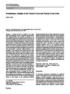
Endothelin-3 and the neural crest 1675 isation resulted in single cell suspensions. Each dish was screened under phase contrast. Single cells, which were not in close proximity to other cells, were marked on the underside of the dish with an inkbased object marker (Nikon). Marked clones were re-screened on the same day and any containing more than one cell were excluded from the analysis. Approximately 30% of marked cells, upon re-evaluation the following day, were found to have died. Clone formation was examined at regular intervals to monitor any encroachment by neighbouring colonies. Cultures were grown for 14 days. The formation of a mixed clone is shown in Fig. 1. The resulting colonies were quantified and fixed as described below. Immunocytochemistry Cultures to be analysed were washed with CMF-PBS and fixed on ice with 4% paraformaldehyde in PBS for one hour. Cells were then rinsed three times with 1% bovine serum albumin (BSA) in PBS for 30 minutes. Pigmented colonies (containing melanocytes only), unpigmented colonies (containing no melanocytes) and mixed colonies (containing both pigmented and unpigmented cells) were scored under phase contrast and bright field optics (Sieber Blum and Cohen, 1980). Sensory-like cells were identified as being AC4 positive (SieberBlum, 1989). This antibody recognises SSEA-1 (stage specific embryonic antigen-1). This is present on developing spinal ganglia in frozen sections of quail embryos and co-localises with substance P in neural crest cultures (Sieber-Blum, 1989). Adrenergic cells were iden-
tified using anti-dopamine-β-hydroxylase antibody. MelEM labels melanoblasts and melanocytes of neural crest origin and also stains cells reactive for β-III tubulin (Nataf et al., 1993; Langtimm-Sedlak et al., 1996). SMP reacts in vivo and in vitro with embryonic and adult Schwann cells and oligodendrocytes from E5 avian embryos (Dulac et al., 1988, 1992). 1E8 detects the protein Po and is an early marker of the Schwann cell lineage (Bhattacharyya et al., 1991). Neurofilament 200 binds to intermediate filaments in neurons (Liem and Hutchinson, 1982; Karlsson et al., 1987). MelEM and 1E8 were diluted 1:10 and SMP was used undiluted. The monoclonal antibody AC4 (a kind gift from Dr T. M. Jessell, and Developmental Studies Hybridoma Bank), was used at 1:2.5. Anti-DBH polyclonal antiserum (Biogenesis) was used at 1:100. MelEM, 1E8 or SMP were obtained as hybridoma cells from the Developmental Studies Hybridoma Bank. Neurofilament 200 (Sigma) was used at 1/500. Primary antibodies were left on overnight and then the cells were washed with 1% BSA/PBS. For immunofluorescence, secondary antibodies were fluorescein linked sheep anti-mouse 1:25 and Texas Red linked donkey anti-rabbit 1:50 (Amersham). Secondary antibodies were pooled and added for 1 hour in the dark. Alternatively, a Vectastain ABC mouse IgG kit (Vector) based on an immunoperoxidase detection system and VIP chromogen (Vector), was used to detect antibody binding. Cultures were washed with PBS before mounting with Aqua Mount (BDH).
Fig. 1. Mixed-clone formation in control medium. (A) Day 0, single cell marked. Bar, 150 µm. (B) Day 2 (same scale as A). (C) Day 4. Bar, 300 µm. (D) Day 10. Bar, 500 µm.
1676 J. G. Stone, L. I. Spirling and M. K. Richardson
RESULTS
control
100
EDN3-supplemented 80
60
40
20
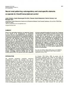
General observations The dose-response curve indicates that the maximum effect on population growth was at 1 µg/ml (Fig. 2). In order to be consistent with Lahav et al. (1996) we chose the submaximal dose of 100 ng/ml. We examined whether this dose had general mitogenic effects on other embryonic cell types. As can be seen from Fig. 3 there is no stimulation of population growth in retinal pigment epithelial cells or limb mesoderm cells. This is in contrast with the dramatic increase in cell numbers in truncal crest cultures grown in the presence of EDN3. Thus among the cell types examined, the mitogenic action of EDN3 was specific for neural crest cells. EDN3 inhibits loss of developmental potential in primary culture Primary cultures of trunk neural crest were maintained for 10 days in control or EDN3-supplemented medium and then replated into control medium for analysis. This replating stage is essential for two reasons: (1) to disperse the cells; (2) because cells do not differentiate in EDN3-supplemented medium and 35
30
25
Cell no. X 10,000
120
Cell no. (x10,000)
Statistical analysis In experiments with MelEM, 1E8 and SMP, we used a Poisson heterogeneity test. We compared the number of positively stained colonies between control and EDN3 treated cultures. To look at neuronal and melanocyte markers (colonies designated according to the P, S, A nomenclature) we merged day 2 and 4 cultures to form a single ‘early’ category; and day 8 and 10 cultures to form a ‘late’ category. This was necessary to bring colony numbers to a significant level for statistical analysis to be made. We then classified colonies according to whether they contained 0, 1, 2 or 3 identified cell types. A Chi squared test was used to look at changes in the number of cell types per colony between early and late cultures, and between control and treated cultures.
20
15
10
5
0 0
0. 1
1
10
100
1000
10,000
Concentration of EDN3 (ng/ml)
Fig. 2. Dose response curve for cells grown in different concentrations of EDN3 in secondary culture.
0 trunk crest
RPE
limb st.25
Tissue Type
Fig. 3. Population growth in cultures of truncal crest, retinal pigmented epithelium and limb mesenchyme. Cells were grown for 8 days in control medium with or without 100 ng/ml EDN3. They were then trypsinized and counted. RPE: retinal pigmented epithelium.
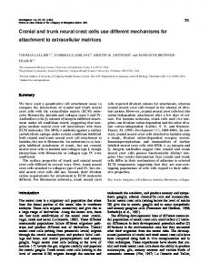
so their developmental potential cannot be analysed. This experiment has the advantage that EDN3 treatment begins immediately on removing cells from the embryo. In primary cultures in control medium (Fig. 4), large numbers of differentiated melanocytes were seen from day 4 and aggregates of neurofilament-positive cells from day 10 (as described by Cohen and Konigsberg, 1975). Cells were replated into control medium from primary cultures at seven days to determine the precursor types remaining. Because many cells in the primary cultures had differentiated, few remained which were capable of giving rise on replating to typical neural crest colonies. Thus the replating efficiency was low (18%). Of the colonies which did form, the majority (72.5%) were unpigmented. Pigment colonies constituted only 23.8% and mixed colonies 3.7% (Table 1). Primary cultures in EDN3-supplemented medium behaved quite differently to control cultures (Fig. 4). They did not give rise to fully differentiated melanocytes, although grey melanosomes were seen in many cells from day 8. Aggregates of neurofilament-positive cells were not observed, although rare isolated neurofilament-positive cells were seen; however the cultures were too dense to make quantitation of these cells feasible. In contrast to control cultures, primary cultures grown in EDN3 gave rise to a majority of pigment colonies (73.8%) and a threefold higher percentage of mixed colonies (12.6%). Unpigmented colonies constituted only 13.6% of the total. Many more undifferentiated colony-forming cells were present and so the plating efficiency was high (84.3%). Immunochemistry was used to characterise the colonies observed upon replating. MelEM detects early melanoblasts, melanocytes and β-III tubulin+ crest cells. The latter have been interpreted to be uncommitted neural crest precursors (Langtimm-Sedlak et al., 1996). The most striking difference was that cells re-plated from control cultures gave rise to a predominance of colonies lacking neural crest markers (MelEM−
Endothelin-3 and the neural crest 1677
Fig. 4. Photographs of primary cultures of trunk neural crest grown in (A) control medium and (B) medium supplemented with EDN3. Note that in the absence of exogenous EDN3 cells differentiate into melanocytes. In the presence of EDN3 cells remain unpigmented or with a few scattered melanosomes (100 ng/ml). Bar, 300 µm.
unpigmented colonies, 70.3%; Table 2). These unpigmented colonies also lacked the Schwann cell markers 1E8 and SMP (Table 2; Fig. 5). A very different pattern of colonies was seen in cells re-plated from EDN3-treated primary cultures. MelEM− unpigmented colonies constituted only 5.8% of total colonies. Mixed colonies containing 1E8+ cells were more abundant after EDN3 treatment than in controls (6.1% compared with 1.3%, respectively). Statistical analysis showed the differences between control and EDN3 cultures to be highly significant (P