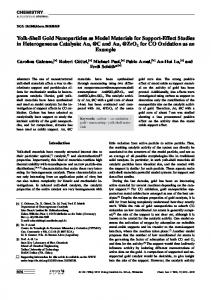
Wan et al. Nanoscale Research Letters (2017) 12:535 DOI 10.1186/s11671-017-2313-4
NANO IDEA
Open Access
The Preparation of Au@TiO2 Yolk–Shell Nanostructure and its Applications for Degradation and Detection of Methylene Blue Gengping Wan, Xiange Peng, Min Zeng, Lei Yu, Kan Wang, Xinyue Li and Guizhen Wang*
Abstract This paper reports the synthesis of a new type of Au@TiO2 yolk–shell nanostructures by integrating ion sputtering method with atomic layer deposition (ALD) technique and its applications as visible light-driven photocatalyst and surface-enhanced Raman spectroscopy (SERS) substrate. Both the size and amount of gold nanoparticles confined in TiO2 nanotubes could be facilely controlled via properly adjusting the sputtering time. The unique structure and morphology of the resulting Au@TiO2 samples were investigated by using various spectroscopic and microscopic techniques in detail. It is found that all tested samples can absorb visible light with a maximum absorption at localized surface plasmon resonance (LSPR) wavelengths (550–590 nm) which are determined by the size of gold nanoparticles. The Au@TiO2 yolk–shell composites were used as the photocatalyst for the degradation of methylene blue (MB). As compared with pure TiO2 nanotubes, Au@TiO2 composites exhibit improved photocatalytic properties towards the degradation of MB. The SERS effect of Au@TiO2 yolk–shell composites was also performed to investigate the detection sensitivity of MB. Keywords: Au@TiO2, Yolk–shell nanostructure, Surface plasmon resonance, Photocatalysis, SERS
Background Heterogeneous metal/semiconductor nanocomposites have attracted tremendous research interest by virtue of their unique physic-chemical properties and potential applications in solar energy conversion [1], biomedicine [2], surface-enhanced Raman scattering [3], lightemitting diodes [4], and environmental remediation [5]. Motivated by their various applications, a vast number of efforts have been paid to design and modulate the compositions, nanostructures, and dimensions of such materials [6–8]. For example, Yin et al. [9] synthesized ZnO/Ag and ZnO/Pd hybrid nanostructures and found that the deposition of Ag or Pd onto ZnO tremendously improved photocatalytic activity of ZnO. Sun et al. [10] demonstrated that Au-Fe3O4 nanoparticles with nanoscale interactions between Au and Fe3O4 exhibited a rich variety of magnetic, physical, and chemical properties. * Correspondence:
[email protected] Key Laboratory of Advanced Materials of Tropical Island Resources (Hainan University), Ministry of Education, Haikou 570228, People’s Republic of China
In recent years, significant advances in the controlled synthesis of metal/semiconductors applied to photocatalysis have been made due to the increasingly serious environmental problems such as air pollution [11, 12] and potential technical applications in energy conversion [13]. Among the various metal/semiconductor composites that have been proposed, those involving TiO2 and nano Au are the most practical as such heterostructure has strong localized surface plasmon resonance (LSPR) in the visible spectrum range and makes it a new kind of wide-spectrum-response photocatalyst [14–16]. Another advantageous function of Au/TiO2 nanocomposites is that Au nanoparticles work as electron storage, effectively reducing the recombination of photoexcited electron-hole pairs, and eventually increasing the quantum yield of photocatalysis [17, 18]. Some innovative investigations based on Au/TiO2 composite system applied in degradation of organic dyes, solar water splitting, and conversion of organic compounds have demonstrated their efficient visible light photocatalytic
© The Author(s). 2017 Open Access This article is distributed under the terms of the Creative Commons Attribution 4.0 International License (http://creativecommons.org/licenses/by/4.0/), which permits unrestricted use, distribution, and reproduction in any medium, provided you give appropriate credit to the original author(s) and the source, provide a link to the Creative Commons license, and indicate if changes were made.
Wan et al. Nanoscale Research Letters (2017) 12:535
features, indicating a crucial role of the plasmonic effects of Au played in Au/TiO2 system [17, 19, 20]. However, one of the main limitations for the Au/TiO2 nanocomposites translated into practical applications is the poor stability of the supported gold catalysts. The outstanding properties presented in the original nanoparticles may weaken as they tend to agglomerate and grow into larger particles under a variety of reaction conditions [21, 22]. And in some other cases, it has been proved that Au nanoparticles deposited on the surfaces of TiO2 are likely to undergo corrosion or dissolution during a catalytic reaction [23]. The design and construction of core–shell and yolk–shell structured composites are considered as an effective method to address these issues. Gong et al. [24] reported the fabrication of gold nanorod@TiO2 yolk–shell catalysts with different aspect ratios of gold nanorod through a seed-mediated method. The multicomponent hybrid nanocomposites also present the enhanced photocatalytic activities in the oxidation reaction of benzyl alcohol. Zaera and coworkers [21] reported on the synthesis and characterization of a new Au@TiO2 yolk–shell-nanostructured catalyst, showing a promoting activity comparable to those observed with more conventional Au/ TiO2 catalysts but an improved stability against sintering. Kim et al. [25] synthesized core–shell plasmonic nanostructures consisting of Au–TiO2 supported on SiO2 spheres in dye-sensitized solar cells (DSSCs), which exhibited observably enhanced power conversion efficiencies of ~ 14%. Despite tremendous research efforts have been made, the facile synthesis of Au@TiO2 composites with a well-defined core–shell/yolk–shell structure still remains a challenge for mass application. Recently, many studies confirmed that controlled chirality at the nanoscale might induce a greater LSPR effect because a multihelical chiral nanostructure can give rise to induced birefringence at the microscopic scale and generate the Kerr effect caused by an induced electric field at the macroscopic scale [26–28]. In this study, the Au@TiO2 yolk–shell nanocomposites with helical fiber-like structure have been successfully synthesized by a controllable and facile strategy. The gold nanoparticles loaded on the surface of carbon nanocoils (CNCs) were produced by ion sputtering. The TiO2 films with highly uniform and controlled thickness could be integrated steady on the surface of gold nanoparticles by an atomic layer deposition (ALD) technology. Followed by an annealing step, the Au@TiO2 nanocomposites were obtained. The above-developed method can also be extended to fabricate other metal (Pt, Ag)@TiO2 yolk–shell nanocomposites with a helical nanostructure. As a representative photocatalyst, the photocatalytic activities of obtained Au@TiO2 nanocomposites were evaluated by degradation of methylene blue (MB) under visible light irradiation. In addition, the surface-enhanced
Page 2 of 9
Raman spectroscopy (SERS) activities of Au@TiO2 nanocomposites were also investigated through detection of MB. Experimental Synthesis of Au@TiO2
CNCs used as templates were prepared by chemical vapor deposition method as reported previously. Briefly, acetylene and copper nanoparticles were used as the carbon source and the appropriate catalysts, respectively. The growth of CNCs was carried out at atmospheric pressure in a horizontal quartz tube. A ceramic plate containing the copper catalysts was placed in the reactor. After the tube was heated to 250 °C in vacuum, acetylene was introduced into the reactor [29–31]. After the apparatus was cooled to room temperature, the asprepared CNCs were obtained. The obtained CNCs were dispersed in ethanol under ultrasonic stirring and then daubed uniformly on the surface of a glass slide. After being dried in ambient air, the Au layer was deposited by an ion sputtering instrument (Hitachi, E-1010). The size and thickness of Au films were determined by discharge current and sputtering time. In this step, the discharge current was 10 mA and the sputtering time varied from 30 to 120 s. The obtained samples were marked as CNCs@Au-x, in which x refers to the sputtering time (seconds). Subsequently, the samples were dispersed in ethanol by ultrasonic agitation and then spread onto a quartz wafer to be coated with TiO2 by ALD process. ALD is a kind of vaporphase coating preparation technique and can achieve precise thickness control and excellent uniformity of films [32–36]. ALD process was carried out in a hotwall, flow-type ALD reactor at 145 °C with titanium tetraisopropanolate (TTIP) and deionized H2O used as the titanium and oxygen precursors, respectively. Finally, after the ALD process, the above-coated nanocoils were calcined at 450 °C for 2 h in air under ambient pressure to remove the carbon cores and the helical TiO2-coated Au yolk–shell structures were obtained. For comparison, the pure TiO2 helical tube was also collected by calcinated TiO2-coated CNCs without sputtering Au and is denoted as TiO2 in the following discussion. Material Characterization
X-ray diffraction (XRD) patterns were recorded on a Bruker D8 Advance diffractometer with copper Kα (λ = 0.154056 nm) radiation source. Scanning electron microscopy (SEM) images were acquired with a Hitachi S-4800 microscope. Transmission electron microscopy (TEM), selected area electron diffraction (SAED), and high-resolution TEM (HRTEM) images were obtained using a JEOL JEM-2100 microscope instrument operated at 200 kV. X-ray photoelectron spectroscopy (XPS) data were acquired using a PHI5000 Versaprobe-II spectrometer
Wan et al. Nanoscale Research Letters (2017) 12:535
with a monochromatic Al Kα (1486.6 eV) source. Optical absorption spectra were recorded using a PerkinElmer Lambda 750s UV–Vis–NIR absorption spectrophotometer. The Raman scattering spectra were recorded on a Renishaw Invia Reflex Laser Raman spectrometer. The excitation wavelength was 514 nm from an air-cooled argon ion laser with an effective power of 2 mW.
Photocatalytic Activities Evaluation
The photocatalytic activities of catalysts were investigated by the photodegradation of MB dyes in aqueous solutions using the procedure as described below. Two milligrams of catalyst was spread uniformly into a 100mL photoreactor equipped with circulating cooling water pipes. Then, 20 mL of 0.01 mg/mL MB solutions was added into the photoreactor. Before photoirradiation, the system was ultrasonically mixed for 2 min and bidirectional magnetic stirred for 30 min both in the dark in order to balance the adsorption–desorption between the photocatalysts and MB. The above 100-mL photoreactor containing suspension was then irradiated under a 300 W xenon lamp (Beijing Perfectlight Technology Co. Ltd., PLS-SXE300C) with cutoff filters so that wavelengths of light between 420 and 780 nm reached the solutions. During the process of photocatalytic reaction, the irradiation intensity was ~ 154 mW cm−2 and the cooling water was kept flowing to dispel thermal effect of the system. At the time intervals of every 10 min for a total time of 90 min, a portion (1 mL) of the suspensions was pipetted and immediately diluted to 3 mL, and 2 mL supernate was collected after centrifugal separation. Eventually, the residual concentration of MB in the supernate was analyzed by using an UV–Vis–NIR spectrophotometer at the solution’s characteristic wavelength (λMB = 664 nm).
Page 3 of 9
Results and Discussion Morphology and Phase Structure Analysis
Figure 1a displays a schematic preparation flow of Au@TiO2 yolk–shell heterostructure, including Au sputtering, TiO2 coating, and calcination processes. Figure 1b–e shows typical TEM images corresponding to the above every procedure. The CNCs used as the starting template in this work have uniform fiber diameter, coil diameter, and coil pitch, and the average diameter of the fiber is about 80 nm (Additional file 1: Figure S1). After the Au sputtering treatment, the outer layer of CNCs was coated with numerous uniform Au nanoparticles as shown in Fig. 1c. As seen from the TEM image shown in Fig.1d, by applying 200 ALD cycles for TiO2 deposition, a uniform TiO2 coating with a thickness of about 8 nm is coated on the surface of Au/CNCs. Generally, the anatase phase of TiO2 has much better photocatalytic performance than that of rutile [37, 38]. For this reason, we chose 450 °C as a proper calcination temperature to remove the carbon cores and get the final Au@TiO2 yolk–shell structure. As displayed in Fig.1e, the TiO2 nanotubes with encapsulated Au nanoparticles and free space were formed. After all processing steps, the elegant helical morphology of the starting CNCs can be well maintained. The crystallinity and structures of all samples were measured by XRD. As observed in Fig. 2a, the diffraction peaks for pure TiO2 sample can be ascribed to wellcrystallized anatase phase (JCPDS 21-1272), without additional impurity peaks. For Au/TiO2, the additional diffraction peaks in Fig. 2b–e can be well indexed to the face-centered cubic (FCC) Au (JCPDS 01-1174), which conformed the successful coating of Au nanoparticles on the surface of CNCs by ion sputtering. The TiO2 (004) peak at 38.2° has large overlap with the Au (111) peak at 38.3°. It is interesting that a weak peak located at 35.5
Fig. 1 a Schematic illustration of the synthetic process of Au-x@TiO2. b–e TEM images reveal the morphological evolution
Wan et al. Nanoscale Research Letters (2017) 12:535
Fig. 2 XRD patterns. a TiO2. b Au-30@TiO2. c Au-50@TiO2. d Au-80@TiO2. e Au-120@TiO2
degrees in Fig. 2b–e can be indexed to the (020) plane of γ-Ti3O5, indicating that the Ti/O atomic ratio is not exactly 1/2 for Au/TiO2. In present work, the strong reducing action of carbon fiber and Au nanoparticles under high temperature likely induces the production of oxygen vacancies and lower oxidation states of titanium. In addition, due to the decrease of relative content for TiO2, it can be observed that all TiO2 diffraction peaks become weaker with the increased sputtering time from 30 to 120 s.
Page 4 of 9
Figure 3 shows the TEM images of TiO2 and Aux@TiO2 with different Au sputtering time (x signifies sputtering time, x = 30, 50, 80, 120). For TiO2, it can be observed that the sample displays a helical tubular structure similar to that of the CNC templates. No collapse of the shell materials occurred during the annealing process to remove the carbon cores. The TiO2 shell is about 8-nm thick after 200 cycles. On account of a larger atomic number of Au compared to that of Ti in Au@TiO2, Au nanoparticles show a darker contrast resulting in clearly visible yolk–shell morphology. The average diameter of Au nanoparticles clearly increases with the increased sputtering time. It amounts to about 4.5, 5.5, 10.5, and 20.5 nm corresponding to the sputtering time of 30, 50, 80, and 120 s, respectively (Additional file 1: Figure S2, a2d2). As shown in Fig. 3b–d, the homogeneous TiO2 thin film with about the thickness of 8 nm is also obtained for Au-30@TiO2, Au-50@TiO2, and Au-80@TiO2 nanocomposites with the same ALD TiO2 deposition. However, the thickness of TiO2 shell for Au-120@TiO2 declines to about 5 nm (Fig. 3e), which can be ascribed to the influence of large size and significant conglomerations of Au nanoparticles. The detailed microscopic structures of the TiO2 and Au-30@TiO2 nanocompositions were further investigated by HRTEM. As observed in Fig. 4a–b, both TiO2 shells and Au nanoparticles are well crystallized assigned to anatase TiO2 (101) (0.3565 and 0.3501 nm) and Au (111) (0.2399 nm) crystalline lattices, respectively. It should be noted that the interface in Au/TiO2 yolk–shell nanostructures is clearly visible (Fig. 4b) because of the
Fig. 3 TEM images. a TiO2. b Au-30@TiO2. c Au-50@TiO2. d Au-80@TiO2. e Au-120@TiO2
Wan et al. Nanoscale Research Letters (2017) 12:535
Page 5 of 9
Fig. 4 HRTEM images of a TiO2 and b Au-30@TiO2, in which the top right inset in b shows the SAED patterns of Au-30@TiO2 nanostructure. High-resolution XPS of c Ti 2p and d Au 4f of Au-30@TiO2
different contrast. Such rich interface is important for the following photocatalysis application as it may provide the access for hot electron transportation from Au nanoparticles to TiO2 upon LSPR excitation [20]. The inset in Fig. 4b displays the SAED pattern recorded on Au-30@TiO2 nanostructure. The clear diffraction rings can be attributed to (101) and (211) crystal planes of anatase TiO2 and (220) and (111) crystal planes of Au, respectively, in agreement with the XRD results. In order to analyze the chemical state of Au and acquire in-depth fundamental information on the interaction of Au with TiO2, Au30@TiO2 nanocomposite was further investigated by XPS measurements. The high-resolution spectra of Ti 2p and Au 4f are presented in Fig. 4c and d, respectively. As displayed in Fig. 4c, two peaks with the binding energy at approximately 458.4 and 464.2 eV can be assigned to Ti 2p3/2 and Ti 2p1/2 spin–orbit components of Ti4+, respectively [39]. Figure 4d shows the Au 4f XPS spectrum with two peaks appeared at 83.6 and 87.4 eV for Au 4f7/2 and Au 4f5/2 levels, respectively, suggesting that Au species exist as metallic state. The relative negative shift (0.4 eV) of Au 4f7/2 peak in comparison of bulk Au (4f7/2 at 84.0 eV) can be attributed to the electron transfer from oxygen vacancies of the TiO2 to Au, which confirms the strong Au/TiO2 interaction [40, 41].
Figure 5 shows the UV–Vis diffuse reflection spectra of the TiO2 and Au-x@TiO2 nanostructures. An intense absorption band below 400 nm is observed for all these samples, which can be owed to the large band gap of anatase TiO2 [42]. Compared with TiO2, it can be found that the Au-x@TiO2 has not only a similar absorption below 400 nm but also the enhanced absorption range from 400 to 800 nm with a broad absorption peak at about 580 nm arisen from the LSPR effect of Au
Fig. 5 UV–Vis absorption spectra of TiO2 and Au-x@TiO2
Wan et al. Nanoscale Research Letters (2017) 12:535
nanoparticles [43]. These results indicate that a better photocatalytic activity for Au-x@TiO2 can be expected under visible light irradiation, especially for the Au80@TiO2 with stronger absorption intensity. The slight shift of the LSPR absorption for Au@TiO2 nanostructures with different sputtering time is also reasonable since Au nanoparticle is sensitive to its size and surrounding environment [24, 42]. These observations declare that the Au-x@TiO2 photocatalysts can possess a tunable light-harvesting range through adjusting the shape, diameter, and morphology of Au nanoparticles [44]. Photocatalytic Activity
Removal of organic pollutants from wastewater produced from industry and households has attracted much attention [45–48]. MB is frequently employed as targeted pollutant to evaluate the catalytic efficiency in photocatalytic reactions because the blue color of MB from the absorption at 664 nm would fade gradually with the degradation process [49, 50] and can be easily monitored by UV–Vis absorption spectra. The photocatalytic activities of the TiO2 and Au-x@TiO2 composites were evaluated by monitoring MB dye’s absorbance at 664 nm to detect the degradation rate under visible light (420 to 780 nm) irradiation. The changes of relative MB concentration versus irradiation time upon the different catalysts are presented in Fig. 6a. For comparison, the photocatalytic activity of pure TiO2 nanotubes was first examined. It can be found that about 60% of MB was degraded with TiO2 as the photocatalyst under visible light irradiation for 90 min. The relatively low photocatalytic efficiency of TiO2 is due to its poor absorption ability of visible light. Compared with the above blank experiment, the Aux@TiO2 photocatalysts exhibit higher degradation efficiency and the degradation efficiency for Au-80@TiO2 amounts to about 90% under the same experimental conditions. The promotive photocatalytic properties can be ascribed to increased electron-hole generation rate due to the presence of hetero-interface and the corresponding
Page 6 of 9
plasmon-enhanced light absorption [51, 52]. It is known that both high-energy plane (200) of Au and the thickness of TiO2 shells are important parameters affecting the activity [24, 53]. Among Au-x@TiO2 photocatalysts, with the increased of sputtering time, Au (200) peak exhibits more high-energy planes, as shown in corresponding XRD peak intensity. In addition, Au-120@TiO2 with thinner TiO2 shell (5 nm) is unable to provide enough reaction sites for the consumption of electrons. Thus, based on the appropriate and similar thickness of TiO2 shell over different Au-x@TiO2, Au-80@TiO2 shows the highest activity. As heterogeneous catalysts, the reusability of catalyst is also very important in practical application. We performed three consecutive operations to investigate the reusability of the Au-80@TiO2. As shown in Fig. 6b, no noticeable deactivation is observed, indicating excellent durability of Au-80@TiO2. TEM image of Au-80@TiO2 (Additional file 1: Figure S3) after recycling of three times reveals that helical yolk–shell structures of catalysts are well maintained, which further confirms that the confined effect of TiO2 nanotubes can prevent Au loss and thus enhances the stability of catalysts. Based on the above results, we propose a photocatalytic process for MB degradation using helical Au@TiO2 nanostructures (Fig. 7). Under visible light irradiation, hot electrons are produced by the LSPR effect of Au nanoparticle inside the TiO2 nanotube. Subsequent electrons would transfer from Au to the conduction band of TiO2. The degradation of adsorbed MB would start from holes (•Au + ) because the holes can scavenge the surface adsorbed water, generating highly reactive hydroxyl radical species [24, 51, 54]. Simultaneously, the electron injected into the conduction band of TiO2 may be trapped by oxygen molecules to form reactive superoxide radicals •O−2 . Then, it can further react with H+ to yield active •HO−2 and •OH radicals. Finally, the organic pollutants may be destroyed by these forming radicals. In this work, it is believed that polarized light rotated by the helical chiral Au@TiO2 structure can accelerate the excitation of LSPR, which
Fig. 6 a Evaluation of MB concentration versus reaction time in different conditions. b Recyclability of the photocatalytic degradation of MB aqueous solution using Au-80@TiO2 with three cycles
Wan et al. Nanoscale Research Letters (2017) 12:535
Page 7 of 9
performance, implying that Au nanoparticles contacted with TiO2 nanoparticles may form a large number of hot spots, which can facilitate to effective SERS enhancement [55]. To explore the influence of varying concentrations of MB solution on the detection ability of Au-30@TiO2, Raman measurement was also carried out. As presented in Fig. 8b, the intensity of Raman signal is decreased with the decrease of MB concentrations ranging from 10−4 to 10−6 M. The discernable Raman signal of 10−6 M MB with the Raman band varying from 900 to 1500 cm−1, indicating that Au-30@TiO2 acted as SERS substrate, can detect the concentrations of MB as low as 10−6 M, which shows potential applications for detecting pollutants [56]. Fig. 7 Schematic representation for the mechanism of photocatalytic degradation of MB over Au@TiO2
further enhance the photocatalytic activity of helical Au@TiO2. In addition, the adsorbed MB molecule may be excited and transfers an electron to the conduction band of TiO2 as the pure TiO2 nanotubes show a little photocatalytic activity under visible light irradiation. Thus, the photosensitization effect of MB should also lead to a small part of decomposition of MB. SERS activity
To exploit the multifunctional application of such catalysts, we carried out the further experiments by using Au-x@TiO2 as SERS substrates to detect the MB molecules adsorbed on the surface of gold nanoparticles. As we can see from Fig. 8a, upon probed with 1.0 × 10⁻⁵ M MB solution, SERS activity of the as-prepared substrate decreases with the increase of Au sputtering time from 30 to 120 s. This result indicated that Au-30@TiO2 has the most excellent SERS
Conclusions In this study, we have successfully synthesized Au@TiO2 yolk–shell heterogeneous nanocomposites with helical coil-like morphology and investigated their multifunctional use including photocatalysis and the SERS effect. The visible photocatalysis degradation of MB displays that the obtained Au-x@TiO2 composite with the Au nanoparticles sputtering time of 80 s shows the highest photocatalytic performance because of the increased light absorption and the restriction of the recombination of the photoexcited electron-hole pairs by the LSPR effect of Au nanoparticles. Raman measurements suggest that the Au-x@TiO2 can be used as efficient SERSactive substrates. Considering its fascinating properties and features, the novel heterogeneous nanocomposite may provide inspiration in various areas, including water splitting and solar cells. Furthermore, the helical yolk– shell Au@TiO2 model system studied here can be extended to the design of other heterostructures, such as Ag@TiO2, Au@ZnO, and Au@NiO, for application in solar conversion.
Fig. 8 a The SERS spectra of 1.0 × 10⁻⁵ M MB solution collected on the substrates with different Au-x@TiO2. b The SERS spectra of MB with different concentrations collected on the Au-30@TiO2 substrate
Wan et al. Nanoscale Research Letters (2017) 12:535
Additional file Additional file 1: Supporting information. Figure S1. SEM images of CNCs. Figure S2. TEM images and the size distribution analysis of Au nanoparticles of (a1 and a2) Au-30@TiO2; (b1 and b2) Au-50@TiO2; (c1 and c2) Au-80@TiO2; (d1 and d2) Au-120@TiO2. Figure S3. TEM image of the Au-80@TiO2 after photocatalytic reaction. (DOC 11017 kb) Acknowledgements This work was supported by the National Natural Science Foundation of China (11564011, 51362010, 21706046), the Natural Science Foundation of Hainan Province (514207, 514212), the Scientific Research Projects of Colleges and Universities of Hainan Province (HNKY2014-14), and the Open Foundation of Key Laboratory of Advanced Materials of Tropical Island Resources (Hainan University), Ministry of Education (AM2017-20). Authors’ Contributions GPW carried out the overall experiment and wrote the manuscript. XEP, MZ, and LY participated in supervising this study and revising the manuscript. KW and XYL helped with TEM studies. GZW provided the guidance and assistance for the whole work. All authors read and approved the final manuscript. Competing Interests The authors declare that they have no competing interests.
Publisher’s Note Springer Nature remains neutral with regard to jurisdictional claims in published maps and institutional affiliations. Received: 27 May 2017 Accepted: 11 September 2017
References 1. Wu HS, Sun LD, Zhou HP, Yan CH (2012) Novel TiO2-Pt@SiO2 nanocomposites with high photocatalytic activity. Nanoscale 4:3242–3247 2. Xu CJ, Xie J, Ho D, Wang C, Kohler N, Walsh EG, Morgan JR, Chin YE, Sun SH (2008) Au-Fe3O4 dumbbell nanoparticles as dual-functional probes. Angew Chem 47:173–176 3. Li XH, Chen GY, Yang LB, Jin Z, Liu JH (2010) Multifunctional Au-coated TiO2 nanotube arrays as recyclable SERS substrates for multifold organic pollutants detection. Adv Funct Mater 20:2815–2824 4. Chen C, Chen JW, Zhang J, Wang S, Zhang W, Liang RL, Dai JN, Chen CQ (2016) Ag-decorated localized surface plasmon enhanced ultraviolet electroluminescence from ZnO quantum dot-based/GaN heterojunction diodes by optimizing MgO interlayer thickness. Nanoscale Res Lett 11:480 5. Lu JW, Zhang P, Li A, Su FL, Wang T, Liu Y, Gong JL (2013) Mesoporous anatase TiO2 nanocups with plasmonic metal decoration for highly active visible-light photocatalysis. Chem Commun 49:5817–5819 6. Dahl M, Liu YD, Yin YD (2014) Composite titanium dioxide nanomaterials. Chem rev 114:9853–9889. 7. Cai JB, Wu XQ, Li SX, Zheng FY, Zhu LC, Lai ZH (2015) Synergistic effect of double-shelled and sandwiched TiO2@Au@C hollow spheres with enhanced visible-light-driven photocatalytic activity. ACS Appl Mater Interfaces 7:3764–3772 8. Cai JB, Wu XQ, Li SX, Zheng FY (2017) Controllable location of Au nanoparticles as cocatalyst onto TiO2@CeO2 nanocomposite hollow spheres for enhancing photocatalytic activity. Appl Catal B Environ 201:12–21 9. He WW, Wu HH, Wamer WG, Kim HK, Zheng JW, Jia HM, Zheng Z, Yin JJ (2014) Unraveling the enhanced photocatalytic activity and phototoxicity of ZnO/metal hybrid nanostructures from generation of reactive oxygen species and charge carriers. ACS Appl Mater Interfaces 6:15527–15535 10. Yu H, Chen M, Rice PM, Wang SX, White RL, Sun SH (2005) Dumbbell-like bifunctional Au-Fe3O4 nanoparticles. Nano Lett 5:379–382 11. Wu XF, Song HY, Yoon JM, Yu YT, Chen YF (2009) Synthesis of core−shell Au@ TiO2 nanoparticles with truncated wedge-shaped morphology and their photocatalytic properties. Langmuir 25:6438–6447 12. Wang M, Han J, Xiong H, Guo R, Yin Y (2015) Nanostructured hybrid shells of r-GO/AuNP/m-TiO2 as highly active photocatalysts. ACS Appl Mater Interfaces 7:6909–6918
Page 8 of 9
13. Dillon RJ, Joo JB, Zaera F, Yin Y, Bardeen CJ (2013) Correlating the excited state relaxation dynamics as measured by photoluminescence and transient absorption with the photocatalytic activity of Au@TiO2 core–shell nanostructures. Phys Chem Chem Phys 15:1488–1496 14. Seh ZW, Liu SH, Low M, Zhang SY, Liu ZL, Mlayah A, Han MY (2012) Janus Au-TiO2 photocatalysts with strong localization of plasmonic near-fields for efficient visible-light hydrogen generation. Adv Mater 24:2310–2314 15. Kimura K, Naya S-I, Jin-nouchi Y, Tada H (2012) TiO2 crystal formdependence of the Au/TiO2 plasmon photocatalyst’s activity. J Phys Chem C 116:7111–7117 16. Brennan LJ, Purcell-Milton F, Salmeron AS, Zhang H, Govorov AO, Fedorov AV, Gun'ko YK (2015) Hot plasmonic electrons for generation of enhanced photocurrent in gold-TiO2 nanocomposites. Nanoscale Res Lett 10:38 17. Kowalska E, Mahaney OOP, Abe R, Ohtani B (2010) Visible-light-induced photocatalysis through surface plasmon excitation of gold on titania surfaces. Phys Chem Chem Phys 12:2344–2355 18. Wu L, Li F, Xu YY, Zhang JW, Zhang DQ, Li GS, Li HX (2015) Plasmoninduced photoelectrocatalytic activity of Au nanoparticles enhanced TiO2 nanotube arrays electrodes for environmental remediation. Appl Catal B Environ 164:217–224 19. Sakthivel S, Shankar MV, Palanichamy M, Arabindoo B, Bahnemann DW, Murugesan V (2004) Enhancement of photocatalytic activity by metal deposition: characterisation and photonic efficiency of Pt, Au and Pd deposited on TiO2 catalyst. Water Res 38:3001–3008 20. Pu YC, Wang GM, Chang KD, Ling YC, Lin YK, Fitzmorris BC, Liu CM, Lu XH, Tong YX, Zhang JZ, Hsu YJ, Li Y (2013) Au nanostructure-decorated TiO2 nanowires exhibiting photoactivity across entire UV-visible region for photoelectrochemical water splitting. Nano Lett 13:3817–3823 21. Lee I, Joo JB, Yin YD, Zaera F (2011) A yolk@shell nanoarchitecture for Au/ TiO2 catalysts. Angew Chem 123:10390–10393 22. Corma A, Garcia H (2008) Supported gold nanoparticles as catalysts for organic reactions. Chem Soc Rev 37:2096–2126 23. Subramanian V, Wolf E, Kamat PV (2001) Semiconductor-metal composite nanostructures. To what extent do metal nanoparticles improve the photocatalytic activity of TiO2 films? J Phys Chem B 105:11439–11446 24. Li A, Zhang P, Chang XX, Cai WT, Wang T, Gong JL (2015) Gold nanorod@TiO2 yolk-shell nanostructures for visible-light-driven photocatalytic oxidation of benzyl alcohol. Small 11:1892–1899 25. Jang YH, Jang YJ, Kochuveedu ST, Byun M, Lin ZQ, Kim DH (2014) Plasmonic dye-sensitized solar cells incorporated with Au–TiO2 nanostructures with tailored configurations. Nanoscale 6:1823–1832 26. Liu SH, Han L, Duan YY, Asahina S, Terasaki O, Cao YY, Liu B, Ma LG, Zhang JL, Che SN (2012) Synthesis of chiral TiO2 nanofiber with electron transitionbased optical activity. Nat Commun 3:1215 27. Wang DW, Li Y, Puma GL, Wang C, Wang PF, Zhang WL, Wang Q (2013) Ag/AgCl@helical chiral TiO2 nanofibers as a visible-light driven plasmon photocatalyst. Chem Commun 49:10367–10369 28. Zhang C, Li Y, Wang DW, Zhang WL, Wang Q, Wang YM, Wang PF (2015) Ag@helical chiral TiO2 nanofibers for visible light photocatalytic degradation of 17α-ethinylestradiol. Environ Sic Pollut R 22:10444–10451 29. Qin Y, Zhang ZK, Cui ZL (2003) Helical carbon nanofibers prepared by pyrolysis of acetylene with a catalyst derived from the decomposition of copper tartrate. Carbon 41:3072–3074 30. Wang GZ, Ran G, Wan GP, Yang P, Gao Z, Lin SW, Fu C, Qin Y (2014) Sizeselective catalytic growth of nearly 100% pure carbon nanocoils with copper nanoparticles produced by atomic layer deposition. ACS Nano 8: 5330–5338 31. Wang GZ, Gao Z, Tang SW, Chen CQ, Duan FF, Zhao SC, Lin SW, Feng YH, Zhou L, Qin Y (2012) Microwave absorption properties of carbon nanocoils coated with highly controlled magnetic materials by atomic layer deposition. ACS Nano 6:11009–11017 32. Wang GZ, Gao Z, Wan GP, Lin SW, Yang P, Qin Y (2014) High densities of magnetic nanoparticles supported on graphene fabricated by atomic layer deposition and their use as efficient synergistic microwave absorbers. Nano Res 7:704–716 33. Wang GZ, Peng XE, Yu L, Wan GP, Lin SW, Qin Y (2015) Enhanced microwave absorption of ZnO coated with Ni nanoparticles produced by atomic layer deposition. J Mater Chem A 3:2734–2740 34. Wan GP, Wang GZ, Huang XQ, Zhao HN, Li XY, Wang K, Yu L, Peng XE, Qin Y (2015) Uniform Fe3O4 coating on flower-like ZnO nanostructures by atomic layer deposition for electromagnetic wave absorption. Dalton T 44:18804–18809
Wan et al. Nanoscale Research Letters (2017) 12:535
35. Yu L, Wan GP, Peng XE, Dou ZF, Li XY, Wang K, Lin SW, Wang GZ (2016) Fabrication of carbon-coated NiO supported on graphene for high performance supercapacitors. RSC Adv 6:14199–14204 36. Yu L, Wan GP, Li XY, Wang K, Peng XE, Wang GZ (2016) Highly effective synthesis of NiO/CNT nanohybrids by atomic layer deposition for high-rate and long-life supercapacitors. Dalton T 45:13779–13786 37. Liu G, Yu JC, Lu GQM, Cheng H-M (2011) Crystal facet engineering of semiconductor photocatalysts: motivations, advances and unique properties. Chem Commun 47:6763–6783 38. Murdoch M, Waterhouse GIN, Nadeem MA, Metson JB, Keane MA, Howe RF, Llorca J, Idriss H (2011) The effect of gold loading and particle size on photocatalytic hydrogen production from ethanol over Au/TiO2 nanoparticle. Nature Chem 3:489–492 39. Chen J-J, Wu JCS, Wu PC, Tsai DP (2010) Plasmonic photocatalyst for H2 evolution in photocatalytic water splitting. J Phys Chem C 115:210–216 40. Tsukamoto D, Shiraishi Y, Sugano Y, Ichikawa S, Tanaka S, Hirai T (2012) Gold nanoparticles located at the interface of anatase/rutile TiO2 particles as active plasmonic photocatalysts for aerobic oxidation. J Am Chem Soc 134:6309–6315 41. Ding DW, Liu K, He SN, Gao CB, Yin YD (2014) Ligand-exchange assisted formation of Au/TiO2 Schottky contact for visible-light photocatalysis. Nano Lett 14:6731–6736 42. Zhang N, Liu SQ, Fu XZ, Xu YJ (2011) Synthesis of M@TiO2(M = Au, Pd, Pt) core-shell nanocomposites with tunable photoreactivity. J Phys Chem C 115:9136–9145 43. Zhou XM, Liu G, Yu JG, Fan WH (2012) Surface plasmon resonancemediated photocatalysis by noble metal-based composites under visible light. J Mater Chem 22:21337–21354 44. Tian N, Zhou ZY, Sun SG, Ding Y, Wang ZL (2007) Synthesis of tetrahexahedral platinum nanocrystals with high-index facets and high electro-oxidation activity. Science 316:732–735 45. Yu SJ, Wang XX, Yao W, Wang J, Ji YF, Ai YJ, Alsaedi A, Hayat T, Wang XK (2017) Macroscopic, spectroscopic, and theoretical investigation for the interaction of phenol and naphthol on reduced graphene oxide. Envir Sci Technol 51:3278–3286 46. Yao W, Yu SJ, Wang J, Zou YD, Lu SS, Ai YJ, Alharbi NS, Alsaedi A, Hayat T, Wang XK (2017) Enhanced removal of methyl orange on calcined glycerol-modified nanocrystallined Mg/Al layered double hydroxides. Chem Eng J 307:476–486 47. Zhang SW, Fan QH, Gao HH, Huang YS, Liu X, Li JX, Xu XJ, Wang XK (2016) Formation of Fe3O4@MnO2 ball-in-ball hollow spheres as a high performance catalyst with enhanced catalytic performances. J Mater Chem A 4:1414–1422 48. Zou YD, Wang XX, Ai YJ, Liu YH, Ji YF, Wang HQ, Hayat T, Alsaedi A, Hu WP, Wang XK (2016) β-Cyclodextrin modified graphitic carbon nitride for the removal of pollutants from aqueous solution: experimental and theoretical calculation study. J Mater Chem A 4:14170–14179 49. Kongkanand A, Kamat PV (2007) Electron storage in single wall carbon nanotubes. Fermi level equilibration in semiconductor–SWCNT suspensions. ACS Nano 1:13–21 50. Takai A, Kamat PV (2011) Capture, store, and discharge. Shuttling photogenerated electrons across TiO2 silver interface. ACS Nano 5:7369–7376 51. Zhou N, Polavarapu L, Gao NY, Pan YL, Yuan PY, Wang Q, Xu QH (2013) TiO2 coated Au/Ag nanorods with enhanced photocatalytic activity under visible light irradiation. Nanoscale 5:4236–4241 52. Wang M, Han J, Xiong H, Guo R (2015) Yolk@shell nanoarchitecture of Au@r-GO/TiO2 hybrids as powerful visible light photocatalysts. Langmuir 31:6220–6228 53. Wadams RC, Yen C-W, Butcher DP, Koerner H, Durstock MF, Fabris L, Tabor CE (2014) Gold nanorod enhanced organic photovoltaics: the importance of morphology effects. Org Electron 15:1448–1457 54. Zhu SY, Liang SJ, Gu Q, Xie LY, Wang JX, Ding ZX, Liu P (2012) Effect of Au supported TiO2 with dominant exposed {001} facets on the visible-light photocatalytic activity. Appl Catal B Environ 119:146–155 55. Li WY, Camargo PHC, Lu XM, Xia YN (2008) Dimers of silver nanospheres: facile synthesis and their use as hot spots for surface-enhanced Raman scattering. Nano Lett 9:485–490 56. Wang T, Hu XG, Dong SJ (2008) A renewable SERS substrate prepared by cyclic depositing and stripping of silver shells on gold nanoparticle microtubes. Small 4:781–786
Page 9 of 9