|
Received: 4 December 2017 Accepted: 24 August 2018 DOI: 10.1111/desc.12743
PAPER
The reciprocal relation between sleep and memory in infancy: Memory-dependent adjustment of sleep spindles and spindle-dependent improvement of memories Manuela Friedrich1,2 | Matthias Mölle3 | Angela D. Friederici2 | Jan Born4 1 Institute of Psychology, HumboldtUniversity Berlin, Berlin, Germany
Abstract
2
Sleep spindle activity in infants supports their formation of generalized memories
Department of Neuropsychology, Max Planck Institute for Human Cognitive and Brain Sciences, Leipzig, Germany 3
Center of Brain, Behavior and Metabolism (CBBM), University of Lübeck, Lubeck, Germany 4
Institute of Medical Psychology and Behavioral Neurobiology and Center for Integrative Neuroscience, University of Tübingen, Tubingen, Germany
during sleep, indicating that specific sleep processes affect the consolidation of memories early in life. Characteristics of sleep spindles depend on the infant’s developmental state and are known to be associated with trait-like factors such as intelligence. It is, however, largely unknown which state-like factors affect sleep spindles in infancy. By varying infants’ wake experience in a within-subject design, here we provide evidence for a learning-and memory-dependent modulation of infant spindle activity. In a lexical-semantic learning session before a nap, 14- to 16-month-old
Correspondence Manuela Friedrich, Institute of Psychology, Humboldt-University Berlin, Berlin, Germany. Email:
[email protected]
infants were exposed to unknown words as labels for exemplars of unknown object
Funding information Deutsche Forschungsgemeinschaft, Grant/ Award Number: FR 1336/2-1 and FR 1336/2-2
ries. Central–parietal fast sleep spindles increased after the encoding of unknown
categories. In a memory test on the next day, generalization to novel category exemplars was tested. In a nonlearning control session preceding a nap on another day, the same infants heard known words as labels for exemplars of already known categoobject–word pairings compared to known pairings, evidencing that an infant’s spindle activity varies depending on its prior knowledge for newly encoded information. Correlations suggest that enhanced spindle activity was particularly triggered, when similar unknown pairings were not generalized immediately during encoding. The spindle increase triggered by previously not generalized object–word pairings, moreover, boosted the formation of generalized memories for these pairings. Overall, the results provide first evidence for a fine-tuned regulation of infant sleep quality according to current consolidation requirements, which improves the infant long-term memory for new experiences. KEYWORDS
consolidation, generalization, learning, memory, sleep, sleep spindles
1 | I NTRO D U C TI O N
been identified as part of the neural processes that result in the sleep-dependent consolidation of memories. In adults, an increas-
Sleep supports the formation of long-term memory. During the last
ing number of studies show that sleep spindles are correlated with
two decades, certain components of the sleep architecture have
improvements in subsequent memory performance (e.g., Clemens,
This is an open access article under the terms of the Creative Commons Attribution-NonCommercial License, which permits use, distribution and reproduction in any medium, provided the original work is properly cited and is not used for commercial purposes. © 2018 The Authors. Developmental Science Published by John Wiley & Sons Ltd. Developmental Science. 2018;e12743. https://doi.org/10.1111/desc.12743
wileyonlinelibrary.com/journal/desc | 1 of 12
|
FRIEDRICH et al.
2 of 12
Fabo, & Halasz, 2005; Gais, Mölle, Helms, & Born, 2002; Lustenberger, Wehrle, Tüshaus, Achermann, & Huber, 2015; Schabus et al., 2004, 2008; Tamminen, Payne, Stickgold, Wamsley, & Gaskell, 2010). Sleep spindles are transient oscillations at a frequency of 11–15 Hz with a duration of at least 0.5 s and an initially waxing and then waning amplitude (De Gennaro & Ferrara, 2003). They appear in NonREM sleep (NonREM for “non-rapid eye movement”) and are most prominent in sleep stage 2. Beyond their supposed role in maintaining sleep, spindles are thought to be involved in the reactivation of recent memories during sleep and to be mainly responsible for the sleep-dependent plasticity in the neocortex (Bergmann, Mölle, Diedrichs, Born, & Siebner,
RESEARCH HIGHLIGHTS • Characteristics of infant daytime naps depend on previous wake experience. • Infant’s extensive encoding of unknown stimuli triggers extra spindle activity. • Infant’s encoding-related spindle increase supports generalization of memories. • Spindle-dependent generalizations are retained in infant memory till the next day.
2012; Latchoumane, Ngo, Born, & Shin, 2017; Mölle, Marshall, Gais, & Born, 2002; Niethard, Burgalossi, & Born, 2017; Rasch & Born, 2013; Rosanova & Ulrich, 2005; Steriade, 1999).
2005b, 2008; Junge, Cutler, & Hagoort, 2012; Parise & Csibra, 2012;
Sleep spindles first emerge within the second month of life and are
Rämä, Sirri, & Serres, 2013; Von Koss Torkildsen, Syversen, Simonsen,
consistently observed in infants of the ninth postnatal week. During
Moen, & Lindgren, 2007; Von Koss Torkildsen et al., 2007b). In line
early ontogeny, characteristics of sleep spindles undergo rapid devel-
with the looking preference to target objects in the behavioral study
opmental changes, such as an increase in spindle density from 1.5 to
on lexical–semantic generalization (Horváth et al., 2016), the N400
3 months, which is followed by a relatively long period of individu-
generalization effect emerged first in the memory test, and only when
ally stable spindle density (Louis, Zhang, Revol, Debilly, & Challamel,
infants slept after the encoding session (Friedrich et al., 2015, 2017).
1992). While frontal spindles are particularly thought to reflect as-
In these studies, thus, infants had generalized the newly encoded
pects of brain maturation, central and parietal spindles are found to
memories off-line during the postencoding nap, and not immediately
be more stable during development (Scholle, Zwacka, & Scholle, 2007;
during encoding.
Shinomiya, Nagata, Takahashi, & Masumura, 1999). Sleep is the predominant state in infants, and its importance for
The strength of the generalization effect in the memory test of the ERP study was, moreover, related to the amount of fast sleep spindles
early development is unchallenged (for a recent review, see Grigg-
over central and parietal brain regions, which evidences the involve-
Damberger, 2017). Longitudinal research has shown, for instance, that
ment of infant sleep spindles in the sleep-dependent generalization
sleep maturation predicts memory development. In particular, the in-
of early memories. Given the overall age range of 10 months in these
dividual ratio of daytime sleep to nighttime sleep is negatively related
studies, the relation between sleep spindles and memory generaliza-
to an infants’ later language outcome (Dionne et al., 2011). However,
tion appears to be independent of developmental trends in spindle
despite the fact that daytime sleep decreases with development, the
characteristics. But then, the question arises why some infants gen-
growth in vocabulary in a certain period increases with the frequency
erate higher spindle activity and are able to generalize new experi-
of daytime naps (Horváth & Plunkett, 2016), a finding that points to
ences better than others. One possible reason is that the capability to
the timely consolidation of daytime experience during sleep.
generate sleep spindles represents a physiological index of intelligence
Indeed, experimental studies have provided evidence that sleep
(Fogel & Smith, 2011). Sleep spindles are relatively stable in an indi-
supports the retention and reorganization of memories even in in-
vidual and their trait-like characteristics are related to an individual’s
fancy (Friedrich, Wilhelm, Born, & Friederici, 2015; Friedrich, Wilhelm,
perceptual, cognitive, and learning abilities (Bódizs et al., 2005; Fogel,
Mölle, Born, & Friederici, 2017; Gómez, Bootzin, & Nadel, 2006;
Nader, Cote, & Smith, 2007; Schabus et al., 2006, 2008). Individual
Horváth, Hannon, Ujma, Gombos, & Plunkett, 2018; Horváth, Liu, &
abilities as reflected in spindle characteristics may affect stimulus
Plunkett, 2016; Horváth, Myers, Foster, & Plunkett, 2015; Hupbach,
processing already in infancy, as it appeared to be the case for visual
Gomez, Bootzin, & Nadel, 2009; Konrad, Herbert, Schneider, Lorek,
habituation in 3-month-olds (Horváth et al., 2018). In the study with
& Seehagen, 2015; Konrad, Herbert, Schneider, & Seehagen, 2016;
6- to 8-month-old infants, however, not only spindle activity itself, but
Seehagen, Konrad, Herbert, & Schneider, 2015; Simon et al., 2017).
also its individually normalized local increase over the relevant cen-
For the consolidation of early lexical–semantic memories, a benefit
tral–parietal regions with reference to remaining regions was related
of sleep has been demonstrated by analyzing the looking behavior in
to the generalization of the category–word pairings (Friedrich et al.,
16-month-olds (Horváth et al., 2015, 2016) and by measuring event-
2017), which suggests that trait-like differences in spindle activity do
related potentials (ERPs) in groups of 6- to 8- and 9- to 16-month-olds
not fully explain the spindle-related improvement in memory in these
(Friedrich et al., 2015, 2017). In the ERP studies, the generalization of
studies.
new object–word pairings to previously experienced similar object–
In adults, sleep spindles are also state-dependent, since they
word pairings was indicated by the so-called N400 component that re-
vary with the current consolidation requirements. Spindle density,
flects a semantic processing stage (Kutas & Federmeier, 2011; Kutas &
in particular, increases after learning when compared to a nonlearn-
Hillyard, 1980) and is taken as evidence for the presence of lexical–se-
ing control task (Gais et al., 2002; Mölle, Eschenko, Gais, Sara, &
mantic memories in infants and toddlers (Friedrich & Friederici, 2005a,
Born, 2009). Schabus and colleagues found that this learning-related
|
3 of 12
FRIEDRICH et al.
spindle increase affects the subsequent memory performance in-
informed consent before participation. The study was approved by
dependent of individual intellectual abilities (Schabus et al., 2008).
the ethics committee of the Humboldt University of Berlin.
Overall, the pattern of findings in adults points to a reciprocal re-
Infants of the present study varied in their socio- economic
lationship between sleep and memory: not only do current sleep
background, with about half of the parents having a university (or
spindles enhance the consolidation of recently encoded memories,
equivalent) degree, and half a lower professional qualification. All
but also is the amount of current spindle activity enhanced by the
infants were born in the 37th to 42nd week of pregnancy with a birth
encoding of new memories.
weight ranging from 2,480 g to 4,230 g (3,518 ± 493 g). They had no
In the present study we asked whether this fine-tuned regulation
known visual or hearing deficits and no major sleep problems. As
of sleep spindle activity in response to consolidation requirements is
typical for the investigated age group, all infants were habitual nap-
functional already in infancy. By applying a within-subject design to
pers. According to parental reports, they usually napped between 1
14- to 16-months-old infants we tested, whether the massed expo-
and 3 hr (2 ± 0.64 hr) during the day, and slept 9–12 hr (11 ± 1.11 hr)
sure to new category–word pairings increases infant sleep spindles
during the night.
in a subsequent nap, and if so, whether this encoding-related spindle increase is related to the infant’s memory on the next day.
2.2 | Procedure In a within-subject-design, infants participated in three laboratory
2 | M E TH O DS
sessions, each taking place on a different day (Figure 1). An additional task was applied after the control nap, but not reported here.
2.1 | Participants
In the learning task on the first day, infants heard new words while
The experimental design was applied to 47 monolingual infants
seeing exemplars of unknown object categories. In the memory test
from ~14 to 16 months of age. Of these, 30 infants (mean age
session on the following day, generalization to novel category exem-
469 days ± 30 days, 15 female) contributed to the final analyses.
plars was tested. In the nonlearning control session on a third day
Data from 17 infants were excluded from analyses because of too
(about a week later), infants heard known names for exemplars of
few artifact-free trials in one of the experimental conditions (n = 7),
already known object categories. In each experimental session, in-
due to very noisy event-related potential (ERP) responses (n = 3), due
fants were exposed to 128 individual object–word pairs. The three
to lack of interest in the visual stimuli (n = 2), because infants did not
sessions lasted each for 7 min.
fall asleep after the experimental session (n = 2), or due to technical
In the learning session, 64 exemplars belonging to eight ini-
problems (n = 3). When analyzing the data of the learning session,
tially unknown similarity-b ased categories (eight exemplars per
three infants were additionally excluded due to their low number
category) were presented once together with a pseudoword as
of artifact-free trials and resulting noisy ERPs. All parents gave
category name in the consistent pairing condition. In order to
Learning
NAP
Memory Test
Day 1
UNKNOWN Pairings
CORRECT Pairings
Pünel
...
Pünel
...
Bofel
... ...
Bofel
...
Zuser
...
...
Zuser
...
Zuser
... ...
Auto
...
Bofel
...
Hund
...
Pünel
...
Bofel
...
Ball
Zuser
Pünel
...
KNOWN Pairings
Bofel
...
NAP
Day 3
INCORRECT Pairings
Pünel
...
Bofel
...
≈ 7 days
Zuser
Zuser
...
No Learning
Day 2
Pünel
...
Hund
...
Auto
... ...
Ball
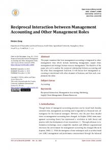
F I G U R E 1 Experimental design. In the learning session on the first day, infants heard unknown pseudowords as names for exemplars of unknown similarity-based object categories. In the memory test on the following day, generalization was tested by presenting novel category exemplars in both correct and incorrect pairings, that is, in same category–word pairings as in the learning session or in different pairings. In the nonlearning control session about a week later, infants heard known words as names for exemplars of known categories. Subsequent to the learning and control sessions infants napped. (For a detailed description, see Section 2)
|
FRIEDRICH et al.
4 of 12
assess immediate generalization of the object–word pairings while controlling for repetition effects, additional eight objects and
2.3 | Stimuli
eight words were presented each eight times, but not consistently
Visual stimuli were colored illustrations of single objects (Figure 1).
paired, such that the formation of stable object–word pairings was
In the learning session, eight exemplars of each of eight different
prevented.
similarity-based object categories were presented. In the memory
In the memory test on the next day, each four novel exemplars
test, four additional exemplars of each category were presented. For
of a category were presented in order to test for the presence
the nonlearning session, pictures of eight different exemplars for
of generalized memories of the category–word pairings. In the
each of the selected eight categories were chosen.
correct pairing condition, categories and words were paired as in
In the nonlearning session, eight words naming the known
the learning session on the previous day. In the incorrect pairing
basic-level categories were used as auditory stimuli. In the learn-
condition, the same exemplars and the same words as in the cor-
ing session, eight disyllabic pseudowords were taken as names
rect pairing condition were presented, but in different pairings
for the new categories. Pseudowords were phonotactically legal
that violated the category–word pairings of the learning session.
in German, were stressed on the first syllable, had a consonant–
Each individual pairing was presented once. For the case that in-
vowel onset, and had typical masculine or neuter endings. All au-
fants did not show generalized memories, after the presentation
ditory stimuli were spoken slowly by a female speaker, digitized at
of novel exemplars, four old exemplars of each category were
a rate of 44.1 kHz, and presented through loudspeaker with mod-
presented with the correct and incorrect words in order to as-
erate intensity.
sess learning in the test phase compared to initial learning. Since generalized memory turned out to be present in the first half of the test phase, memory and new learning may have interfered in
2.4 | Sleep recordings and sleep spindle analyses
the second half, therefore we did not include these data in the
Infants’ sleep was recorded using a portable amplifier (SOMNOscreen
analyses.
EEG 10–20, Somnomedics, Kist, Germany). EEG recordings were ob-
In the nonlearning control session, 64 exemplars belonging to
tained with electrodes attached at F3, FZ, F4, C3, C4, P3, PZ, P4,
eight basic-level categories (eight exemplars per category) were each
left, and right mastoids, referenced to CZ (positions according to
presented twice together with their correct word label. Categories
the International 10–20 system), filtered between 0.03 and 35 Hz,
were known to be acquired very early in infancy, the word labels
and sampled at 256 Hz. Electrooculographic and electromyographic
were (in German): Auto (car), Ball (ball), Hund (dog), Eimer (pail), Keks
recordings were bipolar from electrodes close to the eyes and at
(cookie), Löffel (spoon), Schuh (shoe), Vogel (bird). Infant’s compre-
the chin, respectively. Off-line, EEG signals were rereferenced to
hension of these words was assessed by parental ratings. On aver-
the average potential at left and right mastoid electrodes. EEG re-
age, infants comprehended seven of the eight words.
cordings were visually scored according to standard criteria (Grigg-
During the experimental sessions, infants sat on the mother’s or
Damberger et al., 2007; Rechtschaffen & Kales, 1968). For each nap,
father’s lap in a sound-attenuated room. In each trial a colored pic-
total sleep time (TST) and the time spent in the different sleep stages
ture of a single object appeared on the screen for 3,200 ms. After an
(1, 2, slow wave sleep, and REM sleep) were determined.
interval of 800 ms postpicture onset, the German indefinite article
Periods of arousal were excluded and power spectral analysis
ein (masculine/neuter) was presented to direct the children’s atten-
of the EEG signal was performed using fast Fourier transformation
tion to the acoustically presented word that followed the article pre-
for the remaining periods of NonREM sleep. The spectra were cal-
sentation after 900 ms.
culated for successive 8-s (2,048 data points) artifact-free intervals
For both the learning and the nonlearning sessions, infants were
using a Hanning window to taper the data. Average power was cal-
scheduled at a time when they were expected to take a nap within
culated first over all bins in the frequency range of interest; then
the next hour. In 26 of 30 infants, the learning and nonlearning tasks
averages were calculated for the succeeding 8-s intervals.
were applied before noon. On average, the learning session ended
For the detection of discrete sleep spindles, the EEG of all
at 10:54 (SD 1:18) and the nonlearning session at 10:48 (SD 1:12).
artifact-free NonREM epochs was low-p ass filtered (32 Hz) and
After the learning and the nonlearning sessions, infants were pre-
down- s ampled (128 Hz). The spindle detection algorithm and
pared for polysomnographic recordings (5–10 min) and laid down in
criteria were adopted from Mölle et al. (2009). First, for each
a baby crib or in their pram. When necessary, infants were held by
subject, the individual spindle peak frequency was identified
their parent until they fell asleep. Also if needed, infants were fed
in the NonREM sleep power spectra of all channels (learning:
or diapered before laying down for sleep. Sleep onset latency from
14.12 ± 1.07 Hz, nonlearning: 14.08 ± 1.10 Hz, across all subjects
the end of preparation (M ± SD: 24.8 ± 24.2 min) did not significantly
and channels; t 29 = 0.875, p = 0.389 for the comparison of learning
differ between the naps following the learning and nonlearning ses-
vs. nonlearning). The EEG signal was then filtered with a band-
sions (t29 = −1.033, p = 0.310). After the learning session, infants
pass width of 3 Hz centered on the detected individual peak fre-
slept for 63.1 ± 23.7 min, and after the nonlearning session, for
quency. A root mean-s quare (RMS) representation of the filtered
55.7 ± 19.8 min. Total sleep time did not significantly differ between
signal was calculated using a sliding window of 0.2 s with a step
the naps (t29 = 1.517, p = 0.140).
size of one sample. Additional smoothing was performed with a
|
5 of 12
FRIEDRICH et al.
TA B L E 1 Sleep characteristics during the nap after the learning task and during the nap after the nonlearning control session. stage 2 sleep, slow wave sleep, REM sleep, and TST in minutes, spindle peak frequency in Hz, and spindle density in number per 30 s
20 Hz (−3 dB cut-off frequencies of 0.62 and 19.88 Hz). Trials with potential fluctuations exceeding a standard deviation of 80 μV within a sliding window of 500 ms at any electrode site were rejected. ERPs were analyzed time-locked to word onset. For each con-
Postlearning nap
Nonlearning control nap
dition, epochs of 1,200 ms from word onset were averaged. A min-
M
SD
M
SD
imum of 10 artifact-free trials for each condition (correct, incorrect pairing), were required for an individual average to be included in
Stage 2 sleep
31.93
14.64
25.99
11.40
Slow wave sleep
20.30
8.63
20.27
10.80
0.78
2.75
0.00
0.00
TST
63.08
23.74
55.67
19.85
Spindle peak frequency
14.12
1.07
14.08
1.10
sites were combined into regions- of- i nterests (ROIs). The av-
Spindle number frontal
117.80
53.53
100.83
44.10
eraged ERPs at F7, F3, and T7 formed the left fronto-temporal
Spindle number central–parietal
100.44
46.37
79.62
35.87
region (LFT); at F8, F4, and T8 the right fronto-temporal region
Spindle density frontal
1.19
0.24
1.20
0.20
C4, and CP6 the right central region (RC); at P3, P7, and O1 the
Spindle density central–parietal
1.03
0.24
0.96
0.24
left parieto-occipital region (LPO) and at P4, P8, and O2 the right
REM sleep
further analyses. On average, 19 (SD = 6) trials contributed to an ERP condition. Trial numbers did not differ between conditions (t29 = 1.570, p > 0.127). For the statistical analyses of the ERP data, lateral electrode
(RFT); at FC3, C3, and CP5 the left central region (LC); at FC4,
parieto-occipital region (RPO). Memory effects were evaluated by ANOVAs with the within-
sliding-window average of 0.2 s size and one sample point step
subject factors Pairing (correct vs. incorrect), Hemisphere (left vs.
size. Time frames were considered as spindle intervals if the RMS
right), and Region (fronto-temporal, central, parieto-occipital), which
signal during NonREM sleep exceeded a threshold of 1.5 standard
were performed for the ERP mean amplitudes within two time
deviations of the filtered signal (learning: 5.90 ± 1.36 μV, nonlearn-
windows (200–600 ms, 600–1,000 ms). For midline sites, analog
ing: 6.09 ± 1.23 μV, across all subjects and channels; t 29 = 1.183,
ANOVAs were performed with Pairing and Region (FZ, CZ, PZ). To
p = 0.246 for learning vs. nonlearning) in an individual channel of
assess the impact of the learning-related increase in spindle activity
a subject for 0.5–5 s and if the largest value within the frame was
on the infants’ memory, the increases in spindle number and spindle
greater than 2.5 standard deviations of the filtered signal (learn-
density were defined by the individual differences in spindle num-
ing: 9.84 ± 2.26 μV, nonlearning: 10.15 ± 2.04 μV; t 29 = 1.183,
ber/density between postlearning and control nap and included as
p = 0.246 for learning vs. nonlearning). Two succeeding spindles
covariates into repeated measure ANCOVAs. In all AN(C)OVAs de-
were counted as one spindle when the interval between the end
grees of freedoms were adjusted according to Greenhouse–Geisser
of the first spindle and the beginning of the second spindle was
whenever they were >1.
shorter than 0.5 s and the resulting spindle was not longer than 5 s.
Subsequent to interactions of Pairing with Spindle increase, the spatial maximum of the memory effect was tested for signifi-
In the previous studies, memory generalization was particu-
cance by one-s ample t-test and taken for the correlation analysis
larly related to sleep spindles over central and parietal brain re-
(using Pearson’s correlation coefficient) between spindle increase
gions (Friedrich et al., 2015, 2017). In order to increase statistical
and memory performance. For the correlations of spindle in-
power, here, we analyzed the mean spindle measures across all
crease with the two ERP memory effects, the significance level
central and parietal channels. Analyses included spindle number,
was adjusted to 0.025. In order to further qualify the impact of
spindle density (spindles per 30 s NonREM sleep), peak-to-p eak
spindle increase on the infants’ memory, the ERP memory effects
amplitude, and length. When testing the difference between the
were tested separately in subgroups defined by a median split due
postlearning and the nonlearning control nap for the four spin-
to the individual’s spindle increase. These subgroups did not sig-
dle parameters, the significance level was Bonferroni-a djusted to
nificantly differ in age (t 28 = −1.037, p = 0.308), comprehension of
0.0125.
words in the control condition (t 28 = 0.349, p = 0.730), attention during encoding as indicated by the number of artifact-free tri-
2.5 | ERP data acquisition and analyses Infant memory was assessed by event-r elated potential (ERP) re-
als (t 28 = −0.306, p = 0.763), nor in the typical amount of sleep during the day (t 28 = 1.214, p = 0.235) and night (t 28 = −1.748, p = 0.091). In order to test for differences in immediate general-
sponses to the word stimuli. The EEG was recorded with a station-
ization during the learning phase, Spindle group was included as
ary amplifier (REFA, TMS International, Oldenzaal, Netherlands)
a between-s ubject factor into an ANOVA with the within-s ubject
at 21 electrode sites and digitized online at 500 Hz. Off-line, the
factors Pairing (consistent vs. inconsistent), Hemisphere, and
EEG was rereferenced to the average potential recorded from
Region as well as into the respective midline ANOVA with Pairing
left and right mastoid electrodes and filtered between 0.5 and
and Region.
|
FRIEDRICH et al.
Frontal
(b)
150 130
20 µV
1.35
Spindle density (number/30 s) over frontal brain regions
(a)
Spindle number over frontal brain regions
6 of 12
110 90 70
Spindle number over central and parietal brain regions
130 110 90 70 50
Non-learning
1
0.1 0
5
10
15
Hz
20
25
30
0.85
Learning
1.35
C3 Cz C4 P3 Pz P4
1.25 1.15 1.05 0.95 0.85 0.75
Learning
Non-learning
Learning
150
Increase in spindle density over central-parietal regions
Learning Non-Learning
(c) Increase in spindle number over central-parietal regions
10
0.95
Non-learning
150
100
1.05
Learning Spindle density (number/30 s) over central and parietal brain regions
Non-learning
Central-parietal
1.15
0.75
50 0.4 s
F3 Fz F4
1.25
100 50 0 –50
–100 –50
–25
0
25
50
Increase in stage 2 sleep (min)
0.50
0.25
0
–0.25
–0.50 –50
–25
0
25
50
Increase in stage 2 sleep (min)
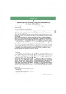
F I G U R E 2 Sleep spindle activity and its increase after learning. (a) EEG power spectra during NonREM sleep (at CZ) and (above) sleep spindles averaged across frontal (F3, FZ, and F4) and across central–parietal (C3, CZ, C4, P3, PZ, and P4) brain regions for the nap after the learning session and the control nap after the nonlearning session (mean ± SEM). (b) Spindle numbers (left panels) and spindle density (right panels) in nonlearning control nap and postlearning nap for spindles over frontal (upper panels) and central–parietal (lower panels) cortex. Learning-induced increases in spindle number and density were significant only for central–parietal spindles but not for frontal spindles. (c) Correlation between the learning-induced increase in time spent in stage 2 NonREM sleep and the learning-induced increase in spindle number (left: r = 0.836, p