IBBJ
Review Article
Winter 2018, Vol 4, No 1
Downloaded from ibbj.org at 20:45 +0330 on Friday October 19th 2018
The Relationship between Glutamate and Multiple Sclerosis Majid Malekzadeh Shafaroudi 1, Hooman Zarei 2, Ali Malekzadeh Shafaroudi 3, Narges Karimi1, Mahmoud Abedini 4* 1. Immunogenetic Research Center, Department of Anatomy & Cell Biology, Faculty of Medicine, Mazandaran University of Medical Sciences, Sari, Iran. 2. Department of Anatomy & Cell Biology, Faculty of Medicine, Student Rese arch Committee, Mazandaran University of Medical Sciences, Sari, Iran. 3. Faculty of Dentistry, Student Research Committee, Mazandaran University of Medical Sciences, Sari, Iran. 4. Department of Neurology, Faculty of Medicine, Mazandaran University of Medical Sciences, Sari, Iran. Submitted 25 Aug 2017; Accepted 28 Sep 2017; Published 29 Oct 2017
Glutamate is the most important excitatory neurotransmitter in the central nervous system which is involved in synaptic transmission, brain development, synaptic plasticity, learning, and memory. Normally, the enzymatic destruction of glutamate does not occur in the synaptic and extracellular space, but glutamate is removed through specific transporter proteins, leading to stabilization of glutamate concentration at non-toxic levels. When extracellular glutamate concentration increases, it could cause excitotoxicity and lead to many diseases of the central nervous system such as neurodegenerative disorders and multiple sclerosis (MS). Trans -glutaminase enzymes produce large quantities of glutamate by deaminating glutamine and consequently activating immune cells, especially lymphocytes. These activated lymphocytes release glutamate abundantly in the lesion location. Also, the expression level of glutamate specific carriers is decreased in the lesion area. This review discusses on the synthesis and release of glutamate, the natural cycle of glutamine/glutamate and glutamate receptors and transporters, and their role in excitotoxicity and finally their relationship with MS. Keywords: Glutamate, multiple sclerosis, excitotoxicity, central nervous system
G
lutamate is a non-essential amino acid that is
low (about 0.5-4 μM). The aggregation of glutamate
known as the most important excitatory
in the cerebrospinal fluid is 1-10 μM, but depending
neurotransmitter, and is uniformly distributed in
on the activity of neurons in the synaptic space, it
large amounts in the central nervous system (CNS)
may vary between 2-1000 mM (4). Since glutamate
structures (1, 2). Appropriate passage of nerve
does not cross the blood-brain barrier (BBB), it is
impulses from glutamatergic synapsis is required for
produced from glucose (by the Krebs cycle) or
organizing the basis of many processes such as
glutamine inside the neurons, and is released into the
memory, and learning in the CNS (3). The amount
synaptic space through the process of exocytosis,
of glutamate in nerve terminals is about 110 mM, of
using synaptic vesicles, and exerts its effects on
which 100 mM is stored in the synaptic vesicles, and
postsynaptic neurons through its receptors (2).
10 mM is present in the cytoplasm of the nerve axon
Glutamate receptors consist of two general
(4). Extracellular glutamate concentration is very
categories: ionotropic receptors that are ion
*Correspondence: Faculty of Medicine. Mazandaran University of Medical Sciences, Sari, Iran. E-mail:
[email protected]
Downloaded from ibbj.org at 20:45 +0330 on Friday October 19th 2018
Shafaroudi M M et al.
channels, and metabotropic receptors that operate
action of glutamate receptors and transporters, as
via G-protein and intracellular signaling processes
well as their relationship with excitotoxicity and MS
(3). Ionotropic receptors include three subunits of N-
disease are examined. Finally, several new
methyl-D-aspartate (NMDA), α-amino-3-hydroxy-
therapeutic methods, together with the efficient
5-methylisoxazole-4-propionic acid (AMPA), and
method of glutamate concentration measurement
kainate. Also, 8 subunits have been identified for the
using carbon nanotube technology, are presented.
metabotropic receptors so far, that are divided into
Synthesis and release of glutamate
three individual groups (1). Excitotoxicity is caused by
increased concentrations of
Glutamate cannot pass the BBB. It is therefore
extracellular
produced from glucose or glutamine in the CNS (16,
glutamate, and is considered as one of the major
17). The natural cycle of glutamate/glutamine was
processes of nerve cells death, and also plays a major
suggested to be the major metabolic pathway in the
role in various diseases such as neurodegenerative
brain (18, 19). This cycle starts with a calcium-
disorders, ischemia, trauma and multiple sclerosis
dependent release of glutamate, which results in a
(MS) (5). This toxicity is due to postsynaptic
20-fold acceleration in the amount of synaptic
glutamate receptors stimulation by a large amount of
glutamate release (20). After transferring the
extracellular glutamate. Each of these receptors can
depolarization messages to postsynaptic neurons,
somehow get
process of
glutamate is led out of the synaptic space by
excitotoxicity, and cause neuron damage or death.
dedicated transport proteins, so the next impulse can
For example, activation of NMDA receptors causes
be created (21). Glutamate is transmitted to CNS
neuronal death by letting-in large amounts of Ca2+
supporting cells which are mainly astrocytes, and
ions into the cell. Other aforementioned receptors
also a small amount of glutamate is released into the
that cause nerve cells death via excitotoxicity are
blood capillaries (16). After entering the astrocytes,
also important (1, 6). Extracellular glutamate
and by using an enzyme known as glutamine
concentration adjustment takes place by glutamate
synthetase, glutamate is converted into its inactive
transporter proteins. So far, five human excitatory
form, glutamine. Glutamine passes through the
amino acid transporters (EAAT1-5) have been
membrane of astrocytes and enters the cytoplasm of
described (7). These proteins are present in the CNS
neurons as well as the mitochondrial intermembrane
and other soft tissues, and are also able to increase
space (21, 22). Finally, in the presence of
the concentration of intracellular glutamate up to
transglutaminase, glutamine is transformed into
10,000 times more than the extracellular fluid.
glutamate, and packed in synaptic vesicles (21, 22).
Therefore, these transporter proteins are required to
In a study performed in 2005 the concentration of
maintain the concentration of glutamate at a non-
glutamate
toxic level. Recent studies showed that impairment
glutamine, N-acetyl-aspartate, myoinositol, choline,
in the function of transporter proteins can be
and creatine) was measured in various parts of brain
effective in neurological diseases development (7-
of MS patients and control group using the magnetic
10). MS is an inflammatory and demyelination
resonance spectroscopy (MRS). The results showed
disease of CNS (11). Axonal loss and neuro-
an important increase of glutamate concentration in
degeneration are caused in the progressive phase of
acute MS lesions. This increase is likely related to
the disease (12, 13). In MS, the release of glutamate
the production of glutamate by activated immune
and glutamate transporters, as well as receptors
cells adjacent to the lesion area (21). Another study
release or signaling, are disproportionate (14, 15). In
in 2012 emphasized on glutamate release increase in
this article, the structure, synthesis, and release of
the axonal injury site, demyelination, and also the
glutamate in the CNS, and also the mechanism of
active role of microglia and leukocytes in creating
involved
in the
and
other
metabolites (including
Int. Biol. Biomed. J. Winter 2018; Vol 4, No 1
2
Downloaded from ibbj.org at 20:45 +0330 on Friday October 19th 2018
Glutamate in Multiple Sclerosis
glutamate. It suggested that balancing glutamate
receptors is not well specified, but there is
release and blocking its receptors, could be used as
agreement that the main reason is due to the influx
a new possible treatment for MS (12). Also, it was
of calcium ions. Simultaneously, the entry of ions
shown that glutamate and glutamine were important
like sodium and manganese, and exit of potassium is
biomarkers in progressive MS disease (23).
also linked with this process (28, 29). In a study on
Glutamate receptors and excitotoxicity
animal models of MS, it has been shown that
Ionotropic glutamate receptors
antagonists of kainate and AMPA receptors
Ionotropic glutamate
receptors comprise
improved disease symptomatic, and also increased
NMDA, AMPA, and kainate that are ion channels,
oligodendrocytes survival, and reduced axonal
and are named after their specific agonist (2, 19).
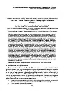
injury (24) (Fig. 1).
Ionotropic glutamate receptors generally have three transmembrane hydrophobic domains (M1, M3, and M4) and a concave circle inwards (M2) that form the channel. All subtypes of this receptor have four similar domains: extracellular amino-terminal domain (ATD), extracellular ligand-binding domain (LBD), transmembrane domain (TMD) and an intracellular carboxyl-terminal domain (CTD). The CTD is the most variable portion among the ionotropic glutamate receptors, as AMPA receptors and the NR1 subgroup of NMDA receptors have a
Figure 1. Glutamate/Glutamine cycle in the CNS.
relatively short CTD domain, while NR2 subgroups of NMDA receptors have larger CTD domains (24).
One of the atypical neurotransmitters that
The glutamate metabolism increase in the synaptic
adjusts brain and neuron activity, and is also
space may create neuronal damage, causing acute
necessary for the proper organization of the
and chronic diseases of the CNS. Due to the
vertebrate nervous system is nitric oxide (NO) (30).
activation of cell receptors, especially NMDA
NO can be produced from several sources in the
receptors, a large number of ions accumulate within
CNS such as cerebral vessel endothelium, microglia
the cell which could lead the neurons towards
and astrocytes, non-adrenergic non-cholinergic
dendritic apoptosis, and deadly degenerative
nerves, and glutamatergic neurons (31). NMDA
processes (25, 26). An increase in glutamate
glutamate receptors activation results in a high
metabolizing enzymes and glutamate transport
amount of NO release. Researches have claimed that
proteins (EAAT1) is observed in the process of
NO modulates the neurotoxicity of glutamate. NO is
excitotoxicity in astrocytes and macrophages,
a reactive free radical that neutralizes free radical
revealing the protective role of these cells in terms
formations related to neurotoxicity, and by detaining
of excitotoxicity states, but probably they are not
glutamate-induced apoptosis, NO pathways could
able to adjust the concentration of glutamate in
prevent the oxidative damage to neurons (32).
sufficient quantities due to a huge increase in
The NMDA receptor can be activated by
extracellular glutamate concentration and severe
successive messages coming from two different
stimulation in glutamate receptors (27). The role of
cells. Thus, it differs from other receptors (33). The
other receptors such as kainate is also important in
message of the first cell sensitizes the membrane of
causing excitotoxicity. The molecular basis of
the cell containing NMDA receptors, and through
glutamate excitotoxicity associated with kainate
the second message, glutamate activates the
3
Int. Biol. Biomed. J. Winter 2018; Vol 4, No 1
Shafaroudi M M et al.
Downloaded from ibbj.org at 20:45 +0330 on Friday October 19th 2018
receptors. When the two messages converge in this
Metabotropic receptors
manner, the NMDA receptor imports a large number
Metabotropic receptors consist of 8 subunits
of calcium ions into the neuron (34). This influx of
which are classified into three groups, and are
ions causes long-term changes in the neuronal
associated with secondary messengers in the cell (2).
membrane which is called long-term potentiation
For example, when metabotropic receptors, which
(LTP). Such mechanisms in which synapses are
are located in the postsynaptic membrane of
strengthened by two convergent messages, provide
neurons, are stimulated, phospholipase C is
an explanation for linking separate incidents in
activated leading to the formation of inositol 1, 4, 5
memory (35, 36). It has been proved that the uptake
triphosphate (IP3), and calcium release from
of glutamate increases in the hippocampus during
intracellular reservoirs (40-42). These receptors are
early long-term potentiation (E-LTP) and late long-
generally associated with G proteins, and launch
term potentiation (L-LTP). Recent studies have also
different mechanisms in neurons. For instance, an
indicated that both astrocytes and glutamate 1
intra-cerebroventricular
(GLT-1) (EAAT2) transporters play an important
metabotropic receptor agonist (S)-3,5-Dihydroxy-
role in glutamate uptake during L-LTP (37).
phenylglycine (DHPG) on broiler chicks, resulted
In fact, in LTP, NMDA receptors are activated
in
a
(ICV)
significant increase in
injection
of
food intake,
sequentially and the concentration of intracellular
while
calcium increases in the postsynaptic cell which
associated protein (AIDA), a group I metabotropic
eventually leads to the activation of calmodulin
receptor antagonist, considerably reduced the food
kinase II. This will increase the expression of
intake. These studies specify the direct or indirect
AMPA receptors on the cell membrane, and
role of receptors, on food intake and appetite (43).
strengthen synaptic connections between neurons (38).
injecting axin interactor dorsalization-
Studies have proved the influence of group I and II metabotropic glutamate receptors in central
Synaptic plasticity has an important role in
nervous system diseases such as MS (44). An excess
recovering demyelinated lesions, and also in the
of axonic metabotropic glutamate receptor 1
healing process of MS. It has been proved that
(mGluR1) expression was observed in chronic and
modulating LTP could increase automatic neuron
acute MS lesions. Also, increased expression of
recovery, and therefore lead to a clinical treatment
mGluR5 and mGluR2/3 within the MS lesions was
for MS. Physical rehabilitation and a number of
observed in astrocyte cells (44). In another study on
drugs promote LTP. Therefore, they could speed up
active lesions, the immune reactivity (IR) of
the synthesis of myelin, and enhance clinical
the mGluR8 receptor which is a group III
recovery (39) (Fig. 2).
metabotropic glutamate receptor was observed in microglia/macrophage cell lineages (45). In chronic and passive lesions, a lesser amount of IR positive GluR8 was observed in macrophage-like cells. Also, no IR positive GluR4 receptors was observed in MS lesions in microglia/macrophage cell lines (46). However, a number of reactive astrocytes expressed both mGluR4 and mGluR8 receptors in the margins of lesions, revealing the importance of metabotropic receptors in CNS diseases (46). Various roles were demonstrated for mGluR4
Figure 2. Different groups of glutamate receptors.
in glial cells (47). It was shown that this receptor
Int. Biol. Biomed. J. Winter 2018; Vol 4, No 1
4
Downloaded from ibbj.org at 20:45 +0330 on Friday October 19th 2018
Glutamate in Multiple Sclerosis
can have a protective role, and increase the
the absence of active microglia, there was no
durability of oligodendrocyte cells, as cinnabarine
EAATs reduction (54).
acid - a mGluR4 agonist - balances the activity of
One study showed that beta-lactam antibiotics
the immune system and protects against MS.
such as ceftriaxone, increase the activity of
Perhydrocyclopentanophenanthrene is a positive
glutamate transporters (similar to EAAT2) by
modulator of mGluR4, and reduces the severity and
stimulating the GLT-1 gene in an animal model, thus
recurrence of the disease (47).
reducing the extracellular glutamate concentration
Glutamate transporters
(55). The effect of the drug deferred the loss of
The concentration of extracellular glutamate is
muscle power and body weight in mice with
kept in levels lower than toxicity values, using
amyotrophic lateral sclerosis (ALS). So it seems that
dedicated transport proteins which are located inside
these antibiotics, bring satisfactory results for the
the membrane of glial cells surrounding the
treatment of neurological disorders (55).
synapses,
and
presynaptic
and
postsynaptic
Another point in this regard is that an excessive
terminals (48). These proteins are EAATs which co-
increase in glutamate transporters will also cause
transmit glutamate with at least two
Na+
ions and a
proton in the same direction, and a single
K+
neurological
disorders.
For
example,
in
ion in
schizophrenia excessive activation of astrocytes
the opposite direction (49). Therefore, they are
glutamate transporters will become extremely out of
called Na+
dependent transporters which have a high
reach in the prefrontal cortex (56). However,
tendency to glutamate. These transporters are
compensatory increase in dopaminergic fibers
located throughout the CNS, and so far 5 sodium-
activity inside the ventral tegmental area which is
dependent transporters with a high tendency to
due to reduced prefrontal cortex inhibition, and
glutamate have been cloned from human tissues
losing control of the limbic system, cause worsening
which consist of EAAT1-5. EAAT1 and EAAT2 are
of the patient's condition and appearance of the
generally located in astrocytes, while EAAT3 and
positive symptoms of the disease such as
EAAT4
hallucination and delirium (56).
are mainly deployed in postsynaptic
membranes, and EAAT5 is located in retinal
In MS, increased expression of glutamate
ganglion cells, photoreceptors, and bipolar cells (50-
transporters including EAAT2 and EAAT1 occurs.
52). EAATs placed on the plasma membrane
This is considered as a regulatory response of glial
of neurons and glial cells contribute to ending
cells to high concentrations of extracellular
quickly the glutamate’s accomplishment, and
glutamate (7).
preserve its extracellular concentration below
Therapeutic approaches
excitotoxic values (50).
MS is an inflammatory disease of the CNS
In 1996, by knocking out and assessing the
which comes along with demyelination and
effects of the three glutamate transporters that have
destruction of oligodendrocytes and axonal damage
been identified in mice, it was concluded that these
and destruction. The main cause of the disease
transporter proteins are involved in regulating the
remains unknown. However, evidences suggest that
concentration of glutamate, and the failure of either
it progresses genetically among people who are
of them can lead to excitotoxicity and neuronal
faced with environmental factors (57-59). The
degeneration (53).
autoimmune T cells are involved in the development
When examining the brain of MS patients,
of early staged lesions of MS (60). While the central
it was observed that in areas with a reduction in
target of the T cells is uncertain, several studies have
these transporters, there were also activated
pointed to myelin antigens. Documents indicate the
microglia, and even in the demyelinated cortex in
involvement of T helper cells with Th17 phenotype,
5
Int. Biol. Biomed. J. Winter 2018; Vol 4, No 1
Downloaded from ibbj.org at 20:45 +0330 on Friday October 19th 2018
Shafaroudi M M et al.
while previous studies expressed that T helper type
reducing excitotoxicity induced by glutamate is one
1 cells played the main role in this disease (60).
of the therapeutic effects that recently have been
Th17 cells probably produce and release high levels
taken into consideration (69).
of glutamate in pathological conditions (61). In
Some studies have demonstrated that AMPA
addition, the role of other immune cells such as B
receptor antagonists reduce the symptoms and
lymphocytes and a whole range of immune
pathological
responses against a limited number of antigens
degeneration in an animal model of MS (71, 72).
within the brain are considered to be important (62).
Also, AMPA receptor antagonists help to reduce
Unfortunately, to date, no decisive cure has
both acute and chronic lesions of MS (63). Drugs
been found for MS, but
changes,
especially
neuronal
anti-inflammatory,
such as alampanel and perampanel are non-
immunosuppressive, and immunomodulatory drugs
competitive AMPA receptor antagonists, and
slow the process of the disease and improve the
NBQX, PNQX, YM-90K and recently ZK200775
symptoms (63). New studies which focus on
are listed among competitive AMPA receptor
glutamate excitotoxicity that occurs in MS, and
antagonists (74-77).
many other disorders of the CNS are capable of introducing
new
therapeutic
approaches
by
inhibiting the process of excitotoxicity (63).
NMDA
receptor
antagonists
such
as
memantine was approved as a treatment for Alzheimer disease (78). This drug competitively
Interferon-beta (IFN-β) in two different forms
inhibits glutamate's activity by binding to its surface
is used to treat MS patients at present (64, 65). When
receptor (79). NMDA receptor light antagonists
IFN-β is attached to its own surface receptor, it
such as amitriptyline are helpful in treating
activates the cascade signaling pathways inside the
conditions such as excitotoxicity (80).
cell by inhibiting the activity and proliferation of T-
Kainate receptor antagonists such as ethanol,
cells (Th2), and altering the cytokine profile in the
6-cyano-7-nitroquinoxaline-2,3-dione
nervous system, and also affect the migration
6,7-dinitroquinoxaline-2,3-dione
of leukocytes from the BBB as well as the
tezampanel, have
expression of neurotropic factors, which leads to
effects (81).
(CNQX),
(DNQX)
and
anti-glutamate excitotoxicity
immune-modulatory effects (66, 67). Beyond these
Researchers have also shown that group II and
known effects, this drug reduces excitatory
III metabotropic glutamate receptor agonists have a
postsynaptic currents by affecting glutamate through
protective neuronal
a new postsynaptic mechanism that requires calcium
presynaptic release of glutamate, while group I
in nucleus striatum that is susceptible to degenerate
agonists singly may cause excitotoxicity (82). Group
in the progression of MS (68, 69). In fact,
I metabotropic glutamate receptors are mainly
this mechanism is based on intracellular calcium
located in the postsynaptic membrane, and due to
Ca2 +/
their activity, G proteins are activated, causing
calmodulin-dependent protein kinase II (CAMK II).
phospholipase C activation, which consequently
This protein kinase is associated with GluN2A
catalyzes the production of IP3 and diacylglycerol
which is one of the subunits of NMDA glutamate
(DAG) (83). IP3 stimulates the release of calcium
receptors that has a major role in the influx of
ions within the cell and DAG activates protein
calcium ions into the cell, and causing excitotoxicity
kinase C, which also increases the amount of
(69). Therefore, a decrease in the concentration of
intercellular calcium (40, 83). Group II receptors
intercellular calcium or inhibition of CAMK II,
which have been sighted on presynaptic and
prevents reducing excitatory synaptic tolerance by
postsynaptic membranes bind to G proteins that
IFN-β (70-73). Relatively, the impact of the drug on
negatively adjust the action of adenylyl cyclase (40).
concentration and
the
activation
of
effect by
reducing the
Int. Biol. Biomed. J. Winter 2018; Vol 4, No 1
6
Downloaded from ibbj.org at 20:45 +0330 on Friday October 19th 2018
Glutamate in Multiple Sclerosis
Group III metabotropic glutamate receptors are
glutamate based on chromatography or capillary
principally positioned in the presynaptic membrane,
electrophoresis, but these methods are very time-
where they operate as auto-receptors and bind
consuming and require expensive equipments (91,
to G proteins to reduce the activity of adenylyl
92). Recently, stabilizing glutamate oxidase or
cyclase (41).
glutamate
dehydrogenase
on
electrodes
for
It was proven that loss of interaction between
designing amperometric biosensors has attracted
GluR2 which is a subunit of AMPA receptors,
many researchers to itself. Since glutamate
and
glyceraldehyde-3-phosphate dehydrogenase
dehydrogenase sensors require NAD+ as a cofactor,
(GAPDH) can reduce excitotoxicity (84). In 2015,
therefore most of the glutamate biosensors are based
metabotropic glutamate receptor subunits were
on glutamate oxidase (93, 94). The accuracy of these
introduced as pharmacological targets for the
sensors is related to the production of hydrogen
treatment of MS (44). A moderate increase in the
peroxide by the reaction (95). Polypyrrole (PPy) is
expression of glutamate transporters has been
used as a selective permeability membrane for
reported to
rejecting interferences in these biosensors. The films
stabilize
the
concentration of
extracellular glutamate, showing therefore the
are
formed
from
aqueous
buffers
under
therapeutic value of the glutamate transporters (7).
electrochemical control at physiological pH (94).
Recently, oral medications based on small
The second stage involves the sedimentation of
molecules that can directly cross through the BBB,
multi-walled carbon nanotubes from an aqueous
and have a protective effect on neurons have
suspension by electrophoresis. This layer acts as a
attracted enormous interest (85). One of these drugs
backup for the removal of enzymes. The next step is
is dimethyl fumarate (DMF) that is processed to
the sedimentation of glutamate oxidase on carbon
monomethyl fumarate (MMF) and fumarate within
nanotubes, and the final stage consists of fixing a
the cells, and increases the activity of Nrf2
thin layer of polyurethane (PU) to increase the
transcription factor. This factor manages the
resistance of the sensor as well as increasing its
expression of antioxidant proteins, and anti-toxicity
linear range. The accuracy of this sensor is 3.84
enzymes. MMF also inhibits the release of
nA/μMmm²,the reaction time is less than 8 seconds
glutamate from pathogenic Th17 lymphocytes. Due
and the linear range is up to 500 μM (94).
to the anti-neuronal excitotoxicity effects of DMF, it
In another study in 2009, using the mechanism
was suggested as an oral medication for treating
of fixing glutamate oxidase enzymes on the
MS (85).
electrodes, and assessing the amount of hydrogen
In vivo measurement of glutamate in biological
peroxide, the galvanic technique of sol-gel was used
fluids
for seating glutamate oxidase enzymes on the silica
It has been reported that a number of drugs,
gel surrounding the electrodes to achieve more
environmental pollutants, and some foodstuffs
accurate assessment, and controlling real-time
highly stimulate glutamate receptors, and increase
glutamate
the release of glutamate (86). Also, in some diseases
amperometric detection of hydrogen peroxide by
of the CNS like strokes or Parkinson's disease, an
glutamate oxidase in the presence of glutamate in
increase occurs in the concentration of glutamate in
the test environment. The accuracy of this method is
the cerebrospinal fluid (87-90). Therefore, its
279.4 ± 2.0 μA (mmol L−1)−1 cm−2
measurement would be of great significance in both
(96). Diagnosis depends
range is from 0.5 to 100 µmol
L-1
on
the
and its linear
(96).
biological and pharmaceutical sciences, and also in
Nanofibers carbon which directly grow on a
food industries. So far, many ways have been
non-crystalline tetrahedron carbon (tetrahedral
provided for measuring the concentration of
amorphous carbon, ta-C), can quickly detect H2O2
7
Int. Biol. Biomed. J. Winter 2018; Vol 4, No 1
Shafaroudi M M et al.
(