Molecular Biology of the Cell Vol. 14, 4015– 4027, October 2003
The Role of mVps18p in Clustering, Fusion, and Intracellular Localization of Late Endocytic Organelles Viviane Poupon,* Abigail Stewart,* Sally R. Gray,* Robert C. Piper,† and J. Paul Luzio*‡ *Cambridge Institute for Medical Research and Department of Clinical Biochemistry, University of Cambridge, Addenbrooke’s Hospital, CB2 2XY Cambridge, United Kingdom; and †Department of Physiology and Biophysics, University of Iowa, Iowa City, Iowa 52242 Submitted January 24, 2003; Revised May 10, 2003; Accepted May 27, 2003 Monitoring Editor: Juan Bonifacino
Delivery of endocytosed macromolecules to mammalian cell lysosomes occurs by direct fusion of late endosomes with lysosomes, resulting in the formation of hybrid organelles from which lysosomes are reformed. The molecular mechanisms of this fusion are analogous to those of homotypic vacuole fusion in Saccharomyces cerevisiae. We report herein the major roles of the mammalian homolog of yeast Vps18p (mVps18p), a member of the homotypic fusion and vacuole protein sorting complex. When overexpressed, mVps18p caused the clustering of late endosomes/lysosomes and the recruitment of other mammalian homologs of the homotypic fusion and vacuole protein sorting complex, plus Rab7interacting lysosomal protein. The clusters were surrounded by components of the actin cytoskeleton, including actin, ezrin, and specific unconventional myosins. Overexpression of mVps18p also overcame the effect of wortmannin treatment, which inhibits membrane traffic out of late endocytic organelles and causes their swelling. Reduction of mVps18p by RNA interference caused lysosomes to disperse away from their juxtanuclear location. Thus, mVps18p plays a critical role in endosome/lysosome tethering, fusion, intracellular localization and in the reformation of lysosomes from hybrid organelles.
INTRODUCTION Delivery of endocytosed macromolecules to lysosomes, in mammalian cells, has been proposed to require maturation processes, “kiss and run” events, and/or direct fusion of late endosomes and lysosomes (reviewed in Storrie and Desjardins, 1996; Luzio et al., 2000; Mullins and Bonifacino, 2001). Direct fusion has been formally demonstrated in a cell-free content mixing assay (Mullock et al., 1998), and there is evidence from electron microscopic studies that the same process occurs in intact cells (Futter et al., 1996; Bright et al., 1997). Fusion results in the formation of hybrid organelles from which lysosomes are reformed by a maturation process involving removal of some membrane proteins, probably by vesicular traffic, and condensation of lumenal content (Pryor et al., 2000). At the molecular level, analogous proteins and processes seem to control fusion of yeast vacuoles as well as mammalian late endosomes and lysosomes. Fusion events among these compartments require N-ethylmaleimide-sensitive factor, soluble N-ethylmaleimide-sensitive factor attachment proteins and Rab GTPases (Mullock et al., 1998, 2000; Wickner and Haas, 2000). Yeast vacuole fusion depends on a set of soluble N-ethylmaleimide-sensitive factor attachment protein receptor (SNARE) proteins, including Vam3p, whereas late endosome fusion events require syntaxin 7, its closest mammalian homolog (Antonin et al., 2000; Mullock et Article published online ahead of print. Mol. Biol. Cell 10.1091/ mbc.E03– 01– 0040. Article and publication date are available at www.molbiolcell.org/cgi/doi/10.1091/mbc.E03– 01– 0040. ‡ Corresponding author. E-mail address:
[email protected].
© 2003 by The American Society for Cell Biology
al., 2000; Ward et al., 2000). Both systems also rely on a calcium/calmodulin-dependent process that probably acts after SNARE complex formation (Peters and Mayer, 1998; Holroyd et al., 1999; Pryor et al., 2000). SNARE complex formation is preceded by a so-called tethering reaction, which can link organelles over distances of ⱖ25 nm (Pfeffer, 1999). For late endosomes and lysosomes, this type of tethering reaction has been partly characterized as fine striations between adjacent late endosomes and lysosomes in morphological studies on cultured mammalian cells (van Deurs et al., 1995; Futter et al., 1996). For yeast homotypic vacuolar fusion, a number of “late-acting” vacuolar protein sorting (Vps) proteins seem to control tethering because they are localized to sites of fusion, interact with relevant SNAREs and Rab GTPases, but act before SNARE complex formation in vitro. These proteins are encoded by the class B* and class C phenotypic class of VPS genes and include Vps11p, Vps16p, Vps18p, and the Sec1like protein Vps33p, as well as the subcomplex comprised of Vps41p/Vam2p and the GTPase exchange factor Vps39p/ Vam6p. All of these factors can physically interact in a large homotypic fusion and vacuole protein sorting (HOPS) or class C Vps complex (Rieder and Emr, 1997; Sato et al., 2000; Seals et al., 2000; Wurmser et al., 2000). The complex may also play a role in tethering/docking at earlier stages of vesicular transport to the vacuole (Srivastava et al., 2000; Peterson and Emr, 2001). Animal homologs of these proteins have been identified, providing candidates for tethering late endosomes and lysosomes. Indeed, loss-of-function mutations in the Drosophila melanogaster genes VPS18, VPS33, and VPS41 (respectively known as deep orange, carnation, and light) perturb the formation of the lysosome-like pigment granule in
4015
V. Poupon et al.
the eye (Warner et al., 1998; Sevrioukov et al., 1999). Mammalian homologs of the class C Vps proteins localize to the endocytic pathway and can associate with the late endosomal/lysosomal-localized syntaxin 7 (Huizing et al., 2001; Kim et al., 2001). Finally, overexpression of the mVam6p/ Vps39p subunit of the mVam6p/Vps39p–Vam2p/Vps41p subcomplex alters late endosomal fusion in mammalian cells (Caplan et al., 2001). We have investigated the function of mVps18p in the mammalian late endocytic pathway. Our data show that mVps18p acts as a mammalian tethering and/or docking factor that promotes aggregation and fusion of late endosomes/lysosomes in vivo and that its presence is required for the tethering function of mVps39p. Our data suggest that in addition to, or as a consequence of its tethering functions, mVps18p also participates in the reformation of lysosomes from the hybrid organelles that result from the fusion of late endosomes with lysosomes, and plays a role in the intracellular positioning of lysosomes. MATERIALS AND METHODS Cells and Antibodies Normal rat kidney (NRK) and HeLa cells were grown as described previously (Ihrke et al., 2000). Phalloidin-tetramethylrhodamine B isothiocyanate was from Sigma-Aldrich (St. Louis, MO). Monoclonal antibody (mAb) to rat lgp120 (GM10), polyclonal antibody (pAb) to rat M6PR tail (1001), pAb to rat lgp110 (580), and mAb to rat TGN38 (2F7.1) were as described previously (Grimaldi et al., 1987; Horn and Banting, 1994; Reaves et al., 1996). mAb to EEA1 was from BD PharMingen (San Diego, CA). mAb to human Lamp1 (H4A3) was from Developmental Studies Hybridoma Bank (University of Iowa, Iowa City, IA). mAbs to RhoA, RhoB, Rac, and Cdc42 were from H. Mellor (University of Bristol, Bristol, United Kingdom). pAb to RILP was from I. Jordens (The Netherlands Cancer Institute, Amsterdam, The Netherlands). mAbs to dynactin p50 and p150glued were from BD Biosciences (San Jose, CA). mAb to ezrin was from A Bretscher (Oregon State University, Corvallis, OR). pAbs to MyoIb (Tu 30), MyoIc (Tu 45), and Myosin IX (Tu 66) were from M. Bahler (University of Mu¨nster, Mu¨nster, Germany). pAb to Myosin II (Kayneed) was from J. Kendrick-Jones (LMB, Cambridge, United Kingdom). pAbs to Myosin V (9) and Myosin VI (2401) were from F. Buss (Cambridge Institute for Medical Research, Cambridge, United Kingdom). pAbs to mouse mVps11p, mVps18p, and mVps33p were raised by immunizing rabbits with 1 mg of glutathione S-transferase–tagged protein corresponding, respectively, to the C-terminal part of mVps11p (aa 758 –935), and of mVps18p (aa 766 –921), and full length mVps33bp. The antibodies were immunoaffinity purified and their specificity tested by immunoblotting. AntimVps11p, anti-mVps18p, and anti-mVps33p were highly specific, whereas anti-mVps33p recognized both isoforms a and b. Texas Red-conjugated goat anti-mouse and anti-rabbit immunoglobulins for immunofluorescence microscopy and mAb 11E5 to green fluorescent protein (GFP) used for immunoblotting were obtained from Molecular Probes (Madison, WI).
Generation of the GFP Constructs Mouse Vps18 cDNA was obtained by polymerase chain reaction (PCR) amplification of a Marathon (BD Biosciences Clontech, Palo Alto, CA) ready mouse liver cDNA with a forward primer, including the starting ATG of human Vps18p and a reverse primer containing the stop codon for mouse Vps18p. The forward primer was designed from human expressed sequence tag (EST) IMAGE clone ID 4461817 (GenBank accession no. AW956323). The reverse primer was designed from clone PIR003, a partial clone identified by screening of a mouse brain cDNA library (Origene Technologies, Rockville, MD). The sequence of mVps18p is coded in a genomic region on mouse chromosome 2, identified as Mus musculus WGS supercontig (GenBank accession no. NW_000178). The gene corresponds to nucleotides 18577752– 18586330, and mRNA is formed of nucleotides (18577752. . . 18577843, 18578866. . . 18579006, 18581410. . . 18581502, 18581632. . . 18583505, 18585610. . . 18586330). The cDNA of murine Vps39 corresponded to EST IMAGE clone ID 3492875 (GenBank accession no. BE282898) cloned in pCMVSPORT6 and was obtained from UK HGMP Resource Center (Hinxton, Cambridge, United Kingdom). The different mVps18p or mVps39p cDNA fragments described in this study were obtained by PCR with a 5⬘ primer with a KpnI site, and a 3⬘ primer with a BamHI site, and then introduced into the pEGFPC1 vector (BD Bio-
4016
sciences Clontech). mVps39 Nter, ⌬ citron homology domain (CNH), and CNH were designed to be similar, respectively, to human Vps39 (or hVamp6p) constructs ⌬CT, ⌬CNH, and ⌬CLH⫹CT described by Caplan et al. (2001). A stable NRK cell line expressing full-length GFP-mVps18, was established using ⌬pMEP as the expression vector, and selection and expression were as described by Ihrke et al., (2000).
Transient Transfection, Dextran Uptake, Wortmannin Treatment, and Immunofluorescence For transient transfection experiments, cells were grown on coverslips to 60% confluence, transfected with 2 g of plasmid DNA by FuGENE (Roche Diagnostics, Lewes, United Kingdom) and used 24 –72 h later. Dextran uptake was performed by incubating transiently transfected cells with 1 mg/ml Texas Red-dextran (Molecular Probes) for 1 h. For wortmannin treatment (Sigma-Aldrich), the cells were incubated at 37°C for 45 min in RPMI medium-1% fetal calf serum containing 100 nM wortmannin. In both cases the cells were then washed in phosphate-buffered saline (PBS), fixed, and permeabilized in methanol for 5 min at ⫺20°C before processing for immunofluorescence microscopy. For all other immunofluorescence microscopy, cells were permeabilized before fixation to wash out the cytosol containing soluble overexpressed proteins by incubation with 0.05% saponin in cytosolic buffer (80 mM PIPES, pH 6.8, 5 mM EGTA, 1 mM MgCl2) for 1 min. The cells were then fixed and permeabilized in methanol as described above, or fixed with 4% formaldehyde in phosphate-buffered saline and processed for immunofluorescence microscopy as described previously (Poupon et al., 1999). Cells were observed with a confocal microscope (MRC1024; Bio-Rad, Hercules, CA) or an Axiophot microscope (Carl Zeiss, Jena, Germany) equipped with a charge-coupled device camera (Figure 8).
Electron Microscopy After an endocytic uptake of bovine serum albumin (BSA)-gold (5 nm) for 4 h at 37°C, followed by incubation in conjugate-free medium for 20 h (Reaves et al., 1996; Bright et al., 1997), cells were prepared for ultrastructural analysis by using transmission electron microscopy as described previously (Bright et al., 1997). Sections (70 nm) were observed in a Philips CM100 electron microscope. The mean area of the organelles containing 5-nm gold was measured using the online facility of the Philips CM100 microscope at a magnification of 4600⫻ as described previously (Wettey et al., 2002). For immunogold electron microscopy, the cells were prepared as described previously (Bright et al., 1997) and observed in a Philips CM100 at 80 kV.
Biochemical Procedures To prepare whole cell extracts from a stably transfected NRK cell line expressing GFP-mVps18p, or from NRK cells transiently transfected with GFPmVps18p or GFP-mVps39p, the cells were scraped into extraction buffer containing 50 mM Tris-HCl, pH 7.4, 150 mM NaCl, 1 mM EDTA, 0.5% Triton X-100, and protease inhibitor cocktail (complete; Roche Diagnostics). After sonication, the lysate was centrifuged at 5000 ⫻ g for 10 min, and the supernatant processed for immunoprecipitation by using mAb 3B6 to GFP (Molecular Probes) coupled to protein A-Sepharose beads (Amersham Biosciences UK, Little Chalfont, Buckinghamshire, United Kingdom), and/or immunoblotting as described previously (Poupon et al., 1999).
RNA Interference Small inhibitory RNAs (siRNAs) matching a selected region of mVps18 cDNA sequence were purchased from Dharmacon Research (Lafayette, CO): sense mVps18 siRNA, 5⬘(219)-GGAUACACUGCUCCGCAUUdTdT-3⬘; antisense mVps18 siRNA, 5⬘(237)-AAUGCGGAGCAGUGUAUCCdTdT-3⬘ (data for these are shown in Figures 8 and 9). In addition, siRNAs matching a different region of mVps18 cDNA were synthesized using an Ambion silencer siRNA construction kit [Ambion (Europe), Huntingdon Cambridgeshire, United Kingdom]: sense mVps18.3 siRNA, 5⬘(499)-GGACAGAUCUUUGAAGCAGdTdT-3⬘; antisense mVps18.3 siRNA, 5⬘(517)-CUGCUUCAAAGAUCUGUCCdTdT-3⬘. siRNAs were fluorescently labeled using the silencer Cy3 siRNA labeling kit [Ambion (Europe)], and the single strands purified by ethanol precipitation. Transfections were performed on NRK cells plated at 50,000 cells/well the day before, by using OligofectAMINE reagent (Invitrogen, Carlsbad, CA) with siRNA at a final concentration of 100 nM. Forty-eight hours posttransfection, the cells were trypsinized and diluted to be replated at 50,000 cells/well in a 24-well tissue culture plate containing 11-mm coverslips. Another transfection was performed and 72 h later the cells were washed and the coverslips processed for immunofluorescence microscopy, whereas cells in adjacent wells were processed for SDS-PAGE analysis.
Molecular Biology of the Cell
mVps18p in Endocytic Organelle Fusion
Figure 1. The overexpression of mVps18p causes clustering of lysosomes. (A) NRK cells grown on coverslips were transiently transfected and allowed to express GFP-mVps18p for 48h (a– d). Cells were permeabilized before fixation to wash out the cytosol and then labeled with anti-lgp120 mAb GM10 (a and b) or anti-TGN38 mAb 2F7.1 (c and d), and a Texas Red-labeled secondary antibody. Cells were then observed using a confocal microscope. a and c, green fluorescence emitted by GFP; b and d, red fluorescence emitted by Texas Red. Bar 10 m. (B) NRK or HeLa cells grown on coverslips were transiently transfected and allowed to express GFP-mVps18 (left) or GFP-mVps39 (right) for 24, 48, and 72 h. Cells were labeled as in A with the anti-lgp120 mAb GM10. One hundred transfected cells were observed in each case, and the percentage of transfected cells with clearly visible lysosomal clustering was scored. Bar 10 m. (C) NRK cells were transiently transfected and allowed to express GFP-mVps18p (left) or GFP-mVps39p (right) for 48 h. Cell lysates were immunoprecipitated (IP) with mAb 3B6 to GFP coupled to protein A-Sepharose beads. After elution, proteins were separated by SDS-PAGE and immunoblotted with mAb 11E5 to GFP or pAbs to mVps11p and mVps18p. The control panels are blots with the mAb to GFP after IP of lysates from pEGFPC1 transfected NRK cells. nd, not done. Vps11, 18, and 39 refer to mVps11p, mVps18p, and mVps39p, respectively.
RESULTS Overexpression of mVps18p Causes the Clustering of Late Endocytic Organelles To assess whether mVps18p could play a similar role in late endocytic organelle tethering to its yeast counterpart, we looked at the effects of overexpressing a GFP-tagged mammalian homolog of Vps18p (GFP-mVps18p) in NRK cells. After transient transfection and further growth for defined periods, cells were permeabilized with saponin to release the large cytosolic pool of expressed GFP-mVps18p, before fixation and examination by indirect immunofluorescence by using a confocal microscope. By 24 h after transfection, GFP-mVps18p had a punctate appearance showing some colocalization with lysosomal integral membrane glycoproteins (lgps) but also extensive colocalization with the early endosomal marker EEA1 (our unpublished data). By 48 h, in
Vol. 14, October 2003
many of the transfected cells, clusters of organelles labeled with antibodies to lgps were observed (Figure 1 A, a and b, and B for lgp120; data for lgp110 not shown), colocalizing with the remaining GFP-mVps18p. Such late endocytic organelle clusters were not seen in the untransfected cells (Figure 1A, a and b). There was no effect of overexpression of GFP-mVps18p on the intracellular localization of the Golgi marker TGN38 (Figure 1A, c and d). The effect on clustering of lgp-positive organelles was maximal 48 h after transfection (Figure 1B), but, even at this time, was clearly observed in only one-fifth of the cells. When nonpermeabilized cells were examined, it was clear that, by immunofluorescence microscopy, clustering was only obvious in those cells with the highest concentrations of GFP-mVps18p (our unpublished data). A similar effect on clustering of lgppositive organelles was observed in HeLa cells (Figure 1B) or
4017
V. Poupon et al.
by overexpression of GFP-mVps39p in NRK and HeLa cells (Figure 1B). The effect of overexpression of GFP-mVps39p was in agreement with the previous report of Caplan et al. (2001). The Clustered Organelles Recruited Mammalian Homologs of the HOPS Complex Components Mammalian homologs of the HOPS complex proteins have been reported to form a large hetero-oligomeric complex (Kim et al., 2001). Thus, we raised polyclonal antibodies, suitable for immunofluorescence, against several of these proteins to examine their recruitment to both mVps18p- and mVps39p-clustered lysosomes. In transiently tranfected NRK cells, expressing GFP-mVps18p (Figure 2, a and d) or GFP-mVps39p (Figure 2, e–j) for 48 h, we observed, by indirect immunofluorescence, recruitment of mVps11p and mVps33p to the GFP-mVps18p–positive organelle clusters and of mVps11p, mVps18p, and mVps33p to the GFPmVps39p–positive organelle clusters (Figure 2). In all cells in which clustered lgp-positive organelles were observed, these GFP-tagged mVps proteins were present in clusters. Anti-GFP antibodies were used to immunoprecipitate lysates from NRK cells overexpressing GFP-mVps18p or GFPmVps39p and after SDS-PAGE, we immunoblotted using our polyclonal antibodies. Although blotting signals were weak, immunoprecipitates of GFP-mVps18p were shown to contain mVps11p, and immunoprecipitates of GFPmVps39p contained mVps11pand mVps18p (Figure 1C). The Clustered Organelles Recruited Actin and Actinassociated Proteins Because actin filaments and associated proteins have been implicated as being involved in membrane traffic between endosomes and lysosomes (van Deurs et al., 1995; Durrbach et al., 1996a,b; Barois et al., 1998; Raposo et al., 1999; Bonangelino et al., 2002, Wickner, 2002; Eitzen et al., 2002), we also examined the recruitment of such proteins to mVps18pclustered organelles. In the transfected NRK cells, actin was present in the GFP-mVps18p–positive organelle clusters (Figure 3, a and b), as was ezrin (Figure 3, c and d), a member of the ezrin-radixin-moesin group of proteins, which are closely related membrane-cytoskeleton linkers that play a role in the Rho and Rac signaling pathway (Bretscher., 1999; Mangeat et al., 1999). Because Rho, Rac, and Cdc42 are small GTPases that have been shown to regulate the cytoskeleton through the assembly and disassembly of actin filaments (review in Stamnes, 2002), we also tested their presence but did not observe any obvious recruitment to the clustered organelles (our unpublished data). Actin was also recruited to GFP-mVps39p–positive clusters in transfected cells (our unpublished data). We also examined the effect of treatment with latrunculin B (25 M, 1 h), to depolymerize actin, on the GFP-mVps18p– clustered organelles. Clusters were not dispersed and there was no discernable effect on the presence of actin associated with the clusters even though actin filaments elsewhere in the cells were disrupted (our unpublished data). In contrast to the recruitment of actin to the clustered organelles, we were unable to detect a change in microtubule distribution in GFP-mVps18p–transfected cells, or any obvious dynein– dynactin complex recruitment (our unpublished data). Nevertheless, the Rab7 effector RILP was recruited to the clustered organelles (Figure 3, e and f). This protein is known to control transport of late endocytic organelles along microtubules through recruitment of dynein-
4018
Figure 2. The lysosome clusters contain mammalian homologs of the HOPS complex components. NRK cells transiently transfected for 48h and expressing GFP-mVps18p (a– d) or GFP-mVps39p (e–j) were processed for confocal fluorescence microscopy as in Figure 1, by using pAb anti-mVps11p (a and b, g and h), anti-mVps18p (e and f), and anti-mVps33p (c and d, i and j) as primary antibodies. a, c, e, g, and i, GFP; b, mVps11p; d, mVps33p; f, mVps18p; h, mVps11p; and j, mVps33p. Bar, 10 m.
dynactin motors and causes lysosomal clustering when overexpressed (Cantalupo et al., 2001; Jordens et al., 2001). We also tested a panel of antibodies raised against members of different classes of unconventional myosins. We observed recruitment of Myo Ib, Ic, V, and IX, but not Myo Id, Ie, II, or VI, to the GFP-mVps18p–positive clusters in transfected cells (Figure 4). Two distinct fluores-
Molecular Biology of the Cell
mVps18p in Endocytic Organelle Fusion
Figure 3. Actin, ezrin, and RILP are recruited into lysosome clusters. NRK cells transiently tranfected for 48 h and expressing GFPmVps18p (a–f) were processed for confocal fluorescence microscopy, as in Figure 1, using Texas Red-labeled phalloidin (a and b), or pAb anti-ezrin (c and d), or pAb anti-Rilp (e and f). a, c, and e, GFP; b, phalloidin; d, ezrin; and f, RILP. Bar, 10 m.
cence patterns were observed for different myosins recruited to the GFP-mVps18p–positive organelle clusters. Staining of Myo Ic and myosin V completely colocalized with the GFP-mVps18p in the organelle cluster (Figure 4, c and d; g and h), but staining of Myo Ib and myosin IX gave the appearance of a shell surrounding the clusters (Figure 4, a and b; k and l). For the data shown in Figures 3 and 4, all cells in which clustered lgp-positive organelles were observed showed accumulation of the cytosolic proteins we identified as being recruited to the clusters. Functional and Morphological Assessment of mVps18pinduced Organelle Clusters To determine whether the clusters of lysosomes observed in GFP-mVps18p–transfected NRK cells were still accessible to fluid phase markers, the cells were allowed to internalize Texas Red-dextran for various times before fixation and fluorescence microscopy. At time points ranging from 15 min to 4 h, we observed no differences between control and transfected cells in the delivery of Texas Red-dextran to
Vol. 14, October 2003
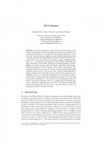
lysosomes (Figure 5 A, 1-h time point). These data are consistent with those obtained from cells overexpressing human Vps39p by Caplan et al. (2001). We took advantage of the ability to load the mVps18pinduced lysosomal clusters with fluid phase endocytic markers to analyze their structure in more detail. A stable NRK cell line inducible for GFP-mVps18p expression was allowed to internalize BSA-5 nm gold, with a procedure that results in all of the gold being localized as aggregates in dense core lysosomes (⬃85–90%) and hybrid organelles (⬃10 –15%) in untransfected control NRK cells (Bright et al., 1997). After standard fixation and resin embedding, cell sections were analyzed by transmission electron microscopy. In the uninduced control cells, 90% of the label was in electron-dense organelles with the characteristic morphology of dense core lysosomes, exactly as in untransfected control cells (Figure 5B). In the induced cells, only 64% of the label was in electron-dense organelles, and the area profile of these showed that many were smaller than in control cells (Figure 5B). The label present in electron-lucent organelles in the induced cells was mostly in organelles with larger areas than any labeled structures in uninduced control cells (Figure 5B). Images of the organelles are shown in Figure 6. In the stably transfected induced cells, clusters of electronlucent organelles were easily observed often mixed with dense core organelles containing aggregated 5-nm gold (Figure 6a). Striations were often observed between closely apposed electron lucent organelles (Figure 6b). These had similar morphology to the tethers between late endosomes and lysosomes described by Futter et al. (1996). Sometimes, a rim of aggregated protein fibrils gave the appearance of a shell surrounding the clustered organelles (Figure 6a). The size of the fibrils in these rims was consistent with the rims being actin enriched, suggesting a basis for the recruitment of some myosin classes to a shell around clustered organelles (see above). In standard 70-nm sections, many of the electron-lucent organelles in the clusters contained no 5-nm gold, presumably because it was out of the plane of section (e.g., Figure 6a). By immunoelectron microscopy, many of the clustered structures in stably transfected induced cells were labeled with antibodies to either the cation-independent mannose 6-phosphate receptor (MPR) or lgp120, with some organelles being labeled with both (Figure 6c). When antibodies to GFP were used at concentrations that did not label the general cytoplasm, some labeling was still observed in the region of the organelle clusters consistent with an enrichment of GFP-mVps18p (Figure 6d). In a further electron microscopy experiment (our unpublished data), we preloaded lysosomes, in a stable NRK cell line inducible for GFP-mVps18p expression, with 5-nm gold (Bright et al., 1997) and then switched on expression by adding 5 M cadmium chloride. After 48 h, and still in the presence of 5 M cadmium chloride, late endocytic compartments were loaded with BSA-10 nm gold by endocytic uptake for 4 h followed by a 20-h chase. Mixing of the two sizes of gold was observed in electron-dense and electron-lucent organelles consistent with overexpression of mVps18p not inhibiting fusion events with previously loaded lysosomes. Overexpression of mVp18p Results in Loss of Mannose 6-Phosphate Receptors Although we were able to detect some MPR by immunoelectron microscopy in clustered organelles in stably transfected cells overexpressing GFP-mVps18p, we had previously noticed that immunofluorescence staining of MPR in transiently transfected cells was much reduced (our unpub-
4019
V. Poupon et al.
Figure 5. Endocytic uptake of dextran and BSA-gold. (A) NRK cells transiently transfected with GFP-mVps18p were incubated with Texas Red-dextran for 1 h and then fixed for confocal microscopy. a, GFP; b, Texas Red-dextran. Bar, 10 m. (B) Stably transfected NRK cells, either uninduced or expressing GFP-mVps18p (as a result of induction with 10 M cadmium chloride for 48 h), were incubated with BSA-gold 5 nm for 4 h followed by a 20-h chase (in the presence of 10 M cadmium chloride) to label late endocytic organelles. Histograms show the area profile of electron dense and electron lucent organelles containing 5-nm gold in control (uninduced) cells and cells expressing GFP-mVps18p.
lished data). Using the inducible NRK cell line expressing GFP-mVps18p, we confirmed that immunofluorescence staining of MPR was greatly reduced in cells expressing GFP-mVps18p (Figure 7A, b), compared with uninduced cells (Figure 7A, a). With the ⌬pMEP vector used to create the stable NRK cell line, levels of expression of induced
Figure 4. Mysosin proteins are recruited into lysosome clusters. NRK cells transiently tranfected for 48 h and expressing GFPmVps18p (a–i) were processed for confocal fluorescence microscopy as in Figure 1, by using pAbs to MyoIb (a and b), MyoIc (c and d),
4020
Myosin II (e and f), Myosin V (g and h), Myosin VI (I and j) and Myosin IX (k and l) revealed by secondary Texas Red antibodies. a, c, e, g, i, and k, GFP; b, MyoIb; d, MyoIc; f, Myosin II; h, Myosin V; j, Myosin VI; and l, Myosin IX. Bar, 10 m.
Molecular Biology of the Cell
mVps18p in Endocytic Organelle Fusion
Figure 6. Electron microscopy of clustered late endocytic organelles. Stably transfected NRK cells expressing GFP-mVps18p were incubated with BSA-gold (5 nm) to load late endocytic organelles and then processed for conventional transmission electron microscopy (a and b) or immunoelectron microscopy (c and d). a, cluster of electron lucent organelles and dense core lysosomes surrounded by proteinaceous shell (open arrows). *(a, upper left quadrant), protein aggregates; small arrows, 5-nm gold; arrow heads, fine striations between adjacent organelles. Bar, 1 m. b, enlargment (2.25⫻) of fine striations shown in a between arrow heads. c, immunogold labeling of lgp120 (15-nm gold, arrowheads) and MPR (10-nm gold, large arrows). Small arrows, 5-nm gold. Bar, 250 nm. d, immunogold labeling of lgp120 (15-nm gold, arrowheads) and GFP (10-nm gold, large arrows). Small arrows, 5-nm gold. Bar, 250 nm.
protein can be increased by the addition of increasing concentrations of cadmium chloride (Ihrke et al., 2000). Immunoblotting showed that with increasing concentrations of GFP-mVps18p, there was a greater reduction in the concentration of MPR in the NRK cells, compared with the change in lgp120 (Figure 7B). These data are consistent with overexpression resulting in the trapping of MPR in late endocytic organelles containing active acid hydrolases, and therefore increasing its degradation. The change of lgp120 concentration may also be a consequence of increased time spent in active hydrolyzing organelles, with the extensive glycosylation of lgp120 providing greater protection than for MPR.
Vol. 14, October 2003
Overexpression of mVps18p Overcomes the Effect of Wortmannin to Swell Late Endocytic Organelles Through its ability to inhibit phosphatidyl inositol 3-kinases, wortmannin has a variety of effects on the function and morphology of endocytic compartments in mammalian cells. These include the swelling of late endocytic organelles, which results mainly from wortmannin inhibition of membrane traffic out of late endocytic compartments (Kundra and Kornfeld, 1998). The net effect of wortmannin to swell late endocytic compartments is particularly marked in NRK cells, where swollen organelles with areas ⬎2 m2 have been observed (Reaves et al., 1996; Bright et al., 2001). In the present study, we found that overexpression of GFP-
4021
V. Poupon et al.
Figure 7. Functional characterization of clusters of late endocytic organelles. (A) Overexpression of GFP-mVp18p disrupts MPR expression. Stably transfected NRK cells were not induced (a), or induced to express GFP-mVps18p for 48 h with 5 M cadmium chloride (b), before processing for confocal fluorescence microscopy by using an anti-MPR pAb. a and b, MPR. (B) Immunoblotting of MPR. Stably transfected NRK cells were induced for 48 h with increasing amounts of cadmium chloride, ranging from 0.1 to 10 M to stimulate expression of GFP-mVps18p. a, expression of MPR and lgp120; the ratios of densities of MPR compared with lgp120 bands are shown (the densities of lgp120 bands relative to density in noninduced cells are 89, 89, and 78%, and of MPR bands relative to density in noninduced cells are 76, 71, and 51% after induction, respectively, with 0.1, 1, and 10 M cadmium chloride). b, expression of GFP-mVps18p and endogenous mVps18p were assessed in total cell lysates by immunoblotting. (C) Wortmannin treatment. NRK cells, transiently transfected for 48 h with GFP-mVps18p, were incubated with wortmannin for 45 min. The cells were fixed and processed for confocal fluorescence microscopy as described in MATERIALS AND METHODS. a, GFP, b, lgp120.
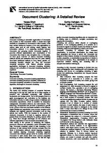
mVps18p prevented the formation of swollen organelles in response to wortmannin treatment (Figure 7C). Similar results were obtained in cells overexpressing GFP-mVps39p (our unpublished results). These data imply a role for mVps18p and associated proteins in traffic out of late endocytic organelles as well as in membrane fusion and delivery of endocytosed material to lysosomes. Reduction of the Intracellular Concentration of mVps18p by RNA Interference Results in Redistribution of Lysosomes Within the Cell In many cells, including NRK cells, lysosomes tend to be concentrated in a juxtanuclear position near the microtubule
4022
organizing center, though such concentrations are easily distinguished from the clusters observed when overexpressing mVps18p or mVps39p. The juxtanuclear concentration is the result of minus end-directed transport along microtubules mediated by a dynein-dynactin motor recruited by the Rab 7 effector RILP (Jordens et al., 2001). The filamentous actin network and Myo1b (myosin 1␣) play a retention role in intracellular localization of lysosomes (Cordonnier et al., 2001). Because overexpression of mVps18p resulted in recruitment of actin, Myo1b, and RILP to lysosome clusters (Figure 3, e and f), we reasoned that reduction of the concentration of mVps18p in NRK cells might result in redistribution of lysosomes away from the microtubule organizing center. We transfected NRK cells with short interfering oligonucleotides (siRNAs) under conditions that resulted in ⬃50% transfection efficiency and ⬃50% reduction in mVps18p as determined by immunoblotting (Figure 8a, inset). Transfected cells were identified by the use of fluorescently tagged oligonucleotides. Cells were viewed by indirect immunofluorescence, with a conventional upright epifluorescence microscope to see easily the juxtanuclear concentration of lysosomes in the nontransfected cells (in contrast to confocal microscopy used for all fluorescent images shown in Figures 1–5, 7, and 10). We observed that lgp-positive organelles were distributed throughout the cytoplasm of transfected cells (Figure 8a). There was much less effect on the distribution of MPR-positive organelles (Figure 8b) and no effect on TGN38 (Figure 8c). After RNA interference, the distributed lgp-positive organelles remained accessible to endocytosed Texas Red-dextran (our unpublished data). The RNA interference experiments were repeated with a second pair of siRNAs matching a different region of mVps18 cDNA and resulted in the same effects (our unpublished data). When NRK cells were transfected with siRNAs matching mVps18cDNA before transient transfection with a plasmid encoding GFP-mVps39p, lgp-positive organelles were still dispersed away from the juxtanuclear region and were not clustered (Figure 9, a and b). In contrast, when cells overexpressing GFP-mVps39p were transfected with mVps18 siRNAs, no disruption of clustered lgp-positive organelles was observed (Figure 9, c and d). These data are consistent with the presence of mVps18p being required for the tethering function of mVps39p in mammalian cells. Delineation of Functional Domains in mVps18p Mouse Vps18p shares 96% amino acid identity with its human homolog. A search with BLAST, National Center for Biotechnology Information Conserved Domain Search, and 3D-PSSM programs confirmed that it contains, as does its human homolog (Huizing et al., 2001; Kim et al., 2001), a clathrin homology (CLH) repeat domain at amino acids 638 –769, a RING-H2 finger domain within amino acids 853– 947, and two coiled-coil domains within amino acids 853– 878 and 802– 848 (Figure 10A). We decided to delineate the functional domains in mVps18p by investigating which were necessary for lysosomal localization, for induction of lysosomal clustering, and the prevention of wortmanninmediated swelling of late endocytic organelles. A set of GFPtagged deletion constructs of mVps18p, as well as mVps39p (Figure 10A), were transiently transfected in NRK cells. Transfection efficiency and expression of GFP, observed by fluorescence microscopy, were similar for all constructs. The RING-H2 domain, or the coiled-coil domain close to it, were not able to associate to lysosomes by themselves (images not shown). However, a construct containing both,
Molecular Biology of the Cell
mVps18p in Endocytic Organelle Fusion
crease in the proportion of transfected cells showing clustered lysosomes (Figure 10B a– c, and C). Caplan et al. (2001) observed that the N-terminal twothirds of human Vps39p contains a CNH and a CLH domain. A construct containing both domains colocalized with and caused clustering of lysosomes, but not constructs containing a single domain (Caplan et al., 2001). We observed similar results with equivalent mouse Vps39p constructs, but also made a construct, mVps39Cter, containing the CLH domain and an additional 20 amino acids at the N terminus that colocalized with lysosomes to some extent and caused some clustering (Figure 10A; and C, images not shown). The additional 20 amino acids are not predicted to display any obvious structural feature. For both mVps18p and mVps39p constructs, the extent of inhibition of the wortmannin effect on swelling of late endocytic organelles correlated with the amount of lysosomal clustering observed (Figure 10A). DISCUSSION
Figure 8. Reduction of intracellular mVps18p by RNA interference causes redistribution of lysosomes. NRK cells transiently transfected with mVps18p siRNA oligonucleotides (a– c) were processed for indirect immunofluorecence as in Figure 1, by using anti-lgp120 mAb GM10 (a), anti-MPR pAb1001 (b), or anti-TGN38 mAb 2F7.1 (c). Cells were observed using a conventional upright epifluorescence microscope. Stars, transfected cells. Bar, 10 m. a, inset, expression of mVps18p was assessed on total cell lysates of untransfected (left) and transiently transfected (right) NRK cells by immunoblotting.
although not the CLH domain, colocalized with lgp120 but did not cause clustering (Figures 7C and 10B, d–f). A construct containing only the CLH domain also colocalized with lgp120 (images not shown), and its overexpression induced the clustering of lysosomes to some extent (Figure 10C). A construct consisting of the C-terminal third of the protein, and containing all three domains, led to a significant in-
Vol. 14, October 2003
Our data are consistent with the hypothesis that the mammalian homologs of the yeast HOPS/class C Vps complex proteins orchestrate the recruitment of much of the cytosolic protein machinery for efficient late endosome-lysosome fusion together with proteins required to maintain the morphological and functional integrity of late endocytic organelles. In our initial experiments we found that overexpression of mVps18p caused clustering of late endocytic organelles, including lysosomes, and recruitment of other mammalian homologs of the yeast HOPS/class C Vps complex proteins to the organelle clusters. Our data show that overexpressing mVps18p results in an increase in the amount of this protein associated with late endocytic organelles and recruitment to these of other mammalian HOPS complex homologs. This is consistent with the study of Kim et al. (2001), who showed, by coimmunoprecipitation and gel filtration, that human Vps18p forms a large heteroligomeric complex with other HOPS complex homologs and interacts with syntaxin 7 (Antonin et al., 2000). It is also consistent with the proposed function of the HOPS complex in yeast for both homotypic vacuole fusion (Seals et al., 2000) and vesicle fusion with the vacuole (Sato et al., 2000). The clusters of late endocytic organelles that we observed contained many that were enlarged and electron lucent, in agreement with experiments of Caplan et al. (2001) on mVps39p. They suggested that late endosomes and lysosomes first cluster and then fuse to generate large vacuoles. The observed decrease in cellular MPR content after overexpression of mVps18p may also be explained by increased fusion of late endosomes and lysosomes leading to the entrapment and degradation of MPRs in the resultant hybrid organelles. Our data show that overexpression of mVps18p, or mVps39p, recruits many other proteins to the late endocytic organelle clusters, in addition to other mammalian homologs of HOPS complex components. The recruitment of actin is of major interest because this has been widely reported to have a role in delivery to lysosomes in cell free systems, living cells (Kolset et al., 1979; van Deurs et al., 1995; Durrbach et al., 1996b; Jahraus et al., 2001) and in homotypic vacuole fusion in vitro (Eitzen et al. 2002). Interestingly, the proteinaceous, actin rich “shell” observed around clustered late endocytic organelles in our experiments may have an effect on the morphology of the organelles, because previous studies have shown that the swelling of phagocytic vacuoles can be constrained by surrounding cytoskeletal, actin-rich
4023
V. Poupon et al.
Figure 9. Reduction of intracellular mVps18p by RNA interference prevents mVps39p-induced clustering of lysosomes. NRK cells transiently transfected with mVps18p siRNA oligonucleotides then with a plasmid encoding GFPmVps39p for 48h (a and b) or vice versa (c and d) were processed as in Figure 1, by using pAb 580 to lgp110 (a and c) and observed using a conventional upright epifluorescence microscope as in Figure 8. Single stars, cells transfected with mVps18 siRNAs. Double stars, cells transfected with both mVps18 siRNAs and the plasmid encoding GFP-mVps39p. Bar 10 m. a and c, lgp110; b and d, GFP.
networks (Reeves et al., 2002). The presence of ezrin in the clusters implies attachment of actin filaments to the surrounding membranes (Bretscher, 1999) and the specific subset of myosins likely provide a means of moving organelles toward, or away from, each other. Myo Ib (also known as myosin I␣) has been shown to be involved in delivery from endosomes to lysosomes (Raposo et al., 1999) and myosin V has been implicated in the local movement of melanosomes, which are lysosome-like organelles (Wu et al., 1998). Myosin IX has been proposed to move toward the minus end of actin filaments (Inoue et al., 2002), only the second myosin, along with myosin IV to do so (Buss et al., 2001). Given the absence of myosin VI from clustered lysosomes in our experiments, myosin IX is clearly a candidate motor protein to be involved in actin-dependent movement away from late endocytic organelles. Filamentous actin and Myo1b have been proposed to play a role in the intracellular distribution of lysosomes, and expression of a nonfunctional Myo1b lacking the ATP binding site affects their motility along microtubules (Cordonnier et al., 2001). The net direction of lysosome movement is toward the minus end of microtubules (Bucci et al., 2000), mediated by a dynein dynactin motor recruited by the Rab 7 effector RILP (Cantalupo et al., 2001; Jordens et al., 2001). Our data, both from overexpressing mVps18p and its knockdown by RNA interference, suggest that the mammalian
4024
homologs of the yeast HOPS complex components may play a role in recruiting the cytoskeletal motors required for intracellular lysosome movement and localization. The recruitment of RILP when overexpressing mVps18p does not necessarily imply that it acts upstream of Rab7. Caplan et al. (2001) showed that overexpression of mVps39p induces lysosome clustering and fusion even in the presence of a dominant-negative Rab7, implying that the mammalian HOPS complex acts downstream or independently of Rab7. Our RNA interference experiments suggest that mVps18p functions upstream of mVps39p, with its presence being necessary for mVps39p function. This is consistent with the model proposed by Sato et al., 2000, for the function of the HOPS/class C Vps complex in docking/fusion of cargo vesicles to the vacuole in yeast. It is interesting to note that in yeast, in addition to effects on vacuole fusion events, the HOPS complex proteins function at multiple stages of the vacuolar transport pathway (Srivastava et al., 2000; Peterson and Emr, 2001). Our observation that, at low levels of expression, GFP-mVps18p partially colocalizes with EEA1 was consistent with other data from one of our laboratories showing partial colocalization of endogenous mammalian HOPS complex proteins, including mVps18p, with EEA1 (Richardson and Piper, unpublished data) These experiments raise the possibility that mVps18p and associated mammalian HOPS complex components also func-
Molecular Biology of the Cell
mVps18p in Endocytic Organelle Fusion
Figure 10. Delineation of functional domains in mVps18p and mVps39p. (A) Schematic representation of full-length mVps18p and mVps39p, and various deletion constructs. The columns on the right indicate whether constructs colocalize with lgp120, cause clustering of lysosomes, and prevent the effect of wortmannin on swelling of late endocytic organelles. (B) Expression of mVps18p deletion constructs. NRK cells transiently transfected for 48 h with GFP-tagged deletion constructs of mVps18p (a–f) were processed for confocal fluorescence microscopy, as in Figure 1, by using mAb antilgp120 (a–f) as a primary antibody. Herein, are shown the expression of two representative constructs, GFP-mVps18Cter (a– c), and GFP-mVps18(CC⫹RH2) (d–f). a and d, GFP; b and e, lgp120; c and f, merge of green and red channels. (C) Quantification of the effect of mVps18p and mVps39p constructs on localization and lysosome clustering. NRK (left) or HeLa cells (right) were transiently transfected for 48 h with all the GFP-tagged constructs of mVps18p and mVps39p and then processed for confocal fluorescence microscopy as in Figure 1, by using mAb anti-lgp120 for NRK cells and mAb anti-lamp1 for HeLa cells. For each construct, ⬎100 cells were counted and sorted into three categories, according to colocalization with and clustering of lysosomes.
tion at other membrane traffic steps in mammalian cells as well as in the late endocytic pathway. In addition, or alternatively, it is possible that recruitment of the HOPS complex commences early in the endocytic pathway before exerting its functions on late endocytic organelles. The fact that endocytosed dextran can still be delivered to dispersed lgp-positive organelles after knockdown of mVps18p by RNA interference suggests either that knockdown is incomplete in the transfected cells and/or that this protein is not essential for endosome-lysosome fusion. From the present experiments, we cannot rule out
Vol. 14, October 2003
the possibility that mVps18p and associated proteins play a role in increasing the efficiency of fusion of late endocytic organelles rather than being absolutely required for the fusion process itself. Not only does the mammalian HOPS complex play a role in recruiting cytosolic machinery for efficient fusion and intracellular movement of late endocytic organelles but also, we suggest, for vesicular traffic out of these organelles. Little is known about such machinery but Rab9 and the mammalian homologs of the yeast retromer complex proteins have been suggested to be involved (Pfeffer, 2001). The clear effect
4025
V. Poupon et al.
of overexpressing mVps18p (or mVps39p) to prevent the gross swelling of late endocytic organelles after wortmannin treatment suggests that this is the case. Although the effects of wortmannin on the endocytic pathway are multiple and complex, the gross swelling of late endocytic organelles has been attributed to net inhibition of retrograde traffic from them (Reaves et al., 1996; Kundra and Kornfeld, 1998; Bright et al., 2001). An attractive hypothesis for how the wortmannin effect may be overcome is suggested by experiments on early endosomes, where excess Rab 5-GTP can overcome the wortmannin-induced release of the tether protein EEA1 which is normally recruited to the endosome membrane by binding to both Rab 5 and PtdIns 3-phosphate (Simonsen et al., 1998). Thus, we suggest that overexpression of mVps18p may be sufficient to overcome a PtdIns 3-phosphate requirement to recruit cytosolic proteins necessary for vesicular traffic out of late endocytic organelles, for example in the reformation of lysosomes from hybrid organelles. This function of mVps18p may ensure that lysosome reformation is tightly coupled to late endosome-lysosome fusion. Our studies have identified two functionally important domains within mVps18p, the CLH and the RING-H2 domains, which are both important for recruitment to the late endocytic organelles. In the clathrin heavy chain, there are seven CLH domains, required for homo-oligomerization, each consisting of ⬃140 amino acids organized in multiple alpha helical repeats (Ybe et al., 1999). In yeast Vps41p/ Vam2p (Darsow et al., 2001) and human Vps39p/hVam6p (Caplan et al., 2001), the CLH motifs have been proposed to mediate protein–protein interactions leading to homo- or hetero-oligomerization. The RING-H2 finger domain is a subfamily of the RING finger motif, also present in Vps11p and Vps41p (Caplan et al., 2001; Huizing et al., 2001, Kim et al., 2001). It is important for the biological function of Vps18p in both yeast and Drosophila, because point mutations of the conserved cysteines within the motif lead to perturbations in the morphology of late endocytic organelles (Emr and Malhotra, 1997, Sevrioukov et al., 1999). RING finger motifs have been implicated in both protein–protein interactions (Borden and Freemont, 1996) and lipid binding, e.g., the FYVE domain of EEA1 which binds to PtdIns 3-phosphate (Stenmark et al., 1996, Lawe et al., 2002). Mammalian Vps11p and Vps41p also contain a RING-H2 domain, which in the latter case (Ward et al., 2001) has been shown to mediate membrane association of the protein, but may also be involved in interactions with other proteins required for tethering and/or fusion. Because coiled-coil regions are also potentially involved in homo- or hetero-oligomerization, mVps18p is a protein composed of several domains that may be involved in protein–protein interactions. In conclusion, our studies implicate mVps18p as a mammalian tethering and/or docking factor which promotes aggregation and fusion of late endosomes/lysosomes in vivo. Further studies of its two functional domains, their potential regulation and the characterization of the proteins interacting with these domains, including the other components of the mammalian HOPS complex, should provide a better understanding of the mechanisms involved in lysosome fusion and reformation. ACKNOWLEDGMENTS We thank Drs. Folma Buss and Paul Pryor for reagents and much valuable discussion. This work was partly funded by an Medical Research Council program grant to J.P.L., and a Human Frontier Science Program grant to R.P., J.P.L., and Dr. D.R. James. VP was funded by La Fondation Medicale pour la
4026
Recherche and subsequently, as a Wellcome Trust Traveling Fellow. Cambridge Institute for Medical Research is in receipt of a strategic award from the Wellcome Trust.
REFERENCES Antonin, W., Holroyd, C., Fasshauer, D., Pabst, S., Von Mollard, G.F., and Jahn, R. (2000). A SNARE complex mediating fusion of late endosomes defines conserved properties of SNARE structure and function. EMBO J. 19, 6453– 6464. Barois, N., Forquet, F., and Davoust, J. (1998). Actin microfilaments control the MHC class II antigen presentation pathway in B cells. J. Cell Sci. 111, 1791–1800. Bonangelino, C.J., Chavez, E.M., and Bonifacino, J.S. (2002). Genomic screen for vacuolar protein sorting genes in Saccharomyces cerevisiae. Mol. Biol. Cell 13, 2486 –2501. Borden, K.L., and Freemont, P.S. (1996). The RING finger domain: a recent example of a sequence-structure family. Curr. Opin. Struct. Biol. 6, 395– 401. Bretscher, A. (1999). Regulation of cortical structure by the ezrin-radixinmoesin protein family. Curr. Opin. Cell Biol. 11, 109 –116. Bright, N.A., Reaves, B.J., Mullock, B.M., and Luzio, J.P. (1997). Dense core lysosomes can fuse with late endosomes and are re-formed from the resultant hybrid organelles. J. Cell Sci. 110, 2027–2040. Bright, N.A., Lindsay, M.R., Stewart, A., and Luzio, J.P. (2001). The relationship between lumenal and limiting membranes in swollen late endocytic compartments formed after wortmannin treatment or sucrose accumulation. Traffic 2, 631– 642. Bucci, C., Thomsen, P., Nicoziani, P., McCarthy, J., and van Deurs, B. (2000). Rab 7, a key to lysosome biogenesis. Mol. Biol. Cell 11, 467– 480. Buss, F., Luzio, J.P., and Kendrick-Jones, J. (2001). Myosin VI, a new force in clathrin mediated endocytosis. FEBS Lett. 508, 295–299. Cantalupo, G., Alifano, P., Roberti, V., Bruni, C.B., and Bucci, C. (2001). Rab-interacting lysosomal protein (RILP): the Rab7 effector required for transport to lysosomes. EMBO J. 20, 683– 693. Caplan, S., Hartnell, L.M., Aguilar, R.C., Naslavsky, N., and Bonifacino, J.S. (2001). Human Vam6p promotes lysosome clustering and fusion in vivo. J. Cell Biol. 154, 109 –122. Cordonnier, M.N., Dauzonne, D., Louvard, D., and Coudrier, E. (2001). Actin filaments and myosin I alpha cooperate with microtubules for the movement of lysosomes. Mol. Biol. Cell 12, 4013– 4029. Darsow, T., Katzmann, D.J., Cowles, C.R., and Emr, S.D. (2001). Vps41p function in the alkaline phosphatase pathway requires homo-oligomerization and interaction with AP-3 through two distinct domains. Mol. Biol. Cell 12, 37–51. Durrbach, A., Collins, K., Matsudaira, P., Louvard, D., and Coudrier, E. (1996a). Brush border myosin-I truncated in the motor domain impairs the distribution and the function of endocytic compartments in an hepatoma cell line. Proc. Natl. Acad. Sci. USA 93, 7053–7058. Durrbach, A., Louvard, D., and Coudrier, E. (1996b). Actin filaments facilitate two steps of endocytosis. J. Cell Sci. 109, 457– 465. Eitzen, G., Wang, L., Thorngren, N., and Wickner, W. (2002). Remodeling of organelle-bound actin ir required for yeast vacuole fusion. J. Cell Biol. 158, 669 – 679. Emr, S.D., and Malhotra, V.V. (1997). Membranes and sorting. Curr. Opin. Cell Biol. 9, 475– 476. Futter, C.E., Pearse, A., Hewlett, L.J., and Hopkins, C.R. (1996). Multivesicular endosomes containing internalized EGF-EGF receptor complexes mature and then fuse directly with lysosomes. J. Cell Biol. 132, 1011–1023. Grimaldi, K.A., Hutton, J.C., and Siddle, K. (1987). Production and characterization of monoclonal antibodies to insulin secretory granule membranes. Biochem J. 245, 557–566. Holroyd, C., Kistner, U., Annaert, W., and Jahn, R. (1999). Fusion of endosomes involved in synaptic vesicle recycling. Mol. Biol. Cell 10, 3035–3044. Horn, M., and Banting, G. (1994). Okadaic acid treatment leads to a fragmentation of the trans-Golgi network and an increase in expression of TGN38 at the cell surface. Biochem. J. 301, 69 –73. Huizing, M., Didier, A., Walenta, J., Anikster, Y., Gahl, W.A., and Kramer, H. (2001). Molecular cloning and characterization of human VPS18, VPS 11, VPS16, and VPS33. Gene 264, 241–247.
Molecular Biology of the Cell
mVps18p in Endocytic Organelle Fusion Inoue, A., Saito, J., Ikebe, R., and Ikebe, M. (2002). Myosin IXb is a singleheaded minus-end-directed processive motor. Nat. Cell. Biol. 4, 302–306.
coproteins suggests a role for phosphoinositide 3-kinase activity in regulating membrane traffic late in the endocytic pathway. J. Cell Sci. 109, 749 –762.
Ihrke, G., Gray, S.R., and Luzio, J.P. (2000). Endolyn is a mucin-like type I membrane protein targeted to lysosomes by its cytoplasmic tail. Biochem. J. 345, 287–296.
Reeves, E.P., Lu, H., Jacobs, H.L., Messina, C.G., Bolsover, S., Gabella, G., Potma, E.O., Warley, A., Roes, J., and Segal, A.W. (2002). Killing activity of neutrophils is mediated through activation of proteases by K⫹ flux. Nature 416, 291–297.
Jahraus, A., Egeberg, M., Hinner, B., Habermann, A., Sackman, E., Pralle, A., Faulstich, H., Rybin, V., Defacque, H., and Griffiths, G. (2001). ATP-dependent membrane assembly of F-actin facilitates membrane fusion. Mol. Biol. Cell 12, 155–170. Jordens, I., Fernandez-Borja, M., Marsman, M., Dusseljee, S., Janssen, L., Calafat, J., Janssen, H., Wubbolts, R., and Neefjes, J. (2001). The Rab7 effector protein RILP controls lysosomal transport by inducing the recruitment of dynein-dynactin motors. Curr. Biol. 11, 1680 –1685. Kim, B.Y., Kramer, H., Yamamoto, A., Kominami, E., Kohsaka, S., and Akazawa, C. (2001). Molecular characterization of mammalian homologues of class C Vps proteins that interact with syntaxin-7. J. Biol. Chem. 276, 29393– 29402. Kolset, S.O., Tolleshaug, H., and Berg, T. (1979). The effects of colchicine and cytochalasin B on uptake and degradation of asialo-glycoproteins in isolated rat hepatocytes. Exp. Cell. Res. 122, 159 –167. Kundra, R., and Kornfeld, S. (1998). Wortmannin retards the movement of the mannose 6-phosphate/insulin-like growth factor II receptor and its ligand out of endosomes. J. Biol. Chem. 273, 3848 –3853. Lawe, D.C., Chawla, A., Merithew, E., Dumas, J., Carrington, W., Fogarty, K., Lifshitz, L., Tuft, R., Lambright, D., and Corvera, S. (2002). Sequential roles for phosphatidylinositol 3-phosphate and Rab5 in tethering and fusion of early endosomes via their interaction with EEA1. J. Biol. Chem. 277, 8611– 8617. Luzio, J.P., Rous, B.A., Bright, N.A., Pryor, P.R., Mullock, B.M., and Piper, R.C. (2000). Lysosome-endosome fusion and lysosome biogenesis. J. Cell Sci. 113, 1515–1524. Mangeat, P., Roy, C., and Martin, M. (1999). ERM proteins in cell adhesion and membrane dynamics. Trends Cell Biol. 9, 187–192. Mullins, C., and Bonifacino, J.S. (2001). The molecular machinery for lysosome biogenesis. Bioessays 23, 333–343. Mullock, B.M., Bright, N.A., Fearon, C.W., Gray, S.R., and Luzio, J.P. (1998). Fusion of lysosomes with late endosomes produces a hybrid organelle of intermediate density and is NSF dependent. J. Cell Biol. 140, 591– 601. Mullock, B.M., et al. (2000). Syntaxin 7 is localized to late endosome compartments, associates with Vamp 8, and Is required for late endosome-lysosome fusion. Mol. Biol. Cell 11, 3137–3153. Peters, C., and Mayer, A. (1998). Ca2⫹/calmodulin signals the completion of docking and triggers a late step of vacuole fusion. Nature 396, 575– 80. Peterson, M.R., and Emr, S.D. (2001). The class C Vps complex functions at multiple stages of the vacuolar transport pathway. Traffic 2, 476 – 486. Pfeffer, S.R. (1999). Transport-vesicle targeting: tethers before SNAREs. Nat. Cell Biol. 1, E17–E22. Pfeffer, S.R. (2001). Membrane transport: retromer to the rescue. Curr. Biol., 11, R109 –111. Poupon, V., Begue, B., Gagnon, J., Dautry-Varsat, A., Cerf-Bensussan, N., and Benmerah, A. (1999). Molecular cloning and characterization of MT-ACT48, a novel mitochondrial acyl-CoA thioesterase. J. Biol. Chem. 274, 19188 –19194. Pryor, P.R., Mullock, B.M., Bright, N.A., Gray, S.R., and Luzio, J.P. (2000). The role of intraorganellar Ca(2⫹) in late endosome-lysosome heterotypic fusion and in the reformation of lysosomes from hybrid organelles. J. Cell Biol. 149, 1053–1062. Raposo, G., Cordonnier, M. N., Tenza, D., Menichi, B., Durrbach, A., Louvard, D. and, Coudrier, E. (1999). Association of myosin I alpha with endosomes and lysosomes in mammalian cells. Mol. Biol. Cell 10, 1477–1494. Reaves, B.J., Bright, N.A., Mullock, B.M., and Luzio, J.P. (1996). The effect of wortmannin on the localisation of lysosomal type I integral membrane gly-
Vol. 14, October 2003
Rieder, S.E., and Emr, S.D. (1997). A novel RING finger protein complex essential for a late step in protein transport to the yeast vacuole. Mol. Biol. Cell 8, 2307–2327. Sato, T.K., Rehling, P., Peterson, M.R., and Emr, S.D. (2000). Class C Vps protein complex regulates vacuolar SNARE pairing and is required for vesicle docking/fusion. Mol. Cell 6, 661– 671. Seals, D.F., Eitzen, G., Margolis, N., Wickner, W.T., and Price, A. (2000). A Ypt/Rab effector complex containing the Sec1 homolog Vps33p is required for homotypic vacuole fusion. Proc. Natl. Acad. Sci. USA 97, 9402–9407. Sevrioukov, E.A., He, J.P., Moghrabi, N., Sunio, A., and Kramer, H. (1999). A role for the deep orange and carnation eye color genes in lysosomal delivery in Drosophila. Mol. Cell 4, 479 – 486. Simonsen, A., Lippe, R., Christoforidis, S., Gaullier, J.M., Brech, A., Callaghan, J., Toh, B.H., Murphy, C., Zerial, M., and Stenmark, H. (1998). EEA1 links PI(3)K function to Rab5 regulation of endosome fusion. Nature 394, 494 – 498. Srivastava, A., Woolford, C.A., and Jones, E.W. (2000). Pep3p/Pep5p complex: a putative docking factor at multiple steps of vesicular transport to the vacuole of Saccharomyces cerevisiae. Genetics 156, 105–122. Stamnes, M. (2002). Regulating the actin cytoskeleton during vesicular transport. Curr. Opin. Cell. Biol. 14, 428. Stenmark, H., Aasland, R., Toh, B.H., and D’Arrigo, A. (1996). Endosomal localization of the autoantigen EEA1 is mediated by a zinc-binding FYVE finger. J. Biol. Chem. 271, 24048 –24054. Storrie, B., and Desjardins, M. (1996). The biogenesis of lysosomes: is it a kiss and run, continuous fusion and fission process? Bioessays 18, 895–903. van Deurs, B., Holm, P.K., Kayser, L., and Sandvig, K. (1995). Delivery to lysosomes in the human carcinoma cell line HEp-2 involves an actin filamentfacilitated fusion between mature endosomes and preexisting lysosomes. Eur. J. Cell Biol. 66, 309 –323. Ward, D.M., Pevsner, J., Scullion, M.A., Vaughn, M., and Kaplan, J. (2000). Syntaxin 7 and VAMP-7 are soluble N-ethylmaleimide-sensitive factor attachment protein receptors required for late endosome-lysosome and homotypic lysosome fusion in alveolar macrophages. Mol. Biol. Cell 11, 2327–2333. Ward, D.M., Radisky, D., Scullion, M.A., Tuttle, M.S., Vaughn, M., and Kaplan, J. (2001). hVPS41 is expressed in multiple isoforms and can associate with vesicles through a RING-H2 finger motif. Exp. Cell. Res. 267, 126 –134. Warner, T.S., Sinclair, D.A., Fitzpatrick, K.A., Singh, M., Devlin, R.H., and Honda, B.M. (1998). The light gene of Drosophila melanogaster encodes a homologue of VPS41, a yeast gene involved in cellular-protein trafficking. Genome 41, 236 – 43. Wettey, F.R., Hawkins, S.F., Stewart, A., Luzio, J.P., Howard, J.C., and Jackson, A.P. (2002). Controlled elimination of clathrin heavy-chain expression in DT40 lymphocytes. Science 297, 1521–1525. Wickner, W. (2002). Yeast vacuoles and membrane fusion pathways. EMBO J. 21, 1241–1247. Wickner, W., and Haas, A. (2000). Yeast homotypic vacuole fusion: a window on organelle trafficking mechanisms. Annu. Rev. Biochem. 69, 247–75. Wu, X., Bowers, B., Rao, K., Wei, Q., and Hammer, J.A. (1998). Visualization of melanosome dynamics within wild-type and dilute melanocytes suggests a paradigm for myosin V function in vivo. J. Cell Biol. 143, 1899 –1918. Wurmser, A.E., Sato, T.K., and Emr, S.D. (2000). New component of the vacuolar class C-Vps complex couples nucleotide exchange on the Ypt7 GTPase to SNARE-dependent docking and fusion. J. Cell Biol. 151, 551–562. Ybe, J.A., Brodsky, F.M., Hofmann, K., Lin, K., Liu, S.H., Chen, L., Earnest, T.N., Fletterick, R.J., and Hwang, P.K. (1999). Clathrin self-assembly is mediated by a tandemly repeated superhelix. Nature 399, 371–375.
4027